DEVELOPMENT AND STRUCTURAL BASIS OF A TWO-MAB COCKTAIL FOR TREATING SARS-COV-2 INFECTIONS
←
→
Page content transcription
If your browser does not render page correctly, please read the page content below
ARTICLE
                  https://doi.org/10.1038/s41467-020-20465-w                OPEN

                  Development and structural basis of a two-MAb
                  cocktail for treating SARS-CoV-2 infections
                  Chao Zhang1,7, Yifan Wang2,7, Yuanfei Zhu3,7, Caixuan Liu2,7, Chenjian Gu3,7, Shiqi Xu1,7, Yalei Wang1, Yu Zhou1,
                  Yanxing Wang2,4, Wenyu Han2, Xiaoyu Hong2, Yong Yang1, Xueyang Zhang1, Tingfeng Wang1, Cong Xu2,
                  Qin Hong2, Shutian Wang2, Qiaoyu Zhao2, Weihua Qiao1, Jinkai Zang1, Liangliang Kong5, Fangfang Wang5,
                  Haikun Wang 1, Di Qu3,6, Dimitri Lavillette 1, Hong Tang1, Qiang Deng 3 ✉, Youhua Xie 3 ✉,
                  Yao Cong 2,4 ✉ & Zhong Huang 1 ✉
1234567890():,;

                  The ongoing pandemic of coronavirus disease 2019 (COVID-19) is caused by severe acute
                  respiratory syndrome coronavirus 2 (SARS-CoV-2). Neutralizing antibodies against SARS-
                  CoV-2 are an option for drug development for treating COVID-19. Here, we report the
                  identification and characterization of two groups of mouse neutralizing monoclonal anti-
                  bodies (MAbs) targeting the receptor-binding domain (RBD) on the SARS-CoV-2 spike (S)
                  protein. MAbs 2H2 and 3C1, representing the two antibody groups, respectively, bind distinct
                  epitopes and are compatible in formulating a noncompeting antibody cocktail. A humanized
                  version of the 2H2/3C1 cocktail is found to potently neutralize authentic SARS-CoV-2
                  infection in vitro with half inhibitory concentration (IC50) of 12 ng/mL and effectively treat
                  SARS-CoV-2-infected mice even when administered at as late as 24 h post-infection. We
                  determine an ensemble of cryo-EM structures of 2H2 or 3C1 Fab in complex with the S trimer
                  up to 3.8 Å resolution, revealing the conformational space of the antigen–antibody complexes
                  and MAb-triggered stepwise allosteric rearrangements of the S trimer, delineating a pre-
                  viously uncharacterized dynamic process of coordinated binding of neutralizing antibodies to
                  the trimeric S protein. Our findings provide important information for the development of
                  MAb-based drugs for preventing and treating SARS-CoV-2 infections.

                  1 CAS  Key Laboratory of Molecular Virology & Immunology, Institut Pasteur of Shanghai, Chinese Academy of Sciences, University of Chinese Academy of
                  Sciences, Shanghai, China. 2 State Key Laboratory of Molecular Biology, National Center for Protein Science Shanghai, Shanghai Institute of Biochemistry and
                  Cell Biology, Center for Excellence in Molecular Cell Science, Chinese Academy of Sciences, University of Chinese Academy of Sciences, Shanghai, China.
                  3 Key Laboratory of Medical Molecular Virology (MOE/NHC/CAMS), Department of Medical Microbiology and Parasitology, School of Basic Medical

                  Sciences, Shanghai Medical College, Fudan University, Shanghai, China. 4 Shanghai Science Research Center, Chinese Academy of Sciences, 201210
                  Shanghai, China. 5 The National Facility for Protein Science in Shanghai (NFPS), 201210 Shanghai, China. 6 BSL-3 Laboratory of Fudan University, School of
                  Basic Medical Sciences, Shanghai Medical College, Fudan University, Shanghai, China. 7These authors contributed equally: Chao Zhang, Yifan Wang,
                  Yuanfei Zhu, Caixuan Liu, Chenjian Gu, Shiqi Xu. ✉email: qdeng@fudan.edu.cn; yhxie@fudan.edu.cn; cong@sibcb.ac.cn; huangzhong@ips.ac.cn

                  NATURE COMMUNICATIONS | (2021)12:264 | https://doi.org/10.1038/s41467-020-20465-w | www.nature.com/naturecommunications                                   1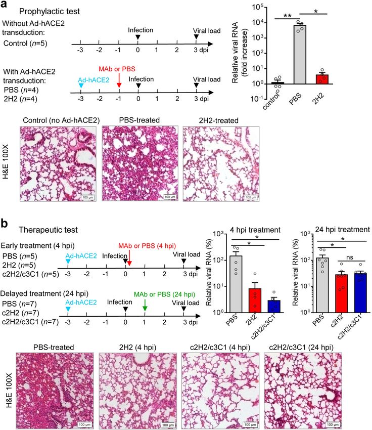
ARTICLE                                                               NATURE COMMUNICATIONS | https://doi.org/10.1038/s41467-020-20465-w

T
        he ongoing pandemic of coronavirus disease 2019                         In this study, we identified and comprehensively characterized
        (COVID-19) is caused by a newly identified coronavirus                two groups of mouse anti-SARS-CoV-2-neutralizing MAbs.
        named severe acute respiratory syndrome coronavirus 2                MAbs 2H2 and 3C1, representing the two antibody groups,
(SARS-CoV-2; formerly designated 2019-nCoV)1–3. Individuals                  respectively, targeted distinct epitopes on RBD and were com-
infected with SARS-CoV-2 may develop severe respiratory                      patible in formulating a noncompeting antibody cocktail. A
manifestations and even death with a fatality rate of ~5% (ref. 4).          humanized version of the 2H2/3C1 cocktail was found to
Extensive efforts have been made to rapidly develop vaccines and             synergistically neutralize authentic SARS-CoV-2 infection
therapies against SARS-CoV-2 (refs. 5,6).                                    in vitro and effectively treat SARS-CoV-2-infected mice when
   SARS-CoV-2 is an enveloped, single-stranded, positive-sense               given at as late as 24 h.p.i. Moreover, we captured an ensemble of
RNA virus belonging to the Betacoronavirus genus within the                  cryo-EM structures of SARS-CoV-2 S trimer in complex with
Coronaviridae family7. Spike (S) protein protrudes from the                  the Fab of 2H2 or 3C1 up to 3.8 Å resolution, revealing the
surface of the spherical virions and mediates virus entry into host          MAb-triggered stepwise allosteric rearrangements of the S tri-
cells. It consists of an ectodomain comprised of the S1 receptor-            mer to coordinate the binding of neutralizing antibodies tar-
binding subunit and the S2 membrane fusion subunit, a trans-                 geting the SARS-CoV-2 RBD, also providing structural basis for
membrane domain, and a short intracellular tail. The S1 subunit              MAbs 2H2 and 3C1 as noncompeting antibody cocktail. Our
mainly consists of the N-terminal domain (NTD) and the C-                    findings provide important information for the development of
terminal domain (CTD). The CTD directly engages the cellular                 MAb-based drugs for preventing and treating SARS-CoV-2
receptor, human angiotensin-converting enzyme 2 (ACE2), and                  infections.
functions as the receptor-binding domain (RBD)1,8–11. RBD is
composed of the core structure and the receptor-binding motif
(RBM; residues 439–506) that is responsible for directly engaging            Results
the ACE2 receptor10,11. The S protein on the SARS-CoV-2 virion               Isolation and characterization of SARS-CoV-2-neutralizing
forms trimers. It has been recently shown that two different                 MAbs. We attempted to generate SARS-CoV-2-neutralizing
states of the trimeric SARS-CoV-2 S glycoprotein exist, called               MAbs from mice immunized with a recombinant protein con-
“closed” (receptor-inaccessible) and “open” (receptor-accessible)            taining the SARS-CoV-2 RBD fused with a C-terminal mouse IgG
states12–17. In the closed state, all three RBDs are in down con-            Fc (RBD-mFc) by using the conventional hybridoma technology.
formation, whereas for the open state only a single RBD is in up             Culture supernatants from the resulting hybridoma clones were
position, which is thought to be less stable12,13,17.                        screened for RBD binding, blockade of ACE2 binding to immo-
   Neutralizing antibodies play a major role in the antiviral                bilized RBD, and neutralization of SARS-CoV-2 pseudovirus
immunity and have been shown to be a viable option for devel-                (Supplementary Fig. 1). The results showed that 31 hybridoma
oping therapies against viral infections18,19. Recently, human               clones strongly bound SARS-CoV-2 RBD, among which 12 were
monoclonal antibodies (MAbs) with neutralization effects on                  found to cross-react with the recombinant RBD of SARS-CoV. A
SARS-CoV-2 have been identified by a number of groups20–31.                   total of nine hybridoma clones (#1, #4, #5, #11, #16, #25, #28, #29,
These MAbs possess varied neutralization potency and receptor                and #31) exhibited strong competition with ACE2 for binding to
blocking ability. Among them, some MAbs were found to be                     SARS-CoV-2 RBD, and six of them (clones #11, #16, #25, #28,
therapeutic effective in mouse models of SARS-CoV-2 infection                #29, and #31) were able to potently neutralize SARS-CoV-2
when the antibody treatment was initiated no later than 12 h post            pseudovirus (containing a luciferase reporter gene) infection.
infection (h.p.i.)20,21,26,29. It remains unknown whether these              Consistently, five (#16, #25, #28, #29, and #31) out of the nine
MAbs, when given at a delayed time point after virus challenge,              ACE2-competing hybridoma clones showed strong neutralizing
will still be efficacious. The possibility of antibody resistance due         activity against SARS-CoV-2 pseudovirus containing the GFP
to the emergence of virus escape mutants is another concern for              reporter, while the other four clones (#1, #4, #5, and #11)
developing MAb-based treatment. For example, a recent study                  exhibited weak inhibitory effect (Supplementary Fig. 2). Based on
reports that escape mutants rapidly appeared following in vitro              the results from the two neutralization assays, we selected the five
passaging of replicating VSV-SARS-CoV-2-S virus in the pre-                  strongly neutralizing MAb clones (#16, #25, #28, #29, and #31),
sence of individual MAbs, whereas treatment with a non-                      designated 3C1, 2H2, 2G3, 3A2, and 8D3, respectively, for sub-
competing, two-MAb cocktail did not generate escape mutants30.               sequent in-depth studies. Isotyping assay showed that MAb 3A2
Therefore, it is important to discover more powerful anti-SARS-              is IgG2b, while the other four MAbs belong to IgG1 class
CoV-2 MAbs for formulating an MAb-based therapy with an                      (Fig. 1b). The coding sequences for these MAbs were determined
extended therapeutic window and minimized risk of developing                 and analyzed using IgBLAST32, and the results showed that
drug resistance.                                                             antibody variable regions of the five clones were derived from
   The epitopes of the newly identified anti-SARS-CoV-2-neu-                  different germline genes (Supplementary Fig. 3), indicating that
tralizing MAbs were found to be located on the S protein, par-               these five MAbs were distinct clones.
ticularly its RBD, as defined by structural studies of MAbs in                   Purified MAbs were firstly evaluated for binding to SARS-
complex with recombinant RBD protein, using either crystal-                  CoV-2 RBD and SARS-CoV RBD protein by ELISA. All of the
lization or cryo-EM approaches20,24,27,28, suggesting that block-            five anti-SARS-CoV-2 MAbs, but not the irrelevant isotype
ade of the interaction between RBD and the ACE2 receptor is the              control MAb, dose-dependently bound SARS-CoV-2 RBD with
main mechanism for MAb-mediated neutralization. However,                     half maximal effective concentration (EC50) ranging from 8.4 to
these studies still limit us from comprehensive understanding of             21.6 ng/mL (Fig. 1a). In addition, MAb 3C1 cross-reacted with
the structural basis for neutralizing MAb binding and function,              SARS-CoV RBD with EC50 of 31.4 ng/mL, whereas the other four
primarily because the S protein on the SARS-CoV-2 virion forms               anti-SARS-CoV-2 MAbs did not regardless of the antibody
trimers that may exist in at least two different conformational              concentration (Supplementary Fig. 4a). The MAbs were then
states (“closed” and “open”), with distinct positioning and con-             assessed for binding affinities to different antigens, including
formation of their three RBD subunits12,13. Some important                   recombinant SARS-CoV-2 RBD, SARS-CoV-2 S trimer, and
questions remain unaddressed, e.g., the occupancy of antibodies              SARS-CoV RBD, by bio-layer interferometry (BLI) assay. The five
and the global effect of antibody binding in the context of S                anti-SARS-CoV-2 MAbs showed high binding affinities to SARS-
trimers.                                                                     CoV-2 RBD with equilibrium dissociation constants (KD)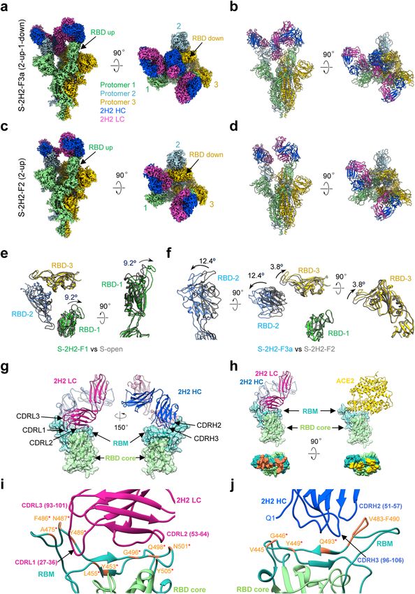
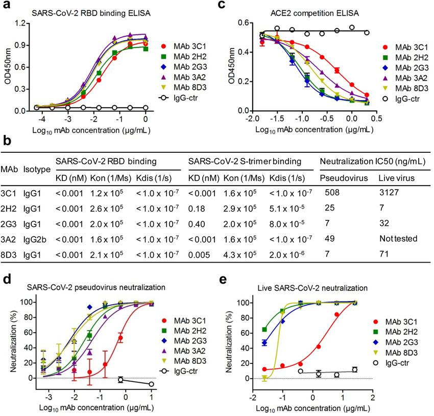
NATURE COMMUNICATIONS | https://doi.org/10.1038/s41467-020-20465-w                                                                          ARTICLE

Fig. 1 Binding properties, receptor-binding inhibitory activity, and neutralization activity of the MAbs. a Reactivities of anti-SARS-CoV-2 MAbs to the
SARS-CoV-2 RBD measured by ELISA. Data are mean ± SEM of triplicate wells. Zika virus (ZIKV)-specific MAb 5F8 served as IgG1 isotype control (IgG-
ctr) and was used as a control in all subsequent experiments. b Isotypes, binding affinities, and neutralization activity of the MAbs. Binding affinities of the
MAbs to immobilized SARS-CoV-2 RBD and S trimer were determined by bio-layer interferometry (BLI). c Competition between the MAbs and ACE2 for
binding to SARS-CoV-2 RBD was measured by ELISA. Biotinylated ACE2-hFc fusion protein was tested for the ability to bind to immobilized RBD in
presence of the MAbs, and the signal was detected using HRP-conjugated streptavidin. Data are mean ± SEM of triplicate wells. d The MAbs neutralized
SARS-CoV-2 pseudovirus infection in vitro. The purified MAbs were fourfold serially diluted and evaluated for neutralization of murine leukemia virus
(MLV) pseudotyped with SARS-CoV-2 spike protein. Luciferase activity was measured 2 days after infection. Results shown are representative of two
independent experiments. Data are expressed as mean ± SEM of five replicate wells. e The MAbs neutralized authentic SARS-CoV-2 infection in vitro.
Serially diluted purified MAbs were subjected to live SARS-CoV-2 virus neutralization assay. After 48 h culture, viral RNA in cells were detected by RT-
qPCR. Data are mean ± SEM of triplicate wells.

and to SARS-CoV-2 S trimer with KD values ranging from
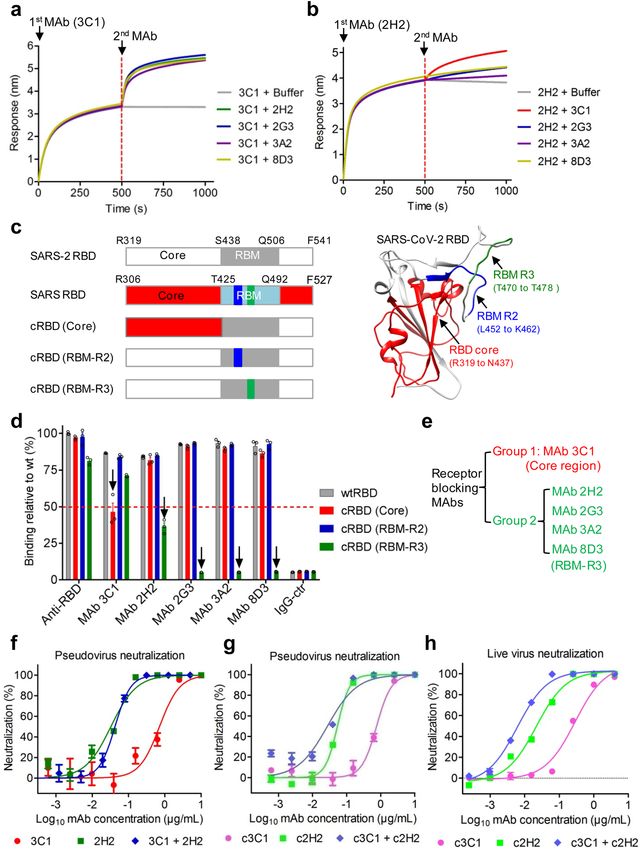
ARTICLE                                                              NATURE COMMUNICATIONS | https://doi.org/10.1038/s41467-020-20465-w

Selection and humanization of a noncompeting two-antibody                   four MAbs led to significant increases in BLI signals, suggesting
cocktail. Combined use of two or more antiviral MAbs targeting              that 3C1 targets an epitope distinct from the binding sites of the
distinct epitopes may increase therapeutic efficacy and reduce               other MAbs. In the second experiment, immobilized SARS-
the risk of acquiring drug resistance30. Therefore, we attempted            CoV-2 RBD was saturated with MAb 2H2, followed by incu-
to identify a noncompeting MAb pair from the five individual                 bation with a second MAb or dissociation in the buffer. The
neutralizing MAbs. BLI-based antibody competition assays were               result showed that 3C1 produced the highest additional BLI
performed. In the first experiment, immobilized SARS-CoV-2                   signal (Fig. 2b), implicating that 3C1 and 2H2 recognize distinct
RBD was saturated with 3C1 (first antibody) and then allowed to              epitopes on RBD; in contrast, incubation with 2G3, 3A2, or 8D3
interact with a second antibody or dissociate in the buffer. As             resulted in only a slight increase in BLI signals, suggesting that
shown in Fig. 2a, 3C1 (100 nM) hardly dissociated in the buffer,            the epitopes of these three antibodies may overlap with that of
showing high binding affinity; incubation with each of the other             the MAb 2H2.

4              NATURE COMMUNICATIONS | (2021)12:264 | https://doi.org/10.1038/s41467-020-20465-w | www.nature.com/naturecommunications
NATURE COMMUNICATIONS | https://doi.org/10.1038/s41467-020-20465-w                                                                       ARTICLE

Fig. 2 Antibody competition, epitope mapping, and generation of antibody cocktail. a, b Antibody binding competition assay. Antibody competition for
binding to SARS-CoV-2 RBD was measured by BLI. Immobilized RBD was first saturated with the first antibody MAb 3C1 (a) or MAb 2H2 (b), and then a
second MAb (MAb names were shown after the plus sign) or dissociation buffer (control) was added and allowed to react with the RBD. c Diagrams of
chimeric RBD mutants (cRBD). cRBD (core), the N-terminal residues R319 to N437 of core region in the SARS-CoV-2 RBD were mutated into the
corresponding part of SARS-CoV. cRBD (RBM-R2) and cRBD (RBM-R3), residues L452 to K462, and residues T470 to T478 of RBM region in the SARS-
CoV-2 RBD were separately substituted by the corresponding residues of SARS-CoV. The positions of the mutated amino acids are shown in the wild-type
RBD crystal structure (PDB: 6M0J; right panel). d Reactivities of the MAbs to wild-type (wt) and mutant SARS-CoV-2 RBD proteins measured by ELISA.
RBD-mFc immune sera (anti-RBD) served as positive control. The downward arrow indicates that substitutions in RBD mutants significantly reduced the
binding of the MAbs compared to wild-type RBD. The reactivity level of wild-type SARS-CoV-2 RBD and anti-RBD sera was set to 100%, and the red
dashed line represents 50% reduction relative to wild type. Data are mean ± SEM of triplicate wells. Each symbol represents one well. e Grouping of the
MAbs. Group 1, MAb16-3C1; group 2, the other MAbs. Antibody epitopes were shown in brackets. f Neutralization activity of the murine 2H2/3C1 cocktail.
2H2 alone, 3C1 alone, and the 2H2/3C1 (1:1) cocktail were serially diluted and evaluated for neutralization of SARS-CoV-2 pseudovirus. g Neutralization
activity of the chimeric MAb cocktail against SARS-CoV-2 pseudovirus. c2H2 alone, c3C1 alone, and the c2H2/c3C1 (1:1) cocktail were serially diluted and
assessed for neutralization of SARS-CoV-2 pseudovirus. For f and g luciferase activity was measured 2 days after infection. Data are expressed as mean ±
SEM of five replicate wells. h Neutralization activity of the chimeric MAb cocktail against authentic SARS-CoV-2. Serially diluted purified MAbs were
subjected to live SARS-CoV-2 virus neutralization assay. After 48 h culture, viral RNA in cells were detected by RT-qPCR. Data are mean ± SEM of triplicate
wells. For f–h, for MAb cocktails the concentration on the x-axis is that of the 2H2 or c2H2 antibody.

   To roughly map the antibody epitopes, we designed six                       efficiently bound to recombinant SARS-CoV-2 RBD with KD
chimeric RBD mutants by replacing a domain/fragment of SARS-                   values
ARTICLE NATURE COMMUNICATIONS | https://doi.org/10.1038/s41467-020-20465-w
FcγR-expressing cell lines was minimal. Moreover, treatment Fab, with two RBDs in the up conformation (protomer 1 and 2),
with serially diluted (ranging from 10 to 0.000128 μg/mL) 2H2 or and the third RBD remaining down but can still engage with a
3C1 antibody did not significantly affect pseudovirus entry of the 2H2 Fab (protomer 3, Fig. 4a, b). While in S-2H2-F3b structure,
two cell lines (Supplementary Fig. 8a, b). Similarly, the huma- all the three RBDs are up and each binds with a Fab (Supple-
nized antibodies c2H2 and c3C1 did not show any enhancing mentary Fig. 10d).
effects on SARS-CoV-2 pseduovirus entry regardless of the These four cryo-EM structures with increasing number of
antibody concentration (Supplementary Fig. 8a, b). Collectively, associated Fabs may represent the snapshots during conforma-
these results demonstrate that, in the assay system we tested, tional transitions of the S trimer to gradually coordinate the
MAbs 2H2 and 3C1 did not promote ADE. binding of more 2H2 Fabs. In S-2H2-F1 state, we observed a
further 9.2° outward tilt of the up RBD-1 induced by the first
associated 2H2 Fab (Fig. 4e). Surprisingly, our S-2H2-F3a
In vivo prophylactic and therapeutic efficacies of the neu-
structure suggested although RBD-3 is in the down configuration,
tralizing MAbs. To evaluate the protective efficacy of our MAbs,
it can still bind a 2H2 Fab with a slight 3.8° upward tilt of RBD-3
we developed in house a mouse model of authentic SARS-CoV-2
and a further 12.4° outward tilt of RBD-2 to coordinately
infection, in which wild-type Balb/c mice were intranasally
accommodate the binding of the third Fab (Fig. 4f). Collectively,
inoculated with hACE2-encoded adenovirus 5 (Ad5-hACE2) to
this ensemble of cryo-EM structures revealed the conformational
allow expression of the hACE2 receptor in the lung, followed by
space of the S trimer as a dynamic allosteric machinery to
intranasal infection with live SARS-CoV-2 3 days later. This
coordinate the binding of more 2H2 Fabs.
model permitted efficient SARS-CoV-2 infection and replication
Our structures show that 2H2 Fab is bound on the top of RBD
in the mouse lung; in contrast, only baseline levels of viral RNA
(Fig. 4a–d). Inspection of the better resolved S-2H2-F3a structure
were detected in the wild-type mice without Ad5-hACE2 inocu-
revealed that the CDRH2 and CDRH3, together with all the three
lation after live virus challenge. Consistently, hematoxylin and
light chain complementarity-determining regions (CDRs) of 2H2
eosin (H&E) staining assay showed that severe interstitial pneu-
form contacts with the RBD domain, particularly the RBM region
monia was observed in the Ad5-hACE2-treated mice, but not in
of the S protein (Fig. 4g and Supplementary Table 2), with the
the mice without Ad5-hACE2 treatment (Fig. 3a). The prophy-
buried 2H2–RBD interaction surface area ranging from ~1320 to
lactic efficacy of MAb 2H2 was examined by intraperitoneally
1369 Å2. The RBM also mediates the binding of S protein to
(i.p.) injecting 10 mg/kg (body weight) antibody into the Ad5-
human ACE2 (Fig. 4h), the receptor for both SARS-CoV-2 and
hACE2-treated mice 24 h before SARS-CoV-2 infection. Analysis
SARS-CoV1,14. Among these interactions with RBD, the light
of the viral load of the mouse lungs and histopathological exam-
chain of 2H2 Fab contributes more than the heavy chain does
ination at 3 days post infection (d.p.i.) showed that 2H2 pre-
(Fig. 4i, j). Specifically, CDRL2 (residues 53–64), which touches
treatment could almost completely neutralize SARS-CoV-2
the top “palm” of RBD, contributes the most to the interaction,
infection, reducing viral load by ~1600 fold as compared to the
possibly forming contacts with six residues (Y453, L455,
control (PBS) pretreatment (Fig. 3a). To assess the therapeutic
loop496–501, and Y505) in RBM. Both CDRL1 and CDRL3
efficacy, Ad5-hACE2-treated mice were i.p. injected with 20 mg/kg
contact the RBM loop486–489 located on the other edge of the
of murine 2H2 antibody or 40 mg/kg of the c2H2/c3C1 (1:1,
RBM, and CDRL1 also likely contacts A475 (Fig. 4i and
20 mg/kg each) cocktail at 4 h.p.i., and mouse lungs were collected
Supplementary Table 2). As for the heavy chain, CDRH2 and
at 3 d.p.i. for qRT-PCR and H&E analysis. As shown in Fig. 3b,
CDRH3 mainly contact the RBM loop483–490 (Fig. 4j and
injection of 2H2 resulted in significant decrease (by ~17.8-folds) in
Supplementary Table 2). In addition to the CDRs, Q1 from the
viral load in the mouse lung and less severe lung lesions as
heavy chain of 2H2 Fab presumably interacts with V445 and
compared to the control (PBS) treatment, indicating a strong
G446 in RBM (Fig. 4j).
therapeutic effect for 2H2; in addition, treatment with the c2H2/
Furthermore, the epitope of 2H2 Fab on RBD would mostly
c3C1 cocktail appeared to be more effective than 2H2 alone in
overlap with the binding sites of ACE2 on RBD (13 out of the 17
reducing viral loads in mouse lungs.
total ACE2–RBD binding sites, PDB ID: 6M0J)10, which could
We also assessed the therapeutic potential of single c2H2
lead to severe clash and spatial hindrance between ACE2 and 2H2
antibody and the c2H2/c3C1 cocktail administered at a delayed
Fab (Fig. 4h). Our structural data are in line with the observed
(24 h.p.i.) time point. Both c2H2 and the c2H2/c3C1 cocktail
high potency of 2H2 on blocking the interaction between RBD
treatments could significantly reduce viral loads as compared to
and ACE2 (Fig. 1c).
the control (PBS) treatment (Fig. 3b). Together, the above data
demonstrate that the c2H2/c3C1 cocktail has high therapeutic
efficacies in vivo.
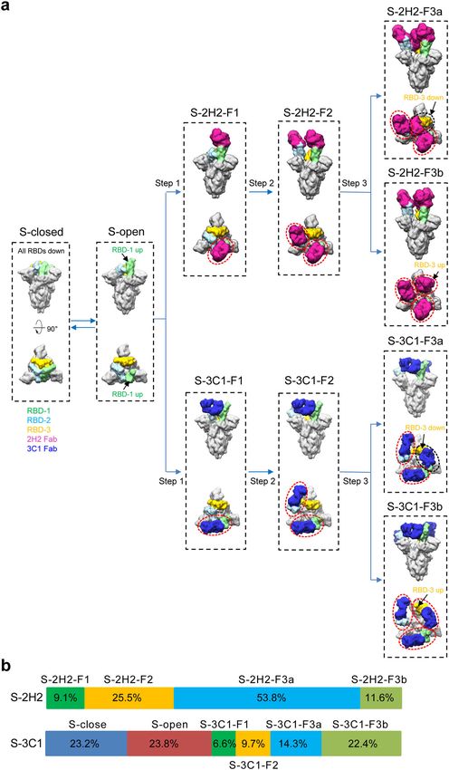
Structural snapshots of the S trimer in complex with 3C1 Fab.
To disclose the molecular basis of 3C1-mediated neutralization of
Structural snapshots of the S trimer in complex with 2H2 Fab. SARS-CoV-2, we determined four cryo-EM structures of SARS-
To investigate the molecular basis of 2H2-mediated neutralization CoV-2 S trimer in complex with the 3C1 Fab at distinct con-
of SARS-CoV-2, we resolved four cryo-EM structures of the formational states, including S-3C1-F1 (with one Fab), S-3C1-F2
stabilized SARS-CoV-2 trimeric S glycoprotein in complex with (with two Fabs), and S-3C1-F3a/S-3C1-F3b (with three Fabs;
2H2 Fab in distinct conformational states, termed S-2H2-F1 Fig. 5a–d and Supplementary Fig. 11). Among these structures, S-
(associated with one Fab), S-2H2-F2 (with two Fabs), and S-2H2- 3C1-F3b was better resolved to 4.3 Å resolution, and the other
F3a/S-2H2-F3b (with three Fabs; Fig. 4a–d, Supplementary Fig. 9, three were at 5.6–7.5 Å resolution range (Fig. 5a–d and Supple-
and Supplementary Fig. 10a–f). Among these structures, S-2H2- mentary Fig. 12a–g). In the S-3C1-F3b structure, all the three
F3a and S-2H2-F2 were better resolved to 3.8 and 4.3 Å resolu- RBDs are in the up conformation and each of them associates
tion, respectively (Fig. 4a–d and Supplementary Fig. 10e–f). In the with a 3C1 Fab (Fig. 5a). Still, the S-3C1-F3b structure appears
S-2H2-F1/S-2H2-F2 structures, there are one/two RBDs in the up asymmetric especially in the associated RBD-3C1 Fab region.
configuration with each bound with a 2H2 Fab, while the Indeed, all the three up RBDs in this structure exhibit an addi-
remaining RBDs are in the down conformation without Fab tional outward tilt relative to the up RBD in the open state S
binding (Fig. 4c, d and Supplementary Fig. 10c). Our S-2H2-F3a trimer likely induced by 3C1 binding, with RBD-1 outward tilt
structure reveals that each of the three RBDs binds with a 2H2 the most (50.8°, Fig. 5e). Due to the all up configuration of the
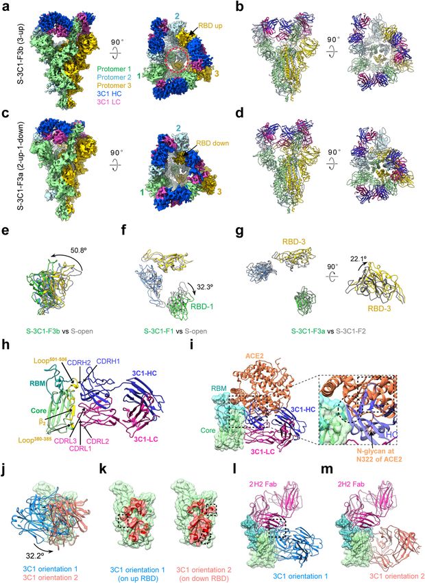
6 NATURE COMMUNICATIONS | (2021)12:264 | https://doi.org/10.1038/s41467-020-20465-w | www.nature.com/naturecommunicationsNATURE COMMUNICATIONS | https://doi.org/10.1038/s41467-020-20465-w ARTICLE Fig. 3 Protective efficacy of MAb 2H2 and the chimeric antibody cocktail against authentic SARS-CoV-2 infection in mice. a, b In vivo prophylactic efficacy (a) and therapeutic efficacy (b) of MAb 2H2, c2H2, and/or the c2H2/c3C1 cocktail against SARS-CoV-2 infection. Upper left panel: study outline. Upper right panel: qRT-PCR analysis of viral RNA copies present in lung tissues after 3 days of infection. Lower panel: H&E staining of lung tissue sections at 3 d.p.i. For a, qPCR results are shown as fold increase relative to wide-type Balb/c group (without Ad5-hACE2 treatment). For b, qPCR results are expressed as viral RNA levels in different antibody treatment groups relative to that in the PBS control group. For top right panels in a and b, each symbol represents one mouse. Error bars represent SEM. Statistical significance was determined by a two-tailed Student’s t test and indicated as follows: ns not significant; *p < 0.05; **p < 0.01. For a, p value between the control group and the PBS group (Ad-hACE2 transduction) is 0.0073; p value between the PBS group and the 2H2 group is 0.0169. For early treatment experiment in b, p value between the PBS group and the 2H2 group is 0.0488; p value between the PBS group and the c2H2/c3C1 group is 0.0418. For delayed treatment experiment in b, p value between the PBS group and the c2H2 group is 0.0183; p value between the PBS group and the c2H2/c3C1 group is 0.0205; p value between the c2H2 group and the c2H2/c3C1 group is 0.7803. NATURE COMMUNICATIONS | (2021)12:264 | https://doi.org/10.1038/s41467-020-20465-w | www.nature.com/naturecommunications 7
ARTICLE                                                              NATURE COMMUNICATIONS | https://doi.org/10.1038/s41467-020-20465-w

RBDs and their outward tilt, the originally covered S2 subunits             (RBD-1 and RBD-2) and each binds a 3C1 Fab, while the
are now exposed when visualized from the top (Fig. 5a, indicated            remaining RBD-3 in the down conformation can still bind a 3C1
with red circle), which might be beneficial for the release of               Fab (Fig. 5c). In addition, we also captured the S-3C1-F1/S-3C1-
S1 subunits and the subsequent transformations of the S trimer              F2 structures with one/two RBDs up, and each of the up RBDs
from the prefusion state to postfusion state. Furthermore, in the           binds with a 3C1 Fab, while the remaining down RBDs have no
S-3C1-F3a map, there are two RBDs in the up configuration                    associated Fab (Supplementary Fig. 12e, f).

8              NATURE COMMUNICATIONS | (2021)12:264 | https://doi.org/10.1038/s41467-020-20465-w | www.nature.com/naturecommunications
NATURE COMMUNICATIONS | https://doi.org/10.1038/s41467-020-20465-w                                                                       ARTICLE

Fig. 4 Cryo-EM structures of the SARS-CoV-2 S trimer in complex with 2H2 Fab. a, b Side and top views of the S-2H2-F3a cryo-EM map (a) and pseudo
atomic model (b). RBD-1 and RBD-2 are in up configuration, while RBD-3 is down, with each of the RBDs bound with a 2H2 Fab. Protomer 1, 2, and 3 are
shown in light green, powder blue, and gold, respectively. This color scheme is followed throughout. Heavy chain and light chain of 2H2 Fab in royal blue
and violet red, respectively. c, d Side and top views of the S-2H2-F2 cryo-EM map (c) and pseudo atomic model (d), with two up RBDs (RBD-1 and RBD-2)
each bound with a 2H2 Fab. e, f 2H2 Fab-induced conformational changes of the S trimer. Shown is the structural comparation of RBDs between S-2H2-F1
(in color) and S-open (dim gray) (e), and between S-2H2-F3a (in color) and S-2H2-F2 (dim gray) (f). g 2H2 Fab mainly binds to the RBM (light sea green
surface) of RBD, with major involved structural elements labeled. RBD core is rendered as light green surface. h 2H2 Fab (left) and ACE2 (right, gold, PDB:
6M0J) share overlapping epitopes on RBM (second row) and would clash upon binding to the S trimer. i, j The involved regions/residues forming potential
contacts between the light chain (in violent red, i) or heavy chain (in royal blue, j) of 2H2 and the RBD-1 of S-2H2-F3a. Asterisks highlight residues also
involved in the interactions with ACE2. Note that considering the local resolution limitation in the RBD-2H2 portion of the map due to intrinsic dynamic
nature in these regions, we analyzed the potential interactions that fulfill criteria of both < 4 Å side chain distance cutoff and
ARTICLE                                                               NATURE COMMUNICATIONS | https://doi.org/10.1038/s41467-020-20465-w

MAbs targeting the RBD of either MERS-CoV or SARS-CoV could                  IgG1 Fc could enhance SARS2-CoV-2 pseudovirus entry of FcR-
enhance pseudovirus entry into FcR-expressing cell lines, including          expressing K562 or THP-1 cells regardless of the antibody con-
THP-1 (ref. 41). The ADE assay described in that study was                   centration (Supplementary Fig. 8). We should mention that the
modified and then used in the present study to evaluate the ADE               same cell lines have been shown to support anti-DENV-E antibody-
potential of our anti-SARS-CoV-2 MAbs. We found that neither                 triggered ADE of DENV infection in previous studies38–40. Our
the murine antibodies nor the chimeric antibodies with human                 data appear contradictory to the results from previous studies on

10              NATURE COMMUNICATIONS | (2021)12:264 | https://doi.org/10.1038/s41467-020-20465-w | www.nature.com/naturecommunications
NATURE COMMUNICATIONS | https://doi.org/10.1038/s41467-020-20465-w                                                                           ARTICLE

Fig. 5 Cryo-EM structures of the SARS-CoV-2 S trimer in complex with the 3C1 Fab. a, b S-3C1-F3b cryo-EM map (a) and pseudo atomic model (b).
All the three RBDs are up and each of them binds with a 3C1 Fab. The heavy chain of the 3C1 Fab in medium blue and light chain in violet red. c, d S-3C1-F3a
cryo-EM map (c) and pseudo atomic model (d). There are two up RBDs and one down RBD, with each bound with a 3C1 Fab. e Structural alignment of the
three up RBDs of S-3C1-F3b (in color) and the only up RBD from S-open (gray), suggesting 3C1 induced outward tilt of the RBDs within the S trimer.
f, g Conformational comparation between S-3C1-F1 and S-open (f), as well as between S-3C1-F3a and S-3C1-F2 (g). h RBD/3C1 interaction interface (take
RBD-3/3C1 of S-3C1-F3b as an example), with major involved structural elements labeled. i ACE2 (coral, PDB: 6M0J) would clash with the heavy chain of
3C1 Fab (blue). They share overlapping epitopes on the RBM (dotted black circle); additionally, the framework of 3C1-VH would clash with ACE2 (dotted
black frame), which could be enhanced by the presence of an N-linked glycan at site N322 of ACE2. j 3C1 showed two distinct orientations to bind RBD
within S trimer, i.e., adopting orientation 1 to associate with up RBD while orientation 2 with down RBD. k Contact footprint variations of 3C1 on up RBD
(left) compared with that on down RBD (right), with unique epitopes indicated by dotted black frame. l–m Potential simultaneous binding of RBD by 2H2
and 3C1 cocktail. In 3C1 orientation 1, 3C1 and 2H2 could have minor clash (indicated by black frame, l); while in origination 2, there is no clash between 3C1
and 2H2 Fabs (m).

the anti-MERS-CoV or anti-SARS-CoV MAbs (Wan et al.41).                          and ACE2, destabilization of the virion is possibly another neu-
Although the mechanism underlying such a contradiction remains                   tralization mechanism for our MAbs.
to be elucidated, we speculate that SARS-CoV-2 may require an                        In this study, we captured four distinct structural states for
unidentified host factor for MAb-bound pseduovirus or virion to                   each of the S protein/Fab complex (Figs. 4–6), allowing us to
enter FcR-bearing cells, and such a host factor is lacking in the K562           glimpse the main features of the dynamic process of conforma-
and THP-1 cells used in the present study, whereas MERS-CoV and                  tional transitions induced by Fab binding, i.e., from binding with
SARS-CoV may not need the assistance of the same host factor for                 one Fab to with three Fabs making all the RBDs occupied. We
entry of FcR-expressing cells. It is also possible that MAb-induced              should emphasize that the Fab-induced conformational changes
drastic conformational changes of SARS-CoV-2 S proteins (as                      in S trimer cannot be observed in the RBD/Fab crystal structures,
shown in Fig. 6) may cause premature shedding of S1 and exposure                 highlighting the advantage of cryo-EM in capturing dynamic
of the FP before the virion reach the cell surface, thereby abolishing           conformational shifts in macromolecular complex-Fab recogni-
viral infectivity. Nonetheless, our study demonstrated that MAbs                 tion. It has been hypothesized that for SARS or MERS, the up
2H2 and 3C1 do not promote ADE in vitro at least not in the assay                conformation (active state) of the RBD is required for the binding
system we used. In addition, our mouse challenge experiments                     of neutralizing MAbs directed at the receptor-binding site43,44.
showed that the 2H2 antibody or the c2H2/c3C1 cocktail were able                 However, here for both Fabs, we observed a state (S-2H2-F3a or
to neutralize, but not enhance SARS-CoV-2 infection in vivo                      S-3C1-F3a) with RBD-3 in the down conformation, but can still
(Fig. 3). Together, these data demonstrate proof-of-concept for the              associate with a Fab. This suggests that for the antibody targeting
application of our MAbs as a safe and effective treatment option                 RBD domain, if only the epitope is exposed and there is enough
against SARS-CoV-2 infection. We should point out that c2H2 and                  space to accommodate the Fab, the RBD can be “grasped” by the
c3C1 are mouse–human chimeras, and therefore for future human                    Fab regardless it is in the up or down conformation.
use the antibodies will need to be further humanized by grafting                     Altogether, we propose a model of stepwise binding of 2H2/
their CDRs into a suitable human MAb backbone.                                   3C1 Fabs to the RBD domain of the SARS-CoV-2 S trimer
   We have recently showed that in the ligand-free condition,                    (Fig. 6). Take 2H2 as an example, in step 1, the only up RBD in
SARS-CoV-2 S trimer dominantly adopts a stable tightly closed                    the S-open state binds one 2H2 Fab first, leading to the S-2H2-F1
ground prefusion conformation (no RBD up), with only a minor                     state. Our recent study suggested that in the S-open state, the
population of the particles in the open conformation with one                    RBD-2 adjacent to the up RBD-1 has encoded intrinsic dynam-
RBD up14. Here, our structural data showed that binding of 3C1                   ics14. Thus, once RBD-1 bound with a Fab, the resulting outward
or 2H2 MAbs could trigger dramatic conformational transitions                    tilt of RBD-1 (Figs. 4e and 5f) could break the allosteric con-
of the S trimer (especially in the S1 subunit region, Supplemen-                 strains originally imposed on RBD-2, leading to an up config-
tary Fig. 12h) from the tightly closed ground prefusion state to                 uration of RBD-2. This is in line with a recent study of S trimer
the unstable, loosely packed open state with two or three RBDs up                on intact SARS-CoV-2 virion, showing that there is a minor
and released FP. In another word, the prefusion S protein is                     population of the S trimer with the RBD-2 also in the up con-
destabilized to some extent by the 2H2/3C1 Fabs. Note that the                   formation45. In step 2, the transiently up RBD-2 can be quickly
SARS-CoV-2 S protein used in this study was stabilized (tenta-                   trapped by 2H2 Fab, resulting in the S-2H2-F2 form with each of
tively in the prefusion conformation) by furin cleavage site                     the two up RBDs bound with a Fab. Consequently, the original
mutation and two consecutive proline mutations13,14. Therefore,                  steric hindrance could be released to allow RBD-3 to expose its
it is very likely that our Fabs could induce even further con-                   buried epitope and also leave enough space to accommodate the
formational changes toward the postfusion state in the wild-type                 binding of the third 2H2 Fab. Indeed, we observed a further 12.4°
S protein. Upon ACE2 receptor binding, SARS-CoV S protein                        outward tilt of RBD-2, releasing the space for the third Fab
undergoes similar conformational changes with more than one                      (Fig. 4f). Interestingly, in step 3, our data suggested that there are
RBD up and to transit to the postfusion state42. As for SARS-                    two possible reaction pathways. In pathway one, the down RBD-3
CoV-2, we recently also observed ACE2 receptor-triggered tran-                   with exposed epitope can now bind a Fab, forming S-2H2-F3a
sitions of S trimer toward fusion-prone or postfusion states14; and              with each of the RBDs (two up and one down) bound with a Fab.
combined with our current results, it appears the untwisting of S1               In pathway two, after the RBD-1 and RBD-2 are all up releasing
induced by ACE2 receptor/MAb binding could release the ori-                      the original allosteric constrains, RBD-3 has more chance to be
ginally packed FP and induce an early exposure of FP. Therefore,                 transiently up, which could be encoded in the S trimer con-
MAb-induced transition of S protein from prefusion to fusion or                  formational space or triggered by certain external factors. This up
postfusion states, accompanied by premature release of S1, and                   RBD-3 can then be trapped by a Fab and retained in the up
exposure of the cleavage site and FP, may disrupt the integrity of               conformation, forming S-2H2-F3b with each of the three up
the virion and render the virus defective. Hence, our study sug-                 RBDs bound with a Fab. The two pathways in step 3 could take
gests that, besides blockade of the interaction between the virus                place simultaneously.

NATURE COMMUNICATIONS | (2021)12:264 | https://doi.org/10.1038/s41467-020-20465-w | www.nature.com/naturecommunications                                     11
ARTICLE                                                                  NATURE COMMUNICATIONS | https://doi.org/10.1038/s41467-020-20465-w

Fig. 6 A proposed model of stepwise binding of 2H2/3C1 Fabs to the RBD of SARS-CoV-2 S trimer. a 2H2 and 3C1 Fabs appear to follow similar pathway
to induce generally comparable conformational transitions of the S trimer to neutralize the virus. RBD-1, RBD-2, and RBD-3 are colored in light green, light
blue, and gold, respectively; 2H2 and 3C1 Fab in violent red and medium blue, respectively. Red ellipsoid and black ellipsoid indicate Fab bound to up RBD
and down RBD, respectively. The maps of S-2H2 and S-3C1 complexes shown here were generated by lowpass filtering of the corresponding models to
10 Å resolution. b Population distribution for the S-2H2 and S-3C1 dataset.

   Strikingly, our structural data further suggest that for the 3C1            adopted by other MAbs targeting similar regions of RBD. Thus,
Fab, although its epitope mostly locates on the side of RBD,                   our structural study reveals that binding of neutralizing MAbs to
distinct from that of 2H2, it appears to follow similar pathway to             SARS-CoV-2 S trimer is a well-coordinated dynamic process
induce generally comparable conformational transitions of the S                involving stepwise allosteric conformational changes of the S
trimer. We therefore postulate that this procedure might also be               trimer, and also sheds light on the structural basis for MAbs 2H2

12                NATURE COMMUNICATIONS | (2021)12:264 | https://doi.org/10.1038/s41467-020-20465-w | www.nature.com/naturecommunications
NATURE COMMUNICATIONS | https://doi.org/10.1038/s41467-020-20465-w                                                                                     ARTICLE

and 3C1 as noncompeting antibody cocktail. These structural                           conjugated anti-mouse IgG (Sigma; diluted 1:10,000 in 1% milk/PBST) was added
information enhances our understanding of anti-SARS-CoV-2                             and incubated at 37 °C for 1 h. After washes and color development, absorbance
                                                                                      was monitored at 450 nm.
antibody-mediated neutralization and protection.                                          For receptor competition assay, microplates (Nunc) were coated at 4 °C
                                                                                      overnight with 40 ng/well of HEK 293F-expressed SARS-CoV-2 RBD and then
Methods                                                                               blocked with 5% milk/PBST. After washing with PBST, 25 μL of hybridoma culture
Cells and viruses. SP2/0 mouse myeloma cells were grown in RPMI 1640 medium           supernatants or serially diluted purified MAbs were mixed with 25 μL (20 ng) of
(Gibco, Thermo Fisher, USA) supplemented with 10% fetal bovine serum (FBS;            unlabeled or biotinylated ACE2-hFc, and the mixtures were added to the wells and
Gibco) at 37 °C. African green monkey kidney VeroE6 cells were cultured in            incubated at 37 °C for 2 h. The corresponding secondary antibodies, HRP-
DMEM (Gibco, USA) supplemented with 10% FBS. HEK 293 F suspension cells               conjugated anti-human IgG (Abcam, USA; diluted 1:8,000 in 1% milk/PBST) or
(Thermo Fisher) were grown in FreeStyle 293 expression medium (Gibco). Expi-          HRP-conjugated streptavidin (Life Technologies, USA), were added and incubated
CHO-S™ cells (Thermo Fisher) were grown in ExpiCHO expression medium                  at 37 °C for 1 h. After washes and color development, absorbance was monitored at
(Gibco). SARS-CoV-2 clinical isolate nCoV-SH01 (GenBank: MT121215.1)46 was            450 nm.
expanded in VeroE6 cells and virus titers were expressed as plaque forming units          To determine the isotypes of the MAbs, hybridoma culture supernatants were
(PFU) per mL. All the infection experiments were performed in the biosafety level-    tested by sandwich ELISA using the SBA Clonotyping system/HRP kit (Southern
3 (BSL-3) laboratory of Fudan University.                                             Biotech, USA) according to manufacturer’s instructions.

Recombinant proteins and antibodies. For mouse immunization, recombinant              Pseudovirus neutralization assay. Murine leukemia virus (MLV)-based SARS-
SARS-CoV-2 RBD (residues R319 to F541) fused with a C-terminal mouse IgG1 Fc          CoV-2 S pseudoviruses were prepared as follows: HEK 293 T cells grown in 10-cm
tag (RBD-mFc) was purchased from Sino Biological Inc (Beijing, China). For            dish were cotransfected using PEI (polysciences) with 10 μg of MLV Gag-Pol
antibody screening and characterization, several recombinant proteins were pro-       packaging plasmid, 10 μg of transfer plasmid containing a luciferase or EGFP
duced in our laboratory. Specially, to prepare SARS-CoV-2 RBD, RBD DNA                reporter gene, and 2 μg of plasmids encoding either wild-type or mutant (D614G) S
fragment corresponding to residues V320 to G550 derived from SARS-CoV-2               proteins. The cells were incubated with the transfection mixture for 4 h. After
strain Wuhan-Hu-1 (GenBank ID: MN908947.3) was codon optimized and cloned             washing once with DMEM, fresh DMEM medium supplemented with 10% FBS
into a modified pcDNA3.4 vector that contains interleukin-10 (IL-10) signal            was added and incubated at 37 °C for 48 h. The culture supernatant was harvested,
sequence and a C-terminal His tag, yielding plasmid pcDNA3.4-SARS-2 RBD. To           filtered through 0.45 µm filters and either used immediately or frozen at −80 °C.
express SARS-CoV RBD, RBD gene fragment corresponding to rsidues R306 to                  For pseudovirus neutralization assay, VeroE6 cells or HEK 293 T cells stably
I520 derived from SARS-CoV strain Tor2 (GenBank ID: AAP41037.1) was codon             overexpressing human ACE2 receptor were plated into 96-well or 48-well plates
optimized and cloned into the expression secretion vector pSecTag2A (Invitrogen,      and grown overnight. A total of 90 μL of the pseudovirus was mixed with 45 μL of
USA), yielding plasmid pSecTag2A-SARS-RBD. To generate ACE2, DNA fragment             antibody samples (hybridoma culture supernatants or serially diluted purified
encoding the extracellular domain of human ACE2 (residues Q18 to S740) was            MAbs), and the mixtures were incubated at 37 °C for 1 h, and then added to the
cloned into a modified pcDNA3.4 vector that contains IL-10 signal sequence and         plates. After 2 h, the pseudovirus/antibody mixtures were removed and the cells
C-terminal human IgG1 Fc and His tag, yielding plasmid pcDNA3.4-ACE2-hFc.             were washed once with DMEM, followed by the addition of fresh culture medium.
To prepare SARS-CoV-2 S protein, mammalian codon-optimized gene coding S              At 48 h after infection, luciferase activity was measured using the luciferase assay
ectodomain (residues 1–1208) with proline substitutions at residues 986 and 987, a    system (Promega), or GFP expression resulting from pseudovirus infection was
“GSAS” substitution at the furin cleavage site (residues 682–685) was cloned into     analyzed by flow cytometry using a FACSCelesta flow cytometer (BD
vector pcDNA3.1+. A C-terminal T4 fibritin trimerization motif, a TEV protease         Biosciences, USA).
cleavage site, a FLAG tag and a His tag were cloned downstream of the SARS-CoV-
2 S glycoprotein ectodomain. Primer information is listed in Supplementary
Table 4. The above four plasmids were separately transfected into HEK 293 F           Determination and analysis of MAb sequences. For antibody sequencing, total
suspension cells using polyethylenimine (PEI; PolySciences, USA). The super-          RNA was extracted from hybridoma cells and first strand cDNA is prepared using
natants were harvested after 4–5 days of culture and His-tagged proteins were         M-MLV reverse transcriptase (Promega) and MAb isotype-specific primers. DNA
purified using Ni-NTA resin (Millipore, USA) according to manufacturer’s pro-          fragments encoding antibody variable regions were amplified individually from
tocol. To prepare biotinylated proteins, purified ACE2-hFc fusion protein or SARS-     cDNA using mouse Ig-primer set (Novagen, Merck, Germany) and Premix Ex Taq
CoV-2 S trimer protein were dialyzed against PBS and then labeled with EZ-Link™       reagent (Takara, Japan), followed by DNA sequencing.
Sulfo-NHS-LC-LC-Biotin (Thermo Fisher) followed by purification using Zeba™                The closest mouse immunoglobulin V, D, and J germline genes and positions of
spin desalting column (Thermo Fisher), according to manufacturer’s instructions.      CDRs were identified using IgBLAST32.
    MAb 5F8 is an IgG1 antibody against E protein of zika virus47, serving as
isotype control.
                                                                                      Bio-layer interferometry assay. To measure binding affinities of the MAbs, BLI
                                                                                      experiments were performed using an Octet Red96 instrument (Pall FortéBio,
Preparation of MAbs. The animal studies were approved by the Institutional            USA) following manufacturer’s instructions. Briefly, in one experiment, His-tagged
Animal Care and Use Committee at the Institut Pasteur of Shanghai. The mice           RBD proteins of SARS-CoV-2 or SARS-CoV were immobilized to Ni-NTA bio-
were kept in the SPF (specific pathogen free) animal facility with controlled tem-     sensors (Pall FortéBio) until saturation. In another experiment, biotinylated SARS-
perature (20–26 °C), humidity (40–70%), and lighting conditions (12 h light/12 h      CoV-2 S trimer protein was immobilized to streptavidin (SA) biosensors (Pall
dark cycle).                                                                          FortéBio) until saturation. For both experiments, the antigen-immobilized bio-
    To generate MAbs, female BALB/c mice aged 6–8 weeks were each primed with         sensors were transferred to wells containing MAb samples at varying concentra-
100 μg of RBD-mFc protein (Sino Biological) formulated with 0.5 mg of aluminum        tions for a 500-s association step. The sensors were then transferred to dissociation
hydroxide adjuvant (Invivogen, USA) and 25 μg of CpG oligonucleotides (Sangon         buffer (0.01 M PBS supplemented with 0.02% Tween 20 and 0.1% bovine serum
Biotech, China) via the i.p. route on day 0. The mice were boosted via the            albumin) for a 500-s dissociation step.
subcutaneous route on day 8 with RBD-mFc (50 μg/mouse) emulsified with                     For antibody competition assay, the antigen-immobilized biosensors were then
Freund’s complete adjuvant (Sigma), and on day 13 with RBD-mFc (50 μg/mouse)          dipped into the wells containing 15 μg/mL (100 nM) of the first MAb for a 500-s
emulsified with Titermax adjuvant (Sigma). On day 22, one mouse was injected           association period. The sensors were then transferred to wells containing
with 75 μg of HEK 293F-expressed RBD protein in PBS in a tail vein. On day 26,        dissociation buffer or 15 μg/mL of the second MAb samples and incubated for 500
splenocytes were isolated and fused with SP2/0 cells using polyethylene glycol 1450   s. For all BLI assays, data analysis was performed using Octet data analysis software
(Sigma). Fused cells were then selected in a hypoxanthine, aminopterine, and          version 11.0 (Pall FortéBio).
thymidine (HAT; Sigma) medium. Eight days later, hybridoma supernatants were
screened for their ability to bind to RBD protein and to block the ACE2-hFc/SARS-
CoV-2 RBD binding by ELISA, as described below. ELISA-positive hybridoma cells        Authentic virus neutralization assay. A total of 200 PFU (50 µL) of live SARS-
were cloned by limiting dilution method and the resulting monoclonal cell lines       CoV-2 virus (nCoV-SH01 strain) was mixed with 50 µL of fourfold serially diluted
were expanded. Purified MAbs were prepared from ascitic fluids using HiTrap™            purified MAbs and incubated at 37 °C for 1 h. The mixtures were then added to
Protein G HP column (GE Healthcare, USA).                                             confluent VeroE6 cells grown in 96-well plates. After 48 h of incubation at 37 °C,
                                                                                      culture supernatants were harvested for viral RNA isolation and cells were fixed for
                                                                                      immunofluorescence analysis.
ELISA. To determine binding properties of the antibodies, ELISA plates (Nunc,             RNA was extracted from culture supernatants using TRIzol reagent (Invitrogen,
USA) were coated with 100 ng/well of HEK 293F-expressed SARS-CoV-2 RBD or             USA). Reverse transcription quantitative PCR (RT-qPCR) was carried out in an
SARS-CoV RBD (purchased from Kactus Biosystems (Shanghai, China) or pro-              MXP3000 thermal cycler (Stratagene, USA) using Verso SYBR Green 1-Step
duced in the laboratory) at 4 °C overnight. The plates were then blocked with 5%      qRT-PCR Kit Plus ROX Vial (Thermo Fisher) according to the manufacturer’s
milk in PBS-Tween 20 (PBST). After washing with PBST, 50 μL of hybridoma              protocol. The primers that target SARS-CoV-2 N gene spanning nt 608–706 are as
culture supernatants or serially diluted purified MAbs were added to the wells and     follows: forward primer, 5′-GGGGAACTTCTCCTGCTAGAAT-3′; reverse primer,
incubated at 37 °C for 2 h. After washing, horseradish peroxidase (HRP)-              5′-CAGACATTTTGCTCTCAAGCTG-3′.

NATURE COMMUNICATIONS | (2021)12:264 | https://doi.org/10.1038/s41467-020-20465-w | www.nature.com/naturecommunications                                                 13
ARTICLE NATURE COMMUNICATIONS | https://doi.org/10.1038/s41467-020-20465-w
For immunofluorescence assays, cells were fixed in 4% paraformaldehyde SuperReal PreMix Plus SYBR Green kit (Tiangen) and the SARS-CoV-2 N gene-
solution, and permeabilized with 0.2% Triton X-100 (Thermo Fisher). Next, the specific primers, as described above.
cells were incubated overnight at 4 °C with a mouse polyclonal antibody against N Mouse lungs were fixed in 4% paraformaldehyde solution. Tissue paraffin
protein prepared in house, followed by incubation with Alexa Fluor 488-labeled sections (2–4 μm in thickness) were stained with H&E. The slices were observed
donkey anti-mouse IgG secondary antibody (1:1000, Thermo Fisher) at 37 °C for with Olympus microscope.
1 h. Cell nuclei were stained with DAPI (Thermo Fisher). Finally, the images were
recorded by fluorescence microscopy (Thermo Fisher).
SARS-CoV-2 S/Fab complex formation. Purified 2H2/3C1 IgG was dialyzed
against PBS (HyClone, USA), and then incubated with papain (300:1 W/W) in the
Mapping of MAb epitopes with RBD mutants by ELISA. For antibody epitope presence of 20 mM L-cysteine and 1 mM EDTA for 3 h at 37 °C. The reaction was
mapping, a series of SARS-CoV-2 RBD mutants were constructed. In all RBD quenched by adding 20 mM iodoacetamide. Fab was purified by performing ion-
mutants, residues from the core or RBM regions of SARS-CoV-2 RBD were exchange chromatography using a HiTrap DEAE FF column (GE Healthcare).
replaced with the corresponding residues of SARS-CoV to make the chimeric Purified SARS-CoV-2 S was incubated in a 1:4 molar ratio with 2H2 or 3C1 Fab
proteins (Supplementary Fig. 6). Specially, for mutant RBD (Core), residues R319 on ice for 2 h before being subjected to purification by size-exclusion
to N437 in the core region were mutated; for mutant RBD (RBM-R1), residues chromatography, using a Superose 6 increase 10/300 GL column (GE Healthcare)
S438 to G446 in the RBM region were substituted; for mutant RBD (RBM-R2), in 20 mM Tris-HCl pH 7.5, 200 mM NaCl, and 4% glycerol. The complex peak
residues L452 to K462 in the RBM region were replaced; for mutant RBD (RBM- fractions were concentrated and assessed by SDS–PAGE and negative-stain
R3), residues T470 to T478 in the RBM region were substituted; for mutant RBD electron microscopy (NS-EM).
(RBM-R4), residues N481 to F486 in the RBM region were mutated; for mutant
RBD (RBM-R5), residues F490 to V503 in the RBM region were replaced. All
Negative-stain sample preparation, data collection, and initial model building.
mutant plasmids were constructed based on the plasmid pcDNA3.4-SARS-2 RBD
For the NS sample, a volume of 5 μL of the S-2H2 sample was placed on a glow-
by using the Mut ExpressTM II Fast Mutagenesis Kit V2 (Vazyme, China)
discharged copper grid for 30 s. Excess sample on the grid was blotted off using
according to the manufacturer’s protocol. The resulting mutated plasmids were
filter paper, and a volume of 5 μL of 0.75% UF (Sigma-Aldrich) was added to wash
separately transfected into HEK 293 F cells using PEI. After 5 days of culture, His-
the grid. After blotting, another volume of 5 μL of 0.75% UF was placed on the grid
tagged proteins were purified from the culture supernatants using Ni-NTA resin
again for one minute to stain.
(Millipore).
The S-2H2 sample was imaged on a Tecnai G2 Spirit 120 kV transmission
The RBD mutants were tested for reactivity with the MAbs by ELISA. Briefly,
electron microscope (Thermo Fisher Scientific) using an Eagle camera at a nominal
microplates were coated at 4 °C overnight with 100 ng/well of individual RBD
magnification of 67,000× (yielding a pixel size of 1.74 Å ). A total of 9884 particles
mutant in PBS. After blocking, the plates were incubated with the mAbs (50 ng/well)
at 37 °C for 2 h, followed by incubation with HRP-conjugated anti-mouse IgG were picked using EMAN2 (ref. 49). All particles were extracted and subjected to
reference-free 2D classification in Relion 3.1 (ref. 50). Then good classes including
(Sigma; diluted 1:10,000 in 1% milk/PBST). After color development, absorbance at
9305 particles were used to generate an initial model in Relion 3.1. For S-3C1
450 nm was determined.
complex, the same procedure was adopted to generate an initial model from 94,606
cleaned up particles.
Generation and characterization of chimeric MAbs. To prepare chimeric MAbs,
DNA fragments encoding variable regions of murine MAbs were cloned into
Cryo-EM sample preparation and data collection for the S-2H2 and S-3C1
modified pcDNA3.4 vectors that contain IL-10 signal sequence and the constant
regions of human IgG1 or kappa chains by using ClonExpress II One Step Cloning complexes. An aliquot (~2.2 μL) of the S-2H2 sample was applied on a glow-
Kit (Vazyme, China). Heavy chain and light chain expression plasmids were discharged holey carbon grid (R1.2/1.3, 200 mesh; Quantifoil) or a graphene oxide-
transiently cotransfected in ExpiCHO-S™ cells (Thermo Fisher) by using the lacey carbon grid (300 mesh, EMR company). The grid was blotted with Vitrobot
ExpiFectamine CHO transfection kit (Gibco). The supernatants were harvested Mark IV (Thermo Fisher Scientific) and then plunged into liquid ethane cooled by
after 14 days of culture and the MAbs were purified using protein G agarose resin liquid nitrogen. To handle the potential preferred orientation problem, for sample
4FF (Yeasen, China), according to manufacturer’s protocol. frozen using holey carbon grid, 0.05% octyl β-D-glucopyranoside (Sigma) or 0.1%
To characterize chimeric MAbs, the recombinant chimeric MAbs were polylysine (Polysciences) was added into the sample or applied on grid before
subjected to BLI and pseudovirus neutralization assays as described above. freezing, respectively. The above-mentioned procedure was also followed to pre-
pare the cryo-EM grids for the S-3C1 complex.
Movies for the cryo-EM samples were collected on a Titan Krios electron
ADE assay. A total of 150 μL of the SARS-CoV-2 pseudovirus was mixed with microscope (Thermo Fisher Scientific) operated at an accelerating voltage of
50 μL of fivefold serially diluted antibody samples, and the mixtures were incubated 300 kV with a nominal magnification of 22,500× (Supplementary Table 1). The
at 37 °C for 2 h. Next, 30,000 THP-1 or K562 cells were plated into 48-well plates, movies were recorded on a K2 Summit direct electron detector (Gatan) operated in
followed by addition of the pseudovirus/antibody mixtures. Three days after the super-resolution mode (yielding a pixel size of 1.02 Å after two times binning)
infection at 37 °C, the cells were transferred to 1.5-mL Eppendorf tubes and and in an automatic manner using SerialEM51. Each frame was exposed for 0.15 s at the
washed once with PBS. Luciferase activity was measured using the luciferase assay dose rate of 8 e−/Å 2·s and the total accumulation time was 6.45 s, leading to a total
system (Promega). accumulated dose of 49.6 e−/Å 2 on the specimen (Supplementary Table 1).
In vivo protection assays. To generate recombinant adenovirus 5 expressing Cryo-EM 3D reconstruction. For both datasets, the motion correction of each
human ACE2 (Ad5-hACE2), hACE2 gene fragment was cloned into the shuttle image stack was performed using the embedded module of Motioncor2 (ref. 52) in
vector pShuttle-CMV48, resulting in plasmid pShuttle-CMV-hACE2. This plasmid Relion 3.1 (ref. 50) and CTFFIND4 was used to determine CTF parameters before
was linearized by PmeI digestion and then used to transform BJ5183-AD-1 cells further data processing53. For the S-2H2 dataset, unless otherwise described, the
(Weidi, China), resulting in plasmid pAd5-hACE2. Next, the pAd5-hACE2 plas- data processing was performed in Relion 3.1 (ref. 50). After automatic particle
mid was linearized with PacI and transfected in HEK 293 cells, to rescue adeno- picking, manual selection, and multiple rounds of reference-free 2D classification,
virus Ad5-hACE2. Ad5-hACE2 was amplified on HEK 293 cells and purified by cleaned up particles remained for further reconstruction with the NS-EM map as
CsCl gradient centrifugation. Adenovirus titer was determined using OD260 assay initial model (Supplementary Fig. 9). After multiple rounds of 2D and 3D classi-
and the titer (virus particles [VP]/mL) can be calculated by multiplying the OD260 fications, we obtained a S-2H2-F3a map from 37,641 particles and a S-2H2-F2 map
reading by 1.1 × 10¹². from 17,819 particles. After CTF refinement and Bayesian polishing, the S-2H2-F3a
Wild-type male Balb/c mice (6–8 week) were raised in pathogen-free isolation and the S-2H2-F2 maps were refined to 3.8 and 4.3 Å resolution, respectively. The
cages in the BSL-3 laboratory of Fudan University and received humane care in overall resolution was determined based on the gold-standard criterion using an
compliance with the guidelines of the Animal Research Ethics Board of Fudan FSC of 0.143. Moreover, the 3D classification also yielded a S-2H2-F1 map from
University. Mice were transduced intranasally with 5 × 10¹⁰ VP of Ad5-hACE2 at 6382 particles and a S-2H2-F3b map from 8083 particles, which were further
−3 d.p.i. To assess the prophylactic efficacy of MAbs, groups of Ad5-hACE2- refined by homogeneous refinement and nonuniform refinement in cryoSPARC54,
transduced mice were injected i.p. with PBS or 10 mg/kg of MAb 2H2 at −1 d.p.i. respectively.
and then infected intranasally with 2 × 10⁵ PFU of SARS-CoV-2. To evaluate the For the S-3C1 dataset, unless otherwise described, the data processing was mainly
therapeutic efficacy of MAb treatment, groups of Ad5-hACE2-transduced mice performed in cryoSPARC54. A total of 1,091,604 particles were picked from original
were infected intranasally with 2 × 10⁵ PFU of SARS-CoV-2. The infected mice micrographs, and all the particles were refined and re-centered against the NS-EM
were injected i.p. with PBS, 20 mg/kg of MAb 2H2, or the chimeric antibody map as initial model in Relion 3.1 (Supplementary Fig. 11). We then loaded these
cocktail (20 mg/kg of c2H2 plus 20 mg/kg of c3C1) at 4 h.p.i., or 20 mg/kg of MAb particles into cryoSPARC for subsequent processing. After 2D classification, we
c2H2, or the chimeric antibody cocktail at 24 h.p.i. For both experiments, all mice obtained a dataset of 416,693 cleaned up particles. After heterogenous refinement, we
were euthanized at 3 d.p.i. and dissected to collect the lungs for viral RNA obtained four classes, among which classes 1, 2, and 4 were further cleaned up by 2D
determination and histopathological examination. classification and nonuniform refined to 3.0, 6.3, and 4.3 Å resolution corresponding
Viral RNA was extracted from ground lung tissue with Trizol reagent to S-close, S-open, and S-3C1-F3b state, respectively. As for class 3, we performed
(Invitrogen) and reverse transcribed using cDNA Synthesis Kit (Tiangen, China) multiple rounds of heterogenous refinement/nonuniform refinement, and eventually
according to the manufacturer’s instructions. Real-time qPCR was performed using obtained another three distinct states, namely, S-3C1-F3a, S-3C1-F1, and S-3C1-F2
14 NATURE COMMUNICATIONS | (2021)12:264 | https://doi.org/10.1038/s41467-020-20465-w | www.nature.com/naturecommunicationsYou can also read