Novel CARMIL2 loss of function variants are associated with pediatric inflammatory bowel disease - Nature
←
→
Page content transcription
If your browser does not render page correctly, please read the page content below
www.nature.com/scientificreports
OPEN Novel CARMIL2 loss‑of‑function
variants are associated
with pediatric inflammatory bowel
disease
Luca Bosa1, Vritika Batura2, Davide Colavito3, Karoline Fiedler2, Paola Gaio1, Conghui Guo2,
Qi Li2, Antonio Marzollo4,5, Claudia Mescoli6, Ryusuke Nambu2,7, Jie Pan2, Giorgio Perilongo1,
Neil Warner2, Shiqi Zhang2, Daniel Kotlarz8, Christoph Klein8, Scott B. Snapper9,10,
Thomas D. Walters2,11, Alberta Leon3, Anne M. Griffiths2,11, Mara Cananzi1,13 & Aleixo M. Mui
se2,11,12,13*
CARMIL2 is required for CD28-mediated co-stimulation of NF-κB signaling in T cells and its deficiency
has been associated with primary immunodeficiency and, recently, very early onset inflammatory
bowel disease (IBD). Here we describe the identification of novel biallelic CARMIL2 variants in three
patients presenting with pediatric-onset IBD and in one with autoimmune polyendocrine syndrome
(APS). None manifested overt clinical signs of immunodeficiency before their diagnosis. The first
patient presented with very early onset IBD. His brother was found homozygous for the same
CARMIL2 null variant and diagnosed with APS. Two other IBD patients were found homozygous for
a nonsense and a missense CARMIL2 variant, respectively, and they both experienced a complicated
postoperative course marked by severe infections. Immunostaining of bowel biopsies showed reduced
CARMIL2 expression in all the three patients with IBD. Western blot and immunofluorescence of
transfected cells revealed an altered expression pattern of the missense variant. Our work expands the
genotypic and phenotypic spectrum of CARMIL2 deficiency, which can present with either IBD or APS,
aside from classic immunodeficiency manifestations. CARMIL2 should be included in the diagnostic
work-up of patients with suspected monogenic IBD.
The CARMIL2 gene (Capping Protein Regulator And Myosin 1 Linker 2, also known as RLTPR) is located on
chromosome 16 and encodes a cell membrane-cytoskeleton-associated protein expressed in many cell types,
including bone marrow and lymphoid tissue, endocrine glands and the gastrointestinal t ract1. CARMIL2 controls
actin polymerization at the barbed end of actin filaments, thus regulating a variety of cell functions related to
membrane-associated actin assembly and signaling (e.g. cell morphology, polarity, protrusion formation and
migration)2–7. Independently of its actin-uncapping function, CARMIL2 is required for CD28-mediated co-
stimulation of NF-κB signaling in T cells, which is important for naive T cells activation, proliferation, maturation
into T memory cells, and differentiation into T helper (Th) and T regulatory cells (Treg). Acting as a scaffold,
CARMIL2 couples CD28 to the CARD11/CARMA1 cytosolic adaptor, thereby activating the NF-κB signaling
1
Department of Woman’s and Child’s Health, University of Padova, 35128 Padua, Italy. 2SickKids Inflammatory
Bowel Disease Centre, The Hospital for Sick Children, 555 University Ave, Toronto, ON M5G 1X8, Canada. 3Research
& Innovation (R&I Genetics) Srl, C.so Stati Uniti 4, 35127 Padua, Italy. 4Pediatric Hematology, Oncology and Stem
Cell Transplant Division, Padova University Hospital, 35128 Padua, Italy. 5Fondazione Citt della Speranza, Istituto
di Ricerca Pediatrica, 35127 Padua, Italy. 6Department of Medicine, Padova University Hospital, 35128 Padua,
Italy. 7Division of Gastroenterology and Hepatology, Saitama Children’s Medical Center, 1‑2 Shintoshin, Chuo‑ku,
Saitama, Saitama 330‑8777, Japan. 8Department of Pediatrics, Dr. von Hauner Children’s Hospital, University
Hospital, LMU Munich, Munich, Germany. 9Division of Gastroenterology, Hepatology and Nutrition, Boston
Children’s Hospital, Harvard Medical School, Boston, MA, USA. 10Division of Gastroenterology, Brigham and
Women’s Hospital, Boston, MA, USA. 11Department of Paediatrics, University of Toronto, The Hospital for Sick
Children, Toronto, ON M5G1X8, Canada. 12Cell Biology Program, Research Institute, The Hospital for Sick Children,
Toronto, ON M5G0A4, Canada. 13These authors contributed equally: Mara Cananzi and Aleixo M. Muise. *email:
aleixo.muise@utoronto.ca
Scientific Reports | (2021) 11:5945 | https://doi.org/10.1038/s41598-021-85399-9 1
Vol.:(0123456789)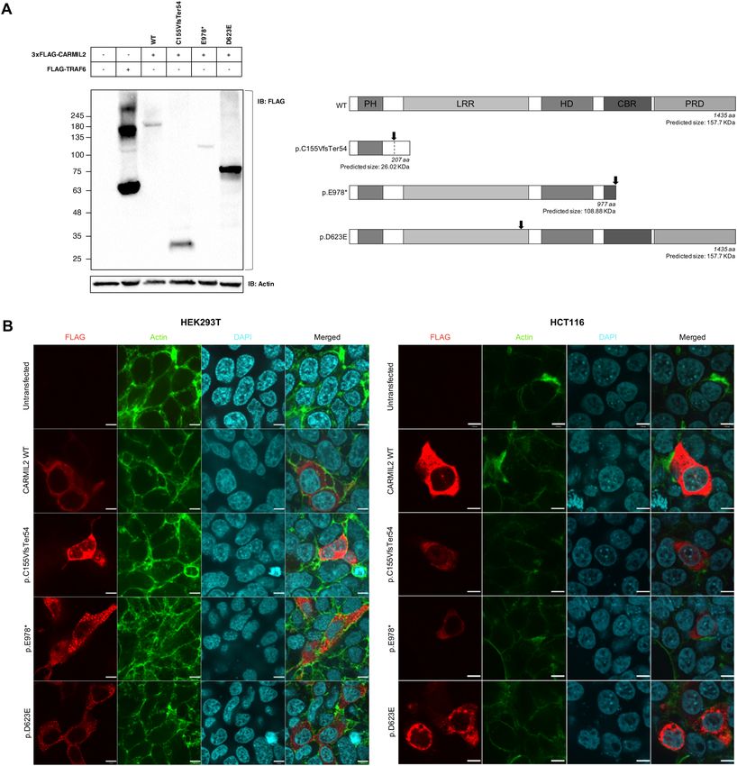
www.nature.com/scientificreports/
pathway8,9. Finally, CARMIL2 has a role in antigen-receptor signaling in B cells, leading to NF-κB activation
after B cell receptor (BCR), but not CD40, l igation10.
CARMIL2 variants have been implicated in human disease. CARMIL2 deficiency has been associated with
an autosomal recessive primary immunodeficiency (Immunodeficiency 58 IMD58 [MIM: 618131]), character-
ized by recurrent and/or chronic bacterial, viral, and fungal infections, cutaneous manifestations including
eczematous dermatitis, and disseminated Epstein–Barr virus-associated smooth muscle tumors9–14. Recently,
biallelic loss-of-function (LoF) variants of CARMIL2 have been linked to very early onset inflammatory bowel
disease (IBD) or IBD-like inflammatory gastrointestinal disorder, with or without clinical manifestations of
immunodeficiency11,13,15–18.
IBD is a spectrum of complex multifactorial immune disorders characterized by chronic intestinal inflam-
mation. Conventional IBD exhibits a polygenic architecture, while Mendelian or monogenic forms of IBD are
caused by rare variants with a large effect on gene f unction19–24. The relative contribution of genetic factors and
the frequency of IBD-causing monogenic variants seem to be inversely related to the age of onset of I BD22,25–28.
A recent study identified rare variants in genes linked to monogenic IBD in 7.8% of subjects in the subgroup
of very early onset IBD (VEOIBD), which includes patients younger than 6 years at disease recognition, and
in 2.3% of children diagnosed with IBD after 6 years of age29. To date, about 70 monogenic defects affecting
intestinal immune-epithelial homeostasis have been associated with I BD29. Among them, defects in T- and
B-cell function and in Tregs and immune regulation can lead to immunodeficiency, autoimmunity, and IBD-
like immunopathology22.
Autoimmune polyendocrine syndromes (APS) are characterized by functional impairment of multiple endo-
crine glands due to loss of immune tolerance toward them in genetically susceptible hosts. Similarly to IBDs, most
APSs result from polygenic predisposition, although they rarely present as part of a broader syndrome with an
underlying monogenic etiology. Monogenic APSs are caused by mutations in genes involved in maintenance of
central (e.g. AIRE) or peripheral (e.g. FOXP3, CTLA4, LRBA, STAT1, STAT3, STAT5b, ITCH, BACH2) immune
tolerance, leading to aberrant Treg function or activation of self-reactive effector T c ells30–33.
Hereby, we describe the identification and characterization of novel CARMIL2 variants in three patients
diagnosed with pediatric-onset IBD and in a child affected by APS, thus expanding the genotypic and phenotypic
spectrum of CARMIL2 deficiency.
Results
Identification of patients with biallelic CARMIL2 variant. In total, 4 patients from 3 unrelated fami-
lies were identified with homozygous variants of CARMIL2 (Fig. 1A). Patient 1 was identified through whole
exome sequencing (WES), performed during the evaluation for suspected monogenic IBD at the University
Hospital of Padova. Patient 2, the eldest brother of Patient 1, was recognized by family segregation analysis and
then further sequenced by WES. Patient 3 and Patient 4 were identified by screening for biallelic CARMIL2 vari-
ants from WES data in a cohort of 1005 pediatric IBD patients enrolled at SickKids, Toronto, as described in the
“Methods”29.
Clinical features of patients with biallelic CARMIL2 variant. Patient 1 was born in Italy to consan-
guineous (first cousins) healthy Moroccan parents, fourth-born of four male sons. At 3.25 years he was diagnosed
with colonic Crohn’s disease, phenotype A1aL2B1G1 according to the Paris classification (Fig. 1B,C, Table 1)25.
Symptoms started when he was 2.6 years old. The patient was induced with prednisone, and after clinical remis-
sion he was maintained with azathioprine. Follow-up endoscopy performed after 18 months revealed histologic
persistent mild total colonic inflammation in biopsies while in clinical remission. At the age of 5 years, the
patient developed Streptococcus pneumoniae pneumonia complicated by sepsis, despite being vaccinated against
pneumococcal disease. Past medical history included eczema in the first months of life. Anti-thyroglobulin anti-
bodies and anti-thyroid peroxidase antibodies were significantly raised, while thyroid function tests and thyroid
ultrasound were unremarkable.
Patient 2 is the eldest brother of Patient 1 (Table 1). The boy was diagnosed with Addison’s disease at 12 years
of age, and shortly after with Hashimoto’s thyroiditis. No other endocrinopathies were detected. He suffered from
difficult-to-treat eczema from a young age. He had no candidiasis, but at the age of 17 he was clinically diagnosed
with extended Tinea corporis skin infection. Laboratory tests were significant for negative anti-adrenal antibodies
and positive anti-thyroid autoantibodies. Plasma assay for very long-chain fatty acids was normal, thereby exclud-
ing peroxisomal disorders. Abdomen magnetic resonance imaging revealed bilateral adrenal hypoplasia. Sanger
sequencing of the AIRE gene was non informative. Thus, Patient 2 was diagnosed with autoimmune polyendo-
crine syndrome type 2 (APS-2), or Schmidt syndrome, whose distinctive feature is Addison’s disease, associated
with at least one among autoimmune thyroid disease and type 1A diabetes mellitus. There was neither history
of significant or recurrent infectious episodes, nor evidence of bowel disease and fecal calprotectin was normal.
Immunological work-up of Patients 1 and 2 revealed a marked increase in the proportion of naïve T and B
cells, with a reduction in memory T and B cells and marginal zone B cells. Patient 1 but not patient 2 had an
increase in the number of double negative T cells (CD3 + CD4-CD8-TCRαβ+) (Supplementary Table S1). Both
patients had negative anti-EBV IgG and IgM and EBV-DNA on peripheral blood. Both patients had low titers
of specific antibody against tetanus and diphtheria toxoids.
Patient 3 is a female diagnosed with ulcerative colitis (UC), type pancolitis (A1bE4S1), at the age of 11 years
old (Table 1). Remission was achieved after induction with prednisone, and maintained first with sulfasalazine,
then with azathioprine. After relapse, she failed infliximab and at the age of 17 years underwent colectomy with
loop ileostomy and J pouch. Her post-operative course was complicated by intestinal leakage causing intraab-
dominal sepsis, stricture at pouch anastomosis and pouchitis. Past medical history included reactive airway
Scientific Reports | (2021) 11:5945 | https://doi.org/10.1038/s41598-021-85399-9 2
Vol:.(1234567890)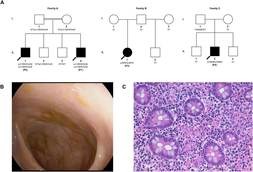
www.nature.com/scientificreports/

                                  Figure 1.  Novel biallelic CARMIL2 variants identified in four patients from three families. (A) Family pedigree
                                  and segregation analysis of the three kindreds. As for Family A, both parents and their second son, who had
                                  myoclonic epilepsy and attention deficit hyperactivity disorder, were heterozygous. (B) Colonoscopy image
                                  of Patient 1, showing only a mild patchy loss of vascular pattern throughout the colon. (C) Histopathologic
                                  findings in a colonic biopsy sample from Patient 1, showing epithelioid granulomas in the lamina propria
                                  (Hematoxylin–Eosin × 400).

                                  disease and an episode of pneumonia. Before IBD onset she had no other significant history of infections or
                                  immune-related diseases. She had no familial history of IBD.
                                      Patient 4 is a male diagnosed with UC, type pancolitis (A1bE4S1), at the age of 15 years (Table 1). The patient
                                  was induced with IV methylprednisolone, became steroid dependent first, then steroid refractory; azathioprine
                                  was thus introduced. He developed azathioprine-induced pancreatitis with pseudocysts, hepatic abscesses and C.
                                  difficile infection. Drain insertion was complicated by peri-sigmoid abscess, colonic-peripancreatic enterocutane-
                                  ous fistula and toxic megacolon. At the age of 16 the patient underwent subtotal colectomy and ileostomy, then
                                  J pouch surgery. Past medical history was unremarkable. Particularly, he had no previous history of infections,
                                  or immune-related diseases. He had no familial history of IBD.

                                  Genetic analysis of patients with biallelic CARMIL2 variant. All four patients were found to have
                                  novel biallelic variants of CARMIL2 (Fig. 2).
                                      Patient 1 and 2 have a homozygous nonsense variant p.Cys155ValfsTer54 of CARMIL2, located on Exon
                                  6 (Table 2). This frameshift mutation is caused by a single nucleotide (cytosine) deletion resulting in a non-
                                  synonymous substitution of cysteine (C) with valine (V) and premature termination of translation after 54
                                  codons (predicted number of amino acids: 207, while wild type CARMIL2 consists of 1435 amino acids). Variant
                                  p.C155VfsTer54 is not reported in literature and no data are available on allele frequency on reference databases
                                  (ExAC, gnomAD, 1000 Genomes database, EVS) (Table 2). Segregation analysis showed that the parents were
                                  heterozygous carriers of the variant.
                                      Patient 3 has a homozygous nonsense CARMIL2 variant p.Glu978*, located on exon 29 of CARMIL2 (Table 2)
                                  resulting in stop-gain nucleotide substitution and premature termination codon. CARMIL2 variant p.E978* is
                                  not reported in literature and is absent from population databases. Patient 3 was recruited as a “singleton”, so
                                  parental DNA was not available for variant segregation analysis.
                                      Patient 4 has a homozygous missense variant p.Asp623Glu, located on exon 21 of CARMIL2 (Table 2). It has
                                  not been reported in literature, and its frequency is extremely low (< 0.001) in population databases. Variant
                                  p.D623E is predicted to be deleterious by the majority of the algorithms examined. Moreover, the aspartic acid
                                  at position 623 is located in a leucine-rich repeat (LRR) domain of CARMIL2 and is part of an evolutionarily

Scientific Reports |   (2021) 11:5945 |                https://doi.org/10.1038/s41598-021-85399-9                                                  3

                                                                                                                                              Vol.:(0123456789)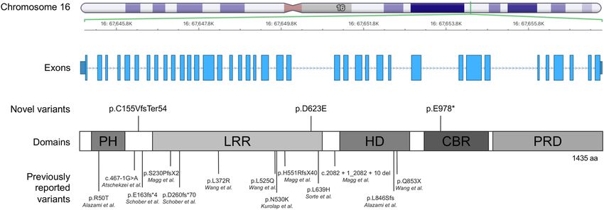
www.nature.com/scientificreports/
Patient 1 Patient 2 Patient 3 Patient 4
Demographics
Age at diagnosis 2 years 12 years 11 years 15 years
Gender Male Male Female Male
Consanguinity Yes Yes No Unknown
Indigenous Canadian (mother),
Parents origin (ethnicity/country) Arab-Berber/Moroccan Arab-Berber/Moroccan Unknown
Israeli-Polish (father)
Gastrointestinal disease
Family history of IBD None None None None
IBD (Paris classification of IBD)25 CD (A1aL2B1G1) UC (A1bE4S1) UC (A1bE4S1)
Chronic diarrhea, ± blood and
Abdominal pain, bloody diarrhea, Abdominal pain, bloody diarrhea,
Symptoms at onset mucus, ± abdominal pain, failure
weight loss fever, weight loss
to thrive
Hyperemic esophagus, small
Gastric erythema and superficial
Normal/mild patchy loss of vascu- ulcers and inflammation of
EGD/IC erosion of duodenal cap/Pancolitis
lar pattern stomach/Severe pancolitis with
with normal terminal ileum
backwash ileitis
None
Altered villous profile and atrophy,
Mild increase of duodenal
mild lymphomonocytic inflamma-
lamina propria mononuclear cells, Normal duodenum, mild chronic
tion and epithelioid microgranu-
moderate antritis with focal acute non-atrophic gastritis, mild
Pathology (upper/lower gastroin- lomas in duodenal bulb/Crypt
activity, moderate chronic body esophagitis/ Chronic colitis with
testinal tract) distortion, mild lymphomonocytic
gastritis/Mild-moderately active mild to moderate acute activity
inflammation, Paneth cell meta-
IBD of entire colon, focal superfi- throughout all biopsies
plasia and epithelioid granulomas
cial acute terminal ileitis
of the colon
Surgery None Colectomy, ileostomy, J pouch Colectomy, ileostomy, J pouch
Other clinical features
Hepatic abscesses, C. difficile
Streptococcus pneumoniae pneu- Tinea corporis, prolonged upper Intestinal leak with intraabdomi-
Infections infection, abdominal abscess with
monia complicated by sepsis respiratory tract infections nal sepsis, pouchitis, pneumonia
enterocutaneous fistula
Addison’s disease, Hashimoto
Endocrine disorders None None None
thyroiditis
Skin Eczema (first months) Eczema None None
Neuropsychiatric disorder (devel-
Others opmental delay and behavioral Depressive disorder Reactive airway disease Unknown
disorders)
Anti-thyroid Abs; negative anti-
Autoantibodies Anti-thyroid Abs, LAC ANCA ANCA
Harmonin antibodies
Table 1. Demographic and clinical features of CARMIL2-deficient patients. EGD
esophagogastroduodenoscopy, IBD inflammatory bowel disease, IC ileocolonoscopy, LAC Lupus anticoagulant.
Figure 2. CARMIL2 gene and protein view with newly identified and previously published variants. Schematic
representation of the intron–exon structure of the CARMIL2 gene, which is located on Chromosome 16. The
three novel variants reported in this study are labelled above the schematic illustration of the protein domains of
CARMIL2, along with the previously described variants below. PH Pleckstrin-Homology domain, LRR Leucine-
Rich Region, HD Helical Dimerization domain, CBR Capping Protein-Binding Region, PRD Proline-Rich
Domain.
Scientific Reports | (2021) 11:5945 | https://doi.org/10.1038/s41598-021-85399-9 4
Vol:.(1234567890)www.nature.com/scientificreports/
Patient 1 Patient 2 Patient 3 Patient 4
Chromosome positiona Chr16:67,646,513 Chr16:67,653,066 Chr16:67,649,569
cDNA change (GenBank: NM_001013838.3) c.462delC c.2932G > T c.1869C > A
Amino acid c hangeb (GenPept: NP_001013860.1) p.Cys155ValfsTer54 p.Glu978* p.Asp623Glu
Exon number 6 29 21
Predicted domain None PRD LRR
In silico evaluation
CADD Unknown 41 26.4
SIFT Pred Unknown Unknown Damaging
Polyphen2 Pred Unknown Unknown Probably damaging
LRT Unknown Neutral Deleterious
Mutation Assessor Unknown Unknown Medium (2.645)
Mutation Taster Disease causing Disease causing Disease causing
FATHMM Unknown Unknown Tolerated
PROVEAN Unknown Unknown Damaging
Population databases
Maximum AAFc 0 0 0.0007
ExAC allele frequency None None 0.0002
N of heterozygous in ExAC None None 26
N of homozygous in ExAC None None None
gnomAD allele frequency None None 0.0002499
gnomAD allele count None None 38
N of homozygous in gnomAD None None None
1000 Genomes None None None
EVS None None 0.0003
Table 2. Genetic features of CARMIL2-deficient patients. AAF alternative allele frequency, AR autosomal
recessive, CADD Combined Annotation Dependent Depletion, EVS Exome Variant Server, ExAC Exome
Aggregation Consortium, FATHMM Functional Analysis through Hidden Markov Models, gnomAD Genome
Aggregation Database, LRR Leucine-Rich Region, LRT likelihood ratio test, PRD Proline-Rich Domain,
PROVEAN Protein Variation Effect Analyzer. a According to human genome assembly GRCh38. b According to
NCBI reference sequence NP_001013860.1. c Represents the maximum AAF of a variant in the databases taken
into account.
constrained region (Supplementary Table S2). The patient was recruited as a “duo”, and only the unaffected father’s
DNA was available for allele segregation analysis. The father was a heterozygous carrier for the same variant.
Sanger sequencing confirmed the presence of the biallelic variant p.C155VfsTer54 in Patient 1 and 2 but failed
for both the variants identified in Patient 3 (p.E978*) and Patient 4 (p.D623E), due to a guanine-cytosine-rich
template (Supplementary Fig. S1). However, careful reexamination of raw WES reads showed high coverage
support for homozygous mutant calls in both candidates. Furthermore, since LoF is the pathogenic mechanism
of CARMIL2 deficiency and tissue immunostaining proved reduced protein expression in both patients, confir-
mation by sequencing was deemed u nnecessary34.
Immunostaining of bowel biopsies in biallelic CARMIL2 variant carriers with inflammatory
bowel disease. Immunostaining on sigmoid formalin-fixed paraffin-embedded (FFPE) sections was per-
formed to assess the expression levels of CARMIL2 in the bowel of the three patients affected by IBD (Patient
1, 3 and 4; Fig. 3). In both normal and IBD control sigmoid sections CARMIL2 was strongly expressed in the
enterocytes of mucosal layer and stromal cells. In line with The Human Protein Atlas, CARMIL2 was mainly
localized in cell cytoplasm, but some nuclear expression was also observed in blood c ells1. In the two patients
with homozygous protein-truncating variants (p.C155VfsTer54 and p.E978*), CARMIL2 protein expression was
nearly absent or remarkably down-regulated in both glandular layer and stromal area. In Patient 4, carrying the
homozygous missense variant p.D623E, CARMIL2 expression in the mucosal layer was significantly weaker in
comparison to controls, while infiltrated blood cells located in the stromal area showed focally positive staining.
This range of staining intensity in different cell types is consistent with previously published immunohistochem-
istry images, showing stronger staining in lymphocytes than in intestinal epithelium18. Cytokeratin 18 staining
pattern in all samples was as expected based on previous r eports35, indicating a preserved tissue architecture.
Functional validation of CARMIL2 variants. Next, we investigated the impact of the novel CARMIL2
variants on protein expression and cellular localization using cellular model systems. Western blotting was used
to analyze the expression of 3xFLAG tagged CARMIL2 variants in HEK293Tcells (Fig. 4). Immunoblotting with
anti-FLAG antibody (Fig. 4A) detected bands corresponding to the predicted molecular weight (MW) both for
CARMIL2 wild type (WT) and for the protein-truncating variants, p.C155VfsTer54 and p.E978*. The MW of
Scientific Reports | (2021) 11:5945 | https://doi.org/10.1038/s41598-021-85399-9 5
Vol.:(0123456789)www.nature.com/scientificreports/
Figure 3. Dual immunofluorescence staining for CARMIL2 protein and CK18 on sigmoid FFPE sections
of healthy control, IBD control, and three biallelic CARMIL2 variant carriers diagnosed with IBD. DAPI
counterstaining was used to visualize nuclei. (A) Composite image, where green staining indicates CARMIL2,
red represents cytokeratin 18 (CK18), a marker for single layer epithelial cells, and blue marks nuclear DAPI
stain. In Patient 1 and Patient 3, carriers of the two protein-truncating variants (respectively p.C155VfsTer54
and p.E978*), CARMIL2 signal was almost absent in sigmoid sections. In Patient 4, carrier of missense variant
p.D623E, immunofluorescence staining was weaker than the controls particularly in the mucosal layer. (B)
Single-label immunofluorescence for CK18 (red). (C) Single-label immunofluorescence for CARMIL2 (green).
p.D623E variant was ~ 80 kDa, much lower than the WT, the only difference being the substitution of a single
amino acid residue from aspartic acid to glutamic acid. This strongly suggested proteolysis of the p.D623E vari-
ant to a lower MW species.
Immunofluorescence staining of transfected cells was performed in both HEK293T cells and HCT116 cells
(Fig. 4B). Immunofluorescence imaging showed that the expression pattern of CARMIL2 WT was diffuse across
the cytoplasm in both cell strains, in line with the existing knowledge of the protein, as reported in The Human
Protein Atlas1. Similarly, CARMIL2 variant p.C155VfsTer54 exhibited cytoplasmic expression, albeit less homoge-
neous. Conversely, CARMIL2 variants p.E978* and p.D623E signal appeared as puncta structures throughout the
cytoplasm of HEK293T cells. For variant p.D623E the puncta staining was consistent in HCT116 cells. Therefore,
this missense variant appeared to form aggregates when expressed in different cell lines. Overall, different lines
of evidence suggest that variant p.D623E is susceptible to mislocalization inside the cell and proteolysis, thus
behaving as a null or non-functional allele.
Discussion
Our study confirms that CARMIL2 deficiency can manifest only with isolated IBD. Unlike more recent s tudies17,18
reporting a very early IBD onset (6 out of 6 patients), two of our patients shared a later age of onset of IBD
symptoms, namely at 11 and 15 years of age. Although the majority of studies on monogenic IBD have focused
on the very early onset population, running the risk of selection bias, recent findings have pointed out that a
genetic disorder should be considered in all patients with pediatric onset IBD29. Resistance to conventional lines
of treatment and complicated disease course, similar to what was reported in Patients 3 and 4, should always
prompt the execution of WES, in line with existing recommendations22. None of our patients presented overt
clinical signs of immunodeficiency before the diagnosis of IBD. Patient 1 developed bacterial sepsis after the
initiation of immunosuppression (i.e., azathioprine). Notably, one patient reported in the study of Magg et al.
died due to septic complications at the age of 4 years while on treatment with azathioprine17. Patient 3 and 4
experienced severe infectious complications after surgery. The need for a surgical treatment appears to be a
common feature of pediatric IBD associated to CARMIL2-deficiency, since it occurred in the majority of the
Scientific Reports | (2021) 11:5945 | https://doi.org/10.1038/s41598-021-85399-9 6
Vol:.(1234567890)www.nature.com/scientificreports/
Figure 4. Functional validation of novel CARMIL2 variants. (A) Western blot analysis of CARMIL2 expression.
Anti-FLAG antibody was used for immunoblotting (left panel). FLAG-TRAF6 served as positive control, β-actin
as loading control. The expected molecular weight of CARMIL2 WT and variants (considering the 3xFLAG
tag) is shown for comparison (right panel). The black arrow points to the position of the mutation inside the
protein. The observed band (~ 180 kDa) of WT CARMIL2 does not correspond to the predicted protein size
(~ 155 kDa); this is consistent with what previously reported and with The Human Protein Atlas (available from
v19.3.proteinatlas.org)1,12. Full-length blots are presented in Supplementary Fig. S2. (B) Immunofluorescence
staining of HEK293T cells (left panel) and HCT116 cells (right panel) transfected with wild type CARMIL2
or the indicated variants. The first three columns represent the immunofluorescence images of FLAG tagged
CARMIL2 (red), actin (green) and nuclei (light blue). The fourth column is a composite image. In HEK293T
cells (left panel) CARMIL2 WT and variant p.C155VfsTer54 display a diffuse cytoplasmic expression pattern,
while variants p.E978* and p.D623E exhibit a more granular pattern of expression. In HCT116 cells (right panel)
expression pattern of all CARMIL2 isoforms is cytoplasmic, although variant p.D623E signal appeared as puncta
structures. Scale bar: 7 µm.
Scientific Reports | (2021) 11:5945 | https://doi.org/10.1038/s41598-021-85399-9 7
Vol.:(0123456789)www.nature.com/scientificreports/
patients reported so far (6 out of 9 patients, including our cases) and was frequently due to a failure of medical
treatment (3 out of 6 patients, including our cases)17,18. A recent study found that progression to surgery can be
itself an indicator of monogenic etiology among pediatric IBD p atients29. There is no consensus with respect to
surgical timing, indications, and strategies in monogenic IBDs, including CARMIL2 d eficiency36. Our results
suggest that CARMIL2-deficient IBD patients harbor a significant risk of life-threatening immunosuppression-
and surgery-related infections. A treatment option for several genetic immunodeficiencies underlying IBD-like
phenotypes is hematopoietic stem cell transplantation (HSCT)22. However, it could be inappropriate or even
harmful if an epithelial barrier defect c oexists23. Herein, we confirm that CARMIL2 protein is expressed in gas-
trointestinal epithelium, but it remains to be clarified if this is relevant to IBD pathogenesis in deficient patients.
Overall, CARMIL2 should be included in the diagnostic work-up of patients with suspected monogenic IBD
regardless of the age at disease onset and of the presence of overt clinical signs of immune deficiency. In fact, the
identification of CARMIL2 deficiency has the potential to influence treatment choice and might improve disease
prognosis, especially by means of prevention and prompt recognition of infectious complications.
Alongside phenotypic differences, CARMIL2 deficiency is characterized by marked genotypic heterogeneity.
The known variants associated with CARMIL2 deficiency have different impact on the protein (e.g. nonsense,
frameshift, missense, etc.) and they are spread along the gene (Fig. 2). The only consistency seems to be the
lack of mutations in the C-terminus portion of the gene. One could speculate that mutations occurring in the
C-terminus, close to end of the coding sequence, may preserve some level of protein function. Therefore, failure to
produce the expected phenotype would lead to missed diagnosis. The lack of obvious genotypic-phenotypic cor-
relation, as well as the inter- and intrafamilial clinical heterogeneity, even among carriers of the same CARMIL2
variant, seems to point towards a contribution of additional environmental, genetic, or epigenetic-modifying
factors in determining the clinical manifestations of CARMIL2 deficiency.
Bioinformatic tools can predict the damaging effects of mutations, but might overestimate them, hence the
importance of experimental validation. Different lines of evidence from the experiments we performed showed
that p.D623E variant behaves as a non-functional allele. Particularly, immunofluorescence of transfected cells
resulted in a granular pattern. Endoplasmic-reticulum-associated protein degradation (ERAD) of p.D623E
protein variant might explain this finding. In fact, the residue 623 of CARMIL2 belongs to an LRR domain, a
structural motif with a horseshoe shape, with an interior parallel beta sheet, an exterior array of helices, and
an hydrophobic core containing many leucine r esidues37. An amino acid substitution in this region may lead
to the exposure of normally hidden hydrophobic patches, usually buried inside the protein to keep the lowest
energy state. Exposed patches might lead to protein aggregation, or they could be recognized as a substrate by
ERAD, a cellular pathway that targets misfolded or mutated proteins, which are retained inside the endoplasmic
reticulum (ER) and targeted for ubiquitination and subsequent proteolytic d egradation38,39. ER retention and
impaired trafficking could explain the staining pattern observed in transfected cells, the proteolysis the lower
than predicted MW. Protein overexpression might make the process more obvious by engulfing the cell. Another,
non-exclusive possibility is that substitution from aspartic acid to glutamic acid leads the protein to be recognized
by a glutamic-acid-specific protease.
This is the first report showing the association between CARMIL2 deficiency and autoimmune endocrinopa-
thies. A monogenic etiology has been proved in a subset of patients with APS-2 and several lines of evidence
support CARMIL2 as a candidate gene30–33,40,41. First, the significance of rare LoF variants of CARMIL2 in human
disease has already been established, consistently with a recessive disease m odel10. This is confirmed by the mod-
est LOUEF (loss-of-function observed/expected upper bound fraction) score (0.552 for CARMIL2, indicating a
degree of intolerance to LoF variants) shown using gnomAD (Genome Aggregation Database) data42. Secondly,
biallelic CARMIL2 LoF variants fully segregate with an immune dysregulation disorder in multiple independent
families with autoantibodies and absence of antigen-specific antibodies to bacterial v accines10,14. This is consistent
with the known B cell phenotype of human CARMIL2 deficiency. Additionally, skin manifestations have been
described in the majority of CARMIL2-deficient patients, and eczema is part of other monogenic APSs9–14,17,18,30.
Thirdly, the function of CARMIL2 is consistent with the known pathogenesis of APS, being expressed in immune
cells and endocrine glands and being essential for development of regulatory T cells and for Th17 differentiation,
similarly to other genes mutated in monogenic APS1,8,9,30–33. Moreover, CARMIL2 interactome include DOCK8,
that is mutated in patients presenting with autoimmune disorders, eczema and compromised Treg f unction9,43.
Notably, CARMIL2-deficient mice and humans did not develop any obvious organ-specific autoimmune disor-
der, despite a reduction in Tregs8,9. This might depend on the coincident defect in effector T cells9. Furthermore,
CARMIL2-deficient mice did not display any intrinsic B cell d efect8–10. Therefore, knockout mice might not be
a suitable model for studying human CARMIL2 deficiency. Remarkably, enteropathy and/or IBD-like intestinal
inflammation, autoimmune endocrinopathy and skin manifestations are common in IPEX and other syndromes
presenting with IPEX-like features, caused by monogenetic defects affecting Treg f unction22,30,44. Hence, CAR-
MIL2 deficiency should be added to the increasing group of IPEX-like syndromes, as already p roposed18.
Our study had some limitations, including that overexpression experiments might have been unreliable for
protein-truncating variants p.C155VfsTer54 and p.E978*. In fact, it can be assumed that in vivo these variants
are targeted by nonsense-mediated mRNA decay45. For this reason, the more intense immunofluorescence signal
of p.C155VfsTer54 compared to WT CARMIL2 in cultured cells probably does not reflect what happens in vivo,
and could have been determined by higher transfection efficiency due to a shorter peptide.
In summary, the phenotypic spectrum of CARMIL2 deficiency is broader than previously known, rang-
ing from severe immunodeficiency to IBD and organ-specific autoimmunity. CARMIL2 should be part of the
diagnostic evaluation of patients with suspected monogenic IBD, even in the absence of obvious signs of immu-
nodeficiency. Genetic diagnosis may be vital in monogenic IBD, to guide specific treatment, prevent surgery
or unnecessary therapies, anticipate complications, and help genetic c ounseling22,23,46–48. Moreover, CARMIL2
Scientific Reports | (2021) 11:5945 | https://doi.org/10.1038/s41598-021-85399-9 8
Vol:.(1234567890)www.nature.com/scientificreports/
deficiency can present exclusively with APS. Further studies are needed to better define IBD immunopathogenesis
in CARMIL2 deficiency and the potential therapeutic utility of HSCT.
Methods
Helsinki guidelines. All human experiments followed the Helsinki Guidelines. Informed consent was
obtained from the participants parents and the study had local ethics board approval at The Hospital for Sick
Children (SickKids), Toronto, Canada (Research Ethics Board: REB1000024905).
Patients. Patient 1 and his brother, Patient 2, were recruited from the Pediatric Gastroenterology, Hepatol-
ogy and Liver Transplantation Unit at the Pediatric Clinic of the University Hospital of Padova. Patient 3 and
Patient 4 were discovered through screening of a large cohort of pediatric IBD patients recruited at SickKids as
previously described29. Children (age < 18) undergoing evaluation for IBD were enrolled over a 13-year period
(2003–2015). Diagnosis of IBD was made according to the recommendations of the Porto c riteria49. Patients
with known primary immunodeficiency, chromosomal abnormalities, syndromic disease or diagnosed with
other forms of monogenic intestinal disease were excluded. After obtaining the caregivers’ consent, clinical data
were recorded, and biological samples were processed and stored in a biobank. WES analyses of blood samples
was thus carried out as detailed below.
For Patient 1 and Patient 2, the phenotype review was carried out at University Hospital of Padova using
electronic medical record software Galileo (NoemaLife, Dedalus). For the SickKids cohort, following variant
prioritization50, each patient deemed to have pathogenetic biallelic CARMIL2 variants was reversed phenotyped,
using clinical data from databases and electronic medical record systems. For all IBD patients phenotype was clas-
sified according to the pediatric modification of the Montreal classification, also known as Paris classification25.
Whole exome sequencing. For Patient 1 and 2, WES and segregation analysis were performed. Genomic
DNA was extracted and purified from whole blood. DNA was prepared for sequencing with the kit SureSe-
lectXT All Exon V5 kit (Agilent). Exome sequencing was performed with next-generation sequencing (NGS)
technology on the Illumina HiSeq 2500 platform, using paired-end 100 bp read. DNA sequences were mapped
and analyzed using as reference genome assembly GRCh38 (Genome Reference Consortium Human Build 38).
For the SickKids cohort, banked genomic DNA isolated from whole blood collected by venipuncture using
a Qiagen Puregene Blood Core Kit was processed for exome capture using the NimbleGen VCRome 2.1 design.
Captured libraries were sequenced on the Illumina HiSeq 2500 platform using paired-end 75 bp reads at the
Regeneron Genetics Center (RGC), Tarrytown, NY, USA. Exome sequencing coverage was 30 × or greater
for > 85% of the bases targeted.
Bioinformatic analysis. For Patient 1, a bioinformatic analysis of exons and close intronic portions of 79
genes associated with IBD was performed (Supplementary Table S3). Genetic variants were analyzed and com-
pared with several population databases (Exome Aggregation Consortium [ExAC], Genome Aggregation Data-
base [gnomAD], 1000 Genomes database, Exome Variant Server [EVS]) and genetic clinical databases (OMIM,
ClinVar, HGMD, GWAS, PGKB, Cosmic). Every variant was also evaluated in silico for its possible effects on
protein structure or function (using Polyphen2, SIFT, VAAST Variant Prioritizer [VVP], Combined Annotation
Dependent Depletion [CADD], Mutation Assessor, Mutation Taster) and for evolutionary conservation (phyloP
score).
For the SickKids cohort, WES data from 2307 participants (1005 index patients, and 1302 parents and sib-
lings) were analyzed using the FORGE (Finding of Rare Disease Genes) pipeline. Raw sequencing reads were
aligned to human reference genome (GRCh38/hg38) using BWA-mem (Burrows-Wheeler Aligner, ver. 0.7.12),
followed by indel realignment using Genome Analysis Toolkit (GATK, ver. 3.5). Five variant callers (GATK
HaplotypeCaller ver. 3.5, Vardict ver. 1.4.6, Varscan ver. 2.3.9, Samtools ver. 1.3, and Freebayes ver. 1.0.0) were
run on the Binary Alignment Map (BAM) files of each family to produce family based Variant Call Format (VCF)
files when at least 2 of the 5 agreed on a called variant. Inheritance modeling on family level VCF files was per-
formed using the GEMINI tool to query for rare (Minor Allele Frequency [MAF] < 0.01) protein coding variants
that fit autosomal recessive, compound heterozygous, de novo, autosomal dominant, and X-linked inheritance
models filters. VarSeq software (Golden Helix) was used to import, filter, and do inheritance modeling on the
variants from each trio. Common variants (MAF > 0.01), defined using publicly available variant databases (ExAC
frequency database [ver. 0.3], 1000 Genomes database [phase 1], and the NHLBI Exome Sequencing Project
V2 Exome Variant Frequencies), were filtered out. Variants were further classified according to whether they
were deemed to be coding. Non-synonymous and unclassified variants were then scored using the database for
non-synonymous functional predictions (dbNSFP 2.8), filtering out variants with CADD score < 10 or no other
damaging score (Polyphen2, SIFT, LRT, Mutation Assessor, Mutation Taster, Functional Analysis through Hid-
den Markov Models [FATHMM], Protein Variation Effect Analyzer [PROVEAN]). Variants were also evaluated
in silico for protein domains in which they were predicted to be located (using Uniprot database and Simple
Modular Architecture Research Tool or SMART database) and for evolutionary conservation of corresponding
amino acid (based on Aminode webtool, available at http://www.aminode.org)51. Interpretation of all detected
CARMIL2 variants was performed according to the American College of Medical Genetics g uidelines52.
Sanger sequencing. For Patient 1, Patient 2 and their family, primers were designed to select and amplify
through polymerase chain reaction (PCR) the region containing the reported mutation within the genomic
DNA. PCR products were then sequenced using cycle sequencing Big Dye Terminator ver. 3.1 (Applied Biosys-
tems) and ABI 3100 Avant automated capillary electrophoresis sequencer (Applied Biosystems).
Scientific Reports | (2021) 11:5945 | https://doi.org/10.1038/s41598-021-85399-9 9
Vol.:(0123456789)www.nature.com/scientificreports/
For candidate patients identified within the SickKids pediatric IBD cohort, sequencing was carried out at The
Centre for Applied Genomics (TCAG, The Hospital for Sick Children, Toronto, Canada), that provides high-
quality capillary-based fluorescent sequencing on dual ABI 3730XL instruments. Different sets of primers were
used to troubleshoot sequencing reactions.
Immunofluorescence histochemical staining of bowel biopsies. Bowel samples were fixed in neu-
tral buffered formalin without methanol and embedded in paraffin using routine protocols (formalin-fixed par-
affin-embedded, FFPE). Tissue samples, including normal control and IBD control, were retrieved from the Unit
of Anatomical Pathology, Medicine Department, University Hospital of Padova and from the Division of Pathol-
ogy, The Hospital for Sick Children, Toronto. Only FFPE with well-preserved tissue architecture were chosen,
to avoid false negative staining. As negative control and disease control, a non-IBD patient sample and an unre-
lated IBD patient sample were used, respectively. Immunofluorescent histochemical staining was performed on
sigmoid FFPE sections as previously d escribed53. Briefly, paraffin-embedded sections were deparaffinized using
xylene and rehydrated with different percentages of ethanol. Antigen retrieval was achieved with high-pressure
cooking in EDTA-borax buffer made with 1 mM EDTA, 10 mM borax, 10 mM boric acid and 0.001% ProClin
300 (Supelco) at pH 8.5. To block non-specific staining, the slides were incubated for 1 h at room temperature
(RT) in 4% bovine serum albumin (BSA) and 20% donkey serum in phosphate-buffered saline (PBS). Slides were
incubated with primary antibodies, including anti-CARMIL2 antibody produced in rabbit (Sigma, HPA041402)
and anti-cytokeratin 18 mouse monoclonal antibody (Abcam), overnight at 4 °C. On the following day, stained
slides were washed 3 times for 5 min with PBS. Secondary antibodies, namely Rhodamine Red-X (RRX) Affin-
iPure F(ab’)2 Fragment Donkey Anti-Rabbit IgG (Jackson ImmunoResearch Laboratories) and Fluorescein
(FITC) AffiniPure F(ab’)2 Fragment Donkey Anti-Mouse IgG (Jackson ImmunoResearch Laboratories), were
incubated at RT in darkness for 2 h, then slides were washed 3 times for 10 min in darkness. RedDot2 Far-
Red Nuclear Stain (Biotium) was used for nuclear counterstaining at a dilution of 1:200. Finally, sections were
mounted overnight with Vectashield antifade mounting medium (Vector Laboratories). Immunostained slides
were imaged using a Leica confocal laser scanning microscope (Leica, TCS-SP8) and LAS-AF software (Leica
Microsystems). Image processing, including color resolution, color separation, and merging of fields, was done
using Adobe Photoshop CS5 software (Adobe Systems Incorporated).
Plasmids. Plasmid containing an insert with human CARMIL2 sequence with 3 tandem FLAG epitopes
(DYKDDDDK) on N-terminus was a gift from John Cooper (Washington University, St. Louis, Addgene plasmid
#118740)5. Mutations of CARMIL2 were generated using site-directed mutagenesis by ACGT Corp. (Toronto,
Canada). Plasmid were expanded through transformation and harvest from Escherichia coli DH10B bacteria as
per standard protocols and purified using EZ-10 Spin Column Plasmid DNA Miniprep Kit (Bio Basic, BS614)
and PureLink HiPure Plasmid Maxiprep Kit (Invitrogen, Thermo FisherScientific, K210007). Positive control
plasmid FLAG-TRAF6 was provided by Muise laboratory (SickKids).
Cell culture and transfection. HEK293T cells and HCT116 cells were provided by Muise laboratory and
maintained in DMEM (Wisent Inc.) containing 10% of heat-inactivated fetal bovine serum (FBS) and antibi-
otic–antimycotics at 37 °C in 5% C O2. HEK293T cells were chosen for the ease of transfection, while HCT116
cells were selected for being a colon cancer cell line, thus closer to intestinal cells. For western blot, HEK293T
cells were grown in a Falcon 6-well plate (Corning) to reach 60–70% confluence and transiently transfected with
1 µg of construct DNA (3xFLAG-CARMIL2 WT or mutation, or FLAG-TRAF6 as positive control) per each
well using PolyJet (SignaGen Laboratories) transfection reagent, according to the manufacturer’s instructions.
Cells were collected for lysis and protein analysis 48 h after transfection. For immunofluorescence, HEK293T
cells and HCT116 cells were transiently transfected for 48 h as described above using 0.5 µg of construct DNA
(3xFLAG-CARMIL2 WT or mutation) per well.
Western blotting. Cells were lysed for 20 min in RIPA buffer (Sigma) supplemented with 1 mM phenyl-
methylsulfonyl fluoride (PMSF), 500 uM sodium fluoride (NaF), 0.2 mM sodium orthovanadate (Na3VO4),
1:500 Protease Inhibitor Cocktail (Sigma-Aldrich, P2714) and 1% Phosphatase Inhibitor Cocktail 3 (Sigma-
Aldrich, P0044). Each sample was sonicated with 5 pulses at 30% amplitude (Q125 Sonicator, Qsonica) and
centrifuged 20 min at 4 °C (Eppendorf Centrifuge 5430 R, Eppendorf). Protein concentration in lysate was
measured using Bradford assay and a BSA protein standard in water (Bio-Rad Protein Assay Dye Reagent Con-
centrate, Bio-Rad; Epoch Microplate Spectrophotometer, BioTek). Samples were resuspended in 1xSDS (sodium
dodecyl sulfate) protein sample buffer (40% glycerol, 240 mM Tris/HCl, 8% SDS, 0.04% bromophenol blue, 5%
beta-mercaptoethanol) and a volume corresponding to 40 ug of protein per sample was loaded onto a 4–20%
gradient gel (Mini-PROTEAN TGX Gels, Bio-Rad). Gel electrophoresis was performed (Mini-PROTEAN Tetra
Vertical Electrophoresis Cell and PowerPac HC High-Current Power Supply, Bio-Rad) in running buffer (25 mM
Tris base, 192 mM glycine, 0.1% SDS, pH 8.3) for approximately 1 h at 200 V. Sample was then transferred to
a nitrocellulose membrane (Amersham Protran, GE Healthcare) using a semi-dry blotting process (Trans-Blot
Turbo Transfer System, Bio-Rad) in transfer buffer (25 mM Tris base, 192 mM glycine, 0.00375% SDS, 20% (v/v)
methanol, pH 8.3) for 12 min at 25 V and RT. Membrane was blocked using 5% skim milk in PBST (PBS with
Triton X-100 0.05%) for 1 h at RT. The immunoblot was then incubated with appropriate primary and second-
ary antibodies diluted in blocking buffer, overnight at 4 °C and for 1 h at RT, respectively. Primary antibod-
ies included monoclonal anti-FLAG M2 antibody produced in mouse (Sigma-Aldrich, F3165), anti-CARMIL2
antibody produced in rabbit (Sigma, HPA041402) and monoclonal anti-β-Actin antibody produced in mouse
(Sigma-Aldrich, A5441). Secondary antibodies included Peroxidase AffiniPure Goat Anti-Mouse IgG (H + L)
Scientific Reports | (2021) 11:5945 | https://doi.org/10.1038/s41598-021-85399-9 10
Vol:.(1234567890)www.nature.com/scientificreports/
(Jackson ImmunoResearch Laboratories, 115-035-146), Peroxidase AffiniPure Goat Anti-Mouse IgG (H + L)
(Jackson ImmunoResearch Laboratories, 115-035-003) and Peroxidase IgG Fraction Monoclonal Mouse Anti-
Rabbit IgG light chain specific (Jackson ImmunoResearch Laboratories, 211-032-171). After incubation with
each antibody, membranes were washed 3 times with PBST for 5–10 min. Blots were imaged using chemilumi-
nescent horseradish peroxidase detection reagent (Immobilon Forte Western HRP substrate, MilliporeSigma
or Clarity Max Western ECL Substrate, Bio-Rad) and imaged through chemiluminescence detection (Chemi-
Doc MP Imaging System and Image Lab software, Bio-Rad). In order to re-examine the same protein sample
with different antibodies, stripping buffer (Restore Western Blot Stripping Buffer, Thermo Scientific, 21059) was
added for 10 min. Membrane was then washed in PBST for 3 times and re-blocked. In total, the experiment was
repeated 3 times. An online tool (available at https://www.bioinformatics.org/sms/prot_mw.html) was used to
calculate the predicted MW of each CARMIL2 variant, taking into account the size of the protein tag (3xFLAG).
Immunofluorescence staining of transfected cells. For immunofluorescence, HEK293T and
HCT116 cells were cultured as described above, seeded at low density on cover slips (Fisherbrand) coated with
poly-d-lysine (Sigma) onto Falcon 24-well plates (Corning) and transfected 1 h after seeding. After 48 h, cells
where fixed in methanol-free 4% paraformaldehyde (PFA) for 30 min, then washed three times with PBS. Cells
were permeabilized with 0.1% Triton X-100 in PBS for 10 min and blocked with 10% goat serum in PBS for
30 min at RT. Cells were incubated with the primary antibody, monoclonal ANTI-FLAG M2 antibody produced
in mouse (Sigma, F1804) diluted in 10% goat serum PBS (1:200) for 1 h at RT. After washing 3 times with
PBS-Tween (0.05%), cells were incubated with Mouse IgG (H + L) Highly Cross-Adsorbed Secondary Antibody,
Alexa Fluor 568 Conjugate (Invitrogen, Thermo Fisher Scientific, A-11031) diluted in 10% goat serum PBS
(1:200) for 45 min at RT, then washed again 3 times. 4′,6-diamidino-2-phenylindole (DAPI) diluted in PBS
(1:2500) was used for nuclear staining, while actin was assessed via phalloidin staining (ActinGreen 488 Ready-
Probes reagent, according to the manufacturer’s instructions); they were incubated together 10 min at RT. Cover
slips were then washed with PBS and mounted onto glass slides (Fisherbrand) with mounting medium (Dako
Fluorescent Mounting Medium, Agilent, S3023). Slides were imaged using a quorum spinning disk confocal
microscope (Olympus IX81) set at 63 × objective magnification (oil imaging medium). Images were analyzed
and deconvolved using Volocity 6.3 software (Perkin Elmer). For each condition, at least 3 images were acquired.
Received: 9 December 2020; Accepted: 1 March 2021
References
1. Uhlén, M. et al. Tissue-based map of the human proteome. Science (80-). 347, 1274 (2015).
2. Matsuzaka, Y. et al. Identification, expression analysis and polymorphism of a novel RLTPR gene encoding a RGD motif, tropo-
modulin domain and proline/leucine-rich regions. Gene 343, 291–304 (2004).
3. Liang, Y., Niederstrasser, H., Edwards, M., Jackson, C. E. & Cooper, J. A. Distinct roles for CARMIL isoforms in cell migration.
Mol. Biol. Cell 20, 5290–5305 (2009).
4. Edwards, M. et al. Capping protein regulators fine-tune actin assembly dynamics. Nat. Rev. Mol. Cell Biol. 15, 677–689 (2014).
5. Lanier, M. H., Kim, T. & Cooper, J. A. CARMIL2 is a novel molecular connection between vimentin and actin essential for cell
migration and invadopodia formation. Mol. Biol. Cell 26, 4577–4588 (2015).
6. Lanier, M. H., McConnell, P. & Cooper, J. A. Cell migration and invadopodia formation require a membrane-binding domain of
CARMIL2. J. Biol. Chem. 291, 1076–1091 (2016).
7. Stark, B. C., Lanier, M. H. & Cooper, J. A. CARMIL family proteins as multidomain regulators of actin-based motility. Mol. Biol.
Cell 28, 1713–1723 (2017).
8. Liang, Y. et al. The lymphoid lineage-specific actin-uncapping protein Rltpr is essential for costimulation via CD28 and the devel-
opment of regulatory T cells. Nat. Immunol. 14, 858–866 (2013).
9. Roncagalli, R. et al. The scaffolding function of the RLT PR protein explains its essential role for CD28 co-stimulation in mouse
and human T cells. J. Exp. Med. 213, 2437–2457 (2016).
10. Wang, Y. et al. Dual T cell- and B cell-intrinsic deficiency in humans with biallelic RLT PR mutations. J. Exp. Med. 213, 2413–2435
(2016).
11. Sorte, H. S. et al. A potential founder variant in CARMIL2/RLTPR in three norwegian families with warts, molluscum contagiosum,
and T-cell dysfunction. Mol. Genet. Genom. Med. 4, 604–616 (2016).
12. Schober, T. et al. A human immunodeficiency syndrome caused by mutations in CARMIL2. Nat. Commun. 8, 1–13 (2017).
13. Alazami, A. M. et al. Novel CARMIL2 mutations in patients with variable clinical dermatitis, infections, and combined immuno-
deficiency. Front. Immunol. 9, 203 (2018).
14. Atschekzei, F. et al. A novel CARMIL2 mutation resulting in combined immunodeficiency manifesting with dermatitis, fungal,
and viral skin infections as well as selective antibody deficiency. J. Clin. Immunol. 39, 274–276 (2019).
15. Linhares, N. D. et al. Exome sequencing identifies a novel homozygous variant in NDRG4 in a family with infantile myofibroma-
tosis. Eur. J. Med. Genet. 57, 643–648 (2014).
16. Linhares, N. D. et al. “Exome sequencing identifies a novel homozygous variant in NDRG4 in a family with infantile myofibroma-
tosis (Linhares et al., 2014)” turns out to be EBV+ leiomyomatosis caused by CARMIL2 mutations. Eur. J. Med. Genet. 61, 106
(2018).
17. Magg, T. et al. CARMIL2 deficiency presenting as very early onset inflammatory bowel disease. Inflamm. Bowel Dis. 25, 1788–1795
(2019).
18. Kurolap, A. et al. A unique presentation of infantile-onset colitis and eosinophilic disease without recurrent infections resulting
from a novel homozygous CARMIL2 variant. J. Clin. Immunol. 39, 430–439 (2019).
19. Mirkov, M. U., Verstockt, B. & Cleynen, I. Genetics of inflammatory bowel disease: Beyond NOD2. Lancet Gastroenterol. Hepatol.
2, 224–234 (2017).
20. Uhlig, H. H. & Schwerd, T. From genes to mechanisms: The expanding spectrum of monogenic disorders associated with inflam-
matory bowel disease. Inflamm. Bowel Dis. 22, 202–212 (2016).
21. Uhlig, H. H. & Muise, A. M. Clinical genomics in inflammatory bowel disease. Trends Genet. 33, 629–641 (2017).
Scientific Reports | (2021) 11:5945 | https://doi.org/10.1038/s41598-021-85399-9 11
Vol.:(0123456789)www.nature.com/scientificreports/
22. Uhlig, H. H. et al. The diagnostic approach to monogenic very early onset inflammatory bowel disease. Gastroenterology 147,
990-1007.e3 (2014).
23. Hashkes, P. J. & Laxer, R. M. Textbook of autoinflammation. Textb. Autoinflamm. https://doi.org/10.1007/978-3-319-98605-0
(2019).
24. Pizarro, T. T. et al. Challenges in IBD research: Preclinical human IBD mechanisms. Inflamm. Bowel Dis. 25, S5–S12 (2019).
25. Levine, A. et al. Pediatric modification of the Montreal classification for inflammatory bowel disease: The Paris classification.
Inflamm. Bowel Dis. 17, 1314–1321 (2011).
26. Silverberg, M. S. et al. Toward an integrated clinical, molecular and serological classification of inflammatory bowel disease: Report
of a Working Party of the 2005 Montreal World Congress of Gastroenterology. Can. J. Gastroenterol. 19(Suppl A), 5A (2005).
27. Gordon, H., Trier Moller, F., Andersen, V. & Harbord, M. Heritability in inflammatory bowel disease: From the first twin study to
genome-wide association studies. Inflamm. Bowel Dis. 21, 1428–1434 (2015).
28. Ruel, J., Ruane, D., Mehandru, S., Gower-Rousseau, C. & Colombel, J. F. IBD across the age spectrum—Is it the same disease?. Nat.
Rev. Gastroenterol. Hepatol. https://doi.org/10.1038/nrgastro.2013.240 (2014).
29. Crowley, E. et al. Prevalence and Clinical Features of Inflammatory Bowel Diseases Associated with Monogenic Variants, Identified
by Whole-exome Sequencing in 1000 Children at a Single Center. Gastroenterology (The American Gastroenterological Association,
2020). https://doi.org/10.1053/j.gastro.2020.02.023.
30. Husebye, E. S., Anderson, M. S. & Kampe, O. Autoimmune polyendocrine syndromes. N. Engl. J. Med. 378, 1132–1141 (2018).
31. Johnson, M. B., Hattersley, A. T. & Flanagan, S. E. Monogenic autoimmune diseases of the endocrine system. Lancet Diabetes
Endocrinol. 4, 862–872 (2016).
32. Michels, A. W. & Gottlieb, P. A. Autoimmune polyglandular syndromes. Nat. Rev. Endocrinol. 6, 270–277 (2010).
33. Eisenbarth, G. S. & Gottlieb, P. A. Medical progress: Autoimmune polyendocrine syndromes. N. Engl. J. Med. 350, 2068–2079
(2004).
34. Beck, T. F., Mullikin, J. C. & Biesecker, L. G. Systematic evaluation of sanger validation of next-generation sequencing variants.
Clin. Chem. 62, 647–654 (2016).
35. Majumdar, D., Tiernan, J. P., Lobo, A. J., Evans, C. A. & Corfe, B. M. Keratins in colorectal epithelial function and disease. Int. J.
Exp. Pathol. 93, 305–318 (2012).
36. Sun, S. et al. Surgical treatment of monogenic inflammatory bowel disease: A single clinical center experience. J. Pediatr. Surg. 54,
2155–2161 (2019).
37. El-Gebali, S. et al. The Pfam protein families database in 2019. Nucleic Acids Res. 47, D427–D432 (2019).
38. Vembar, S. S. & Brodsky, J. L. One step at a time: Endoplasmic reticulum-associated degradation. Nat. Rev. Mol. Cell Biol. 9, 944–957
(2008).
39. Ruggiano, A., Foresti, O. & Carvalho, P. ER-associated degradation: Protein quality control and beyond. J. Cell Biol. 204, 869–879
(2014).
40. Majeroni, B. A. & Patel, P. Autoimmune polyglandular syndrome, type II. Am. Fam. Phys. 75, 667–670 (2007).
41. Eisenbarth, G. S., Wilson, P. W., Ward, F., Buckley, C. & Lebovita, H. The polyglandular failure syndrome: Disease inheritance,
HLA type, and immune function. Studies in patients and families. Ann. Intern. Med. 91, 528–533 (1979).
42. Karczewski, K. J. et al. Variation across 141,456 human exomes and genomes reveals the spectrum of loss-of-function intolerance
across human protein-coding genes. bioRxiv https://doi.org/10.1101/531210 (2019).
43. Alroqi, F. J. et al. DOCK8 deficiency presenting as an IPEX-like disorder. J. Clin. Immunol. 37, 811–819 (2017).
44. Okou, D. T. et al. Exome sequencing identifies a novel FOXP3 mutation in a 2-generation family with inflammatory bowel disease.
J. Pediatr. Gastroenterol. Nutr. 58, 561–568 (2014).
45. Kurosaki, T. & Maquat, L. E. Nonsense-mediated mRNA decay in humans at a glance. J. Cell Sci. 129, 461–467 (2016).
46. Griffiths, A. M. Specificities of inflammatory bowel disease in childhood. Best Pract. Res. Clin. Gastroenterol. https://doi.
org/10.1016/j.bpg.2004.01.002 (2004).
47. Heyman, M. B. et al. Children with early-onset inflammatory bowel disease (IBD): Analysis of a pediatric IBD consortium registry.
J. Pediatr. 146, 35–40 (2005).
48. Paul, T. et al. Distinct phenotype of early childhood inflammatory bowel disease. J. Clin. Gastroenterol. https: //doi.org/10.1097/00004
836-200608000-00004 (2006).
49. Escher, J. C. Inflammatory bowel disease in children and adolescents: Recommendations for diagnosis—The Porto criteria. J.
Pediatr. Gastroenterol. Nutr. 41, 1–7 (2005).
50. Tan, A., Abecasis, G. R. & Kang, H. M. Unified representation of genetic variants. Bioinformatics 31, 2202–2204 (2015).
51. Chang, K. T., Guo, J., Di Ronza, A. & Sardiello, M. Aminode: Identification of evolutionary constraints in the human proteome.
Sci. Rep. 8, (2018).
52. Richards, S. et al. Standards and guidelines for the interpretation of sequence variants: A joint consensus recommendation of the
American College of Medical Genetics and Genomics and the Association for Molecular Pathology. Genet. Med. 17, 405–424
(2015).
53. Pan, J., Thoeni, C., Muise, A., Yeger, H. & Cutz, E. Multilabel immunofluorescence and antigen reprobing on formalin-fixed
paraffin-embedded sections: Novel applications for precision pathology diagnosis. Mod. Pathol. 29, 557–569 (2016).
Acknowledgements
The authors thank all the SickKids patients and their families who have consented and participated in this study
and the health care professionals at The Hospital for Sick Children who care for these IBD patients. The authors
also thank the Italian patients and their family for their participation to the study.
Author contributions
L.B. conceived the project. G.P., A.M.M. and M.C. jointly supervised the work. K.F. and P.G. recruited the
patients, obtained informed consent and collected samples. L.B., K.F., P.G. and A.M. contributed to phenotyping.
C.M. and J.P. processed biopsy samples and performed histological analysis. J.P. carried out immunostaining of
bowel biopsies. D.C., A.L., N.W. and S.Z. managed genotyping. D.C. and N.W. carried out bioinformatic analysis.
L.B., C.G., Q.L. and J.P. designed and performed all the experiments. L.B., V.B., C.G., Q.L. and R.N. contributed to
data collection, analysis and interpretation. L.B., M.C. and A.M.M. wrote the main manuscript text. All authors
critically reviewed the manuscript.
Funding
A.M. is supported by Fondazione Città della Speranza ONLUS (http://cittadellasperanza.org/). CK and DK
are funded by German Research Foundation CRC1054 and the Care-for-Rare Foundation. AMM, CK, SBS
are funded by the Leona M. and Harry B. Helmsley Charitable Trust. AMM, CK, and SBS are funded by NIH
Scientific Reports | (2021) 11:5945 | https://doi.org/10.1038/s41598-021-85399-9 12
Vol:.(1234567890)You can also read