Pseudohyphal differentiation in Komagataella phaffii: investigating the FLO gene family
←
→
Page content transcription
If your browser does not render page correctly, please read the page content below
FEMS Yeast Research, 20, 2020, foaa044

                                                                                        doi: 10.1093/femsyr/foaa044
                                                                                        Advance Access Publication Date: 7 August 2020
                                                                                        Research Article

RESEARCH ARTICLE

Pseudohyphal differentiation in Komagataella phaffii:

                                                                                                                                                   Downloaded from https://academic.oup.com/femsyr/article/20/5/foaa044/5884885 by guest on 21 November 2020
investigating the FLO gene family
Sonakshi De1,2 , Corinna Rebnegger2,3 , Josef Moser1,4 , Nadine Tatto1,2 ,
Alexandra B. Graf1,4 , Diethard Mattanovich1,2,† and Brigitte Gasser1,2,3, *,‡
1
  Austrian Centre of Industrial Biotechnology, Muthgasse 11, 1190 Vienna, Austria, 2 Department of
Biotechnology, BOKU University of Natural Resources and Life Sciences, Muthgasse 18, 1190 Vienna, Austria,
3
  CD-Laboratory for Growth-decoupled Protein Production in Yeast, BOKU University of Natural Resources and
Life Sciences, Muthgasse 18, 1190 Vienna, Austria and 4 School of Bioengineering, University of Applied
Sciences-FH Campus Wien, Muthgasse 11, 1190 Vienna, Austria
∗
  Corresponding author: Department of Biotechnology, BOKU University of Natural Resources and Life Sciences, Muthgasse 18, 1190 Vienna, Austria.
E-mail: brigitte.gasser@boku.ac.at
One sentence summary: Two novel members of the FLO gene family, FLO400 and FLO5-1, are involved in pseudohyphal growth in Komagataella phaffii.
Editor: Vivien Measday
†
  Diethard Mattanovich, http://orcid.org/0000-0002-0907-4167
‡
  Brigitte Gasser, http://orcid.org/0000-0003-2881-6370

ABSTRACT
Many yeasts differentiate into multicellular phenotypes in adverse environmental conditions. Here, we investigate
pseudohyphal growth in Komagataella phaffii and the involvement of the flocculin (FLO) gene family in its regulation. The K.
phaffii FLO family consists of 13 members, and the conditions inducing pseudohyphal growth are different from
Saccharomyces cerevisiae. So far, this phenotype was only observed when K. phaffii was cultivated at slow growth rates in
glucose-limited chemostats, but not upon nitrogen starvation or the presence of fusel alcohols. Transcriptional analysis
identified that FLO11, FLO400 and FLO5-1 are involved in the phenotype, all being controlled by the transcriptional regulator
Flo8. The three genes exhibit a complex mechanism of expression and repression during transition from yeast to
pseudohyphal form. Unlike in S. cerevisiae, deletion of FLO11 does not completely prevent the phenotype. In contrast,
deletion of FLO400 or FLO5-1 prevents pseudohyphae formation, and hampers FLO11 expression. FAIRE-Seq data shows that
the expression and repression of FLO400 and FLO5-1 are correlated to open or closed chromatin regions upstream of these
genes, respectively. Our findings indicate that K. phaffii Flo400 and/or Flo5-1 act as upstream signals that lead to the
induction of FLO11 upon glucose limitation in chemostats at slow growth and chromatin modulation is involved in the
regulation of their expression.

Keywords: Komagataella phaffii; pseudohyphal growth; specific growth rate; FAIRE-Seq; epigenetics; FLO genes; Pichia pastoris

INTRODUCTION                                                                 multiple subpopulations of cells exhibiting different pheno-
                                                                             types from a homogeneous population, thus increasing chances
Yeasts possess the ability to exhibit morphological and
                                                                             of survival either of individual cells or an entire subpopulation.
physiological differentiations upon experiencing adverse envi-
                                                                             Depending on the environmental cue, such differentiations may
ronmental conditions. Such differentiations can give rise to

Received: 25 February 2020; Accepted: 5 August 2020

C FEMS 2020. This is an Open Access article distributed under the terms of the Creative Commons Attribution License

(http://creativecommons.org/licenses/by/4.0/), which permits unrestricted reuse, distribution, and reproduction in any medium, provided the
original work is properly cited.

                                                                                                                                               1
2      FEMS Yeast Research, 2020, Vol. 20, No. 5

include switching from budded yeast form to filamentous form,           each other or to the S. cerevisiae FLO proteins making it diffi-
flocculation, sporulation and programmed cell death (Schneper           cult to predict which of them participate in imparting pseudo-
et al. 2004; Zaman et al. 2008). Cell differentiation is driven         hyphal growth or other adhesion-related phenotypes. As seen
by interconnected complex signaling networks regulated by a             from studies in S. cerevisiae (Govender et al. 2008; Moreno-Garcı́a
plethora of transcription factors and chromatin regulators like         et al. 2018; Westman et al. 2018), knowledge about these genes
histone-modifying enzymes and chromatin remodelers (Jaiswal             can open up paths for engineering strains that are more suited
et al. 2017). Both pathogenic and non-pathogenic yeasts feature         for industrial production processes. Prompted by the observa-
such cell differentiation. For example, in the fungal pathogen          tions reported in the abovementioned studies by Rebnegger et
Candida albicans, signals such as neutral pH, body temperature,         al., we decided to take a closer look at the FLO gene family in
serum, nutrient availability, etc. can trigger a switch from yeast      K. phaffii in order to identify the gene or genes responsible for
to filamentous form, which is necessary for its virulence (Biswas       conferring this pseudohyphal phenotype.
et al. 2007). Also, baker’s yeast Saccharomyces cerevisiae is known
to exhibit differentiation in response to nutrient limitation and

                                                                                                                                               Downloaded from https://academic.oup.com/femsyr/article/20/5/foaa044/5884885 by guest on 21 November 2020
other environmental triggers. Such phenotypic differentiation is        MATERIALS AND METHODS
often driven by families of genes encoding cell-surface proteins,
                                                                        Strains and strain creation
such as the ALS gene family in C. albicans or the FLO gene family
in S. cerevisiae.                                                       All K. phaffii strains used in this study (Table 1) are based on the
    The FLO gene family of S. cerevisiae is known to have five dom-     CBS7435 wild-type strain. The CBS7435 flo8 used here was the
inant members: FLO1, FLO5, FLO9, FLO10 and FLO11 (Teunissen             same strain described in Rebnegger et al. (Rebnegger et al. 2016).
and Steensma 1995; Caro et al. 1997). Out of these, FLO1, FLO5,         All the deletion strains were created using the split-marker cas-
FLO9 and FLO10 share sequence homology, are located adjacent            sette method adapted for K. phaffii (Fairhead et al. 1996; Gasser
to telomeres and are known to promote cell–cell adhesion, com-          et al. 2013). Using this method, the target genes were replaced by
monly called flocculation (Guo et al. 2000). FLO11 on the other         an expression cassette encoding an antibiotic selection marker
hand is known to be responsible for filamentous growth whereby          flanked by around 1000 bp of homologous regions for integra-
cells divide but remain adhered to each other, thus forming a           tion. The homologous flanking regions for creating the dele-
long chain of filament-like structures (Lambrechts et al. 1996; Lo      tion cassette for flo11 and flo5-1 were amplified from the K.
and Dranginis 1998). Filamentous growth can either result in fila-      phaffii genomic DNA using primers containing BsaI restriction
ments spreading on the surface, a phenomenon termed as pseu-            sites and also fusion sites (FS) for Golden Gate Cloning (FS A–
dohyphal growth, or downward extension of filaments into the            B for 5 region and FS C–D for 3 region) (Prielhofer et al. 2017).
solid substrate, known as invasive growth. While both haploid           The marker sequences were amplified from available plasmids
and diploid cells can exhibit filamentous growth, pseudohyphal          using primers for introduction of BsaI and FS B-C. In the case of
growth is more prevalent in diploid cells and invasive growth is        flo400, the homologous region contained multiple BsaI restric-
more prevalent in haploid cells of S. cerevisiae (Wright et al. 1993;   tion sites. Therefore, the primers used for the amplification of
Cullen and Sprague 2000; Song and Kumar 2012).                          these regions were designed to contain BpiI restriction sites and
    In S. cerevisiae, flocculation is triggered by external stressors   fusion sites FS 1–2 and FS 3–4 for 5 and 3 regions, respectively.
like antimicrobials and other chemical agents, temperature or           Accordingly, the marker sequence was amplified with primers to
pH variations (Smukalla et al. 2008). Pseudohyphae formation or         introduce BpiI restriction sites and FS 2–3. All marker cassettes
invasive growth is triggered by nitrogen and/or glucose starva-         were flanked by loxP sites for marker recycling using Cre recom-
tion or exposure to fusel alcohols (Gimeno et al. 1992; Dickinson       binase. Golden Gate cloning was then carried out using BsaI or
1996). Depending on the genetic background and external trig-           BpiI to assemble a vector containing the entire deletion cassette.
ger, the expression of one or more of the FLO family members            The vector was used as a template for PCR to generate two frag-
may be affected. The commonly used laboratory strain S288C is           ments overlapping in the marker gene sequence. Finally, 500 ng
impaired in pseudohyphal growth, flocculation, invasive growth          of these two fragments were transformed into electrocompetent
and biofilm formation because it carries a nonsense mutation in         K. phaffii to construct a single deletion or CBS7435 flo11 cells
Flo8p, which is a key transcriptional activator of the FLO genes        to construct double deletions using the transformation protocol
(Liu et al. 1996). Most of the S. cerevisiae studies related to adhe-   described previously (Gasser et al. 2013). Correct deletions were

sion have been carried out in the 1278b strain that has an              identified by colony PCR using forward and backward primers
intact functional Flo8p and in which only FLO11 is active, while        located outside the flanking homologous regions. For the flo8
the other FLO genes are telomerically silenced (Halme et al. 2004).     and flo11 strains, the marker cassette was removed by trans-
    Not much was known about flocculation and pseudohyphae              forming the cells with 300 ng of circular pKTAC Cre hph encod-
forming behavior in the methylotrophic yeast and popular pro-           ing the Cre recombinase as described by Marx, et al. (Marx et al.
tein production host Komagataella phaffii (syn. Pichia pastoris)        2008). All enzymes used in this study were purchased from New
until Rebnegger et al. reported that K. phaffii switches to pseudo-     England Biolabs (Frankfurt, Germany) except for BpiI which was
hyphal growth and exhibits surface adherence when cultivated            purchased from Thermo Fisher Scientific (Vienna, Austria).
at slow growth rates in glucose-limited chemostats (Rebnegger               To create the FLO11-eGFP, FLO400-eGFP and FLO5-1-eGFP
et al. 2014, 2016). The authors observed that at growth rates of μ      strains, CRISPR/Cas9 mediated homology-directed repair was
= 0.075 h−1 or below, some of the cells changed their morpho-           employed (Gassler et al. 2019). Since all the three proteins
logical appearance adopting an elongated shape or occasion-             contain a signal peptide pre-sequence and a signal peptidase
ally a branched pseudohyphae. As the elongated phenotype per-           cleavage site, the enhanced green fluorescent protein (eGFP)
sisted after switching back to faster growth rates, they specu-         sequence was added internally after the pre-sequence at the
lated that the transition of K. phaffii from yeast to pseudohyphal      amino-terminal as described for S. cerevisiae Flo11 (Lo and
form might be epigenetically regulated (Rebnegger et al. 2014).         Dranginis 1998). To generate the 5 homologous region, the
    Komagataella phaffii also has a FLO gene family but its mem-        region around 1000 bp upstream including the start codon and
bers and their functions are yet to be explored. The proteins           the pre-sequence was selected and amplified by PCR using
encoded by these genes show very low sequence similarity to             genomic DNA as template. Similarly, for the 3 homologous
De et al. 3
Table 1. Strains used in this study.
Strain Genotype Resistance
CBS7435 Wild type
CBS7435 flo8 flo8a None Rebnegger et al. (2016)
CBS7435 flo11 flo11::loxP-natMX-loxP Nourseothricin This study
CBS7435 flo400 flo400::loxP-kanMX-loxP Geneticin This study
CBS7435 flo5-1 flo5-1::loxP-kanMX-loxP Geneticin This study
CBS7435 flo11 flo400 flo11 flo400::loxP-kanMX-loxPb Geneticin This study
CBS7435 flo11 flo5-1 flo11 flo5-1::loxP-kanMX-loxPb Geneticin This study
CBS7435 FLO11-eGFP pFLO11-5 FLO11(69bp)-linker-eGFP-linker-FLO11c None This study
CBS7435 FLO400-eGFP pFLO400-5 FLO400(54bp)-linker-eGFP-linker-FLO400c None This study
CBS7435 FLO5-1-eGFP pFLO5-1-5 FLO5-1(78bp)-linker-eGFP-linker-FLO5-1c None This study
Downloaded from https://academic.oup.com/femsyr/article/20/5/foaa044/5884885 by guest on 21 November 2020
a
flo8 strain has a disrupted PP7435 Chr4-0252 locus with 221 bp of the promoter directly upstream of the ORF and the first 228 bp (76 amino acids) deleted.
b
flo11 flo400 and flo11 flo5-1 strains have a loxP-kanMX-loxP cassette in the FLO400 and FLO5-1 locus, respectively, and an additional loxP sequence in the FLO11
locus.
c
These strains were constructed using CRISPR-Cas9 technique to add the enhanced green fluorescent protein (eGFP) sequence in the native locus after the cleavage
site of the pre-sequence.
region 1000 bp of the gene sequence after the pre-sequence was 0.40 g KCl, 0.0134 g CaCl2 ∗ 2H2 O, 11 g citric acid monohydrate,
amplified. A vector containing the eGFP sequence was used as 735 μL trace element solution, 1 mL biotin stock solution (0.1
a template to amplify the eGFP fragment. The primers used for gL−1 ), supplemented with 2% glucose as carbon source. The
this purpose were designed to add a AAA linker before and after trace salt solution contained (L−1 ) 5.0 g FeSO4 ·7H2 O, 20.0 g ZnCl2 ,
the eGFP sequence. As described above, all the fragments con- 6.0 g CuSO4 ·5H2 O, 3.36 g MnSO4 ·H2 O, 0.82 g CoCl2 ∗ 6H2 O, 0.2 g
tained BsaI sites in case of FLO5-1 or BpiI sites in case of FLO400. Na2 MoO4 ·2H2 O, 0.08 g NaI, 0.02 g H3 BO3 and 5.0 mL H2 SO4 (95–
Additionally, the 5 end and the 3 end of the donor DNA also 98% w/w). The amount of (NH4 )2 HPO4 was adjusted depending
contained an additional restriction site (SapI for FLO5-1-eGFP, on the target concentration of nitrogen (1.58 g for standard con-
and BsmI for FLO400-eGFP constructs) to enable excision of the ditions of 24 mM and 0.0033 g for final nitrogen concentration
donor DNA from the vector. Golden Gate assembly was used of 50 μM). The pH was adjusted to 5.5 with KOH. The medium
to assemble a BB3 vector containing the eGFP coding sequence was sterile filtered and 250 mL of 4% agar was added under ster-
flanked by the 5 and 3 homologous regions. The assembled ile conditions prior to use. For plating of the cells, precultures
vector was transformed into chemically competent Escherichia were diluted to an OD600 units of 0.1, washed with sterile water
coli cells. Sufficient amount of plasmid was extracted from the to remove residual nutrients and spread on the required plate
obtained E. coli clones and sequencing was carried out to confirm (YP agar for glucose limitation and low nitrogen agar for nitro-
that the donor sequence is correct. The donor was then excised gen limitation). As control, cells were simultaneously also plated
out of the vector. Human codon-optimized Cas9 under the con- on YPD agar plates and standard minimal agar plates. The plates
trol of K. phaffii LAT1 promoter and a guide RNA targeting the were incubated at 25◦ C for 3 days and observed for morphologi-
region directly upstream of the start codon under the control cal changes by microscopy.
of the pGAP were expressed from an episomal plasmid vector For cultivation in the presence of fusel alcohols, 10 mL of YPD
(Gassler et al. 2019) and 500 ng of this vector was transformed medium was inoculated with cells to a final OD600 units of 0.1
along with the donor DNA for integration of the eGFP sequence. and either 100 μL of butanol (final concentration of 1%) or 150 μL
Correct integration was checked by PCR using primers binding of isoamyl alcohol (final concentration of 1.5%) was added. The
outside the homologous regions and sequencing of the locus. flasks were incubated at 25◦ C for 3 days with shaking at 180 rpm
All primers used to generate the strains are listed in Table S6 and the cultures were checked daily for morphological changes
(Supporting Information). by microscopy.
Glucose-limited chemostat cultivation was carried out as
described in Rebnegger et al. (Rebnegger et al. 2016). Two dif-
Cultivations and sampling
ferent dilution rates (0.1 h−1 and 0.05 h−1 ) were used and the
All media components used in this work were purchased from cultivations were carried out in 1 L DASGIP benchtop bioreac-
Carl Roth (Karlsruhe, Germany) and Merck (Darmstadt, Ger- tors (SR0700ODLS; Eppendorf, Hamburg, Germany). Precultures
many) unless specified otherwise. Yeast cells were grown in were grown overnight as described in the previous section. Cells
standard yeast peptone (YP) medium (10 gL−1 yeast extract, 20 were then harvested, washed, resuspended in sterile deminer-
gL−1 soy peptone) containing 2% glucose as carbon source (YPD) alized water and finally used to inoculate bioreactors at a final
and antibiotics in case of strains containing a selection marker. OD600 units of 2. The chemostat medium contained (L−1 ) 2 g
All shake flask cultures were grown at 25◦ C and 180 rpm. Cul- citric acid monohydrate, 44 g glucose, 17.4 g (NH4)2 HPO4 , 0.8 g
tures for cryostocks were grown overnight in 100 mL flasks con- MgSO4 ∗ 7H2 O, 2 g KCl, 0.03 g CaCl2 ∗ 2H2 O, 1.94 g trace element
taining 10 mL YPD with or without antibiotics. 10% vol/vol glyc- solution, 0.5 g Pluronic and 3.2 g biotin stock solution (0.1 gL−1 ).
erol was added to 1 mL of overnight culture and stored at −80◦ C. The pH was set to 5.85 with 20% HCl. The trace salt solution used
Precultures for inoculation of chemostats were grown by thaw- was the same as described before.
ing one cryostock of the required strain and adding to 1000 mL After completion of the batch phase, which was indicated by
flasks containing 100 mL YPD with or without antibiotics. a sharp rise in dissolved oxygen concentration, the chemostat
For the nitrogen limitation experiments, minimal medium was initiated at a dilution rate of 0.1 h−1 , then switched to 0.05
agar plates were prepared containing (L−1 ) 0.25 g MgSO4 ∗ 7H2 O, h−1 and finally switched back to 0.1 h−1 . The chemostat was run4 FEMS Yeast Research, 2020, Vol. 20, No. 5
for five resident times at each dilution rate set point to attain Tris–HCl (pH 7.4). These FAIRE samples were treated with DNase-
steady state conditions. The working volume was kept steady free RNase A (30 min, 37◦ C), Proteinase K (1 h, 55◦ C) and finally
at 400 mL by means of a level sensor. Cultures were stirred at incubated overnight at 65◦ C for decrosslinking.
700 rpm and supplied with a constant airflow of 25 SLh−1 to For preparation of input control DNA, the 150 μL clarified
keep the dissolved oxygen concentration above 20%. The cul- lysate removed after sonication was first treated with DNase-
ture temperature was set to 25◦ C and the pH was kept at 5.85 by free RNase A, Proteinase K and decrosslinked overnight at
the addition of 12.5% ammonia solution. At every sampling time 65◦ C before proceeding with Phenol/Chloroform/Isoamyl alco-
point (Fig. 3A), samples were collected for the required analyses hol extraction as described for the FAIRE samples. Both the FAIRE
such as OD600 measurement, FAIRE-Seq, transcriptomics (qRT- DNA and input control DNA were further purified using MinE-
PCR and RNA-Seq) and microscopy. Since the flo8 strain did not lute PCR purification kit (Qiagen, Hilden, Germany), quantified
show any phenotype at the slow growth rate, RNA-Seq samples and run on an agarose gel to check proper sonication efficiency.
for this strain were taken only initially at the fast growth rate Library preparation and DNA sequencing (Illumina HiSeq
and then after switching to the slow growth rate. No samples 2000, paired-end, 50 bp read length) was performed at the VBCF
Downloaded from https://academic.oup.com/femsyr/article/20/5/foaa044/5884885 by guest on 21 November 2020
for RNA-Seq were taken for this strain after switching back to NGS unit (www.vbcf.ac.at). Raw reads were filtered of adapter
the fast growth rate. sequence and low-quality reads. Mapping over the K. phaf-
For formaldehyde fixation of samples for FAIRE-Seq, first the fii CBS7435 genome was carried out with the BWA tool using
OD600 of the culture was measured. Then, fixation solution was default settings (Langmead and Salzberg 2012). Reads with
prepared in a 500 mL shake flask that contained 100 mL PBS mapping qualityDe et al. 5
with kallisto index v0.43.1 (Bray et al. 2016) and the latest K. predicted N-terminal signal peptide, while the others do not.
phaffii CBS7435 annotation (FR839628.1, FR839629.1, FR839630.1, Additionally, all of these proteins except for Flo5-1 carry a GPI
FR839631.1, FR839632.1). Accordingly, the count data of all sam- anchor. Almost all the other remaining Flo proteins, namely
ple reads was calculated with kallisto quant v0.43.1. At the Flo11, Flo400, Bsc1 and the protein products of PP7435 Chr1-
same time pseudobam files were created within the same run 2104 and PP7435 Chr4-0629 carry an N-terminal signal peptide
for each sample using the gtf file of K. phaffii CBS 7435 (Koma- and a C-terminal GPI anchor. Here, an exception is PP7435 Chr3-
gataella phaffii cbs 7435.PicPas Mar2011.38.gtf.gz, October 2016) 1237, which has a predicted GPI anchor but no N-terminal signal
from Ensembl Fungi (Kersey et al. 2018) as reference. peptide. Additionally, some of the proteins encoded by these FLO
Differential expression analysis (Love et al. 2014) for each genes, namely Flo11, Flo5-1, Flo400 and the protein products of
sample comparison was performed with several R v3.3.2 pack- PP7435 Chr1-2104, PP7435 Chr3-1237 and PP7435 Chr4-1013 har-
ages, which are tximport and tximportData v1.2.0 (Soneson et al. bor a flocculin t3 repeat, which is a repeat found in S. cerevisiae
2015), readr v1.1.1 (Wickham, Hester and Francois 2015) and Flo9 close to its C-terminus (Marchler-Bauer et al. 2011). Some
DESeq2 v1.14.1 (Gentleman et al. 2004; Love et al. 2014; Huber et al. of the other Flo proteins, namely, Flo5-2, Bsc1 and the protein
Downloaded from https://academic.oup.com/femsyr/article/20/5/foaa044/5884885 by guest on 21 November 2020
2015). Transcripts with a log2 fold-change below −1 and above 1, product of PP7435 Chr4-0629 carry a Herpes BLLF1 domain close
combined with an adjusted P-value below 0.05 were considered to their C-termini. As characteristic of flocculin proteins, most of
to be significantly expressed. the K. phaffii Flo proteins have a central domain containing mul-
tiple tandem repeats that are primarily rich in threonine and
proline.
Microscopy Analysis of the amino acid sequences show that Flo5-1 and
Flo5-2 share >50% sequence similarity. Apart from this, the Flo
Microscopy was performed with a Zeiss Axio Observer micro-
proteins share very little sequence similarity to each other or to
scope using a LCI Plan-Neofluar 63X (numerical aperture 1.3)
any of the S. cerevisiae FLO gene products. Like in S. cerevisiae,
water immersion objective in differential interference contrast
many of the K. phaffii FLO genes are located adjacent to telom-
(DIC) mode and using the 38 HE eGFP shift free (E) filter set (Carl
eres, except for FLO11, FLO5-1, FLO400, PP7435 Chr1-2104 and
Zeiss, Oberkochen, Germany). Images were processed using
PP7435 Chr4-0629 (Fig. 2). In S. cerevisiae, telomeric silencing of
ImageJ 1.52i software (Schneider et al. 2012)
FLO genes has been described during standard growth condi-
tions (Halme et al. 2004). Therefore, we analyzed the expression
RESULTS levels of all FLO genes in RNA sequencing data of K. phaffii culti-
vated in shake-flasks in minimal medium containing 5 g L−1 glu-
The FLO gene family of K. phaffii cose that was previously carried out in our lab (Ata et al. 2018).
In this condition, some of the FLO genes, namely FLO5-1, FLO5-2,
In K. phaffii CBS7435 (Sturmberger et al. 2016; Valli et al.
FLO300, PP7435 Chr1-2104, PP7435 Chr3-1237, PP7435 Chr4-0629
2016), there are seven genes annotated as FLO genes, namely
and PP7435 Chr4-1013 are clearly expressed (Table S2, Support-
FLO11 (PP7435 Chr2-0267), FLO5-1 (PP7435 Chr3-1389), FLO5-
ing Information), which indicates that the expression of these
2 (PP7435 Chr1-1228), FLO100 (PP7435 Chr1-1587), FLO200
genes in standard growth media is independent of their telom-
(PP7435 Chr3-1226), FLO300 (PP7435 Chr4-1020) and FLO400
eric localization, contrary to S. cerevisiae.
(PP7435 Chr4-0865). Their annotation was based on the fact
Komagataella phaffii also possesses a putative Flo8 homolog
that the proteins they encode carry conserved domains typical
(PP7435 Chr4-0252) that contains an N-terminal LisH domain
to flocculin proteins such as flocculin t3, PA14/GLEYA and
similar to the S. cerevisiae FLO gene master regulator Flo8p.
FLO11 (Fig. 1A; Table S1, Supporting Information). Thereof,
Homology to the S. cerevisiae Flo8p is low outside of the LisH
Flo11 is the putative homolog of S. cerevisiae Flo11, while the
domain (Fig. 1B), and in K. phaffii there is no homolog of the
other proteins have only low sequence similarity to the S.
second FLO transcriptional activator Mss11p, which forms a het-
cerevisiae FLO proteins. Based on reciprocal BLASTp, two of
erodimer with Flo8p in S. cerevisiae (Gagiano et al. 2003). In con-
the proteins had significant sequence similarity with Flo5,
trast to S. cerevisiae Flo8p, the K. phaffii Flo8 contains two pre-
and are thus named Flo5-1 and Flo5-2, while for the others no
dicted nuclear localization signals (NLS) and a putative SSDP
clear S. cerevisiae homolog could be designated and the names
domain at position 568–585 based on amino acid sequence
Flo100-400 were chosen. Additionally, there are six other genes-
homology to fungal Flo8/Som homologs (Lin et al. 2015; Bui et al.
BSC1 (PP7435 Chr1-1549), PP7435 Chr1-2104, PP7435 Chr2-0004,
2019). As described for these fungal species, we conclude that
PP7435 Chr3-1237, PP7435 Chr4-0629 and PP7435 Chr4-1013—
K. phaffii Flo8 is also probably forming homodimers to act as a
that encode for proteins that share low sequence similarity
transcriptional activator. Accordingly, in a K. phaffii strain lack-
with flocculin proteins (Valli et al. 2016). Among these genes,
ing PP7435 Chr4-0252/Flo8, surface growth was much less pro-
even though the protein product of PP7435 Chr2-0004 shares a
nounced and also pseudohyphal growth at slow growth rates
low similarity with Flo5-1 and Flo5-2, no conserved domains,
was absent (Rebnegger et al. 2016).
tandem repeats or glycosylphosphatidylinositol (GPI) anchors
were identified in the protein and hence it was excluded from
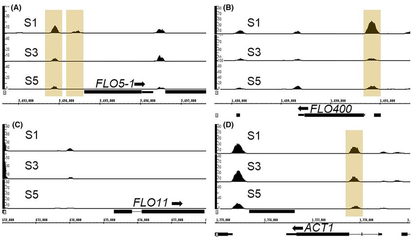
Fig. 1A. Komagataella phaffii CBS7435 forms pseudohyphae and
Flo11 and Bsc1 carry N-terminal FLO11 flocculin domains displays surface adherence only at slow growth rates in
(Fig. 1A) like their S. cerevisiae homologs while the majority of glucose-limited chemostats but not during cultivation
the other Flo proteins carry a PA14/GLEYA domain, which is a under glucose/nitrogen limitation or in presence of
carbohydrate binding domain found in fungal adhesins, con-
fusel alcohols
taining a conserved motif G(M/L)(E/A/N/Q) (Linder and Gustafs-
son 2008). In Flo5-1, Flo5-2, Flo100 and Flo200 the domain is Reported triggers of pseudohyphal growth in S. cerevisiae are
located closer to the N-terminus, while in Flo300 and in the pro- glucose or nitrogen limitation as well as the presence of fusel
tein encoded by PP7435 Chr4-1013 it is located toward the cen- alcohols such as butanol or isoamyl alcohol (Gimeno et al. 1992;
ter or closer to the C-terminus, respectively. Here, among the Dickinson 1996; Cullen and Sprague 2000; Lorenz et al. 2000).
PA14 domain containing Flo proteins, Flo5-1 and Flo5-2 have a To investigate whether these conditions provoke the same6 FEMS Yeast Research, 2020, Vol. 20, No. 5
Downloaded from https://academic.oup.com/femsyr/article/20/5/foaa044/5884885 by guest on 21 November 2020
Figure 1. Schematic representation of the structures of the K. phaffii FLO family proteins. (A) Line diagrams representing the Flo proteins with their predicted N-terminal
signal peptides, identified conserved domains and GPI anchor. (B) Representation of Flo8, master transcription regulator controlling several FLO genes harboring an
N-terminal Lis homology (LisH) domain, a putative single-stranded DNA-binding protein (SSDP) domain and a nuclear localization signal (NLS).
(Figure S1C and D, Supporting Information). Since these obser-
vations indicated that the conditions that trigger pseudohyphal
growth in S. cerevisiae are unable to provoke a similar response
in K. phaffii, we based further experiments on the conditions
where pseudohyphal growth was observed before, namely at
low dilution rates in glucose-limited chemostats (Rebnegger
et al. 2014).
We ran a glucose-limited chemostat first at a high dilution
rate (corresponding to μ = 0.1 h− 1 ) for ∼5 residence times (50 h)
corresponding to ∼7 generations, which serves as a control set-
point. Here, residence time refers to the amount of time taken
to change the entire volume of the chemostat. At this point all
the cells had an ovoid shape (S1 in Fig. 3A). Then the chemostat
Figure 2. Location of the FLO genes on the chromosomes of K. phaffii CBS7435. //
was switched to a low dilution rate (μ = 0.05 h−1 ). At this condi-
indicates the localization of the centromeres. tion, after ∼2 residence times (corresponding to 3 generations),
some cells in the population started to look more elongated (S2
response in K. phaffii, we exposed K. phaffii CBS7435 as well as in Fig. 3A). With increasing number of residence times at this
S. cerevisiae 1278b (serving as a control) to such triggers and low dilution rate, the effect became more pronounced. Addi-
analyzed their morphology by microscopy. tionally, some cells adopted a branched pseudohyphal appear-
We observed that cultivation on YP agar (without glucose) ance (S3 in Fig. 3A). Upon switching to the low dilution rate,
was unable to induce pseudohyphae formation in both S. cere- cells also started growing on the surface of the bioreactors,
visiae and K. phaffii (Figure S1A, Supporting Information). Cul- which became even more pronounced with increasing number
tivation on minimal agar medium containing 24 mM ammonia of residence times (Figure S2, Supporting Information). Strik-
induced pseudohyphal growth in S. cerevisiae but not in K. phaffii. ingly, when the dilution rate was switched back to μ = 0.1
Furthermore, when grown on minimal agar where the nitrogen h−1 , ∼17% of the cells in the population still exhibited an elon-
content was severely reduced (50 μM ammonia) K. phaffii still gated or pseudohyphal phenotype even after 7 generations/5
did not display pseudohyphae formation, while the S. cerevisiae residence times (S4 & S5 in Fig. 3A). This is striking, as at S5
strain was unable to grow under these conditions at all (Figure over 99% of the cells never experienced the slow pseudohyphae-
S1B, Supporting Information). Additionally, when cultivated in triggering growth rate (μ = 0.05 h−1 ) thus indicating that pseu-
the presence of butanol or isoamyl alcohol, K. phaffii again did dohyphae formation upon slow growth in K. phaffii is a heritable
not display any pseudohyphae formation contrary to S. cerevisiae phenotype.De et al. 7
Downloaded from https://academic.oup.com/femsyr/article/20/5/foaa044/5884885 by guest on 21 November 2020
Figure 3. Cultivation at low dilution rate (corresponding to μ = 0.05 h−1 ) in glucose-limited chemostat triggers pseudohyphal growth in K. phaffii, which seems to be
connected to the expression of FLO11. (A) Growth rate profile of the glucose-limited chemostat experiments, sampling time points (S1–S5) and cell morphology of the
wild-type and the flo8 strains. (B) The plot shows the transcript level of FLO11 in the wild-type and the flo8 strains, measured by qRT-PCR, relative to the reference
sample S1 in the wild type.
Pseudohyphal growth of K. phaffii is dependent on the switching the dilution rate back to 0.1 h−1 (sample S4), the
presence of Flo8 expression of FLO11 quickly dropped. However, even after five
residence times (sample S5), the FLO11 expression remained 1.5-
As mentioned before, the S. cerevisiae laboratory strain S288C fold higher compared to S1 (P-value < 0.05). In the flo8 strain,
does not show any adhesion-related phenotype because of a the FLO11 expression remained below detection level, indicating
single nonsense mutation in FLO8 (Liu et al. 1996). To confirm that activation of the gene is under control of Flo8.
that the pseudohyphal phenotype in K. phaffii is also dependent Based on the quantitative PCR results, we considered the
on the putative Flo8 homolog, we cultivated the K. phaffii flo8 possibility that, like in S. cerevisiae, FLO11 is also the sole gene
strain (Gasser et al. 2015) at the same conditions as the wild type. responsible for conferring the pseudohyphal phenotype in K.
In contrast to the wild-type strain, the flo8 strain showed no phaffii. To verify this, we constructed a flo11 strain and culti-
pseudohyphal phenotype and only very reduced surface growth vated it in biological duplicates applying the same chemostat
(Fig. 3A; Figure S2, Supporting Information). Hence the samples cultivation strategy as described before. As a control, we simul-
from the flo8 cultivations were used as controls for further taneously cultivated the wild-type strain. However, we observed
analysis. that unlike in S. cerevisiae, deleting FLO11 did not inhibit pseu-
For ease of understanding, henceforth the chemostat sam- dohyphal growth, although a cell count to compare the percent-
ples will be referred to using their sample names as given in age of pseudohyphal (elongated) to non-pseudohyphal (ovoid)
Fig. 3A (S1, S2, S3, etc.). cells in the wild-type and the flo11 strains revealed that dele-
tion of FLO11 impairs pseudohyphal growth (Fig. 4). After five
residence times at a dilution rate of 0.05 h−1 , ∼17% of the wild-
FLO11 is induced in conditions of pseudohyphal type population showed an elongated or pseudohyphal pheno-
growth, but FLO11 deletion does not prevent type, while only 7.5% of the flo11 population switched to such
pseudohyphae formation in K. phaffii a phenotype (Fig. 4) revealing that unlike in S. cerevisiae, FLO11 is
not the only player in conferring the pseudohyphal phenotype in
In S. cerevisiae, expression of FLO11 is required for pseudohy-
K. phaffii.
phal growth and biofilm formation (Rupp et al. 1999; Cullen and
Sprague 2012) and strains lacking the FLO11 gene are unable to
form pseudohyphae (Lo and Dranginis 1998). To verify whether Quantification of mRNA levels of FLO genes by qRT-PCR
the same gene also plays a crucial role in conferring the pseu-
and RNA-Seq
dohyphal phenotype in K. phaffii, we analyzed the expression
profile of FLO11 during different stages of the chemostat cul- To check the expression patterns of the other FLO genes at fast
tivations by employing quantitative real-time PCR (qRT-PCR) and slow growth, we measured their transcript levels at the dif-
(Fig. 3B). Compared to the reference sample S1 at 0.1 h−1 , FLO11 ferent growth rates in the wild-type as well as the flo8 strains
expression significantly increased at S2 and S3, concurrent with by qRT-PCR and RNA-Seq.
the induction of pseudohyphal growth. Consistent with our qRT-PCR data showed that out of the analyzed genes, BSC1,
observation that with an increasing number of residence times FLO5-1 and FLO400 seemed to be under the control of Flo8 and
at the low dilution rate the pseudohyphal phenotype became showed no expression in the flo8 strain, while FLO5-2, FLO200
more pronounced, expression of FLO11 also got stronger. Upon and PP7435 Chr4-0629 were unaffected by the lack of Flo8 (Fig. 5).8 FEMS Yeast Research, 2020, Vol. 20, No. 5
Four of the genes, BSC1, FLO5-1, FLO200 and FLO400 were down-
regulated at S3 in comparison to the reference sample S1. These
genes stayed repressed even at S5. Considering that >99% of the
cells present at S5 never experienced the slow pseudohyphae-
triggering growth rate, it is speculated that these genes under-
went stable repression.
Next, RNA-Seq was performed on the samples collected dur-
ing the chemostats of the wild-type at S1, S3 and S5 and the
flo8 strains at S1 and S3. Since the pseudohyphal phenotype is
observed only in the wild-type strain but not in flo8, we first
looked at the genes that are differentially regulated between
these strains. As mentioned in Materials and methods, tran-
scripts with a log2 fold-change below −1 and above 1, combined
Downloaded from https://academic.oup.com/femsyr/article/20/5/foaa044/5884885 by guest on 21 November 2020
with an adjusted P-value < 0.05 were considered to be signifi-
cant. Using these criteria, it was seen that 107 genes were upreg-
ulated in the wild-type strain compared to the flo8 strain (Table
S3, Supporting Information). Among the FLO genes this included
FLO11, FLO5-1, FLO400 and PP7435 Chr3-1237 (Table 2). On the
other hand, only 21 genes were downregulated in the wild-type
strain compared to the flo8 strain (Table S3, Supporting Infor-
mation). Analyzing the Gene Ontology terms enriched in this
list of differentially regulated genes using the open source soft-
Figure 4. Microscope pictures of both the wild-type and the flo11 strains at dif-
ware GO::TermFinder (Ashburner et al. 2000; Boyle et al. 2004),
ferent sampling time points. The graph shows the percentage of pseudohyphal
cells in the population for both the wild-type and the flo11 strains (n = 3).
we found that most of them are related to cellular response
to pheromone, reproductive processes and multi-organism pro-
cesses. Interestingly, we also found a big group of unannotated
genes (63 genes) in this list, which are not characterized so far.
The enriched GO terms and the genes belonging to these GO
terms are listed in Table S4 (Supporting Information).
We then looked specifically into the RNA-Seq data of the FLO
genes in the wild-type strain. Table 2 shows the average tran-
scripts per million (tpm) of the three biological replicates of each
sample. The data for ACT1 is also given for comparison. FLO400,
FLO5-1 and PP7435 Chr1-2104 were expressed at levels even
higher than that of ACT1. While the expression of PP7435 Chr1-
2104 did not undergo significant variation at the different stages
of the chemostat cultivation, the expression of FLO400 and FLO5-
1 changed upon switching to the slow growth rate. The high-
est expression levels for these two genes was reached initially
at S1. At S3, both genes were repressed and remained in their
repressed state even after switching back to the fast growth rate
(S5) confirming the pattern of gene expression that we observed
in the qRT-PCR data. PP7435 Chr3-1237 follows a similar expres-
sion and downregulation pattern as FLO400 and FLO5-1 but its
expression level, even at S1, is much lower.
FLO11 reached its highest expression during the slow growth
rate, but even then, the expression level was much lower than
the highest expression levels of FLO400 and FLO5-1. The expres-
sion level of FLO11 was again downregulated after the growth
rate was switched back, but remained >2-fold higher in S5 than
in S1, which reflected the pattern shown by the qRT-PCR data.
In the flo8 strain the expression of FLO400, FLO5-1, FLO11 and
PP7435 Chr3-1237 genes was highly hampered, confirming that
they are under the regulation of Flo8. PP7435 Chr4-0629 was sta-
bly expressed at a low level independent of the dilution rate.
Even though expressed at lower levels compared to FLO400 and
FLO5-1, FLO300 seems to be upregulated at the slow growth rate
Figure 5. qRT-PCR data showing expression pattern of several FLO genes rela- and shows even higher expression upon switching back to the
tive to the reference sample (wild type in S1). Asterisks above the bars denote fast growth rate, however, this expression pattern is similar in
statistical significance of gene expression levels in comparison with the refer-
the flo8 strain.
ence sample (P-values calculated by Student’s t-test; ∗∗ P-value < 0.01 and ∗∗∗ P-
We additionally created three different reporter strains
value < 0.001).
expressing eGFP-tagged versions of Flo11, Flo400 and Flo5-1,
respectively, and carried out similar glucose-limited chemostats
with these strains. The purpose of these reporter strains was toDe et al. 9
Table 2. RNA-Seq data of all FLO genes showing the average transcripts per million (tpm) values of the FLO genes.
Wild type μ = 0.05
Wild type μ = 0.1 h−1 h−1 Wild type μ = 0.1 h−1 flo8 μ = 0.1 h−1 flo8 μ = 0.05 h−1
Gene name S1 S3 S5 S1 S3
FLO11 6.5 58.8 18.1 0.4 0.3
FLO5-1 2156.4 666.3 419.5 47.5 47.9
FLO400 3431.2 1776.4 1029.1 339.4 113.5
BSC1 4.8 4.4 6.1 6.3 4.2
FLO5-2 16.4 12.0 10.7 14.9 12.7
FLO100 0.3 5.3 33.7 0.2 0.0
FLO200 8.1 5.1 5.7 9.7 6.3
FLO300 8.0 42.3 161.0 9.3 25.6
Downloaded from https://academic.oup.com/femsyr/article/20/5/foaa044/5884885 by guest on 21 November 2020
PP7435 Chr1-2104 2942.1 3308.8 3697.5 3977.6 3642.0
PP7435 Chr3-1237 36.0 13.1 17.7 3.1 7.9
PP7435 Chr2-0004 10.9 8.1 7.4 9.9 8.2
PP7435 Chr4-1013 16.3 13.8 14.5 17.8 12.9
PP7435 Chr4-0629 74.5 73.7 67.4 89.6 86.3
ACT1 1726.9 1601.2 1616.6 1704.7 1483.7
follow the change in expression pattern of these proteins and to and S3, and remained in the same state in S5. These peaks were
find out if these proteins are expressed in the entire population either detected in both S3 and S5 but not in S1 (Category I), or
or only in a subpopulation, since pseudohyphal cells only repre- only detected in S1 but not in S3 and S5 (Category II). Screen-
sent a fraction of the population. As expected, both Flo400-eGFP shots of IGB showing representative chromosome regions with
and Flo5-1-eGFP were detected only in the S1 sample. Addition- the different peak patterns are shown in Figure S4A–D (Support-
ally, while Flo400-eGFP is produced in all the cells of the popula- ing Information).
tion, Flo5-1-eGFP was expressed only in a fraction of cells, which Regions corresponding to stable changes occurring in the
was ∼11% of the population (Figure S3, Supporting Information). chromatin after switching to the slow growth rate and return-
Even though a similar N-terminal internal GFP tagging of Flo11 ing to the fast growth rate (S1 vs S3 & S5) according to the
has been described in S. cerevisiae (Lo and Dranginis 1996), unfor- FAIRE-Seq data were further analyzed. As explained above, Cat-
tunately the Flo11-eGFP expressing K. phaffii strain showed no egory I includes regions that were stably closed (nucleosome-
observable fluorescence, which was most probably due to the bound) upon switching to the slow growth rate, while Category
general low expression levels of this protein. II contains regions that were stably opened (nucleosome-free)
upon switching to the slow growth rate (Table S5 and Figure
S4E and F, Supporting Information). The number of peaks in
FAIRE-Seq analysis indicates involvement of chromatin
category I was much lower (around 25) compared to category
modulation in the regulation of FLO5-1 and FLO400
II (around 350). In category I, peaks upstream of two flocculin
It has been reported that in S. cerevisiae some of the members genes, namely, FLO400 and FLO5-1 were detected, which means,
of the FLO gene family are epigenetically silenced (Halme et al. the peaks upstream of these genes were detected only in sample
2004). The most well studied member in that regard is FLO11. S1 but not in S3 or S5, which indicate that the regions upstream
The FLO11 promoter, one of the largest described in S. cerevisiae of FLO400 and FLO5-1 undergo stable changes upon exposure to
with a size of around 3.5 kb, has binding sites for many differ- slow growth rate conditions. Screenshots of the MACS peak call-
ent regulators and its expression is controlled by multiple lev- ing data for these regions as visualized on IGB for the wild-type
els of conventional and epigenetic regulation (Madhani and Fink sample are shown in Fig. 6A and B (Stein et al. 2002; Zhang et al.
1997; Bumgarner et al. 2009; Octavio et al. 2009). The epigenetic 2008). This is in accordance with the changes in transcript lev-
regulation of FLO11 involves chromatin modulation via histone els of these two genes as seen from the qRT-PCR and RNA-Seq
modifications and nucleosome remodeling via the Rpd3L and data where the expression levels of both the genes are signifi-
Swi/Snf complex (Barrales et al. 2012). To investigate if and how cantly downregulated in S3 and S5 (P-value < 10−6 ) compared to
changes in chromatin accessibility occur at a genome-wide level S1. Other than FLO400 and FLO5-1, no peaks were identified in
of fast and slow growing K. phaffii, we carried out Formaldehyde the proximity of FLO11 (Fig. 6C) or any of the other FLO genes.
Assisted Isolation of Regulatory Elements (FAIRE-Seq) analysis The regions in category II, even though being many more
(Giresi and Lieb 2009) of samples S1, S3 and S5. FAIRE-Seq analy- than category I, did not yield any significantly enriched GO
sis allows for the identification of open (nucleosome-free) chro- terms. We looked at the genes that were proximal to these
matin regions, which include regulatory regions, at a genome- regions to specifically find genes that might be connected to the
wide level during each condition. pseudohyphal phenotype. While we did find some genes like
Peaks in the FAIRE-Seq data correspond to open chromatin MSB2, GTR2, MIT1, RSR1, KSS1 and MIG1-1 that are connected to
or nucleosome-free regions. Upon comparison and visualization the regulation of filamentous growth in S. cerevisiae, on correlat-
of FAIRE-seq peaks between S1, S3 and S5 using the genome ing these genes with the RNA-Seq data no significant upregula-
browser IGB, four different patterns were observed, namely, tion of these genes in S3 compared to S1 was seen.
peaks that were detected at the same level in S1, S3 and S5 (indi- Finally, the FAIRE-Seq data of the flo8 strain was compared
cating no chromatin modulation), peaks that were detected only with that of the wild-type strain at each time point but no sig-
at S3 (indicating open chromatin only at slow growth rate) and nificant differences between the open chromatin regions in the
peaks that underwent stable changes between the samples S1 wild-type and the flo8 strains were observed. This indicates10 FEMS Yeast Research, 2020, Vol. 20, No. 5
Downloaded from https://academic.oup.com/femsyr/article/20/5/foaa044/5884885 by guest on 21 November 2020
Figure 6. Open chromatin regions identified in the proximity of FLO genes by FAIRE-Seq analysis. Screenshot from IGB showing differential peaks upstream of FLO5-1
and FLO400 (highlighted in yellow); such statistically differential peaks are not observed in proximity of FLO11. ACT1 is included here as control where an upstream
open region is detected in all the three samples.
that Flo8 does not play any role in the modification of chromatin the specific trigger for transition into this phenotype as well as
structure. its regulation. Saccharomyces cerevisiae exhibits pseudohyphal
growth in both its diploid and haploid form in response to
nutrient limitation or presence of fusel alcohols. In contrast, K.
Deletion of FLO400 or FLO5-1 prevents pseudohyphal
phaffii, which preferentially exists as a haploid, shows no switch
growth and expression of FLO11 at slow growth rates
to the pseudohyphal phenotype when grown on glucose- or
Based on the regulation patterns observed for FLO400 and FLO5- nitrogen-limited agar plates or when cultivated in the presence
1 during cultivation at fast and slow growth rates, the FAIRE- of fusel alcohols (Figure S1, Supporting Information). N-depleted
Seq data and the fact that both of them are controlled by the K. phaffii were reported to rather undergo mating (Heistinger
Flo8 transcription factor, they were considered as possible can- et al. 2018). Until now, the only condition where K. phaffii has
didates that participate in the pseudohyphal phenotype. Thus, been shown to transition to pseudohyphal growth is at slow
we generated deletion strains of these two genes individually, growth rates in glucose-limited chemostats, first reported in
both in the wild-type as well as in the flo11 strains background. the study by Rebnegger et al. (2014) and subsequently confirmed
Again, we performed chemostat runs as described before with in our experiments. The initiation of the phenotype in K. phaffii
these four strains (flo5-1, flo5-1 flo11, flo400, flo400 flo11), seems to be tightly regulated as the appearance of the first
each in biological duplicates. We observed that in both the flo5- elongated cells did not occur immediately upon switching to
1 and flo400 strains pseudohyphae formation was absent, the slow growth rate conditions. Instead it required at least 2
in the wild-type as well as in the flo11 background (Fig. 7A). residence times—corresponding to ∼3 generations—until the
Strikingly, qRT-PCR for checking FLO11 expression in the flo400 first elongated cells appeared, although after its initiation the
and flo5-1 strains revealed that FLO11 transcript levels were phenotype became stronger with increasing residence times
strongly reduced in both the deletion strains, which indicates at the slow growth rate (Fig. 3A). It could be speculated that
that the presence of both Flo5-1 and Flo400 is necessary for the unlike in S. cerevisiae, K. phaffii requires prolonged exposure to
expression of FLO11 (Fig. 7B). glucose-limited conditions and/or more severe limitation to
initiate a switch in phenotype. This is consistent with the fact
that as a Crabtree-negative yeast, K. phaffii has a high-affinity
DISCUSSION glucose uptake system with a glucose saturation constant (Ks )
Pseudohyphal growth in K. phaffii is triggered under in the range of 9.7 μM at high growth rates (Prielhofer et al.
different environmental conditions compared with S. 2013), which is much lower than those of the high affinity
cerevisiae hexose transporters of S. cerevisiae where the Ks is around 1 mM
(Boles and Hollenberg 1997; Reifenberger et al. 1997).
Our data indicate that pseudohyphal growth behavior in K. Apart from pseudohyphal growth, slow growth rate con-
phaffii differs substantially from that in S. cerevisiae including ditions in the glucose-limited chemostat also lead to surfaceDe et al. 11
Downloaded from https://academic.oup.com/femsyr/article/20/5/foaa044/5884885 by guest on 21 November 2020
Figure 7. FLO5-1 and FLO400 are major players involved in the regulation of pseudohyphal growth. (A) Microscope pictures of flo5-1 and flo400 strains in both wild-
type and flo11 backgrounds at sampling time points S1, S3 and S5. (B) qRT-PCR data for FLO11 expression in the flo400 (right) and flo5-1 (left) strains relative to the
reference sample at S1.
adherence of cells indicating that such prolonged exposure to Flo8 is also an important player in the regulation of adhesion-
glucose limitation might also trigger other cell differentiations, related phenotypes. The non-adhering phenotype of flo8 might
thus leading to the formation of different subpopulations of also provide a beneficial performance in industrial bioprocesses.
cells, namely, ovoid, pseudohyphal and surface adhering cells.
This is not surprising since it is known from S. cerevisiae and FLO11 is not the only major gene responsible for
other yeasts that environmental stress can trigger multiple cel-
conferring pseudohyphal phenotype in K. phaffii
lular differentiations to maximize chances of survival. It has
already been contemplated that the pseudohyphal phenotype qRT-PCR analysis showed that the initiation of expression of
provides an advantage in nutrient-limiting conditions because FLO11 in K. phaffii is in accordance with the appearance and per-
the elongated shape enables yeasts to access distant nutri- sistence of the elongated/pseudohyphal cells. This implication
ents more efficiently (Honigberg 2016). Additionally, the higher was enforced by the observation that in the non-transitioning
surface-to-volume ratio of pseudohyphal cells provides more flo8 strain FLO11 expression cannot be detected (Fig. 3B and
space for absorption of nutrients (Adams et al. 1985). Adher- Table 2). Contrary to S. cerevisiae flo11 strains, the K. phaffii
ence to a foreign surface provides the cells with the capability flo11 strain could still switch to pseudohyphal growth, even
to invade the surface to forage for nutrients (Verstrepen and Klis though the number of cells switching to this phenotype was
2006). lower than in the wild-type strain (Fig. 4). So, it can be said that
The control flo8 strain could not initiate pseudohyphal while in K. phaffii FLO11 does play a role in pseudohyphae forma-
growth and also showed highly reduced surface growth in the tion, it is not the only player and clearly there is the involvement
bioreactor, which confirmed that, like in S. cerevisiae, in K. phaffii of other gene(s).12 FEMS Yeast Research, 2020, Vol. 20, No. 5
FLO400 and FLO5-1 are crucial players required for
Prolonged glucose limitation
initiation of pseudohyphal differentiation in K. phaffii
To identify which other gene(s) might be involved, we inves-
tigated the expression patterns of other FLO genes under the
same conditions. This revealed the genes FLO400 and FLO5-1
(Fig. 5; Table 2), which are expressed initially at the fast growth Flo8
rate, but undergo stable repression upon switching to the slow
growth rate. Both genes were downregulated in the flo8 strain.
FLO5-1 FLO400
Reporter strains expressing Flo400-eGFP and Flo5-1-eGFP con-
firmed this expression pattern, and showed that Flo400 was
expressed in all the cells of the population, while Flo5-1 was Flo5-1 Flo400
expressed in only a subpopulation of cells (Figure S3, Support-
Downloaded from https://academic.oup.com/femsyr/article/20/5/foaa044/5884885 by guest on 21 November 2020
ing Information). It remains to be discovered whether the cells
expressing Flo5-1 in S1 are those that transition to pseudohy-
phal growth in S3. FLO11
Since we found indications of stable repression of FLO5-1
and FLO400 triggered by prolonged glucose limitation, we per-
formed FAIRE-Seq analysis to investigate the involvement of
chromatin organization. Upstream open chromatin regions are Induction of pseudohyphal growth
usually associated with the proficiency of transcriptional acti-
vation of these genes. Open chromatin regions were identified
Figure 8. Scheme representing the proposed interplay of Flo8, Flo400, Flo5-1 and
upstream of FLO5-1 and FLO400 at the initial fast growth rate
Flo11 in the cascade leading to pseudohyphal growth in K. phaffii.
(S1) in accordance to their high expression levels in this condi-
tion. After switching to the slow growth rate, no upstream peaks
were seen anymore at S3 and S5, indicating that these chromatin
regions underwent stable conformational changes, which could expression pattern of FLO400 and FLO5-1 might seem contra-
be a factor regulating the expression pattern observed for these dictory to the observations in the knock-out strains in the first
two genes (Fig. 6A and B). It was shown that these chromatin place, we assume that the presence of Flo400 and/or Flo5-1 is
modulations occurred independently whether Flo8 was present needed to allow for activation—but not maintenance—of FLO11
or not. Thus, the question arises if these stable changes are a expression. While such a regulation mechanism for S. cerevisiae
result of histone modifications in these regions leading to epi- FLO11 is not known, it has been reported that two S. cerevisiae
genetic silencing of these genes as reported for FLO genes in S. signaling mucins, Hkr2 and Msb2 that are homologous to Flo11
cerevisiae. This can be verified by analyzing these regions further, are shed from cells and act as osmosensors in the yeast HOG
for example, using enChIP-MS (Fujita and Fujii 2013) that might pathway (Tatebayashi et al. 2007; Vadaie et al. 2008) and both
help to identify proteins interacting with these regions. of these mucins have been reported to differentially activate
Absence of FLO400 or FLO5-1 was sufficient for eliminating the filamentous growth pathway (Pitoniak et al. 2009). However,
pseudohyphae formation. Unlike the flo8 strain though, these while both Flo5-1 and Flo400 have threonine/proline rich inter-
knock-out strains still displayed surface adherence similar to nal repeat regions that are typical of mucins, unlike mucins nei-
the wild type. So, while not being involved in other morphogenic ther of the two proteins has a transmembrane domain. Interest-
behaviors such as surface adherence, both FLO400 and FLO5- ingly, during secretome analysis the Flo5-1 protein was found
1 seem to play a major role in the transition to pseudohyphal in the supernatants of K. phaffii cells in the later samples of
growth. Further work should aim at verifying if epigenetics and glucose- or methanol-limited fed batch cultivations, where cells
epigenetic factors are indeed involved in the observed chro- tend to adhere to the reactor surface as well (Burgard et al. 2020),
matin modulations. indicating that a mechanism of cellular release might also act in
K. phaffii.
Considering the essential role that both these genes play
in pseudohyphae formation and FLO11 expression, it could be
Flo400 and Flo5-1: actors upstream of FLO11?
speculated that Flo400 and Flo5-1 act as sensing proteins act-
Absence of FLO400 or FLO5-1 prevented transition to pseudohy- ing downstream of FLO8 and upstream of FLO11 in the signaling
phal growth and was found to correlate to lack of FLO11 induc- cascade leading to the formation of pseudohyphal cells (Fig. 8).
tion at slow growth rate. Therefore, it is logical to assume that It is interesting to note here that there is a rather large group
FLO400 and FLO5-1 act in concert with FLO11 to initiate pseudo- of non-annotated genes that seem to be regulated by Flo8 (i.e.
hyphal growth in a subpopulation of K. phaffii. In fact, our obser- 90 out of 209 differentially regulated genes in flo8 compared
vations indicate that FLO400 and FLO5-1 possibly act upstream to the wild-type strain at S3; Table S3, Supporting Information).
in the signal cascade leading to the expression of FLO11 (Fig. 7B) As it is possible that this list includes components of the sig-
since the effect of deleting these two genes is much more pro- nal transduction pathway that leads to cellular differentiation in
nounced than deleting FLO11. Although our experiments reveal response to glucose limitation, characterization of these genes
new information about adhesion-related cellular differentiation might help to provide further insight into the regulation of FLO
in K. phaffii, involvement of chromatin modulation and novel genes and the pseudohyphal phenotype in the future.
players in the regulation mechanism of pseudohyphal differ-
entiation, the mechanism by which Flo400, Flo5-1 and Flo11
SUPPLEMENTARY DATA
interact still remains unclear and the roles of these proteins in
pseudohyphal differentiation can only be speculated. While the Supplementary data are available at FEMSYR online.You can also read