The Role and Clinical Implications of the Retinoblastoma (RB)-E2F Pathway in Gastric Cancer
←
→
Page content transcription
If your browser does not render page correctly, please read the page content below
REVIEW
                                                                                                                                               published: 31 May 2021
                                                                                                                                       doi: 10.3389/fonc.2021.655630

                                              The Role and Clinical Implications of
                                              the Retinoblastoma (RB)-E2F
                                              Pathway in Gastric Cancer
                                              Tianyi Wu and Lizhao Wu *

                                              Department of Pathophysiology, College of Basic Medical Sciences, China Medical University, Shenyang, China

                                              Gastric cancer is the most common malignant tumor in the digestive tract, with very high
                                              morbidity and mortality in developing countries. The pathogenesis of gastric cancer is a
                                              complex biological process mediated by abnormal regulation of proto-oncogenes and
                                              tumor suppressor genes. Although there have been some in-depth studies on gastric
                                              cancer at the molecular level, the specific mechanism has not been fully elucidated. RB
                                              family proteins (including RB, p130, and p107) are involved in cell cycle regulation, a
                                              process that largely depends on members of the E2F gene family that encode
                                              transcriptional activators and repressors. In gastric cancer, inactivation of the RB-E2F
                       Edited by:             pathway serves as a core transcriptional mechanism that drives cell cycle progression, and
         Kanjoormana Aryan Manu,              is regulated by cyclins, cyclin-dependent kinases, cyclin-dependent kinase inhibitors, p53,
     Amala Cancer Research Centre,
                             India
                                              Helicobacter pylori and some other upstream molecules. The E2F proteins are encoded by
                        Reviewed by:
                                              eight genes (i.e. E2F1 to E2F8), each of which may play a specific role in gastric cancer.
                        Martin Fischer,       Interestingly, a single E2F such as E2F1 can activate or repress transcription, and enhance
               Fritz Lipmann Institute
                                              or inhibit cell proliferation, depending on the cell environment. Thus, the function of the E2F
                        (FLI), Germany
                     David N. Arnosti,        transcription factor family is very complex and needs further exploration. Importantly, the
            Michigan State University,        presence of H. pylori in stomach mucosa may affect the RB and p53 tumor suppressor
                         United States
                                              systems, thereby promoting the occurrence of gastric cancer. This review aims to
                  *Correspondence:
                          Lizhao Wu
                                              summarize recent research progress on important roles of the complex RB-E2F
                    lzwu@cmu.edu.cn           signaling network in the development and effective treatment of gastric cancer.
                                              Keywords: gastric cancer, RB-E2F pathway, pocket protein, E2F family, Helicobacter pylori, p53
                   Specialty section:
         This article was submitted to
             Gastrointestinal Cancers,
               a section of the journal
                 Frontiers in Oncology
                                              INTRODUCTION
         Received: 19 January 2021            Gastric cancer (GC) is a common type of gastrointestinal cancers. Worldwide it is the fifth most
           Accepted: 07 May 2021              frequently diagnosed cancer and the third leading cause of cancer death (1). Although activation of
           Published: 31 May 2021
                                              proto-oncogenes and inactivation of tumor suppressor genes are considered as driving forces for
                             Citation:        GC, the pathogenesis of GC is a complex biological process mediated by both abnormal regulation
     Wu T and Wu L (2021) The Role
                                              of multiple genes and environmental insults (2). In recent years, the incidence of GC in western
      and Clinical Implications of the
            Retinoblastoma (RB)-E2F
                                              countries has been reduced, but it is still a serious public health problem in developing countries
          Pathway in Gastric Cancer.          (1). Risk factors include Helicobacter pylori (H. pylori) infection, pickled food, smoking, obesity,
            Front. Oncol. 11:655630.          chronic gastritis, and iron deficiency (3, 4). The most commonly used classification of GC is the
     doi: 10.3389/fonc.2021.655630            two-category classification based on Lauren’s criteria: intestinal type and diffuse type, which are

Frontiers in Oncology | www.frontiersin.org                                         1                                         May 2021 | Volume 11 | Article 655630
Wu and Wu                                                                                                         RB-E2F Pathway in Gastric Cancer

different not only morphologically, but also clinically and                regulation of the RB-E2F pathway, are the most frequently
epidemiologically (5). The intestinal type is highly differentiated        mutated genes in GC, but are rarely mutated in IM (20).
with a distinct premalignant state during cancer development,              However, the exact mechanism of different genomic and
whereas the diffuse type is poorly differentiated lacking obvious          epigenetic alterations between IM and GC and their
premalignant lesions (5).                                                  application value in the prevention of GC still need to be
    It is well known that tumorigenesis is a complex biological            explored (20).
process usually mediated by polygenic mutations. The                           Diffuse gastric cancer (DGC) usually results from pangastritis,
retinoblastoma (RB) gene (i.e. RB1) is the first tumor                      has no atrophy and occurs mainly in younger female patients in
suppressor gene cloned in humans by positional cloning (6). It             low-risk areas (14, 15). DGC is poorly differentiated with
plays an important role in cell cycle regulation by regulating the         stronger metastasis and invasiveness, and is often associated
adenoviral early region 2 binding factor (E2F) transcription               with CDH1 deficiency (21). By exploring the co-expression
factor family (7–10). The RB-E2F pathway not only regulates                network of GC-related genes, an integrative functional
the cell cycle, but is also regulated by the cell cycle (10). In           genomics study group revealed differences between the two
essence, it links the cell cycle to the transcriptional machinery,         major subtypes of GC in transcriptional and epigenetic
and plays a major role in the control of cell growth, apoptosis and        regulations as well as in stem cell characteristics (22). IGC was
differentiation, biological processes that are implicated in cancer        believed to be more affected by E2F-mediated transcription (22).
development (9, 11).                                                       Considering different characteristics of the two subtypes,
    The role of RB family proteins in GC was last reviewed in              development of subtype-specific targeted treatment strategies
2010 (12). Although much progress has since been made in                   for GC deserves more attention.
understanding how the RB-E2F pathway is involved in the                        In addition to the aforementioned Lauren’s classification, GC
pathogenesis of GC, the specific role of E2F family members                 can also be divided into four molecular subtypes based on
and the RB-E2F pathway in GC has not been systematically                   analyzing the data from The Cancer Genome Atlas (TCGA)
reviewed since a review article on the role of E2Fs in cancers of          project (23). The first subtype is Epstein–Barr virus (EBV)-
digestive system was published in 2013 (13). In this review, we            associated GC, accounting for about 10% of GC (24). The
will discuss research progress on the role of RB and E2F family            association between EBV and GC was first recognized in 1990
members as well as their major upstream regulators in the                  (25). EBV has been shown to induce the nuclear export of E2F4
initiation, progression and prognosis of GC. In addition, we               and E2F5 to prevent cell cycle arrest, an action that may have
will also summarize major research findings on how H. pylori                implications for the pathogenesis of GC (26). The second subtype
infection impacts the development of GC by functionally                    is microsatellite unstable GC, accounting for 15–20% of GC. The
disrupting the RB and p53 tumor suppressor systems. Finally,               hallmark of this subtype is microsatellite instability (MSI),
we will discuss major clinical implications of this research               accompanied with increased gene mutation rates (23). The
progress in effective treatment of GC.                                     intracranial histological heterogeneity of GC with MSI was
                                                                           associated with progressive frameshift mutations of TGF-
                                                                           receptor type II and E2F-4 (27). High levels of MSI were more
                                                                           common in IGC and in the antrum, with better differentiation
GASTRIC CANCER                                                             and more lymphoid infiltration (28). The other two molecular
Intestinal gastric cancer (IGC) is thought to be initiated primarily       subtypes of GC are genomic stable (GS) GC and GC with
by H. pylori infection, with higher incidence in older men in              chromosomal instability (CIN) (23), which includes poorly
high-risk areas (14, 15). Well differentiated and poorly                   differentiated endocrine carcinomas that are often accompanied
differentiated gastric adenocarcinomas usually harbor different            with the inactivation of p53- and RB-related pathways (29).
genetic changes, with well-differentiated being more frequently            Interestingly, a similar study based on gene expression profiling
associated with changes in important cancer-related genes such             identified three subtypes of gastric adenocarcinoma: proliferative,
as RB and PTEN (16). IGC has a relatively clear development                metabolic, and mesenchymal, with the proliferative subtype being
process that is called metaplasia-neoplasia-carcinoma sequence             often associated with the activation of E2F-mediated pathway
or Correa’s cascade, from atrophic gastritis to intestinal                 (30). Since patients with different subtypes likely have different
metaplasia (IM) to dysplasia and then to IGC (17). IM is a                 clinical characteristics and molecular basis, they may benefit from
recognized premalignant lesion of gastric mucosa, defined as the            different treatments. Abnormalities of key components in the RB-
replacement of gastric mucosa by epithelial cells with intestinal          E2F pathway identified in patients with GC are summarized in
morphology, and is associated with an increased risk of GC (18,            Figure 1 and Table 1.
19). IM can be either complete (with the large-intestine
phenotype) or incomplete (with the small-intestine
phenotype), with the latter more frequently associated with                THE RB FAMILY
malignant transformation (18). In a 10-year prospective study
published in 2018, it was found that IM cells had both genetic             The RB family consists of three members in humans, which are
and epigenetic mutations that differed from GC cells (20). For             collectively referred to as “pocket proteins” and are involved in the
example, TP53 and ARID1A, which are involved in the                        regulation of the cell cycle (52). They are also involved in many

Frontiers in Oncology | www.frontiersin.org                            2                                     May 2021 | Volume 11 | Article 655630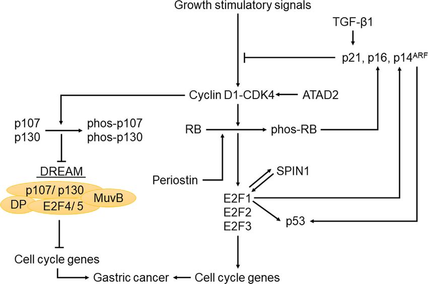
Wu and Wu                                                                                                                       RB-E2F Pathway in Gastric Cancer

 FIGURE 1 | Genetic and epigenetic mutations of genes in key components of the RB-E2F pathway that were identified in different subtypes of gastric cancer.

TABLE 1 | Abnormalities in key components of the RB-E2F pathway in patients with GC.

 Genes                                                                    Alteration prevalence (%)

                 mRNA high          mRNA low        Loss of protein         Positive immunostaining         Methylation           Mutation          Amplification

  E2F1              40 (31)            (−)                (−)                    22.2 (32), 63 (31)              (−)                (−)                 4 (31)
  E2F3                (−)            70 (31)              (−)                           (−)                      (−)                (−)                  (−)
  E2F4                (−)              (−)                (−)                           (−)                      (−)           31–33 (33, 34)            (−)
  E2F6                (−)              (−)                (−)                         46 (35)                    (−)                (−)                  (−)
   RB                 (−)              (−)           33–40 (36, 37)            53–70.2 (38–39–42)             17.9 (43)             (−)                  (−)
  p130                (−)              (−)                (−)                    nucleus: 25 (44)                (−)                (−)                  (−)
                                                                              cytoplasm: 76.05 (44)
cyclin D1          40.5 (45)           (−)                 (−)              37–72 (36–38, 40, 45, 46)            (−)                 (−)               16.6 (45)
 CDK4                 (−)              (−)                 (−)                       61.9 (40)                   (−)                 (−)                  (−)
  p16                 (−)              (−)        22 (47), 49 (36, 37)      27.5–58.3 (40, 41, 48, 49)        72.6 (43)              (−)                  (−)
 p14ARF               (−)              (−)                 (−)                       45.2 (40)                 24 (50)               (−)                  (−)
  p53                 (−)              (−)                 (−)                  39–64 (39, 42, 49)               (−)              44.4 (51)               (−)

(-) represents no published data.

biological processes such as proliferation, differentiation,                        G1/S genes, a process that RB plays a predominant role (69, 70).
senescence, apoptosis, gene regulation, and interact with many                      In the absence of RB and p130, p107 can also repress G1/S genes
other cellular proteins (53, 54). The eponymous member of the                       (69). Under the condition of DNA damage, p130 and p107 can
pocket protein gene family is RB1 or RB, which was named from an                    cooperate to repress the G2/M genes and thus block cell cycle
inherited eye tumor called retinoblastoma (55). The RB gene was                     entry into mitosis (69). In general, when DNA damage leads to p53
mapped on chromosome 13q14.2 (6). RB is widely distributed in                       activation, RB, p130 and p107 cooperatively repress G1/S genes
various tissues and interacts with a large number of transcription                  while p130 and p107 cooperatively repress G2/M genes (69). In
factors and chromatin-remodeling proteins, allowing itself to bind                  mice, pocket proteins have overlapping functions in suppressing
to transcription factors and to modify chromatin structure (56). In                 the development of various types of tumors. For example, RB and
addition to regulating the cell cycle, RB has also been shown to                    p107 worked together to suppress the development of
inhibit apoptosis (57). Consistent with an important role of RB in                  retinoblastoma (71, 72), head and neck cancers (73), and
tumorigenesis, loss of function of RB has been associated with the                  spontaneous skin tumors (74). In addition, RB and p130 worked
development of many human cancers (58–64).                                          together to suppress the development of retinoblastoma (75, 76).
    The second member of the family is p130, which was cloned in
1993 and mapped on chromosome 16q12.2 (65, 66). The third
member of the family is p107, which was mapped on 20q11.2 (67).                     THE E2F FAMILY
Interestingly, the three pocket proteins have overlapping and
interdependent functions (68). In both quiescent and p53                            The E2F family of transcription factors includes 10 members,
activation conditions, RB and p130 can cooperate to repress                         encoded by eight different genes, E2F1–E2F8 (9). E2F3 consists of

Frontiers in Oncology | www.frontiersin.org                                     3                                         May 2021 | Volume 11 | Article 655630
Wu and Wu RB-E2F Pathway in Gastric Cancer
two isoforms, E2F3a and E2F3b, derived from two different its phosphorylation status instead of just its mRNA or protein
promoters (77). Members of the E2F family have both distinct levels. In addition, since RB function is manifested at least in part
and overlapping functions, and are important for various through limiting activities of activator E2Fs, evaluating the RB
biological processes such as cell cycle control, cellular status in GC patient samples will benefit from simultaneously
proliferation and apoptosis (78, 79). E2F1–6 are canonical evaluating the status of activator E2Fs. It is interesting to note
E2Fs, which form heterodimers with dimerization partner that DNA methylation of the RB gene promoter was found in
(DP) proteins (80). E2F7 and E2F8 are atypical E2Fs, which do significantly more GC samples (17.9%) than in normal samples
not bind to DP but have two DNA binding domains (9). All E2F (5.5%) (43), suggesting that RB methylation may also play a role
members can bind to DNA in a sequence-specific manner to in GC.
initiate transcriptional activation or repression of target genes Although little is known about the precise role of p107 in GC,
(80). E2F1-3a are transcriptional activators, whereas E2F3b-8 are cellular localization of p130 seems to play an important role in
transcriptional repressors (9). However, it is worth noting that some aspects of GC. For example, high levels of nuclear
E2F3b can also act as a transcriptional activator (81, 82), even localization of p130 were significantly correlated with lower
though its expression pattern during the cell cycle is similar to grade GC, whereas high levels of cytoplasmic localization of
that of a canonical E2F repressor (77). The functional specificity p130 were significantly correlated with IGC (49). Besides, p130
of E2F-DP complex is determined by the E2F subunit, but in the was localized in the cytoplasm in DGC but in the nucleus in
absence of DP, E2Fs become non-functional (78). normal cells, further supporting an important role of its nuclear
In quiescent cells, pocket proteins can bind to E2F-DP delocalization in the development of GC (44). However, no
heterodimers to repress E2F target genes. It is worth noting correlation has been found between cytoplasmic localization of
that different pocket proteins preferentially bind to different E2F p130 and tumor grade or survival of DGC. Although the
transcription factors (83). RB binds to E2F1, E2F2, and E2F3 to functional consequence of p130 nuclear delocalization on the
form the repressive RB-E2F complex, while p107 and p130 bind development of GC is currently unclear, it is plausible that such
to E2F4 and E2F5 to form the repressive DREAM (DP, RB-like, delocalization promotes the development of GC through
E2F and MuvB) complex (10). E2F6, E2F7 and E2F8 are not inhibiting the function of p130 as a transcriptional modulator.
bound by pocket proteins (9). In G0 and early G1 phases, Further investigations are needed to experimentally determine
hypophosphorylated RB, which is in an activated state, binds the precise role of p130 nuclear delocalization in GC and its
to the pocket domain of E2F1-3 and inhibits E2F-mediated target underlying mechanisms.
gene activation, thereby blocking cell cycle progression at the Since the 2010 review, two significant advances have been
G1/S transition (84). In addition, E2F4 and E2F5 can form made in the understanding of pocket proteins in GC. The first
complexes with p107 and p130 to mediate gene repression interesting and important finding was that p130 was primarily
(84). When cells receive growth stimuli, activation of cyclin localized in the nucleus in normal cells but was mainly localized
dependent kinases (CDKs) leads to the phosphorylation of in the cytoplasm in DGC cells (44). Future studies should be
pocket proteins and collapse of the previously formed RB-E2F directed to understanding the precise role of p130 subcellular
complexes and DREAM complexes (70). The subsequent release localization in GC, the nucleo-cytoplasmic shuttling
of E2F1-3 from those complexes can activate target genes mechanisms, and whether p130 nuclear delocalization
required for cell cycle entry (10, 52). facilitates GC development by impairing p130-mediated
transcriptional repression. In addition, it has been found that
besides RB phosphorylation, RB promoter methylation may also
play a role in the development of GC (43), highlighting the
POCKET PROTEINS IN GASTRIC CANCER importance of epigenetic regulation of pocket proteins in GC. It
Various studies showed that RB plays important roles in the would be interesting to know whether RB promoter methylation
various aspects of GC. However, earlier studies focused on levels are different among various subtypes of GC, or among
evaluations of its protein levels in various contexts of GC different stages of GC development.
appeared to yield seemingly conflicting results. For example,
compared with non-neoplastic tissues, tumors could have higher
(38) or lower (48) levels of RB. In addition, altered RB protein THE E2F FAMILY IN GASTRIC CANCER
levels were more frequent in less-invasive GC than in advanced
invasive GC (85). In univariate and multivariate analyses, Among E2F family members, E2F1 is so far the most widely
positive RB expression was found to be significantly correlated studied in tumors, including GC. It is interesting to note that
with the presence of lymph node metastasis (39). Nevertheless, in vitro different levels of E2F1 had different effects on cell fate:
another study showed that the expression of RB in lymph node low levels of E2F1 could promote cell cycle progression, medium
metastasis was lower than that of the corresponding primary levels of E2F1 could cause cell cycle arrest, and high levels of
tumor (36). These inconsistent data may be related to the fact E2F1 could lead to cell apoptosis (86). Several earlier studies
that RB function is largely dependent on its posttranslational using either transgenic mouse models or in vitro systems showed
regulation (i.e. phosphorylation). Therefore, defining the precise that the role of E2F1 in tumorigenesis was pleiotropic,
role of RB in various processes of GC likely requires evaluation of manifested by the fact that it might either promote or suppress
Frontiers in Oncology | www.frontiersin.org 4 May 2021 | Volume 11 | Article 655630Wu and Wu RB-E2F Pathway in Gastric Cancer tumorigenesis, depending on dominant signaling pathways and activation of various cell death pathways, which may explain cell types (87–91). In GC, E2F1 gene amplification was rare, but the higher sensitivity of samples with high E2F expression to its overexpression was detected in about 40% of patients (31). radiotherapy and chemotherapy (109). Gene expression microarray data and bioinformatic analysis of Given the important role of E2F1 in GC, E2F1 has been public datasets also showed that E2F1 was up-regulated in GC considered as a potential therapeutic target for GC patients (93). (92, 93). In addition, high mRNA levels of E2F1 were related to However, since E2F1 activity is also important for normal poor survival (93). In order to better understand the role of E2F1 cellular proliferation, therapeutically targeting E2F1 may have in biological processes of GC, various research groups significant side effects on normal tissues that are capable of investigated effects of E2F1 overexpression or knockdown on proliferating. In addition, due to the highly overlapping and the tumorigenicity of GC cells. For instance, overexpression of compensatory effects of E2F activators (78), simple targeted E2F1 in MGC-803 GC cell line led to significantly increased intervention of E2F1 may lead to compensational upregulation levels of apoptosis but significantly reduced levels of cellular of other two E2F activators, making such a therapy less effective. proliferation and invasiveness, consistent with the tumor Therefore, E2F1 targeted therapy may require simultaneously suppressor function of E2F1 (94). In addition, overexpression targeting the other two E2F activators to achieve a better clinical of E2F1 suppressed tumor growth and promoted tumor cell outcome. Furthermore, the bidirectional effect of E2F1 on GC apoptosis in nude mice implanted with E2F1-overexpressing suggests that success on the targeted therapy is likely dependent MGC-803 cells (95). Furthermore, adenovirus-mediated on a clear understanding of the predominant oncogenic overexpression of E2F1 in AGS and SNU-1 GC cell lines pathways involved in individual patients. induced apoptosis and reduced cell survival rate (96). On the There are also several studies on E2F4 in GC. E2F4 mutation other hand, E2F1 downregulation by intratumor-injection of was found to be a common and an early event in the occurrence E2F1 shRNA in nude mice engrafted with MGC-803 cells of GC, and might occur in the process of precancerous lesions inhibited tumor growth and promoted apoptosis, accompanied such as IM and dysplasia (33, 34). E2F4 mutation in by up-regulation of PTEN, Caspase-3 and Caspase-9 (97). In gastrointestinal tumors might not be random as it appeared addition, in cisplatin-resistant SGC7901/DDP cells, shRNA- frequently in a microsatellite region at exon 7 with a serine- mediated E2F1 downregulation blocked cell cycle progression, encoding trinucleotide repeat sequence (33, 110). In addition, promoted apoptosis, and increased the sensitivity of cells to E2F4 frameshift mutation was associated with differentiation several chemotherapeutic drugs, suggesting that E2F1 served as grades of GC as frameshift mutation of the microsatellite regions an oncogene and promoted multidrug resistance in GC (98). encoding serine repeats might inhibit the formation of RB-E2F4 Although the dual role of E2F1 in GC is likely context dependent, complex and reduce the level of differentiation (27). mechanisms underlying the seemingly inconsistent results are Furthermore, a study of MSI suggested that E2F4 might be provided from the existing literature. For example, E2F1 is involved in the transformation of gastric adenocarcinoma into considered as a proto-oncogene, and elevated E2F1 levels are squamous cell carcinoma (111). Interestingly, by establishing an sufficient to drive cell proliferation and cell cycle progression (99, E2F-related transcriptional regulatory network, a research group 100), which can also explain E2F1 overexpression corresponding found that target genes regulated by E2F1 and E2F4 showed a to poor prognosis (101). On the other hand, the tumor large number of differential expressions in GC, indicating that suppressive effect of E2F1 can be explained by E2F1-mediated E2F1 and E2F4 might play important roles in tumorigenesis of apoptosis and growth arrest (102–104). E2F1 can inhibit the GC (92). It was found that E2F4 mRNA levels increased with the degradation of p53 by inducing the expression of p14ARF, leading degree of tumor invasion and malignancy (92). Bioinformatic to increased apoptosis and cell cycle arrest (105). In addition, analysis of a Gene Expression Profiling Interactive Analysis E2F1 can also induce p53 independent of p14ARF (103). This also (GEPIA2) dataset representing 408 GC samples and 211 explains why RB-negative tumors tend to be p53 negative, normal tissues showed that there was no difference in average probably to avoid the negative E2F1-p14ARF feedback (106). expression levels of E2F4 between GC samples and normal Interestingly, E2F1 protein levels could also reflect the tissues, but bioinformatics analysis using a completely different sensitivity of GC patients to adjuvant chemoradiotherapy after and consolidated Gene Expression Omnibus (GEO) dataset radical gastrectomy. For example, among postoperative patients representing a much larger sample size (i.e. up to 876 GC receiving adjuvant chemoradiotherapy, the E2F1 immuno- samples) showed that patients with relatively high E2F4 positive group had a higher survival rate than the E2F1 expression had worse survival than those with relatively low immuno-negative group (32). The immunopositivity of E2F1 E2F4 expression (93). As a member of the DREAM complex, might be used as an indicator for good response for adjuvant E2F4 can repress many cell cycle genes (112), which are common chemoradiotherapy and radiotherapy after surgery (107). A key markers of proliferation that can stratify most cancers, including determinant of the efficacy of anticancer therapies is the ability of GC (101). Although both high expression of E2F4 in advanced cancer cells to undergo apoptosis in response to DNA damage GC and its correlation with poor prognosis are seemingly factors (108). The success of radiotherapy or chemotherapy is at contradictory to the repressive role of E2F4 in cell cycle least partly due to the fact that cancer cells are more likely than control, there are existing studies supporting an oncogenic role normal cells to die when induced by DNA damage. Under the of E2F4. For example, E2F4 induced proliferation and promoted condition of DNA damage, E2F1 induces apoptosis through the development of skin tumors in a keratin 5 promoter-driven Frontiers in Oncology | www.frontiersin.org 5 May 2021 | Volume 11 | Article 655630
Wu and Wu                                                                                                       RB-E2F Pathway in Gastric Cancer

E2F4 transgenic mice (113). In addition, E2F4 reduced apoptosis           CDKs, which are in turn regulated by cyclins and cyclin-
in cardiac myocytes (114). However, more investigations should            dependent kinase inhibitors (CKIs) (116, 117). Therefore,
be done to explore the precise role and underlying cellular and           cyclins, CDKs, and CKIs as well as any molecules that regulate
molecular mechanisms of E2F4 in GC initiation and progression,            these three types of proteins may be involved in the pathogenesis
and to determine whether E2F4 overexpression is associated with           of GC.
a specific subtype of GC.                                                      The cyclin D1 protein was almost undetectable in normal
    Compared to E2F1 and E2F4, other E2F family members have              gastric mucosa, but was elevated in about half of GC cases,
been much less studied regarding their potential roles in GC.             indicating that overexpression of cyclin D1 might be an early
Gene expression microarray data showed that mRNA levels of                event in the process of tumorigenesis in GC (45, 46). The p16
E2F2 in GC samples were increased compared with those in                  gene, also known as p16INK4a, is located on chromosome 9p21
normal samples (92). Using Northern blot technique to analyze             (118) and encodes for a protein that is an inhibitor of CDK4
30 GC samples and their corresponding non-neoplastic mucosa,              (119, 120). As a CKI, p16 is able to competitively block the cyclin
a Japanese research group found that mRNA levels of E2F3 were             D1-CDK4 complex by binding to CDK4, an action that inhibits
lower in 70% of GC samples than in normal controls (31). In               CDK4-mediated RB phosphorylation and prevents cell cycle
contrast, bioinformatic analysis of RNA sequencing data from a            progression from G1 to S phase (118). Loss of p16 function
GEPIA2 dataset representing a much larger sample size (i.e. 408           leads to an abnormal increase in cyclin D1-CDK4 complex
GC samples and 211 normal gastric tissues) showed that                    activity, resulting in sustained RB phosphorylation (118). At
expression levels of E2F3, along with E2F2, E2F5, E2F7 and                the same time, phosphorylation of RB in G1 phase results in
E2F8, were significantly higher in GC samples than those in                increased expression of p16 to limit CDK4 activity (118). This
normal tissues (93). These conflicting results on E2F3 expression          negative feedback loop of p16 and RB is critical for normal cell
levels from the two studies may be due to differences in patients’        cycle control to protect cells from abnormal cellular
genetic background (i.e. mostly Japanese vs. mostly Caucasians            proliferation. Therefore, deregulation of key components in the
and African Americans) and/or techniques (i.e. Northern blot vs.          feedback loop is likely associated with the development of GC.
RNA sequencing) used to evaluate E2F3 expression levels.                  For example, various p16 abnormalities have been identified in
Moreover, high levels of E2F2, E2F3, E2F5, E2F6, E2F7 and                 GC patient samples. An early study showed that about 50% of
E2F8 were related to better survivals (93). E2F6 was localized in         GC samples were detected with the loss of p16 expression (36).
the nucleus, and was at high levels in gastric adenocarcinoma             Interestingly, the expression of p16 in distal gastric carcinomas
without lymph node metastasis (35). Similarly, univariate                 was higher than that in gastric cardia carcinomas (40). In GC,
analysis showed that the expression of E2F6 was negatively                abnormal methylation of CpG islands in the promoter region of
correlated with lymph node metastasis, suggesting that E2F6               p16 downregulated p16 (47). Methylation of p16 was present in
might suppress the metastasis of GC (35). Thus it is clear that           about 70% of GC samples, while there was almost no p16
considerably more study is warranted to investigate the role and          methylation in normal samples (43). In addition, methylation
mechanism of E2F2, E2F3, E2F5, E2F6, E2F7 and E2F8 in GC.                 of p16 was found in both IGC and DGC, but had no significant
For example, it would be interesting to know whether all or some          correlation with either tumor staging or histology (50). It is
of the aforementioned upregulated E2F factors (93) are                    worth noting that hypermethylation of p16 significantly
coordinately overexpressed in GC samples.                                 increased in MSI-high GC (121). Furthermore, p16
    Since the summary of the role of E2F transcription factors in         hypermethylation is also very common in EBV-associated GC,
digestive tract malignancies in 2013, much progress has been              and may even be one of the important causes of EBV-associated
made in understanding the roles of E2F family members in GC.              GC (122, 123). The expression levels of p16 and RB were not only
There have been more data to explain the bidirectional effect of          altered in GC, but also negatively correlated (41, 124, 125).
E2F1 on GC. In addition, the relationship between E2F1 and                    Several other upstream regulators in the RB-E2F pathway
better chemoradiotherapeutic response in GC has been                      have also been implicated in GC. For example, transforming
established. It is worth pointing out that the bidirectional effect       growth factor-beta1 (TGF-b1) inhibited GC cell growth by
of E2F1 and its effect on chemoradiotherapeutic sensitivity have          upregulating its downstream target p21, thereby blocking p130
also been found in many other tumors (109, 115). Furthermore,             phosphorylation and preventing aberrant cell cycle progression
the application of bioinformatics has facilitated our understanding       by downmodulating CDK activities (126). In addition, the tumor
of GC-specific genetic alterations in various E2F members as well          suppressor function of periostin was achieved by its induction of
as their prognostic and other clinical implications.                      RB phosphorylation and the subsequent release of E2F1, which
                                                                          activated its target gene p14ARF, leading to the inactivation of
                                                                          MDM2 and the consequential reduced ubiquitination of p53 and
UPSTREAM REGULATORS OF THE                                                E-cadherin (127). Moreover, SPIN1 could form a positive
RB-E2F PATHWAY IN GASTRIC CANCER                                          feedback loop with E2F1 to promote the development of GC
                                                                          (128). Furthermore, ATAD2 knockdown in GC cells led to
Many upstream regulators of the RB-E2F pathway also play                  reduced levels of cyclin D1, cyclin E, E2F1 and RB
important roles in GC. The activities of RB and other pocket              phosphorylation, thus inhibiting proliferation and cell cycle
proteins are mainly regulated by phosphorylation through                  progression (129). Interestingly, decrease of intracellular

Frontiers in Oncology | www.frontiersin.org                           6                                    May 2021 | Volume 11 | Article 655630
Wu and Wu RB-E2F Pathway in Gastric Cancer
chloride ion concentration could increase the level of p21 and by binding to the apoptosis stimulating proteins of p53 (ASPP)
reduce the phosphorylation of CDK2 and RB (130). This effect (133, 134). Like RB, the p53 tumor suppressor also controls cell
led to cell cycle arrest and inhibited the growth of tumor cells, cycle but through independent and interrelated pathways (135).
providing us with a new therapeutic strategy (130). The fact that Therefore, it is not surprising that alterations of TP53 and RB are
many upstream molecules of the RB-E2F pathway have a large common events in human GC. It was reported that TP53 gene
proportion of genetic and epigenetic alterations in GC (Figure 1 mutations were found in about 50% of GC cases (51).
and Table 1) suggests that in addition to the downstream H. pylori is a Gram-negative bacterium that was found in
effectors of RB such as activator E2Fs, its upstream regulators stomach mucosa, and is an important risk factor for GC,
such as p16 and cyclin D1 also play important roles in GC, with equivalent to a type I carcinogen (136). About half of the
the specific regulation network shown in Figure 2. Therefore, population in this world has H. pylori infection, and infection
understanding the status of the upstream regulators of RB will rates in Asian countries are generally higher than those in
not only further help us better understand the functional role of western countries (137). H. pylori could cause abnormal DNA
RB in various process of GC, but may also provide additional methylation and inflammation, which increased the risk of GC
insights on the diagnosis, prognosis and effective treatment (138). However, there was no significant difference in the rates of
of GC. H. pylori infection either between IGC and DGC, or between
proximal and distal tumors (139). Interestingly, high levels of RB
methylation in H. pylori-positive individuals might increase the
risk of GC (140). The proportions of RB tumor suppressor and
HELICOBACTER PYLORI AND THE RB the p53 tumor suppressor pathway abnormalities in H. pylori-
AND P53 TUMOR SUPPRESSOR infected GC were higher than that in non-H. pylori-infected GC
SYSTEMS (42). It was reported that H. pylori infection might first activate
C-MYC and BCL-2 in IM, and then inactivate the RB and p53
The p53 tumor suppressor gene, also known as TP53, was first tumor suppressor pathways in dysplasia, causing a severe
discovered in 1979 (131). In cells under non-stressed condition, imbalance of proliferation and apoptosis in precancerous
p53 is usually present in small amounts (132). However, in the lesions, leading to the occurrence of GC (42).
case of stress, such as hypoxia, DNA damage, proto-oncogene The pathogenicity of H. pylori is mainly due to its flagellum,
activation, radiotherapy and chemotherapy, p53 protein is lipopolysaccharide, vacuolar toxin VacA, and cytotoxin-related
stabilized to initiate a damage response cascade (132). If the gene pathogenicity island (cagPAI) (141–143). VacA could
damage cannot be repaired in time, p53 would induce apoptosis generate a protective intracellular reservoir where H. pylori
FIGURE 2 | The regulatory network of the RB-E2F pathway in gastric cancer. When cells receive growth stimuli, activation of CDKs leads to the phosphorylation of
pocket proteins, collapse of the previously formed RB-E2F complexes and DREAM complexes. The subsequent release of E2F1-3 can activate target genes required
for cell cycle entry. Meanwhile, this pathway is also regulated by upstream and downstream molecules in gastric cancer, such as CKIs, p53, TGF-b1, ATAD2
and SPIN1.
Frontiers in Oncology | www.frontiersin.org 7 May 2021 | Volume 11 | Article 655630Wu and Wu RB-E2F Pathway in Gastric Cancer
survives by usurping lysosomal and autophagy pathways. target genes, leading to the inhibition of apoptosis indirectly
Besides, it was found that gastric epithelial cell apoptosis (155). There is also cross talk between p53 and NF-kB pathway
induced by VacA did not require RB regulation, and occurred that results in reduced apoptosis and the occurrence of tumor
whether or not p53 was expressed (144). The most important (156). Besides, some H. pylori products were associated with the
and widely studied virulence factor of H. pylori strains is RB-E2F pathway in vitro (157, 158). By isolating and cloning
cytotoxin-associated protein (CagA). CagA is introduced into genes encoding for the two secretory H. pylori proteins CagA and
gastric epithelial cells by the type IV secretion system (T4SS), HspB, and transfecting them into AGS cell line, researchers found
leading to the promotion of genetic instability, epithelial– that CagA and HspB directly promoted the growth of GC cells by
mesenchymal transformation and eventual carcinogenesis facilitating G1-S transition of the cell cycle through the
(145). In addition, CagA bound to ASPP2, thereby inhibiting upregulation of cyclin D3 and subsequent RB phosphorylation
the binding of ASPP2 to p53, leading to decreased apoptosis and (158). Interestingly, unknown soluble factor(s) released by H.
promoting the formation of GC (146, 147). pylori in cell culture medium might inhibit RB phosphorylation
H. pylori could activate the PI3K/AKT pathway (148, 149) or by increasing the level of p27, leading to inhibition of cell cycle
the MAPK/ERK pathway (150) to activate the ubiquitin ligases progression in gastric epithelial cells (157). The primary
murine double minute (MDM2, also known as HDM2), which interactions between H. pylori and the RB and p53 tumor
promoted ubiquitination and proteasomal degradation of p53. suppressor pathways are summarized in Figure 3.
On the other hand, p53 can activate MDM2 to form a negative Studies based on clinical practice have shown that treatment
feedback loop that ensures low levels of p53 in unstressed cells of H. pylori reduced the risk of precancerous lesions converting
(151). Related proteins of the p53-MDM2 feedback loop were to GC, but the degree of risk reduction depended on the
distinctly expressed at different stages of GC development (152). population and the extent of damage already present at the
In the case of H. pylori infection, MDM2 expression was found to time of eradication (159–161). The close association between H.
be significantly elevated in the progression from chronic gastritis pylori and the RB and p53 suppressor pathways provides us with
to GC (152). Interestingly, MDM2 was bound to RB through a the possibility of combinational therapy, such as H. pylori
central acidic domain in U20S, C33A, SAOS-2 cells (153), and eradication combined with targeted intervention of MDM2 or
could promote proteasomal degradation of RB in cells of other related molecules, which may greatly improve the
osteosarcoma, cervical cancer, non-small cell lung cancer and therapeutic effect. Interestingly, the positive index of E2F
temperature-sensitive murine ts20 cells (153, 154). nuclear staining was higher in H. pylori-infected gastric
H. pylori could also induce a subtype-specific damage response mucosa than in non-infected gastritis samples, and E2F1 was
mechanism of p53 in a T4SS-dependent manner (155). co-localized with proliferating cell nuclear antigen (PCNA)
Specifically, H. pylori induced D133p53 and D160p53, which (162). The positive index of E2F1 decreased after H. pylori
encode for N-terminally truncated isoforms of p53 protein, successful eradication (162). This study suggests that enhanced
thereby inhibiting the activity of p53. D133p53 also activated expression of E2F may be involved in the occurrence and
the NF-kB pathway and caused up-regulation of its downstream development of H. pylori-infected GC by promoting cell cycle
FIGURE 3 | Regulation of p53 and RB by H. pylori in stomach. The virulence factor CagA is introduced into gastric epithelial cells by T4SS. CagA binds to ASPP2,
thereby inhibiting the binding of ASPP2 to p53, leading to decreased apoptosis. Besides, MDM2 is activated through the AKT/ERK pathway and form a negative
feedback loop with p53. MDM2 can bind to RB and inhibits the function of RB-E2F repressor. Meanwhile, the expression of the isoforms of p53 inhibits the activity
of p53 or activates NF-kB target genes. The outcome is the down-regulation of apoptosis and the occurrence of gastric cancer.
Frontiers in Oncology | www.frontiersin.org 8 May 2021 | Volume 11 | Article 655630Wu and Wu RB-E2F Pathway in Gastric Cancer
progression (162). Therefore, we speculate that E2F-targeted with a single drug are likely to develop drug resistance, but
therapy may be more effective in patients with H. pylori- combinations of drugs are more effective in controlling the
infected gastritis and H. pylori-infected GC, and has the disease. For example, palbociclib had a synergistic effect with
potential to be applied in the prevention and treatment of GC. 5-FU in the treatment of GC cells (169). Combination of human
epidermal growth factor receptor 2 (HER2) inhibitor pyrotinib and
CDK4/6 inhibitor SHR6390 was thought to be a more effective
treatment strategy for HER2-positive metastatic GC (171). In
POTENTIAL THERAPEUTIC TARGETS OF Table 2, we summarize the CKIs currently used in clinical trials
THE RB-E2F PATHWAY IN GASTRIC and preclinical studies for GC. Notably, CDK4/6 inhibitor relies on
CANCER RB to induce cell growth arrest (172). In order to improve
therapeutic efficacy and precision, we need to develop new
Because GC is often asymptomatic in the early to middle stage of therapeutic strategies, such as the use of multiple CDK4/6
the disease progression, it is often diagnosed at an advanced stage inhibitors to enhance cell cycle arrest and selective targeting of
with limited treatment options. Currently the primary treatment RB-deficient tumors (172). Immunotherapy based on immune
strategy for GC is still surgeries, complemented with checkpoint block is being applied in the clinical treatment of
chemotherapy and radiotherapy (163). Most patients still have advanced GC, such as anti-PD-1 therapy (163). However, most
low survival rates and high recurrence rates (163). Therefore, it is GC cases are not sensitive to immune checkpoint inhibitor
particularly important to find more effective treatment strategies monotherapy, so patients may need combinational therapies to
and preventive measures for GC. Through studies on the function improve response to the PD-1 therapy or other immune
of tumor suppressor genes and mechanisms of related pathways checkpoint inhibitors (163). If CDK4/6 inhibitors can be
such as the RB-E2F pathway, we may be able to find novel combined with immunotherapy in the treatment of GC in the
therapeutic targets and develop more effective treatment future, perhaps a better therapeutic effect will be achieved.
strategies for GC. Antagonists of CDK can block the action of Using an E2F promoter-regulated adenovirus carrying the
the cyclin D1-CDK4 complex to target the RB-E2F pathway for p16 gene could combine the apoptosis induced by p16 gene and
cancer therapy (164). Flavopiridol is a broad-spectrum CKI oncolysis induced by virus replication to have antitumor effect on
commonly used in clinical practice of solid tumors (165). A GC (173). This kind of replication-competent adenovirus
phase II clinical trial showed that flavopiridol alone had no (RCAd) provided a new view of cancer therapies (173). At
significant antitumor effect on advanced GC (166), pending present, oncolytic virus has become an active research field on
changes in regimen and combination with other agents. Selective cancer targeted therapies (174). Advances in genetic engineering
CDK4/6 inhibitors palbociclib, ribociclib and abemaciclib have can help scientists create oncolytic viruses that target cancer cells
been developed and are undergoing clinical trials in a variety of with different types of mutations to achieve better therapeutic
cancers (167). Palbociclib is in phase II trials in patients with effects (174). If oncolytic virus and immunotherapy are properly
advanced GC with limited single-agent activity (168). Some of the combined in GC, it is possible to achieve a synergistic anti-cancer
genetic characteristics of GC help us stratify patients for the most effect. However, since there are significant uncertainties on
effective drug therapies. Studies showed that high levels of cyclin potential side effects and viral penetration efficiencies in solid
E protein in GC correspond to increased resistance to palbociclib tumors like GC, such a therapy option still has a long way to go
(169). The methylation of p16 increased the sensitivity of GC cells before it can be used in clinical practice of GC.
to abemaciclib, suggesting that abemaciclib is more effective H. pylori eradication therapy has been widely used in clinical
in patients with hypermethylated p16 (170). Targeted therapies practice and significantly reduced the risk of GC (160, 175).
TABLE 2 | CKIs for clinical trials and preclinical studies in GC.
CKIs Status Subjects Settings Results
Flavopiridol Phase II clinical trial 16 advanced gastric carcinoma single-agent No anti-tumor activity unexpected side effect
(166) patients administration
Palbociclib Phase II clinical trial 29 advanced gastro-esophageal single-agent Limited anti-tumor activity
(168) cancer patients administration
Palbociclib Preclinical research GC cell lines cyclin E Elevated resistance
(169) overexpression
Palbociclib Preclinical research GC cell lines Combined with 5-FU Better anti-tumor effect
(169)
Abemaciclib Preclinical research 146 GC patients & GC cell lines p16 hypermethylation Elevated sensitivity
(170)
SHR6390 Preclinical research GC cell lines & AVATAR mice Combined with Better anti-tumor effect
(171) pyrotinib
SHR6390 Phase I clinical trial fives GC patients Combined with PR in three patients, SD in one patient, PD in one patient
(171) (ongoing) pyrotinib (until June 2020)
PR, partial response; SD, stable disease; PD, progressive disease.
Frontiers in Oncology | www.frontiersin.org 9 May 2021 | Volume 11 | Article 655630Wu and Wu RB-E2F Pathway in Gastric Cancer
An experiment using H. pylori-infected p27-deficient mice data on the dual role of E2F1 in GC came from GC cell lines or
showed that H. pylori eradication through an antibiotic xenograft mouse models, using transgenic mouse models will
combinational therapy could reduce gastric inflammation and likely provide more significant insights on how E2F1 is involved
hinder precancerous lesions such as gastric ulcer and dysplasia, in the various process of GC, including initiation, progression,
thus preventing GC (176). Interestingly, H. pylori induced and drug resistance. In view of the importance of H. pylori in GC
cytoplasmic localization of p27, resulting in loss of tumor and its complex interaction with the RB-E2F pathway, H. pylori
suppressor function of p27 and correspondingly poor eradication therapy combined with targeted therapy may achieve
prognosis of patients (177). CDK4/6 inhibitors play the same more effective therapeutic outcomes. It is worth noting that the
role as p27, so patients with p27 cytoplasmic localization after molecular typing of GC is an important advance as we are now
H. pylori infection may respond better to CDK4/6 inhibitors. In able to tailor our studies to different genetic alterations for each
addition, H. pylori activates many cell cycle-related genes, such as molecular subtype, thereby facilitating precision medicine. In the
E2F1 and cyclin D1 (178), suggesting that current CDK4/6 clinical application of RB-E2F pathway for GC, essentially all
inhibitors and potential RB-E2F targeting agents may be more targeted therapy options remain in preclinical stages or in clinical
effective in H. pylori-infected patients. H. pylori infection can also trials. Therefore, there is an urgent need to facilitate our research
change the epigenetics of cells, such as increased p16 methylation efforts on translating research data into clinical practice.
(179). In this regard, H. pylori-infected GC patients may be more Nevertheless, targeting key components of the RB-E2F
sensitive to abemaciclib (170). pathway for the development of more effective therapies of GC
Intervention of RB-E2F pathway has not been commonly offers both significant promises and challenges to the
used in GC as virtually all such options remain in preclinical scientific community.
stages or in clinical trials. Nevertheless, targeted therapies based
on key components of the RB-E2F pathway, combined with
immunotherapy, oncolytic viral therapy, and/or H. pylori
eradication are likely a viable option for developing more AUTHOR CONTRIBUTIONS
effective treatment strategies for GC. Both authors (TW and LW) prepared, revised and edited this
manuscript. All authors contributed to the article and approved
the submitted version.
CONCLUSIONS AND PERSPECTIVE
In summary, RB-E2F pathway plays important roles in the
occurrence and development of GC. Current understanding on FUNDING
the role and mechanism of the three pocket proteins in GC are
This work was supported by the start-up funds from the China
insufficient, especially for p107. The functional consequences of
Medical University.
epigenetic regulation of RB and cytoplasmic localization of p130,
as well as the cooperative functions of these three pocket proteins
in GC have great potentials to be explored. Most studies
involving E2Fs in GC have focused on E2F1 and E2F4. We ACKNOWLEDGMENTS
need a better understanding of the roles of other E2F family
members in GC. In addition, whether E2F1 can be used as a We would like to acknowledge all members in Dr. Wu’s
viable therapeutic target remain to be determined. Since current laboratory for their critical comments on this manuscript.
REFERENCES 6. Lee WH, Bookstein R, Hong F, Young LJ, Shew JY, Lee EY. Human
Retinoblastoma Susceptibility Gene: Cloning, Identification, and Sequence.
1. Bray F, Ferlay J, Soerjomataram I, Siegel RL, Torre LA, Jemal A. Global Science (1987) 235:1394–9. doi: 10.1126/science.3823889
Cancer Statistics 2018: GLOBOCAN Estimates of Incidence and Mortality 7. Kovesdi I, Reichel R, Nevins JR. Identification of a Cellular Transcription
Worldwide for 36 Cancers in 185 Countries. CA Cancer J Clin (2018) Factor Involved in E1A Trans-Activation. Cell (1986) 45:219–28. doi:
68:394–424. doi: 10.3322/caac.21492 10.1016/0092-8674(86)90386-7
2. Tan P, Yeoh KG. Genetics and Molecular Pathogenesis of Gastric 8. Yee AS, Reichel R, Kovesdi I, Nevins JR. Promoter Interaction of the E1A-inducible
Adenocarcinoma. Gastroenterology (2015) 149:1153–62.e3. doi: 10.1053/ Factor E2F and its Potential Role in the Formation of a Multi-Component
j.gastro.2015.05.059 Complex. EMBO J (1987) 6:2061–8. doi: 10.1002/j.1460-2075.1987.tb02471.x
3. Tsugane S, Sasazuki S. Diet and the Risk of Gastric Cancer: Review of 9. Kent LN, Leone G. The Broken Cycle: E2F Dysfunction In Cancer. Nat Rev
Epidemiological Evidence. Gastric Cancer (2007) 10(2):75–83. doi: 10.1007/ Cancer (2019) 19:326–38. doi: 10.1038/s41568-019-0143-7
s10120-007-0420-0 10. Fischer M, Müller GA. Cell Cycle Transcription Control: DREAM/MuvB
4. Noto JM, Gaddy JA, Lee JY, Piazuelo MB, Friedman DB, Colvin DC, et al. Iron and RB-E2F Complexes. Crit Rev Biochem Mol Biol (2017) 52:638–62. doi:
Deficiency Accelerates Helicobacter Pylori-Induced Carcinogenesis in Rodents 10.1080/10409238.2017.1360836
and Humans. J Clin Invest (2013) 123(1):479–92. doi: 10.1172/JCI64373 11. Hanahan D, Weinberg RA. Hallmarks of Cancer: The Next Generation. Cell
5. Lauren P. The Two Histological Main Types of Gastric Carcinoma: Diffuse (2011) 144(5):646–74. doi: 10.1016/j.cell.2011.02.013
and So-Called Intestinal-Type Carcinoma. An Attempt at a Histo-Clinical 12. Cito L, Pentimalli F, Forte I, Mattioli E, Giordano A. Rb Family Proteins in
Classification. Acta Pathol Microbiol Scand (1965) 64:31–49. doi: 10.1111/ Gastric Cancer (Review). Oncol Rep (2010) 24:1411–8. doi: 10.3892/
apm.1965.64.1.31 or_00001000
Frontiers in Oncology | www.frontiersin.org 10 May 2021 | Volume 11 | Article 655630Wu and Wu RB-E2F Pathway in Gastric Cancer
13. Xanthoulis A, Tiniakos DG. E2F Transcription Factors and Digestive System 32. Lee J, Park CK, Park JO, Lim T, Park YS, Lim HY, et al. Impact of E2F-1
Malignancies: How Much do We Know. World J Gastroenterol (2013) Expression on Clinical Outcome of Gastric Adenocarcinoma Patients With
19:3189–98. doi: 10.3748/wjg.v19.i21.3189 Adjuvant Chemoradiation Therapy. Clin Cancer Res (2008) 14:82–8. doi:
14. Muñoz N, Correa P, Cuello C, Duque E. Histologic Types of Gastric 10.1158/1078-0432.CCR-07-0612
Carcinoma in High- and Low-Risk Areas. Int J Cancer (1968) 3:809–18. 33. Souza RF, Yin J, Smolinski KN, Zou TT, Wang S, Shi YQ, et al. Frequent
doi: 10.1002/ijc.2910030614 Mutation of the E2F-4 Cell Cycle Gene in Primary Human Gastrointestinal
15. Correa P, Sasano N, Stemmermann GN, Haenszel W. Pathology of Gastric Tumors. Cancer Res (1997) 57:2350–3.
Carcinoma in Japanese Populations: Comparisons Between Miyagi 34. Ogata S, Tamura G, Endoh Y, Sakata K, Ohmura K, Motoyama T.
Prefecture, Japan, and Hawaii. J Natl Cancer Inst (1973) 51:1449–59. doi: Microsatellite Alterations and Target Gene Mutations in the Early Stages of
10.1093/jnci/51.5.1449 Multiple Gastric Cancer. J Pathol (2001) 194:334–40. doi: 10.1002/path.895
16. Liu GY, Liu KH, Zhang Y, Wang YZ, Wu XH, Lu YZ, et al. Alterations of 35. Korourian A, Roudi R, Shariftabrizi A, Kalantari E, Sotoodeh K, Madjd Z.
Tumor-Related Genes do Not Exactly Match the Histopathological Grade in Differential Role of Wnt Signaling and Base Excision Repair Pathways in
Gastric Adenocarcinomas. World J Gastroenterol (2010) 16:1129–37. doi: Gastric Adenocarcinoma Aggressiveness. Clin Exp Med (2017) 17(4):505–
10.3748/wjg.v16.i9.1129 17. doi: 10.1007/s10238-016-0443-0
17. Correa P. Human Gastric Carcinogenesis: A Multistep and Multifactorial 36. Feakins RM, Nickols CD, Bidd H, Walton SJ. Abnormal Expression of pRb,
Process–First American Cancer Society Award Lecture on Cancer p16, and Cyclin D1 in Gastric Adenocarcinoma and its Lymph Node
Epidemiology and Prevention. Cancer Res (1992) 52:6735–40. Metastases: Relationship With Pathological Features and Survival. Hum
18. Yuasa Y. Control of Gut Differentiation and Intestinal-Type Gastric Pathol (2003) 34(12):1276–82. doi: 10.1016/j.humpath.2003.07.005
Carcinogenesis. Nat Rev Cancer (2003) 3:592–600. doi: 10.1038/nrc1141 37. Kishimoto I, Mitomi H, Ohkura Y, Kanazawa H, Fukui N, Watanabe M.
19. de Vries AC, van Grieken NC, Looman CW, Casparie MK, de Vries E, Abnormal Expression of p16(INK4a), Cyclin D1, Cyclin-Dependent Kinase
Meijer GA, et al. Gastric Cancer Risk in Patients With Premalignant Gastric 4 and Retinoblastoma Protein in Gastric Carcinomas. J Surg Oncol (2008)
Lesions: A Nationwide Cohort Study in the Netherlands. Gastroenterology 98:60–6. doi: 10.1002/jso.21087
(2008) 134:945–52. doi: 10.1053/j.gastro.2008.01.071 38. Arici DS, Tuncer E, Ozer H, Simek G, Koyuncu A. Expression of
20. Huang KK, Ramnarayanan K, Zhu F, Srivastava S, Xu C, Tan A, et al. Retinoblastoma and Cyclin D1 in Gastric Carcinoma. Neoplasma (2009)
Genomic and Epigenomic Profiling of High-Risk Intestinal Metaplasia 56:63–7. doi: 10.4149/neo_2009_01_63
Reveals Molecular Determinants of Progression to Gastric Cancer. Cancer 39. Songun I, van de Velde CJ, Hermans J, Pals ST, Verspaget HW, Vis AN, et al.
Cell (2018) 33:137–50.e5. doi: 10.1016/j.ccell.2017.11.018 Expression of Oncoproteins and the Amount of Eosinophilic and
21. Becker KF, Atkinson MJ, Reich U, Becker I, Nekarda H, Siewert JR, et al. E- Lymphocytic Infiltrates can be Used as Prognostic Factors in Gastric
Cadherin Gene Mutations Provide Clues to Diffuse Type Gastric Cancer. Dutch Gastric Cancer Group (DGCG). Br J Cancer (1996)
Carcinomas. Cancer Res (1994) 54:3845–52. 74:1783–8. doi: 10.1038/bjc.1996.630
22. Kalamohan K, Periasamy J, Bhaskar Rao D, Barnabas GD, Ponnaiyan S, 40. Xue L, Ouyang Q, Li J, Meng X, Li Y, Xing L, et al. Different Roles for p16
Ganesan K. Transcriptional Coexpression Network Reveals the Involvement (INK) (4a) -Rb Pathway and INK4a/ARF Methylation Between
of Varying Stem Cell Features With Different Dysregulations in Different Adenocarcinomas of Gastric Cardia and Distal Stomach. J Gastroenterol
Gastric Cancer Subtypes. Mol Oncol (2014) 8:1306–25. doi: 10.1016/ Hepatol (2014) 29:1418–26. doi: 10.1111/jgh.12547
j.molonc.2014.04.005 41. Zhou Q, Zou JX, Chen YL, Yu HZ, Wang LD, Li YX, et al. Altered
23. Cancer Genome Atlas Research Network. Comprehensive Molecular Expression of Tumor Suppressors p16 and Rb in Gastric Carcinogenesis.
Characterization of Gastric Adenocarcinoma. Nature (2014) 513 World J Gastroenterol (1997) 3:262. doi: 10.3748/wjg.v3.i4.262
(7517):202–9. doi: 10.1038/nature13480 42. Lan J, Xiong YY, Lin YX, Wang BC, Gong LL, Xu HS, et al. Helicobacter
24. Fukayama M, Hino R, Uozaki H. Epstein-Barr Virus and Gastric Carcinoma: Pylori Infection Generated Gastric Cancer Through P53-Rb Tumor-
Virus-Host Interactions Leading to Carcinoma. Cancer Sci (2008) 99 Suppressor System Mutation and Telomerase Reactivation. World J
(9):1726–33. doi: 10.1111/j.1349-7006.2008.00888.x Gastroenterol (2003) 9(1):54–8. doi: 10.3748/wjg.v9.i1.54
25. Burke AP, Yen TS, Shekitka KM, Sobin LH. Lymphoepithelial Carcinoma of 43. Guo L, Huang C, Ji QJ. Aberrant Promoter Hypermethylation of p16,
the Stomach With Epstein-Barr Virus Demonstrated by Polymerase Chain Survivin, and Retinoblastoma in Gastric Cancer. Bratisl Lek Listy (2017)
Reaction. Mod Pathol (1990) 3:377–80. 118:164–8. doi: 10.4149/BLL_2017_033
26. Ohtani N, Brennan P, Gaubatz S, Sanij E, Hertzog P, Wolvetang E, et al. 44. Cito L, Indovina P, Forte IM, Pentimalli F, Di Marzo D, Somma P, et al.
Epstein-Barr Virus LMP1 Blocks P16ink4a-RB Pathway by Promoting pRb2/p130 Localizes to the Cytoplasm in Diffuse Gastric Cancer. J Cell
Nuclear Export of E2F4/5. J Cell Biol (2003) 162:173–83. doi: 10.1083/ Physiol (2015) 230:802–5. doi: 10.1002/jcp.24805
jcb.200302085 45. Gao P, Zhou GY, Liu Y, Li JS, Zhen JH, Yuan YP. Alteration of Cyclin D1 in
27. Chung YJ, Kim KM, Choi JR, Choi SW, Rhyu MG. Relationship Between Gastric Carcinoma and Its Clinicopathologic Significance. World J
Intratumor Histological Heterogeneity and Genetic Abnormalities in Gastric Gastroenterol (2004) 10:2936–9. doi: 10.3748/wjg.v10.i20.2936
Carcinoma With Microsatellite Instability. Int J Cancer (1999) 82:782–8. doi: 46. Arber N, Gammon MD, Hibshoosh H, Britton JA, Zhang Y, Schonberg JB,
10.1002/(SICI)1097-0215(19990909)82:63.0.CO;2-# et al. Overexpression of Cyclin D1 Occurs in Both Squamous Carcinomas
28. Leung SY, Yuen ST, Chung LP, Chu KM, Wong MP, Branicki FJ, et al. and Adenocarcinomas of the Esophagus and in Adenocarcinomas of the
Microsatellite Instability, Epstein-Barr Virus, Mutation of Type II Stomach. Hum Pathol (1999) 30:1087–92. doi: 10.1016/S0046-8177(99)
Transforming Growth Factor Beta Receptor and BAX in Gastric 90227-7
Carcinomas in Hong Kong Chinese. Br J Cancer (1999) 79:582–8. doi: 47. Song SH, Jong HS, Choi HH, Kang SH, Ryu MH, Kim NK, et al. Methylation
10.1038/sj.bjc.6690092 of Specific CpG Sites in the Promoter Region Could Significantly Down-
29. Pizzi S, Azzoni C, Bassi D, Bottarelli L, Milione M, Bordi C. Genetic Regulate p16(INK4a) Expression in Gastric Adenocarcinoma. Int J Cancer
Alterations in Poorly Differentiated Endocrine Carcinomas of the (2000) 87:236–40. doi: 10.1002/1097-0215(20000715)87:23.0.CO;2-M
30. Lei Z, Tan IB, Das K, Deng N, Zouridis H, Pattison S, et al. Identification of 48. He XS, Rong YH, Su Q, Luo Q, He DM, Li YL, et al. Expression of p16 Gene
Molecular Subtypes of Gastric Cancer With Different Responses to PI3- and Rb Protein in Gastric Carcinoma and Their Clinicopathological
kinase Inhibitors and 5-Fluorouracil. Gastroenterology (2013) 145:554–65. Significance. World J Gastroenterol (2005) 11:2218–23. doi: 10.3748/
doi: 10.1053/j.gastro.2013.05.010 wjg.v11.i15.2218
31. Suzuki T, Yasui W, Yokozaki H, Naka K, Ishikawa T, Tahara E. Expression 49. Mattioli E, Vogiatzi P, Sun A, Abbadessa G, Angeloni G, D’Ugo D, et al.
of the E2F Family in Human Gastrointestinal Carcinomas. Int J Cancer Immunohistochemical Analysis of Prb2/P130, VEGF, Ezh2, p53, p16
(1999) 81:535–8. doi: 10.1002/(SICI)1097-0215(19990517)81:43.0.CO;2-4 Cancer. J Cell Physiol (2007) 210:183–91. doi: 10.1002/jcp.20833
Frontiers in Oncology | www.frontiersin.org 11 May 2021 | Volume 11 | Article 655630You can also read