Brain Insulin Resistance and Hippocampal Plasticity: Mechanisms and Biomarkers of Cognitive Decline - Frontiers
←
→
Page content transcription
If your browser does not render page correctly, please read the page content below
REVIEW
                                                                                                                                                  published: 31 July 2019
                                                                                                                                           doi: 10.3389/fnins.2019.00788

                                            Brain Insulin Resistance and
                                            Hippocampal Plasticity: Mechanisms
                                            and Biomarkers of Cognitive Decline
                                            Matteo Spinelli 1 , Salvatore Fusco 2* † and Claudio Grassi 1,2* †
                                            1
                                             Institute of Human Physiology, Università Cattolica del Sacro Cuore, Rome, Italy, 2 Fondazione Policlinico Universitario
                                            Agostino Gemelli IRCCS, Rome, Italy

                                            In the last decade, much attention has been devoted to the effects of nutrient-related
                                            signals on brain development and cognitive functions. A turning point was the discovery
                                            that brain areas other than the hypothalamus expressed receptors for hormones related
                                            to metabolism. In particular, insulin signaling has been demonstrated to impact on
                                            molecular cascades underlying hippocampal plasticity, learning and memory. Here,
                                            we summarize the molecular evidence linking alteration of hippocampal insulin sensitivity
                           Edited by:       with changes of both adult neurogenesis and synaptic plasticity. We also review the
                    Eugenio Barone,         epidemiological studies and experimental models emphasizing the critical role of brain
    Sapienza University of Rome, Italy
                                            insulin resistance at the crossroad between metabolic and neurodegenerative disease.
                        Reviewed by:
                      Andrea Giaccari,      Finally, we brief novel findings suggesting how biomarkers of brain insulin resistance,
Fondazione Policlinico Universitario A.     involving the study of brain-derived extracellular vesicles and brain glucose metabolism,
Gemelli, Università Cattolica del Sacro
                                            may predict the onset and/or the progression of cognitive decline.
                            Cuore, Italy
                        Viviana Triaca,     Keywords: brain insulin resistance, hippocampus, Alzheimer’s disease, synaptic plasticity, adult neurogenesis
    Italian National Research Council
                            (CNR), Italy
                    João M. N. Duarte,
             Lund University, Sweden
                                            INTRODUCTION
                  *Correspondence:          Since the discovery of insulin almost a century ago, much efforts have been conducted to study
                       Salvatore Fusco
                                            the effects of this hormone on all organs. Insulin was originally shown to act on the brain by
             salvatore.fusco@unicatt.it
                         Claudio Grassi
                                            stimulating the hypothalamic satiety center and inhibiting the feeding behavior (Debons et al.,
              claudio.grassi@unicatt.it     1970). For long time the impact of insulin on other brain areas remained unknown because the
               † Shared   last authorship
                                            central nervous system was considered a non-insulin dependent tissue. In the last decades, the
                                            discovery of insulin receptor (IR) expression in brain areas involved in functions different from
                   Specialty section:       the feeding control, such as learning and memory, has revolutionized this idea and paved the way
         This article was submitted to      for the understanding of how the brain is a highly insulin-sensitive organ (Hill et al., 1986; Zhao
                   Neurodegeneration,       and Alkon, 2001). Brain plasticity, the capability of this organ to undergo structural and functional
               a section of the journal     changes in response to environmental stimuli, is finely modulated by diet and nutrient-dependent
            Frontiers in Neuroscience       hormones including insulin (Mainardi et al., 2015). Accordingly, alteration of insulin signaling
             Received: 30 April 2019        into the central nervous system may accelerate brain aging, affect brain plasticity and promote
             Accepted: 15 July 2019         neurodegeneration (Kullmann et al., 2016).
             Published: 31 July 2019
                                               Here, we review the effects of insulin on hippocampus, a brain area playing a pivotal role in
                             Citation:      learning and memory and primarily affected in Alzheimer’s disease (AD) (Bartsch and Wulff,
     Spinelli M, Fusco S and Grassi C       2015). First, we will describe the effects of insulin on both hippocampal synaptic plasticity and
       (2019) Brain Insulin Resistance
                                            hippocampal adult neurogenesis. In addition, we will illustrate the crosstalk between insulin and
          and Hippocampal Plasticity:
         Mechanisms and Biomarkers
                                            neurotrophin signaling, the impact of insulin on cognitive function and the role of its signaling on
                  of Cognitive Decline.     brain aging. Moreover, we will describe how alteration of brain insulin signaling develops (i.e.,
              Front. Neurosci. 13:788.      brain insulin resistance, hereinafter named BIR) and we will summarize the effects of BIR on
      doi: 10.3389/fnins.2019.00788         hippocampal plasticity, learning and memory along with the link between BIR and AD.

Frontiers in Neuroscience | www.frontiersin.org                                     1                                                  July 2019 | Volume 13 | Article 788
Spinelli et al. Brain Insulin Resistance and Hippocampal Plasticity
Finally, we will discuss novel evidence suggesting that both in the olfactory bulb, hippocampus, neocortex, hypothalamus,
brain-derived extracellular vesicles and brain glucose metabolism and cerebellum, whereas IGF-1R is highly expressed in the
may represent novel biomarkers of BIR able to predict and follow hippocampus, neocortex, and thalamus, with lower expression
up cognitive decline. in the hypothalamus, cerebellum, olfactory bulb, midbrain,
and brainstem (Bruning et al., 2000; Fernandez and Torres-
Aleman, 2012). As reported in other tissues, IRs and IGF-
INSULIN AND BRAIN PLASTICITY 1Rs can heterodimerize in the brain and partially transactivate
their signaling (Bailyes et al., 1997). Moreover, IR and IGF-
Neurons are high energy-consuming cells. Most energy is spent 1R share intracellular signaling machinery, and all major
to generate action and postsynaptic potentials (Howarth et al., components of brain signaling cascades are similar to those
2012), and for the biosynthesis of neurotransmitters (Dienel, present in peripheral tissues, including IR substrate 1 and
2012). Glucose is the main energy source used by brain cells 2 (IRS1 and IRS2, respectively), the major downstream
and its transport across the plasma membrane is mediated phosphoinositide-3-kinase–protein kinase B/Akt (PI3K/Akt)
by a specific family of membrane proteins known as glucose pathway, the downstream effectors target of rapamycin (mTOR)
transporters (GLUTs) (Shepherd and Kahn, 1999). Though and glycogen synthase kinase 3 beta (GSK3β), and the
numerous GLUT isoforms (1–14) have been identified and transcription factors cAMP response element-binding protein
characterized, only some of these transporters are expressed in (CREB) and forkhead box O (FOXO) family (Fernandez and
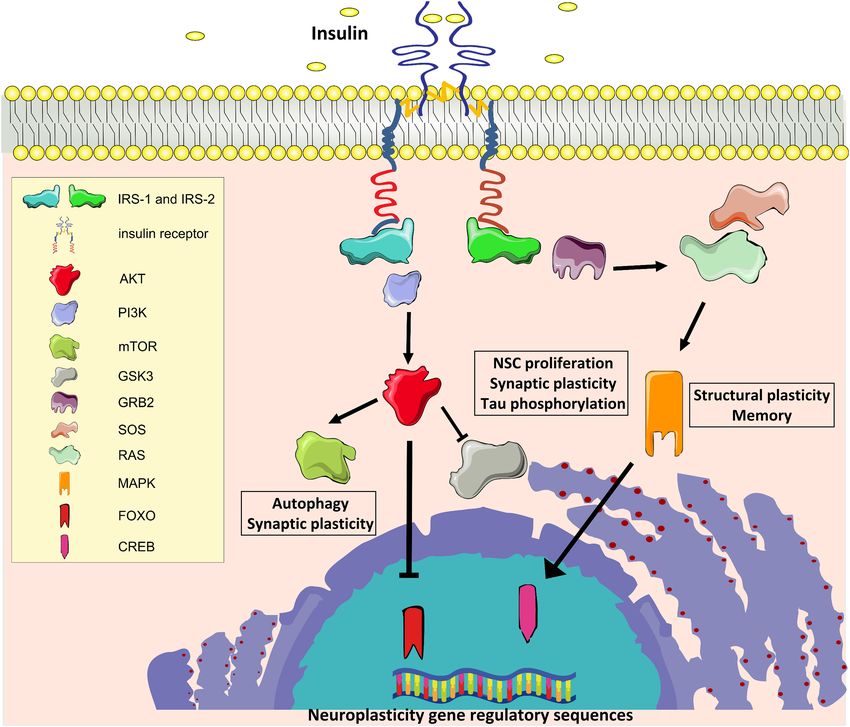
the brain and can be involved in neuronal homeostasis and brain Torres-Aleman, 2012; Figure 1).
function (Duelli and Kuschinsky, 2001). Specifically, the insulin- Many molecules involved in these signaling cascades have
independent transporters GLUT1 and GLUT3 mediate glucose been demonstrated to have key roles in brain functions.
uptake into glial and neuronal cells, respectively (Simpson et al., Activation of PI3-kinase is required for glutamate receptor
2007), suggesting that the impact of insulin on synaptic plasticity insertion at plasma membranes during synaptic plasticity
should be independent of glucose uptake. (Man et al., 2003). GSK3β regulates neural progenitor cell
Moreover, GLUT2 and GLUT4 expression has been proliferation and neuroplasticity, and its activation induces
characterized in specific brain areas: GLUT2 is predominantly hyper-phosphorylation of tau protein that is considered a major
localized in the hypothalamus that regulates food intake (Eny determinant of AD pathogenesis (Salcedo-Tello et al., 2011).
et al., 2008), whereas GLUT4 has been identified in cerebellum, Insulin induces phosphorylation of GSK3β on inhibitory serine
neocortex, and hippocampus, suggesting a role of GLUT-driven 9 residue, thus reducing its enzymatic activity. Moreover, mTOR
glucose uptake in neuronal activity (Vannucci et al., 1998; complex 1 (mTORC1) is fundamental for both protein synthesis
Sankar et al., 2002). GLUT4 is also expressed in astrocytes and and autophagy, which are molecular processes involved in the
insulin stimulation promotes both glucose uptake and glycogen regulation of long-term synaptic plasticity and degradation of
accumulation in astrocyte cultures (Heni et al., 2011). However, misfolded proteins in neurons, respectively (Stoica et al., 2011;
not much data are available in the literature on the role of insulin Son et al., 2012). Insulin/IGF-1 signaling also stimulates the
on astrocytic functions. GLUT5 expression is less relevant and growth factor receptor-bound protein 2- Son of Sevenless-
mainly detected in human and rat brain microglia (Payne et al., Rat sarcoma-mitogen activated protein kinase (Grb2-SOS-Ras-
1997). GLUT6 and GLUT13, which have very low affinity to MAPK) cascade, which plays a pivotal role in cytoskeletal
glucose, are also expressed in the brain, but their role in central modifications underlying the dendritic spine reorganization and
nervous system has yet to be clarified (Joost and Thorens, 2001). memory formation (Adams and Sweatt, 2002).
Conversely, GLUT8 has been shown to drive hippocampal The origin of insulin in the central nervous system is
neuron proliferation during embryogenesis (Membrez et al., controversial. Insulin crosses the blood-brain barrier (BBB) via
2006). However, tissue/cell type specific expression of GLUTs an IR-dependent transport operated by vascular endothelium
in the brain still remains matter of debate. Further, several and its concentration increases after meals (Woods et al.,
growth factors have been reported to modulate the insulin 2003). However, insulin can be also synthesized and secreted by
pathway, GLUT plasma membrane translocation and glucose neurons and adult neural progenitor cells of the hippocampus
uptake by transactivation of the IR downstream effectors (Devaskar et al., 1994; Kuwabara et al., 2011), although no
(Assefa et al., 2017). evidence clearly demonstrated that insulin synthesis in the brain
In this section, we brief the insulin cascade effectors and the is physiologically relevant.
effects of this hormone on both synaptic plasticity and adult However, as described in the next paragraphs, physiological
neurogenesis in the hippocampus along with their impact on levels of insulin play a neurotrophic action on both differentiated
cognitive functions. neurons and neural stem cells (NSCs).
Insulin Signaling in the Brain
Insulin and the insulin-like growth factor 1 (IGF-1) exert Insulin, Synaptogenesis and
their biological effects through two tyrosine kinase receptors, Hippocampal Synaptic Plasticity
the IR and the IGF-1 receptors (IGF-1R), which are closely Modifications of both activity and number of synapses
related and highly distributed throughout the brain (Belfiore are the functional and structural substrates, respectively,
et al., 2009). In the mouse, IR is predominantly expressed of brain plasticity underlying learning and memory
Frontiers in Neuroscience | www.frontiersin.org 2 July 2019 | Volume 13 | Article 788Spinelli et al. Brain Insulin Resistance and Hippocampal Plasticity FIGURE 1 | Insulin signaling. IR, Insulin receptor; IRS1 and IRS2, IR substrate 1 and 2; PI3K, phosphoinositide-3-kinase–protein kinase B; AKT, protein kinase B; mTOR, target of rapamycin; GSK3β, glycogen synthase kinase 3 beta; GRB2, growth factor receptor-bound protein 2; SOS, Son of Sevenless; RAS, Rat sarcoma GTPase protein; MAPK, mitogen activated protein kinase; FOXO, forkhead box O transcription factor; CREB, cAMP response element-binding protein. (Nakahata and Yasuda, 2018). Changes of the synaptic strength, activation (Van Der Heide et al., 2005) and increased membrane either potentiation or depression, and generation of new recruitment of N-methyl-D-aspartate receptors (NMDARs) dendritic spines are causally related to the acquisition and (Skeberdis et al., 2001). Insulin impacts on glutamate receptor consolidation of behavioral modifications. activity by multiple mechanisms. It increases NMDAR-mediated Insulin stimulation of hippocampal neurons induces both currents by enhancing phosphorylation of both NR2A and NR2B presynaptic and postsynaptic effects. Insulin increases basal subunits (Christie et al., 1999; Liu et al., 1995). Insulin treatment neurotransmitter release from presynaptic terminals, as revealed of hippocampal cultures also increases phosphorylation by enhanced frequency of miniature excitatory postsynaptic and clathrin-dependent endocytosis of the GluA1 subunit currents (mEPSCs) (Lee et al., 2011). This effect is paralleled of α-amino-3-hydroxy-5-methyl-4-isoxazolepropionic acid by a Rac1-mediated cytoskeleton rearrangement leading receptors (AMPARs) (Adzovic and Domenici, 2014; Chen et al., to increased density of dendritic spines (Lee et al., 2011). 2014). Downregulation of AMPAR activity in excitatory synapse Moreover, insulin promotes synaptic plasticity by modulating of hippocampal CA1 neurons is fundamental for insulin-induced long-term potentiation (LTP) or long-term depression (LTD) LTD, which is a key step for memory consolidation and flexibility at hippocampal synapses through a metaplastic mechanism. (Ge et al., 2010). IRs have also been demonstrated to modulate Indeed, insulin administration reduces the stimulation frequency type A γ-aminobutyric acid (GABA) receptor activity by threshold required for inducing both LTP and LTD (Van Der regulating both its membrane localization and expression in Heide et al., 2005). Postsynaptic effects are mediated by PI3K inhibitory synapses (Wan et al., 1997). Frontiers in Neuroscience | www.frontiersin.org 3 July 2019 | Volume 13 | Article 788
Spinelli et al.                                                                                   Brain Insulin Resistance and Hippocampal Plasticity

   Furthermore, insulin may impinge on structural features of             effects on neural stem niche based on the timing and the
synapses. For instance, in thalamocortical organotypic slices             duration of stimulation.
this hormone stimulates maturation of silent synapses (Plitzko                Furthermore, calorie restriction has been clearly demonstrated
et al., 2001). Moreover, IR substrate p53 (IRSp53) interacts              to reduce plasma levels of both glucose and insulin, in parallel
with the postsynaptic protein PSD-95 and enhances dendritic               with increasing neurogenesis in the dentate gyrus (Lee et al.,
spine formation (Choi et al., 2005). Interestingly, IRS2 knockout         2002) and counteracting the age-related decline of stem cell
mice show lower activation of NR2B subunits (Martin et al.,               niche (Park et al., 2013). Nutrient deprivation may impact on
2012) and decreased LTP at the CA3–CA1 synapses in parallel               NSC compartment by inducing the expression of the brain-
with higher density of CA1 dendritic spines (Irvine et al.,               derived neurotrophic factor (Bdnf) gene (Maswood et al.,
2011). It is important to underline that these studies did not            2004). In addition, calorie restriction may preserve the NSC
evaluate dendritic spine morphology, therefore our knowledge              capacity to self-renew and differentiate by cell-autonomous
of the effects of IRSs manipulation on structural and functional          mechanisms involving metabolic sensors such as CREB and
plasticity still remains incomplete. Considering the physical             the NAD-dependent histone deacetylase Sirtuin 1 (SIRT1).
and functional interaction between IR and IGF-1R, it is not               In this regard, CREB is a nutrient-dependent transcription
surprising that IGF-1 stimulation can promote plasticity in               factor regulating genes promoting neuronal differentiation and
the hippocampus by increasing spine density of CA1 basal                  survival (Lonze et al., 2002; Fusco et al., 2012b). Moreover,
dendrites in response to physical exercise (Glasper et al.,               SIRT1 is as an epigenetic repressor that modulates adult
2010). Accordingly, IGF-I knockout mice show reduced density              neurogenesis in the subventricular zone and hippocampus
of glutamatergic synapses (Trejo et al., 2007). Importantly,              (Saharan et al., 2013). Calorie restriction also induces the
IR expression and insulin activity in the brain are not                   expression of SIRT1, which has been shown to functionally
restricted to neurons. Insulin has been demonstrated to influence         impinge on CREB-dependent gene expression, thus highlighting
proliferation and metabolism in insulin sensitive glial cells (Heni       a novel molecular link between nutrient-dependent signaling
et al., 2011). Collectively, all the above mentioned evidence             and brain health (Fusco et al., 2012a). Under metabolic and
supports the positive effects of insulin on hippocampal synaptic          oxidative stress, SIRT1 inhibits NSC self-renewal and induces
and structural plasticity.                                                their differentiation (Prozorovski et al., 2008; Ma et al., 2014).
                                                                          In summary, SIRT1 and CREB work as metabolic sensors
                                                                          regulating proliferation and self-renewal of NSCs and controlling
Insulin and Hippocampal Adult                                             their reservoir in the hippocampus (Fusco et al., 2016).
Neurogenesis                                                              Conversely, abolishing the expression of genes encoding the
Hippocampus is one of the brain areas where newborn neurons               insulin-regulated FOXO transcription factors induces hyper-
are generated throughout adulthood (Braun and Jessberger,                 proliferation of neural progenitors and rapid exhaustion of
2014). Specifically, adult neurogenesis occurs in the subgranular         stem cell niche (Renault et al., 2009). Similarly, aberrant
zone of the hippocampus of all mammals including humans                   stimulation of the nutrient-dependent mTOR pathway causes
(Eriksson et al., 1998). NSCs populating this neurogenic                  reduced self-renewal and accelerates NSC differentiation (Magri
niche proliferate and differentiate to generate new neurons               et al., 2011). Together, this evidence confirms that nutrient
(Kempermann et al., 2003). A proper balance between NSC                   related signals control NSC fate under both physiological and
proliferation and their differentiation/maturation underlies the          pathological conditions.
maintenance of both the hippocampal stem cell niche and                       In addition, it is worth mentioning the close similarity
the supply of newborn neurons that integrate into existing                between the intracellular signaling pathways activated by insulin
circuits thus supporting cognitive functions under physiological          and neurotrophins (Reichardt, 2006). In particular, CREB has
conditions and brain repair after injury (Castilla-Ortega et al.,         been shown to play a critical role in the neurotrophin-
2011). Indeed, a growing number of studies indicates that                 triggered effects on neuronal differentiation, survival, and
hippocampal neurogenesis plays a critical role in learning,               plasticity, and it has been also characterized as metabolic
memory, and its impairment has been associated with cognitive             sensor modulated by fasting-related stimuli (Finkbeiner, 2000;
dysfunction in neurodegenerative disorders including AD (van              Altarejos and Montminy, 2011). Moreover, neurotrophic factors
Praag et al., 2002; Taylor et al., 2013).                                 as BDNF, ciliary neurotrophic factor (CNTF), and glial cell-
   Insulin is a key trophic factor for brain development                  derived neurotrophic factor (GDNF) regulate adult neurogenesis
and control of neurogenic niches. Indeed, neuroblast exit                 in multiple stages of NSC maturation and their expression is
from quiescence is regulated by insulin/IGF-I pathway                     affected by overnutrition and metabolic stress (Lindsay et al.,
activation (Chell and Brand, 2010; Sousa-Nunes et al.,                    1994). These evidence emphasizes the role of insulin as growth
2011). Evidence from in vitro and in vivo experiments                     factor for neural niche especially during the early stages of life.
indicate that insulin and IGF-I promote neurogenesis by
modulating NSC proliferation, differentiation, and survival
(Brooker et al., 2000; Åberg et al., 2003). However, a chronic            Insulin Signaling and
hyper-activation of insulin/IGF-I signaling cascades can                  Hippocampus-Dependent Cognitive Task
cause premature depletion of the NSC reservoir (Sun, 2006).               The evidence summarized above suggest that all aspects of
Thus, insulin may produce either trophic or detrimental                   hippocampal plasticity (i.e., functional and structural synaptic

Frontiers in Neuroscience | www.frontiersin.org                       4                                           July 2019 | Volume 13 | Article 788
Spinelli et al. Brain Insulin Resistance and Hippocampal Plasticity
plasticity, and adult neurogenesis) are strongly sensitive to the either upregulation of GABAA receptors or downregulation of
modulation of insulin signaling into the brain. According to AMPA receptors upon insulin treatment (Moosavi et al., 2006).
the key role of hippocampal plasticity in learning and memory, Finally, McNay et al. (2010) demonstrated that endogenous
changes of insulin cascade in the hippocampus markedly affects intrahippocampal insulin signaling was required for memory
cognitive functions. processing. These authors showed that acute injection of
Heterozygous knockout mice for IR display lower preference insulin into the hippocampus at physiological doses enhanced
index in the novel object recognition (NOR) test (Nisticò et al., spatial memory via a PI3K-dependent mechanism (McNay
2012). Accordingly, Zucker rats show lower performance in et al., 2010). Collectively, the results obtained in humans and
the Morris Water Maze (MWM) in parallel with impaired rodents suggest that insulin is fundamental for both memory
insulin sensitivity (Kamal et al., 2013). Moreover, a recent study formation and retention.
performed on Goto-Kakizaki (GK) rats, a model of non-obese
type 2 diabetes (T2D), display spatial memory impairment in
Y-maze task and hippocampal synaptic dysfunction evaluated EFFECTS OF BRAIN INSULIN
by LTP (Duarte et al., 2019). Further, GK rats show a
RESISTANCE ON HIPPOCAMPUS-
reduction of SNAP25 and synaptophysin levels suggesting
synapse degeneration (Duarte et al., 2019). In addition, IRSp53 DEPENDENT FUNCTIONS
knockout mice show impaired learning and memory when
Data reviewed in the previous paragraphs support the view
evaluated in both MWM and NOR tests (Kim et al., 2009).
that changes of either insulin signaling or insulin sensitivity
However, forebrain-specific IRS2 deficiency improved memory
in the hippocampus may alter molecular pathways involved
retention in the MWM task (Irvine et al., 2011), suggesting that
in synaptic plasticity and adult neurogenesis, thereby leading
molecular hubs of insulin signaling may differentially interfere on
to reduced “mindspan” (the maintenance of mental abilities
cognitive behavior.
throughout life) and increased risk of neurodegeneration (Kodl
In agreement with this finding, chronic brain stimulation by
and Seaquist, 2008). Accordingly, while calorie restriction
8-week intranasal insulin administration improved memory in
furthers neuronal survival and improves cognitive function
humans (Benedict et al., 2004). Moreover, lower values of both
(Fusco and Pani, 2013), the excess of nutrients harms the
glycaemia and glycosylated hemoglobin (HbA1c) are associated
brain health and accelerates cognitive decline (Elias et al., 2012;
with better performance in memory tasks in humans (Kerti
Sellbom and Gunstad, 2012). Nutrient excess causes hyper-
et al., 2013). It has been shown that peripheral changes of
activation of insulin signaling in all tissues expressing IR, leading
insulin signaling and sensitivity may affect brain health and
to the desensitization of IR-dependent molecular cascades. BIR
function although the results of these studies are controversial.
decreases the ability of brain cells to respond to insulin and
Learning and memory deficits in MWM have been demonstrated
abolishes both metabolic and cognitive effects of this hormone
in liver-specific IGF1 knockout mice (Trejo et al., 2007).
(Kullmann et al., 2016). Specifically, this deficiency could be
In addition, IGF-I antiserum administration to young rats
caused by lower expression of IR or poor activation of insulin
impairs learning in passive avoidance task (Lupien et al., 2003).
signaling. IR downstream effectors may become insensitive to
Conversely, intraperitoneal injection of insulin impairs retention
the insulin stimulation, resulting in inability of brain cells to
and spatial working memory in a dose-dependent manner
respond to the hormone and leading to impairment of brain
(Kopf and Baratti, 1999; Akanmu et al., 2009). It seems that
plasticity. In the Western world, the incidence of metabolic
the negative impact of peripheral insulin administration on
disorders, including insulin resistance, obesity and T2D, is
cognitive functions may be due to the lowering of blood glucose
increasing at alarming rates in parallel with the prevalence
levels, as indicated by the positive effect of the simultaneous
of cognitive decline (Cukierman-Yaffee, 2009). Obesity and
glucose infusion (Kopf et al., 1998). In line with this hypothesis,
inflammation affect the insulin transport to the brain (Ketterer
peripheral administration of insulin increases verbal memory
et al., 2011) and low expression of IR has been reported in
and attention in healthy subjects under euglycemic conditions
patients with T2D (Kullmann et al., 2016). However, patients
(Kern et al., 2001).
with T2DM and/or obesity showed decreased insulin levels in
To avoid the side effects of systemic insulin administration,
the cerebrospinal fluid despite higher levels of this hormone in
Park et al. (2000) studied the impact of intracerebroventricular
their plasma (Heni et al., 2014). In the following paragraphs,
injection of insulin in rats and found that insulin improved
we will summarize the mechanisms underlying the detrimental
cognitive performance in passive avoidance test. Accordingly,
effects of BIR on hippocampal plasticity and cognition, and the
intrahippocampal injection of insulin into the CA1 region
epidemiological and experimental evidence supporting a link
has been reported to enhance memory of rats in passive
between BIR and AD.
avoidance test (Babri et al., 2007). More importantly, the
effects of intrahippocampal injection of insulin on cognitive
functions seem to be related to its dose. High doses of insulin Alterations of Hippocampal Plasticity in
significantly ameliorate spatial learning and memory in the BIR Models
MWM test, whereas low doses reduce cognitive performance High-fat diet (HFD) is a well-established animal model of
(Moosavi et al., 2006). It has been hypothesized that the negative metabolic disorders (Wong et al., 2016). HFD induces obesity
effects of insulin on spatial memory may be dependent on by compromising β-cell functions, promoting hyper-glycaemia,
Frontiers in Neuroscience | www.frontiersin.org 5 July 2019 | Volume 13 | Article 788Spinelli et al. Brain Insulin Resistance and Hippocampal Plasticity whole-body insulin resistance, and dyslipidemia, and abolished the insulin resistance-dependent impairment of increasing free fatty acids in the blood. Many studies synaptic plasticity (Spinelli et al., 2017). Of course, aberrant have investigated the structural and functional changes of palmitoylation of other zDHHC3 targets (e.g., GABAA RG2) may neuroplasticity in experimental models of insulin resistance contribute to the detrimental effects of HFD on hippocampus- (Fadel and Reagan, 2016). dependent learning and memory. However, our study adds a More specifically, HFD produces detrimental effects on brain new layer to the hippocampal synaptic plasticity regulation by functions including decreased neurogenesis in the dentate gyrus insulin signaling deterioration and proposes a novel molecular (Lindqvist et al., 2006), alteration of BBB integrity (Freeman and mechanism potentially linking BIR and cognitive decline. Granholm, 2012) and changes in both spine density and synapse Other mechanisms underlying fatty acid-driven learning formation (Stranahan et al., 2008a). HFD also impairs insulin deficits involve cholesterol dysmetabolism, oxidative stress, signaling in the hippocampus and reduces the expression of endothelial dysfunctions, and neurotrophin depletion. Mice synaptic proteins PSD-95 and synaptopodin (Arnold et al., 2014). fed with HFD show higher levels of reactive oxygen species However, the most significant effects occur on activity-dependent (ROS), superoxide, and peroxynitrite into the brain, leading synaptic plasticity. Indeed, Zucker rats show impairment in LTP to lower level of brain-derived neurotrophic factor (BDNF) at CA3–CA1 synapses in parallel with loss of insulin sensitivity and impaired cognition performance evaluated by spatial task (Kamal et al., 2013). Moreover, IR heterozygous knockout mice (Wu et al., 2004). Moreover, epidemiological studies showed display normal levels of both basal synaptic transmission and that diets enriched in cholesterol (HCD) were associated with LTP that, however, fails to be consolidated due to reduced Akt poor cognitive performance in humans (Requejo et al., 2003). activation (Nisticò et al., 2012). HCD diet also induced impairment of spatial and working Obesity and T2D have been demonstrated to induce memory due to microglial activation and alteration of the BBB hippocampal insulin resistance through different metabolic integrity in rats (Chen et al., 2018). Interestingly, feeding obese changes including alteration of hypothalamic-pituitary-adrenal rodents with HFD inhibited the transport through the BBB of (HPA) axis leading to elevated levels of glucocorticoids (Plotsky neuroendocrine molecules, such as ghrelin and leptin, which et al., 1992). Accordingly, glucocorticoids stimulation inhibits promote synaptic plasticity and cognitive functions (Banks et al., translocation of GLUT4 to the plasma membrane in the rat 2008; Kanoski et al., 2013; Mainardi et al., 2017). Finally, HFD hippocampus (Piroli et al., 2007). Moreover, Stranahan et al. has been shown to induce activation of microglia and astrocytes, showed that restoring physiological levels of glucocorticoids and increase of pro-inflammatory cytokines/mediators such as in insulin resistant db/db mice rescued the impairment of cyclooxygenase 2, TNF-α, IL-1-β, and IL-6 in the hippocampus hippocampal synaptic plasticity (Stranahan et al., 2008b). of mice (Thirumangalakudi et al., 2008; Duffy et al., 2019). A different model of BIR is obtained by intracerebral injection of In summary, metabolic diseases affecting insulin signaling streptozotocin, which impairs cognitive function by reducing the may impair the synaptic function through a plethora of activity of the neuroprotective protein SIRT1 (Du et al., 2014). molecular mechanisms targeting neurons, astrocytes, endothelial As mentioned before, SIRT1 cooperates with the transcription or inflammatory cells. factor CREB promoting the CREB-dependent expression of the neuroplasticity-related gene Bdnf (Jeong et al., 2012). Cognitive Impairment in BIR Models To better clarify the functional role of hippocampal insulin Epidemiological evidence indicate that metabolic alterations resistance, Grillo et al. (2015) silenced the expression of IR in occurring in T2D, such as hyper-glycaemia and hyper- the hippocampus by injecting lentiviral particles harboring IR insulinaemia, positively correlate with cognitive impairment antisense sequence. This experimental model showed deficits and diabetic patients exhibit higher susceptibility to develop in hippocampal synaptic transmission and spatial learning, dementia (Cukierman-Yaffee, 2009). Dysregulation of glucose in parallel with downregulation of NMDA subunit GluN2B homeostasis increases the risk of dementia in both diabetic and expression and lower phosphorylation of AMPA subunit GluA1, non-diabetic patients (Crane et al., 2013) and is associated with without altering peripheral metabolic parameters (i.e., body reduced hippocampal volume and cognitive decline (Kerti et al., weight, adiposity, and glucose homeostasis) (Grillo et al., 2015). 2013). Furthermore, longitudinal studies demonstrated that also Recently, we described a novel link between BIR and Type 1 diabetes (T1D) patients were affected by mild-severe altered glutamate receptor function underlying the HFD- cognitive impairment related to the age of onset of the disease dependent impairment of hippocampal synaptic plasticity and the microvascular complications (Moheet et al., 2015; (Spinelli et al., 2017). In particular, we found that HFD induced Nunley et al., 2015). Insulin administration is crucial to promote accumulation of palmitic acid and increased FOXO3a-dependent glucose homeostasis in these patients and to reduce the vascular expression of palmitoyl-transferase zDHHC3 leading to complications but it increases the risk for hypoglycemic episodes, GluA1 hyper-palmitoylation in the hippocampus. Accordingly, which negatively impact on cognitive functions (Desrocher and in vitro stimulation of hippocampal neurons with a cocktail Rovet, 2004). However, the role of hypo- or hyper-insulinemia in of insulin and palmitic acid replicated the in vivo molecular T1D-related cognitive alterations has still to be clarified. changes, inhibiting the GluA1 localization at the synaptic Numerous clinical studies revealed worse cognitive membrane and AMPA currents at glutamatergic synapses. performance and earlier age incidence of all-cause dementia Finally, either silencing of zDHHC3 or overexpression of the in subjects with T2D (Davis et al., 2017; Callisaya et al., 2019). palmitoylation-deficient GluA1 mutant in the hippocampus Accordingly, meta-analysis studies showed that in diabetic Frontiers in Neuroscience | www.frontiersin.org 6 July 2019 | Volume 13 | Article 788
Spinelli et al.                                                                                   Brain Insulin Resistance and Hippocampal Plasticity

patients the risk for all types of dementia is increased by 60–73%        are poorly understood. Undoubtedly, micro- and macro-vascular
(Gudala et al., 2013; Chatterjee et al., 2016).                           complications of T2D may increase the risk of cerebrovascular
    However, alteration of brain insulin signaling may negatively         disease, cognitive impairment and vascular dementia (Gorelick
impact on brain function also in the absence of T2D and                   et al., 2011). Moreover, white matter disease, alteration of
before the onset of obesity. Several studies have demonstrated            the BBB and neuro-inflammation may play a pathophysiologic
deficits in hippocampal-dependent learning and spatial memory             role (Hsu and Kanoski, 2014). However, hyper-insulinaemia
associated with Western diet intake (Molteni et al., 2002;                promotes the formation of advanced glucose end products
Kanoski and Davidson, 2010). Interestingly, when Kanoski                  and ROS causing neurotoxicity and brain damage (Brownlee,
and Davidson (2010) investigated both hippocampus-dependent               2001). Despite insulin exerts a neurotrophic role at moderate
and hippocampus-independent memory retention ability after                concentrations, higher levels of the hormone may be associated
different Western diet treatments, they found that only spatial           with increased deposition of amyloid-β (Aβ) in the brain due to
memory impairments occurred after short-term consumption.                 competition for their common and main clearance mechanism,
This suggests that hippocampus is a brain area very sensitive             the insulin-degrading enzyme (Farris et al., 2003). In this regard,
to metabolic stress, and memory impairment may arise before               AD has been defined a form of type 3 diabetes, based on the
the development of diet-induced metabolic alterations in                  evidence of BIR development in the AD brain (Steen et al.,
peripheral tissues. Accordingly, few days of HFD regimen                  2005; Bedse et al., 2015; Sposato et al., 2019). The insulin
were sufficient to cause cognitive impairment in rats evaluated           synthesis decreases during aging and AD progression in brain
with MWM test (Murray et al., 2009). High caloric intake                  areas such as frontal cortex, hippocampus, and hypothalamus
also affected hippocampus-dependent non-spatial learning and              (Frolich et al., 1998). In addition, Aβ inhibits insulin expression
memory tasks and these results were related to changes of the BBB         in astrocytes (Pitt et al., 2017). Together, these studies indicate
integrity. Specifically, high energy diet consumption reduced the         a crosstalk between brain insulin signaling alteration and
expression of tight junction proteins selectively causing increased       Aβ accumulation in neurodegenerative diseases. Accordingly,
blood-to-brain permeability in the hippocampus (Kanoski and               experimental data obtained from neuroimaging and biomarker
Davidson, 2010). The observed learning and memory deficits                studies revealed that T2D patients showed alterations of both
are strikingly similar to the poor task performance and                   brain glucose metabolism and cerebrospinal fluid including
cognitive impairment observed in patients with mild or severe             phosphorylated tau, which are reminiscent of changes observed
metabolic derangements, which strengthens the hypothesis that             in AD (Baker et al., 2011; Moran et al., 2015). In addition,
hippocampal insulin resistance is a key mediator of diet-                 analysis of AD postmortem brains revealed insulin signaling
dependent cognitive alterations. Therefore, cognitive dysfunction         alterations in hippocampal tissues resembling the biochemical
related to HFD or obesity in otherwise healthy individuals may be         features of insulin resistance in parallel with histopathological
due to decreased insulin signaling and development of BIR in the          hallmarks of neurodegeneration (Talbot et al., 2012; Tramutola
hippocampus (McNay et al., 2010).                                         et al., 2015). Moreover, tau is hyper-phosphorylated in the
    Nevertheless, peripheral insulin resistance and diet-induced          brain of NIRKO mice (Schubert et al., 2004) and BIR has
obesity are correlated with some other changes that may                   been associated with tau pathology in AD human brains
cause neurocognitive dysfunction. Indeed, they induce systemic            (Yarchoan and Arnold, 2014).
and central inflammation with high levels of circulating pro-                Interestingly, T2D and AD also share several metabolic
inflammatory interleukins that have been linked to impaired               derangements promoting brain aging. AD patients show hyper-
executive function (Trollor et al., 2012). Obesity also alters HPA        insulinaemia and decreased peripheral insulin sensitivity (Craft
axis causing enhanced secretion of glucocorticoids, which have            et al., 1996), whereas insulin levels in cerebrospinal fluid are
been associated with reduced hippocampal volume, memory                   reduced (Craft et al., 1998). Accordingly, sustained peripheral
impairment and mood alterations (MacQueen and Frodl, 2011).               hyper-insulinaemia can reduce the transport of insulin into the
Moreover, mice specifically lacking the IR into the brain (NIRKO          brain due to the lower expression of IR at the BBB (Schwartz
mice) display changes in dopamine turnover associated with                et al., 1990). Brain insulin uptake is also impaired in both aging
anxiety and depressive-like behaviors (Kleinridders et al., 2015).        and AD independently by T2D (Frolich et al., 1998). Recent
In addition, diet-induced microbiota dysbiosis can impact on the          evidence suggests that insulin may influence Aβ deposition
gut-brain axis, thus promoting insulin resistance and cognitive           and AD-dependent impairment of both synaptic plasticity and
impairment (Daulatzai, 2014). Finally, HFD exposure during                memory formation (Cholerton et al., 2013). Intranasal insulin
early stages of life is associated with impaired learning and             administration has been demonstrated to improve cognitive
spatial memory (Boitard et al., 2012), suggesting that alteration         function in humans (Hallschmid et al., 2007; Reger et al.,
of insulin signaling may negatively influence cognitive function          2008). However, recent data about a clinical trial with mild
at each stage of life.                                                    cognitive impairment (MCI) or moderate AD patients revealed
                                                                          no significant effects of long-term intranasal insulin delivery on
                                                                          cognitive performance in memory task (Craft et al., 2017).
Brain Insulin Resistance, Brain Aging                                        Finally, genetic and experimental data about insulin degrading
and Neurodegenerative Diseases                                            enzyme and, more recently, the Aβ metabolism regulation
While diabetes is known to increase the risk for dementia, the            by sortilin related VPS10 domain containing receptor 1
underlying mechanisms linking insulin resistance, T2D and AD              (SorCS1) gene suggest novel mechanistic links between BIR

Frontiers in Neuroscience | www.frontiersin.org                       7                                           July 2019 | Volume 13 | Article 788
Spinelli et al. Brain Insulin Resistance and Hippocampal Plasticity
and AD (Lane et al., 2010; Wang et al., 2015). Thus, BIR extracellularly secreted via exosomes (Rajendran et al., 2006;
seems to play a pivotal role at the crossroad between Sharples et al., 2008). More importantly, changes of insulin
metabolic and neurodegenerative diseases, independently from resistance molecular markers (i.e., higher serine phosphorylation
the cerebrovascular mechanisms. and lower tyrosine phosphorylation of IRS-1) have been
found in neural-derived exosomes extracted from blood of
AD patients compared to age- and gender-matched patients
BIOMARKERS OF BRAIN INSULIN with frontotemporal dementia or T2D (Kapogiannis et al.,
RESISTANCE 2015). These differences were detectable up to 10 years before
the onset of AD symptoms. Finally, exosomal biomarkers
In view of the close relationship among metabolic diseases, of BIR were associated with higher brain atrophy in AD
BIR and cognitive decline, it is emerging the need to identify patients (Mullins et al., 2017) emphasizing the potential role
biomarkers able to detect BIR before, or possibly even in of brain derived microvesicles as detectors of brain insulin
the absence of, peripheral insulin resistance, that may be signaling and biomarkers of brain damage due to metabolic and
predictive of age- and dementia-related cognitive impairment. neurodegenerative disorders.
Ideal biomarkers should be reliable, simple to measure, non-
invasive and inexpensive (Noel-Storr et al., 2013). In this regard,
the dosage of both Aβ and tau proteins in the cerebrospinal CONCLUSION
fluid is invasive and most likely indicative of a pathology
already under development. For these reasons, in the last Molecules involved in metabolic homeostasis are now recognized
years several studies focused on evaluation of brain glucose to exert a great influence on hippocampal plasticity, and
metabolism and analysis of brain-derived extracellular vesicles alteration of their equilibrium has a strong impact at the
extracted from the blood as biomarkers of BIR and early-phase functional and behavioral levels. Insulin exerts a trophic role
cognitive decline. into the brain and it may also act as a signal of positive
Cerebral glucose metabolism is tightly correlated with metabolic homeostasis promoting neuroplasticity, which is
neuronal activity (Simpson et al., 2007). Therefore, imaging of a high energy demanding process. Insulin plays a pivotal
local brain hypo-metabolism can be used to visualize areas of role in the regulation of central nervous system homeostasis
reduced synaptic activity. The most frequently used method and higher functions such as learning and memory, by
of brain metabolic imaging is positron emission tomography controlling both NSC fate and the activity of neuronal
(PET) with (18 F)fluorodeoxyglucose (FDG) (Cohen and Klunk, network. In this regard, identifying the molecular targets
2014). Reduced cerebral glucose metabolism represents one that underlie the effects of insulin on brain plasticity may
of the earliest signs of AD, and studies in both humans contribute to understand the mechanisms regulating neural
and experimental models suggest that altered brain glucose plasticity in health and metabolic diseases and reveal novel
metabolism is associated with AD progression (Kapogiannis and targets in pathologies characterized by impaired neural
Mattson, 2011; Ishibashi et al., 2015). plasticity, especially AD.
Recent work have identified in GK rats reduced glutamine Actually, we do not yet have an exhaustive understanding
synthesis and impairment of the glutamate-glutamine cycle of how systemic and brain insulin resistance are related to
between astrocytes and neurons, driving to diabetes-induced brain aging and AD, but clinical and experimental evidence
neurodegeneration and cognitive dysfunction (Girault et al., indicates that insulin supplementation can be a therapeutic tool
2017). In a mouse model of AD, impaired glucose transport for patients with cognitive impairment and an added value
through the BBB and decreased cerebral lactate release during in the treatment of dementia (Chapman et al., 2018; Santiago
neuronal activity occur at early stages of the phenotype (Merlini and Hallschmid, 2019). The availability of BDE, such as other
et al., 2011). Dysregulated brain glucose metabolism resembling biomarkers of brain metabolism detectable in the plasma, will
changes observed in AD patients has been observed in metabolic foster clinical studies to identify novel therapeutic approaches for
disorders such as obesity or T2D (Tschritter et al., 2006, personalized medicine in neurodegenerative diseases.
2007). However, whether neuroimaging changes of brain glucose
metabolism anticipate the onset of neurodegeneration or are
related to the development of BIR in the same brain areas remain
AUTHOR CONTRIBUTIONS
still poorly understood. All authors conceived the work, took part to the scientific
More recently, molecular strategies have been developed discussion, and wrote the manuscript.
to selectively isolate brain-derived exosomes (BDE) from
biological fluids (Tschritter et al., 2006, 2007; Fiandaca et al.,
2015; Goetzl et al., 2016). Exosomes are extracellular vesicles FUNDING
carrying information (e.g., proteins, lipids, and nucleic acids)
to distant cells, which are emerging as novel potential This research was supported by the Italian Ministry of University
biomarkers for human diseases (Tkach and Thery, 2016). Several and Research (SIR 2014 RBSI14ZV59 to SF) and Universitá
pathogenic proteins that are involved in neurodegenerative Cattolica del Sacro Cuore intramural grants (Linea D.3.2 2015
diseases, including AD, are loaded into vesicles and then and Linea D.3.2 2017 to CG).
Frontiers in Neuroscience | www.frontiersin.org 8 July 2019 | Volume 13 | Article 788Spinelli et al. Brain Insulin Resistance and Hippocampal Plasticity
REFERENCES Bruning, J. C., Gautam, D., Burks, D. J., Gillette, J., Schubert, M., Orban, P. C.,
et al. (2000). Role of brain insulin receptor in control of body weight and
Åberg, M. A., Åberg, N. D., Palmer, T. D., Alborn, A. M., Carlsson-Skwirut, C., reproduction. Science 289, 2122–2125. doi: 10.1126/science.289.5487.2122
Bang, P., et al. (2003). IGF-I has a direct proliferative effect in adult hippocampal Callisaya, M. L., Beare, R., Moran, C., Phan, T., Wang, W., and Srikanth, V. K.
progenitor cells. Mol. Cell. Neurosci. 24, 23–40. doi: 10.1016/s1044-7431(03) (2019). Type 2 diabetes mellitus, brain atrophy and cognitive decline: a
00082-4 longitudinal study. Diabetologia 62, 448–458. doi: 10.1007/s00125-018-4778-9
Adams, J. P., and Sweatt, J. D. (2002). Molecular psychology: roles for the ERK Castilla-Ortega, E., Pedraza, C., Estivill-Torrús, G., and Santín, L. (2011). When is
MAP kinase cascade in memory. Annu. Rev. Pharmacol. Toxicol. 42, 135–163. adult hippocampal neurogenesis necessary for learning? Evidence from animal
doi: 10.1146/annurev.pharmtox.42.082701.145401 research. Rev. Neurosci. 22, 267–283. doi: 10.1515/RNS.2011.027
Adzovic, L., and Domenici, L. (2014). Insulin induces phosphorylation of the Chapman, C. D., Schioth, H. B., Grillo, C. A., and Benedict, C. (2018). Intranasal
AMPA receptor subunit GluR1, reversed by ZIP, and over-expression of Protein insulin in Alzheimer’s disease: food for thought. Neuropharmacology 136,
Kinase M zeta, reversed by amyloid beta. J. Neurochem. 131, 582–587. doi: 196–201. doi: 10.1016/j.neuropharm.2017.11.037
10.1111/jnc.12947 Chatterjee, S., Peters, S. A., Woodward, M., Mejia Arango, S., Batty, G. D., Beckett,
Akanmu, M. A., Nwabudike, N. L., and Ilesanmi, O. R. (2009). Analgesic, learning N., et al. (2016). Type 2 diabetes as a risk factor for dementia in women
and memory and anxiolytic effects of insulin in mice. Behav. Brain Res. 196, compared with men: a pooled analysis of 2.3 million people comprising more
237–241. doi: 10.1016/j.bbr.2008.09.008 than 100,000 cases of dementia. Diabetes Care 39, 300–307. doi: 10.2337/dc15-
Altarejos, J. Y., and Montminy, M. (2011). CREB and the CRTC co-activators: 1588
sensors for hormonal and metabolic signals. Nat. Rev. Mol. Cell Biol. 12, Chell, J. M., and Brand, A. H. (2010). Nutrition-responsive glia control exit of
141–151. doi: 10.1038/nrm3072 neural stem cells from quiescence. Cell 143, 1161–1173. doi: 10.1016/j.cell.2010.
Arnold, S. E., Lucki, I., Brookshire, B. R., Carlson, G. C., Browne, C. A., Kazi, H., 12.007
et al. (2014). High fat diet produces brain insulin resistance, synaptodendritic Chen, T.-J., Dean-Chuan, W., Hui-Shan, H., and Hsuan-Fang, H. (2014). Insulin
abnormalities and altered behavior in mice. Neurobiol. Dis. 67, 79–87. doi: can induce the expression of a memory-related synaptic protein through
10.1016/j.nbd.2014.03.011 facilitating AMPA receptor endocytosis in rat cortical neurons. Cell. Mol. Life
Assefa, B., Mahmoud, A. M., Pfeiffer, A. F. H., Birkenfeld, A. L., Spranger, J., Sci. 71, 4069–4080. doi: 10.1007/s00018-014-1620-5
and Arafat, A. M. (2017). Insulin-like growth factor (IGF) binding protein-2, Chen, Y., Yin, M., Cao, X., Hu, G., and Xiao, M. (2018). Pro- and anti-inflammatory
independently of IGF-1, induces GLUT-4 translocation and glucose uptake in effects of high cholesterol diet on aged brain. Aging Dis. 9, 374–390. doi: 10.
3T3-L1 adipocytes. Oxid. Med. Cell Longev. 2017:3035184. doi: 10.1155/2017/ 14336/AD.2017.0706
3035184 Choi, J., Ko, J., Racz, B., Burette, A., Lee, J. R., Kim, S., et al. (2005). Regulation of
Babri, S., Badie, H. G., Khamenei, S., and Seyedlar, M. O. (2007). Intrahippocampal dendritic spine morphogenesis by insulin receptor substrate 53, a downstream
insulin improves memory in a passive-avoidance task in male wistar rats. Brain effector of Rac1 and Cdc42 small GTPases. J. Neurosci. 25, 869–879. doi:
Cogn. 64, 86–91. doi: 10.1016/j.bandc.2007.01.002 10.1523/jneurosci.3212-04.2005
Bailyes, E. M., Nave, B. T., Soos, M. A., Orr, S. R., Hayward, A. C., and Siddle, Cholerton, B., Baker, L. D., and Craft, S. (2013). Insulin, cognition, and dementia.
K. (1997). Insulin receptor/IGF-I receptor hybrids are widely distributed in Eur. J. Pharmacol. 719, 170–179. doi: 10.1016/j.ejphar.2013.08.008
mammalian tissues: quantification of individual receptor species by selective Christie, J. M., Wenthold, R. J., and Monaghan, D. T. (1999). Insulin causes
immunoprecipitation and immunoblotting. Biochem. J. 327, 209–215. doi: a transient tyrosine phosphorylation of NR2A and NR2B NMDA receptor
10.1042/bj3270209 subunits in rat hippocampus. J. Neurochem. 72, 1523–1528. doi: 10.1046/j.
Baker, L. D., Cross, D. J., Minoshima, S., Belongia, D., Watson, G. S., and Craft, 1471-4159.1999.721523.x
S. (2011). Insulin resistance and Alzheimer-like reductions in regional cerebral Cohen, A. D., and Klunk, W. E. (2014). Early detection of Alzheimer’s disease using
glucose metabolism for cognitively normal adults with prediabetes or early type PiB and FDG PET. Neurobiol. Dis. 72, 117–122. doi: 10.1016/j.nbd.2014.05.001
2 diabetes. Arch. Neurol. 68, 51–57. doi: 10.1001/archneurol.2010.225 Craft, S., Claxton, A., Baker, L. D., Hanson, A. J., Cholerton, B., Trittschuh,
Banks, W. A., Burney, B. O., and Robinson, S. M. (2008). Effects of triglycerides, E. H., et al. (2017). Effects of regular and long-acting insulin on cognition
obesity, and starvation on ghrelin transport across the blood-brain barrier. and Alzheimer’s disease biomarkers: a pilot clinical trial. J. Alzheimers Dis. 57,
Peptides 29, 2061–2065. doi: 10.1016/j.peptides.2008.07.001 1325–1334. doi: 10.3233/JAD-161256
Bartsch, T., and Wulff, P. (2015). The hippocampus in aging and disease: from Craft, S., Newcomer, J., Kanne, S., Dagogo-Jack, S., Cryer, P., Sheline, Y.,
plasticity to vulnerability. Neuroscience 309, 1–16. doi: 10.1016/j.neuroscience. et al. (1996). Memory improvement following induced hyperinsulinemia in
2015.07.084 Alzheimer’s disease. Neurobiol. Aging 17, 123–130. doi: 10.1016/0197-4580(95)
Bedse, G., Di Domenico, F., Serviddio, G., and Cassano, T. (2015). Aberrant insulin 02002-0
signaling in Alzheimer’s disease: current knowledge. Front. Neurosci. 9:204. Craft, S., Peskind, E., Schwartz, M. W., Schellenberg, G. D., Raskind, M., Porte, D.
doi: 10.3389/fnins.2015.00204 Jr., et al. (1998). Cerebrospinal fluid and plasma insulin levels in Alzheimer’s
Belfiore, A., Frasca, F., Pandini, G., Sciacca, L., and Vigneri, R. (2009). Insulin disease: relationship to severity of dementia and apolipoprotein E genotype.
receptor isoforms and insulin receptor/insulin-like growth factor receptor Neurology 50, 164–168. doi: 10.1212/wnl.50.1.164
hybrids in physiology and disease. Endocr. Rev. 30, 586–623. doi: 10.1210/er. Crane, P. K., Walker, R., and Larson, E. B. (2013). Glucose levels and risk of
2008-0047 dementia. N Engl. J. Med. 369, 1863–1864. doi: 10.1056/nejmc1311765
Benedict, C., Hallschmid, M., Hatke, A., Schultes, B., Fehm, H. L., Born, Cukierman-Yaffee, T. (2009). The relationship between dysglycemia and cognitive
J., et al. (2004). Intranasal insulin improves memory in humans. dysfunction. Curr. Opin. Invest. Drugs 10, 70–74.
Psychoneuroendocrinology 29, 1326–1334. doi: 10.1016/j.psyneuen.2004.04.003 Daulatzai, M. A. (2014). Chronic functional bowel syndrome enhances gut-brain
Boitard, C., Etchamendy, N., Sauvant, J., Aubert, A., Tronel, S., Marighetto, A., axis dysfunction, neuroinflammation, cognitive impairment, and vulnerability
et al. (2012). Juvenile, but not adult exposure to high-fat diet impairs relational to dementia. Neurochem. Res. 39, 624–644. doi: 10.1007/s11064-014-1266-6
memory and hippocampal neurogenesis in mice. Hippocampus 22, 2095–2100. Davis, W. A., Zilkens, R. R., Starkstein, S. E., Davis, T. M., and Bruce, D. G.
doi: 10.1002/hipo.22032 (2017). Dementia onset, incidence and risk in type 2 diabetes: a matched cohort
Braun, S. M., and Jessberger, S. (2014). Adult neurogenesis: mechanisms and study with the fremantle diabetes study phase I. Diabetologia 60, 89–97. doi:
functional significance. Development 141, 1983–1986. doi: 10.1242/dev.104596 10.1007/s00125-016-4127-9
Brooker, G. J., Kalloniatis, M., Russo, V. C., Murphy, M., Werther, G. A., and Debons, A. F., Krimsky, I., and From, A. (1970). A direct action of insulin on
Bartlett, P. F. (2000). Endogenous IGF-1 regulates the neuronal differentiation the hypothalamic satiety center. Am. J. Physiol. 219, 938–943. doi: 10.1152/
of adult stem cells. J. Neurosci. Res. 59, 332–341. doi: 10.1002/(sici)1097- ajplegacy.1970.219.4.938
4547(20000201)59:33.0.co;2-2 Desrocher, M., and Rovet, J. (2004). Neurocognitive correlates of type 1 diabetes
Brownlee, M. (2001). Biochemistry and molecular cell biology of diabetic mellitus in childhood. Child Neuropsychol. 10, 36–52. doi: 10.1076/chin.10.1.
complications. Nature 414, 813–820. doi: 10.1038/414813a 36.26241
Frontiers in Neuroscience | www.frontiersin.org 9 July 2019 | Volume 13 | Article 788Spinelli et al. Brain Insulin Resistance and Hippocampal Plasticity
Devaskar, S. U., Giddings, S. J., Rajakumar, P. A., Carnaghi, L. R., Menon, R. K., Girault, F. M., Sonnay, S., Gruetter, R., and Duarte, J. M. N. (2017). Alterations
Zahm, D. S., et al. (1994). Insulin gene expression and insulin synthesis in of brain energy metabolism in Type 2 diabetic goto-kakizaki rats measured
mammalian neuronal cell. J. Biol. Chem. 269, 8445–8454. in vivo by 13C magnetic resonance spectroscopy. Neurotox. Res. 36, 268–278.
Dienel, G. A. (2012). Fueling and imaging brain activation. ASN Neuro. 4:e00093. doi: 10.1007/s12640-017-9821-y
doi: 10.1042/AN20120021 Glasper, E. R., Llorens-Martin, M. V., Leuner, B., Gould, E., and Trejo, J. L. (2010).
Du, L. L., Xie, J. Z., Cheng, X. S., Li, X. H., Kong, F. L., Jiang, X., et al. (2014). Blockade of insulin-like growth factor-I has complex effects on structural
Activation of sirtuin 1 attenuates cerebral ventricular streptozotocin-induced plasticity in the hippocampus. Hippocampus 20, 706–712. doi: 10.1002/hipo.
tau hyperphosphorylation and cognitive injuries in rat hippocampi. Age 36, 20672
613–623. doi: 10.1007/s11357-013-9592-1 Goetzl, E. J., Mustapic, M., Kapogiannis, D., Eitan, E., Lobach, I. V., Goetzl, L., et al.
Duarte, J. M. N., Skoug, C., Silva, H. B., Carvalho, R. A., Gruetter, R., and (2016). Cargo proteins of plasma astrocyte-derived exosomes in Alzheimer’s
Cunha, R. A. (2019). Impact of caffeine consumption on Type 2 diabetes- disease. FASEB J. 30, 3853–3859. doi: 10.1096/fj.201600756r
induced spatial memory impairment and neurochemical alterations in the Gorelick, P. B., Scuteri, A., Black, S. E., Decarli, C., Greenberg, S. M.,
Hippocampus. Front. Neurosci. 12:1015. doi: 10.3389/fnins.2018.01015 Iadecola, C., et al. (2011). Vascular contributions to cognitive impairment and
Duelli, R., and Kuschinsky, W. (2001). Brain glucose transporters: relationship dementia: a statement for healthcare professionals from the american heart
to local energy demand. News Physiol. Sci. 16, 71–76. doi: 10.1152/ association/american stroke association. Stroke 42, 2672–2713. doi: 10.1161/
physiologyonline.2001.16.2.71 str.0b013e3182299496
Duffy, C. M., Hofmeister, J. J., Nixon, J. P., and Butterick, T. A. (2019). High fat Grillo, C. A., Piroli, G. G., Lawrence, R. C., Wrighten, S. A., Green, A. J., Wilson,
diet increases cognitive decline and neuroinflammation in a model of orexin S. P., et al. (2015). Hippocampal insulin resistance impairs spatial learning and
loss. Neurobiol. Learn. Mem. 157, 41–47. doi: 10.1016/j.nlm.2018.11.008 synaptic plasticity. Diabetes Metab. Res. Rev. 64, 3927–3936. doi: 10.2337/db15-
Elias, M. F., Goodell, A. L., and Waldstein, S. R. (2012). Obesity, cognitive 0596
functioning and dementia: back to the future. J. Alzheimers Dis. 30, S113–S125. Gudala, K., Bansal, D., Schifano, F., and Bhansali, A. (2013). Diabetes mellitus
doi: 10.3233/JAD-2011-111175 and risk of dementia: a meta-analysis of prospective observational studies.
Eny, K. M., Wolever, T. M., Fontaine-Bisson, B., and El-Sohemy, A. (2008). Genetic J. Diabetes Invest. 4, 640–650. doi: 10.1111/jdi.12087
variant in the glucose transporter type 2 is associated with higher intakes of Hallschmid, M., Benedict, C., Born, J., and Kern, W. (2007). Targeting metabolic
sugars in two distinct populations. Physiol. Genomics 33, 355–360. doi: 10.1152/ and cognitive pathways of the CNS by intranasal insulin administration. Expert
physiolgenomics.00148.2007 Opin. Drug Deliv. 4, 319–322. doi: 10.1517/17425247.4.4.319
Eriksson, P. S., Perfilieva, E., Bjork-Eriksson, T., Alborn, A. M., Nordborg, C., Heni, M., Hennige, A. M., Peter, A., Siegel-Axel, D., Ordelheide, A. M., Krebs, N.,
Peterson, D. A., et al. (1998). Neurogenesis in the adult human hippocampus. et al. (2011). Insulin promotes glycogen storage and cell proliferation in primary
Nat. Med. 4, 1313–1317. human astrocytes. PLoS One 6:e21594. doi: 10.1371/journal.pone.0021594
Fadel, J. R., and Reagan, L. P. (2016). Stop signs in hippocampal insulin signaling: Heni, M., Schöpfer, P., Peter, A., Sartorius, T., Fritsche, A., Synofzik, M., et al.
the role of insulin resistance in structural, functional and behavioral deficits. (2014). Evidence for altered transport of insulin across the blood-brain barrier
Curr. Opin. Behav. Sci. 9, 47–54. doi: 10.1016/j.cobeha.2015.12.004 in insulin-resistant humans. Acta Diabetol. 51, 679–681. doi: 10.1007/s00592-
Farris, W., Mansourian, S., Chang, Y., Lindsley, L., Eckman, E. A., Frosch, M. P., 013-0546-y
et al. (2003). Insulin-degrading enzyme regulates the levels of insulin, amyloid Hill, J. M., Lesniak, M. A., Pert, C. B., and Roth, J. (1986). Autoradiographic
beta-protein, and the beta-amyloid precursor protein intracellular domain localization of insulin receptors in rat brain: prominence in olfactory and limbic
in vivo. Proc. Natl. Acad. Sci. U.S.A. 100, 4162–4167. doi: 10.1073/pnas. areas. Neuroscience 17, 1127–1138. doi: 10.1016/0306-4522(86)90082-5
0230450100 Howarth, C., Gleeson, P., and Attwell, D. (2012). Updated energy budgets for
Fernandez, A. M., and Torres-Aleman, I. (2012). The many faces of insulin-like neural computation in the neocortex and cerebellum. J. Cereb. Blood Flow
peptide signalling in the brain. Nat. Rev. Neurosci. 13, 225–239. doi: 10.1038/ Metab. 32, 1222–1232. doi: 10.1038/jcbfm.2012.35
nrn3209 Hsu, T. M., and Kanoski, S. E. (2014). Blood-brain barrier disruption: mechanistic
Fiandaca, M. S., Kapogiannis, D., Mapstone, M., Boxer, A., Eitan, E., Schwartz, links between Western diet consumption and dementia. Front. Aging Neurosci.
J. B., et al. (2015). Identification of preclinical Alzheimer’s disease by a profile of 6:88. doi: 10.3389/fnagi.2014.00088
pathogenic proteins in neurally derived blood exosomes: a case-control study. Irvine, E. E., Drinkwater, L., Radwanska, K., Al-Qassab, H., Smith, M. A., O’Brien,
Alzheimers Dement. 11, 600–607. doi: 10.1016/j.jalz.2014.06.008 M., et al. (2011). Insulin receptor substrate 2 is a negative regulator of memory
Finkbeiner, S. (2000). CREB couples neurotrophin signals to survival messages. formation. Learn. Mem. 18, 375–383. doi: 10.1101/lm.2111311
Neuron 25, 11–14. doi: 10.1016/s0896-6273(00)80866-1 Ishibashi, K., Kawasaki, K., Ishiwata, K., and Ishii, K. (2015). Reduced uptake of
Freeman, L. R., and Granholm, A. C. (2012). Vascular changes in rat hippocampus 18F-FDG and 15O-H2O in Alzheimer’s disease-related regions after glucose
following a high saturated fat and cholesterol diet. J. Cereb. Blood Flow Metab. loading. J. Cereb. Blood Flow Metab. 35, 1380–1385. doi: 10.1038/jcbfm.
32, 643–653. doi: 10.1038/jcbfm.2011.168 2015.127
Frolich, L., Blum-Degen, D., Bernstein, H. G., Engelsberger, S., Humrich, J., Laufer, Jeong, H., Cohen, D. E., Cui, L., Supinski, A., Savas, J. N., Mazzulli, J. R., et al.
S., et al. (1998). Brain insulin and insulin receptors in aging and sporadic (2012). Sirt1 mediates neuroprotection from mutant huntingtin by activation
Alzheimer’s disease. J. Neural. Transm. 105, 423–438. of the TORC1 and CREB transcriptional pathway. Nat. Med. 18:159. doi: 10.
Fusco, S., Leone, L., Barbati, S. A., Samengo, D., Piacentini, R., Maulucci, G., 1038/nm.2559
et al. (2016). A CREB-Sirt1-Hes1 circuitry mediates neural stem cell response Joost, H. G., and Thorens, B. (2001). The extended GLUT-family of sugar/polyol
to glucose availability. Cell Rep. 14, 1195–1205. doi: 10.1016/j.celrep.2015. transport facilitators: nomenclature, sequence characteristics, and potential
12.092 function of its novel members. Mol. Membr. Biol. 18, 247–256. doi: 10.1080/
Fusco, S., Maulucci, G., and Pani, G. (2012a). Sirt1: def-eating senescence? Cell 09687680110090456
Cycle 11, 4135–4146. doi: 10.4161/cc.22074 Kamal, A., Ramakers, G. M., Gispen, W. H., and Biessels, G. J. (2013).
Fusco, S., Ripoli, C., Podda, M. V., Ranieri, S. C., Leone, L., Toietta, G., et al. Hyperinsulinemia in rats causes impairment of spatial memory and learning
(2012b). A role for neuronal cAMP responsive-element binding (CREB)-1 in with defects in hippocampal synaptic plasticity by involvement of postsynaptic
brain responses to calorie restriction. Proc. Natl. Acad. Sci. U.S.A. 109, 621–626. mechanisms. Exp. Brain Res. 226, 45–51. doi: 10.1007/s00221-013-3409-4
doi: 10.1073/pnas.1109237109 Kanoski, S. E., and Davidson, T. L. (2010). Different patterns of memory
Fusco, S., and Pani, G. (2013). Brain response to calorie restriction. Cell Mol. Life. impairments accompany short- and longer-term maintenance on a high-energy
Sci. 70, 3157–3170. doi: 10.1007/s00018-012-1223-y diet. J. Exp. Psychol. Anim. Behav. Process 36, 313–319. doi: 10.1037/a0017228
Ge, Y., Dong, Z., Bagot, R. C., Howland, J. G., Phillips, A. G., Wong, T. P., et al. Kanoski, S. E., Fortin, S. M., Ricks, K. M., and Grill, H. J. (2013). Ghrelin signaling
(2010). Hippocampal long-term depression is required for the consolidation of in the ventral hippocampus stimulates learned and motivational aspects of
spatial memory. Proc. Natl. Acad. Sci. U.S.A. 107, 16697–16702. doi: 10.1073/ feeding via PI3K-Akt signaling. Biol. Psychiatry 73, 915–923. doi: 10.1016/j.
pnas.1008200107 biopsych.2012.07.002
Frontiers in Neuroscience | www.frontiersin.org 10 July 2019 | Volume 13 | Article 788You can also read