Liquid-Liquid Phase Separation in Physiology and Pathophysiology of the Nervous System
←
→
Page content transcription
If your browser does not render page correctly, please read the page content below
The Journal of Neuroscience, 0, 2021 • 00(00):000 • 1

Symposium

Liquid-Liquid Phase Separation in Physiology and
Pathophysiology of the Nervous System
    Yasunori Hayashi,1 Lenzie K. Ford,2                                     Luana Fioriti,3                Leeanne McGurk,4 and          Mingjie Zhang5,6
1                                                                                                                                   2
 Department of Pharmacology, Kyoto University Graduate School of Medicine, Kyoto 606-8501, Japan, Zuckerman Mind, Brain, Behavior Institute,
Columbia University, New York, New York 10027, 3Department of Neuroscience, Mario Negri Institute for Pharmacological Research, Istituto Di
Ricovero e Cura a Carattere Scientifico, Milan 20156, Italy, 4Cell and Developmental Biology, School of Life Sciences, University of Dundee, Dundee
DD1 5EH, United Kingdom, 5Division of Life Science, Hong Kong University of Science and Technology, Kowloon, Hong Kong, China, and 6School
of Life Sciences, Southern University of Science and Technology, Shenzhen 518055, China

Molecules within cells are segregated into functional domains to form various organelles. While some of those organelles are
delimited by lipid membranes demarcating their constituents, others lack a membrane enclosure. Recently, liquid-liquid
phase separation (LLPS) revolutionized our view of how segregation of macromolecules can produce membraneless organelles.
While the concept of LLPS has been well studied in the areas of soft matter physics and polymer chemistry, its significance
has only recently been recognized in the field of biology. It occurs typically between macromolecules that have multivalent
interactions. Interestingly, these features are present in many molecules that exert key functions within neurons. In this
review, we cover recent topics of LLPS in different contexts of neuronal physiology and pathology.
Key words: liquid-liquid phase separation; membraneless organelle; local protein synthesis; synapse; neurodegenerative
diseases

Introduction                                                                                               of P granules, cytosolic protein granules found in germline cells
A neuron has a highly polarized and compartmentalized struc-                                               of Caenorhabditis elegans, revealed that these granules have liq-
ture, which requires precise localization of various cellular com-                                         uid-like properties, including fusion, fission events, changes in
ponents. Molecules synthesized in the cell body must travel                                                size, and reversibility (Brangwynne et al., 2009). At the same
long distances to reach their final destination. Upon reaching                                             time, the molecules undergo constant exchange between the
their destination, the molecules must be retained in an appro-                                             external environment, or dilute phase, and the condensed
priate concentration relative to other factors. Additionally, the                                          phase. This exchange was demonstrated by the photobleaching
molecules may need to be segregated from their immediate                                                   of fluorescently labeled molecules (Brangwynne et al., 2009).
environment, to establish a functional domain. Anomalies in                                                These observations required us to rethink how membraneless
                                                                                                           organelles maintain their shape and constituents.
this process can lead to pathologic outcomes in the brain.
                                                                                                               Subsequently, it was demonstrated that biological macromo-
    Compartmentalization of molecular processes is accom-
                                                                                                           lecules, including proteins and nucleic acids, can condense and
plished by various intracellular organelles that spatially segre-
                                                                                                           self-assemble into protein droplets in vitro (Kato et al., 2012; Li
gate functionally related molecules. Major organelles, such as                                             et al., 2012). Inside the condensate, the molecule can be enriched
the nucleus, endoplasmic reticulum, mitochondria, lysosome,                                                hundreds of folds compared with the original concentration in
endosome, etc., have demarcating membranes. In contrast,                                                   the cellular milieu (Zeng et al., 2018). In the simplest scenario,
there are organelles that lack any demarcating membrane.                                                   the molecules segregate from the solvent because they can exist
These include the nucleoli, chromosomes, ribosomes, centro-                                                more stably in a condensed phase than in a diluted phase, similar
somes, RNA granules, and stress granules. How such organelles                                              to the formation of oil droplets in a water-enriched environment.
maintain their constituent molecules was mostly overlooked in                                              This phenomenon is called liquid-liquid phase separation (LLPS)
early studies using static images. However, a live-imaging study                                           because both diluted and condensed phases still retain properties
                                                                                                           as liquid (Hyman et al., 2014; Banani et al., 2017).
Received June 30, 2020; revised Dec. 17, 2020; accepted Dec. 18, 2020.                                         Importantly, the proteins condensed by the mechanism of
   This work was supported by Kyoto University SPIRITS 2019, Grant-in-Aid for Scientific Research for      LLPS still retain native physiological conformation and functions
Innovative Areas from Ministry of Education, Culture, Sports, Science and Technology, Japan 18H04733 and
18H05434 to Y.H., Simons Foundation Award 510178 and Research Grant Council of Hong Kong AoE-M09-12
                                                                                                           while undergoing exchange between the dilute and condensed
and C6004-17G to M.Z., Human Frontier Science Program Research Grant RGP0020/2019 to Y.H. and M.Z.,        phases. This is unlike more solid protein aggregates where the
Academy of Medical Sciences Springboard Fellowship and University of Dundee start-up grant to L.M., and    constituent proteins can be misfolded and immobile. However,
Telethon Career Award TCP15011 and Alzheimer Association’s Grant AARG-17-505136 to Lu.F.                   LLPS can trigger the aggregation of proteins localized to the con-
   Y.H. was supported by Fujitsu Laboratories and Dwango. The remaining authors declare no competing       densed phase (Hyman et al., 2014; Banani et al., 2017).
financial interests.
   Correspondence should be addressed to Yasunori Hayashi at yhayashi-tky@umin.ac.jp.
                                                                                                               LLPS elucidates a wide variety of cellular functions, such as
   https://doi.org/10.1523/JNEUROSCI.1656-20.2020                                                          transcriptional and translational regulation, metabolism and ca-
Copyright © 2021 the authors                                                                               tabolism, signal transduction, and cellular motility. It is possible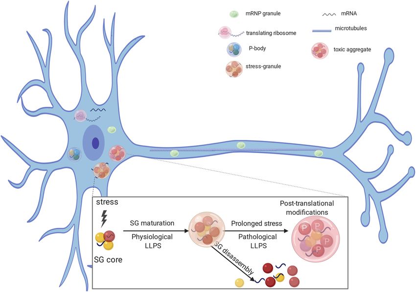
2 • J. Neurosci., 0, 2021 • 00(00):000                                                                             Hayashi et al. · LLPS in Nervous System

that many reported protein-protein interactions mediating these           membraneless organelles are radically different from mem-
cellular functions are actually part of a larger protein interaction      brane-based organelles.
network underlying LLPS. In this review, we discuss the role of               Because of the complexity of interactions between biological
LLPS in neurons, with a focus on local protein synthesis, synaptic        macromolecules, more than two condensates of different composi-
organization, and neurodegenerative disease.                              tion can form at the same time in the same cellular compartment.
                                                                          They can form independently of each other (phase-to-phase) or
Biophysics behind LLPS                                                    one condensate can form inside of another condensate (phase-in-
LLPS has been well studied in the field of soft-matter physics, but       phase) (Kato et al., 2012; Quiroz et al., 2020; Hosokawa et al.,
biologists have only recently discovered its importance and               2021). This might account for subdomains observed in some mem-
implications in divergent cellular functions (Hyman et al., 2014;         braneless organelles, such as core-shell architecture of nucleoli,
Banani et al., 2017). The governing mechanism for forming                 stress granules, and P granules (Kato et al., 2012).
phase-separated condensates in biological systems is multivalent              To observe LLPS in vitro, proteins of interest are purified, flu-
interactions (Li et al., 2012; Banani et al., 2017; Chen et al., 2020).   orescently labeled, mixed, and observed by diffusion interference
Such interactions can occur between molecules with multiple               contrast microscopy or fluorescence microscopy (Fig. 1E).
pairs of specific interactions (e.g., between multidomain scaffold        Photobleaching of a single fluorescent droplet or part of a fluo-
proteins and their binding partners). An increase in multivalency         rescent droplet enables measurements of protein movement
lowers the critical protein concentration required for phase sepa-        within the droplet as well as protein in exchange with diluted
ration (Li et al., 2012). Multivalent interactions can also occur         phase (Feng et al., 2019). These studies enable researchers to
among proteins with intrinsically disordered regions, a region of         understand how protein components regulate LLPS in vitro;
protein without any fixed conformation or domain structure, or            however, it is important to reproduce in vitro studies in the living
with various RNA species. Intrinsically disordered regions are of-        cell.
ten composed of low-complexity amino acids that are rich in
hydrophilic residues (serine, glutamine, glutamate, arginine, and         LLPS and local protein synthesis
lysine) and that can form electrostatic interactions. Aromatic resi-      Membraneless organelles control gene expression, from tran-
dues, such as phenylalanine, tyrosine, and tryptophan, are stacked        scription in the nucleus to local protein synthesis in distal proc-
on each other to form p electron cloud (p -p interaction) or              esses (Martin and Ephrussi, 2009; Hnisz et al., 2017; Langdon
interact with positively charged residues via cation-p interactions.      and Gladfelter, 2018). These organelles circumvent the need for
In contrast, aliphatic residues, such as valine, leucine, and isoleu-     active transport of macromolecules across a membrane, enabling
cine, are less frequently observed in low-complexity domains.             rapid signal transduction. While many of the membraneless or-
Both protein-domain interactions and electrostatic interactions in        ganelles involved in gene expression share the biophysical trait of
the intrinsically disordered region contribute to the formation of        LLPS, each organelle is distinct in its molecular composition and
condensed molecular assemblies with specific and distinct biologi-        function. Here, we focus on neuronal mRNA-containing ribonu-
cal functions via phase separation.                                       cleoprotein (mRNP) granules.
    In a simple two-molecule system, such as a protein in water,               Proteins and mRNAs within neuronal mRNP granules can be
the phase behavior of the solution can be characterized by the            dendritically localized (Kiebler and Bassell, 2006), where their
free energy diagram (Fig. 1A) and the corresponding phase dia-            translation can be regulated at synapses (Knowles et al., 1996;
gram (Fig. 1B). Under conditions relevant to living cells, most           Kohrmann et al., 1999; Krichevsky and Kosik, 2001; Mallardo et
proteins in water form a homogeneous one-phase solution                   al., 2003; Kanai et al., 2004) (Fig. 2). Retrograde and anterograde
because of the tendency of the mixture to increase its entropy            transport of these granules is microtubule-dependent (Knowles
(Fig. 1C). However, on self-interaction, the protein may undergo          et al., 1996; Kohrmann et al., 1999). The movement of mRNAs
LLPS, leading to two distinct phases: a highly condensed phase            to specific distal sites is necessary for synaptic plasticity and the
and a dilute phase (Fig. 1C). In the two-phase mixture, there is          strengthening of neuronal connections, a critical component of
no free energy difference between the condensed and the dilute            cognitive processes, such as long-term memory (Richter and
phases. The diffusion chemical potential (m) of the protein gener-        Lorenz, 2002; Klann and Dever, 2004).
ated by the concentration gradient between the two phases is off-              LLPS of components of neuronal mRNP granules plays essen-
set by the net free energy gain (DDG) of increased binding                tial roles in mRNA trafficking and local protein synthesis
between protein molecules in the condensed phase because of its           (Fig. 2). Work from the Kandel and Lu.F. laboratories posits
higher concentration (i.e., m = DDG). Thus, the phase-separated           a link between LLPS of cytoplasmic polyadenylation element
liquid solution is at a thermodynamic equilibrium. Nonetheless,           binding protein 3 (CPEB3) in trafficking dendrite-bound
protein molecules in the condensed phase can freely exchange              mRNAs that contain cytoplasmic polyadenylation elements
with molecules in the dilute phase (Fig. 1D).                             (CPEs) (Ford et al., 2019). Indeed, neuronal mRNP granules
    The free-energy state of a two-component mixture at any spe-          concentrate a large amount of CPE-containing mRNAs,
cific condition within the phase separation zone (pale blue and           including CaMKIIa (Huang et al., 2003; Martin, 2004). The
blue regions in Fig. 1B; see the corresponding free energy states         CPEs promote cytoplasmic polyadenylation-induced transla-
of the regions in Fig. 1A) dictates that the system will spontane-        tion of the mRNAs in response to synaptic stimulation, such
ously reach to two local minima, corresponding to Ud and Uc.              as NMDA-dependent LTP (Gu et al., 1999; Huang et al.,
Depending on the free energy state, phase separation can occur            2006; Fioriti et al., 2015). Kandel and Lu.F. have shown that
via binodal nucleation (formation of condensed phase requiring            CPEB3 binds CPEs of dendrite-bound mRNAs, providing
a nucleation processes) or spinodal decomposition (rapid and              translational regulation that is necessary for memory persist-
spontaneous phase separation without nucleation) (Fig. 1E). In            ence (Fioriti et al., 2015). Additionally, they found that
a membrane-sealed compartment, exchange of molecules                      CPEB3 undergoes LLPS when bound to its target mRNA and
within and outside of the compartment needs to go through the             is SUMOylated (Ford et al., 2019), suggesting that LLPS plays
membrane bilayer and requires energy (Fig. 1F). Thus,                     a role in translation regulation. Indeed, CPEB3 leaves the
Hayashi et al. · LLPS in Nervous System                                                                                                                  J. Neurosci., 0, 2021 • 00(00):000 • 3

Figure 1. Phase separation illustrated by a simple two-component system. A, Free energy diagram showing phase separation of a two-component system (e.g., a protein indicated by blue
dots; in water indicated by brown dots) under a certain condition. A uniformly mixed system can undergo phase separation by lowering the free energy to its minima, which results in a two-
phase system: a dilute phase (Ud, expressed as fraction volume for the dilute phase) and a condensed phase (Uc, fraction volume for the condensed phase). B, Phase diagram of the two-com-
ponent system constructed by plotting the free energy minima as a function of temperature. Blue curve indicates a sharp boundary (or the threshold concentration) of the system transitioning
from a homogeneous single-phase state to a two-phase state. Within the phase separation region, two modes of phase separation, binodal nucleation and spinodal decomposition, can occur.
C, In a phase-separated two-component system, a thermodynamic equilibrium is reached (i.e., DGd/c = 0). A sharp gradient in the concentration of the blue molecule is established between
the two phases. D, After phase separation, the components of the condensed phase and the diluted phase can freely exchange. However, there is no net flow of components between the two
phases. E, An example of binodal nucleation-induced phase separation forming condensed spherical droplets (left) and an example of spinodal decomposition-induced phase separation forming
worm-like condensed networks (right). F, In sharp contrast to membraneless condensates, spontaneous compartment fusion or materials exchange does not occur in membrane-separated
organelles.

membraneless Processing Body (P body) to join the distally                                       neural morphology in the form of excessively long and thin filo-
located polysome after chemically induced LTP (Ford et al.,                                      podia-like spines and fewer mature spines (Nimchinsky et al.,
2019). This work identifies the movement of phase separated,                                     2001). FMRP is localized to the synapse on metabotropic glu-
translation-dependent components from a repressed state in                                       tamate receptor activation, where it functions to target dendri-
neuronal mRNP granule-like P bodies (Barbee et al., 2006) to                                     tic mRNAs and regulates translation (Jin and Warren, 2003;
an active state at distal ribosomes, and suggests that P bodies                                  Antar et al., 2004). FMRP represses mRNA translation both in
are playing an essential role in this process (Cougot et al.,                                    vivo and in vitro, possibly by blocking ribosome elongation at
2008; Ford et al., 2019).                                                                        the polysome (Zalfa et al., 2006) and/or by miRNA-FMRP
    Fragile X Mental Retardation Protein (FMRP) is another                                       interaction, which would repress translation via the RNA-
well-characterized component of neuronal mRNP granules,                                          induced silencing complex (Zalfa et al., 2006). Experiments
largely studied for its role in the pathogenesis of fragile X syn-                               conducted in vitro using reticulocytes extracts and recombi-
drome, the most commonly inherited form of mental retardation                                    nant FMRP suggest that this translation repression likely
(Jin and Warren, 2003). Disruption of FMRP results in altered                                    occurs within the LLPS state, since FMRP-containing droplets
4 • J. Neurosci., 0, 2021 • 00(00):000 Hayashi et al. · LLPS in Nervous System
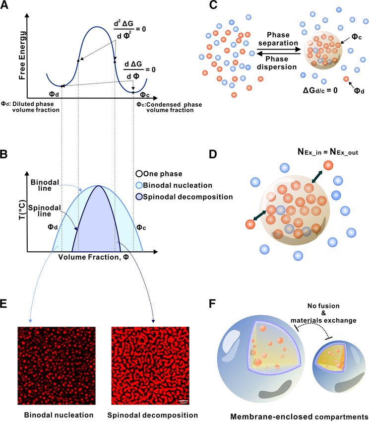
Figure 2. RNA binding proteins are involved in RNA stability (P bodies), mRNA transport (mRNA transport granules), translation, and stress granules (SG) formation. Under transient stress,
protein-protein and RNA interactions form a dense SG core. Several RNA binding proteins can be recruited to SG cores and undergo LLPS forming functional dynamic structures (physiological
LLPS). Under conditions of transient stress, SGs are transiently formed but disassemble after the stress is gone. In case of prolonged stress, and after post-translational modifications, such as
phosphorylation, proteins can become insoluble (pathologic LLPS). The same RNA binding proteins can participate in the formation of nontoxic hydrophobic aggregates and toxic cytoplasmic
inclusions.
can recruit translational repressors and miRNA (Tsang et al., role in regulating axonal local translation (Antar et al., 2004;
2019). However, the same authors do not show direct evidence Kiebler and Bassell, 2006). A coculture system of Aplysia sensory
that only the phase-separated state is capable of repressing presynaptic and motor postsynaptic neurons has been used for
translation in an intact cellular environment. Thus, additional studies of axonal local translation. After stimulation to induce
studies are necessary to clarify whether the ability to repress long-term facilitation, relevant mRNAs, such as sensorin, rapidly
translation is an exclusive property of the condensed phase. concentrate in the presynaptic terminus of sensory neurons
Interestingly, FMRP LLPS is mediated by binding to its mRNA (Lyles et al., 2006). Moreover, live-cell imaging of fluorescent
targets and by post-translational modifications, such as phos- translational reporters revealed accumulation of newly synthe-
phorylation (Tsang et al., 2019). Tsang et al. (2019) predict that sized proteins in the presynaptic terminus (Wang et al., 2009),
additional RNA-binding proteins involved in translational suggesting that local translation occurs in the presynaptic termi-
repression might undergo LLPS to function as translational nus during long-term facilitation.
repressors in neurons. As they are transported along axons to growth cones or pre-
mRNAs in neuronal mRNP granules can also drive LLPS and synaptic structures, RNA-binding proteins and mRNAs form
direct dendritic targeting of mRNP granules. RNA modifies the mRNP granules through LLPS. Translation is suppressed in these
LLPS behavior of RNA-binding proteins (Maharana et al., 2018); granules until they receive extracellular signals that initiate local
and the post-transcriptional state of the RNA, such as secondary translation. FMRP, together with proteins, such as fragile X-
structure, also plays a role in changing LLPS behavior (Langdon related 1 (FXR1) and FXR2, forms FMRP-containing granules
and Gladfelter, 2018; Van Treeck and Parker, 2018). Recently, the (FXGs) by LLPS, which plays an important role in the translation
Jaffery laboratory identified a facilitating role of methylation of control (Antar et al., 2006; Li et al., 2009; Till et al., 2011; Parvin
adenosine at the nitrogen-6 position (m6A) in LLPS in vitro, and et al., 2019; Tsang et al., 2019). These granules are often localized
linked the high abundance of m6A RNA to LLPS of specific mem- near synaptic vesicles (Christie et al., 2009), which may serve as
braneless organelles (Ries et al., 2019). Interestingly, transcripts platforms for local translation at presynaptic structures. The syn-
critical for synaptic organization and function are highly modified aptic vesicle protein synapsin 1 condenses into liquid droplets
with m6A and are translocated to synapse (Merkurjev et al., 2018). and promotes clustering of synaptic vesicles at presynaptic termi-
Like the disrupted neuromorphology seen with FMRP mutations nals (Milovanovic et al., 2018). Because FXGs localize with syn-
(Nimchinsky et al., 2001; Tsang et al., 2019), reducing the levels of aptic vesicles, it is possible that FMRP suppresses local
the protein “m6A reader,” a protein that interacts with m6A- translation to maintain mRNAs and translational machinery at
modified mRNA, caused structural and functional deficits in hip- the synapsin/synaptic vesicle condensate. Once a signal to initiate
pocampal dendritic spines (Merkurjev et al., 2018). translation for synapse formation or plasticity is received, FMRP
Local translation also takes place in axons (Jung et al., 2012; is dephosphorylated and FXGs are dispersed to initiate transla-
Wong et al., 2017; Hafner et al., 2019). Similarly to the local pro- tion. The surrounding phase environment (synapsin/synaptic
tein synthesis in dendrites, RNA-binding proteins play a major vesicles condensate) may affect the process of forming/dispersingHayashi et al. · LLPS in Nervous System J. Neurosci., 0, 2021 • 00(00):000 • 5
membrane proteins. Ca21 comes into the pre-
synaptic terminus through voltage-gated Ca21
channels at the active zone of the presynaptic
membrane. The clustering of the voltage-gated
Ca21 channels is mediated by two active zone
proteins, Rab3-interacting molecule (RIM) and
RIM-Binding Protein (RIM-BP), which interact
with voltage-gated Ca21 channels. RIM has a
proline-rich domain and a PDZ domain, which
interact with three SH3 domains in RIM-BP and
with the PDZ binding motif of the N-type volt-
age-gated Ca21 channels, respectively (Wu et al.,
2019; Wu, 2020). Through these multiple do-
Figure 3. Schematic diagram LLPS at synapses. Synapses contain various unique biological condensates, such as main interactions, RIM, RIM-BP, and voltage-
active zones and PSD. In a presynaptic bouton (light blue), the reserve pool of synaptic vesicles (SV) can form molec-
gated Ca21 channels can phase separate and
ular condensates via coacervating with the synapsin condensates. The docked pool of synaptic vesicles instead coats
the surface of active zone condensates formed by proteins, including RIM, RIM-BP, and ELKS. In the postsynaptic
form clusters at the active zone (Wu et al., 2019).
neuron (purple) and both in excitatory and inhibitory synapses, formation of PSD assemblies may also involve phase Wu et al. (2020) demonstrated that purified syn-
separation of synaptic scaffold proteins interacting with neurotransmitter receptors. aptic vesicles coat the surface of the RIM/RIM-
BP condensates either in solution or tethered to
membrane bilayers by the cytoplasmic tail of
FXGs by LLPS. However, further studies at higher resolution are voltage-gated Ca21 channels, forming a new
necessary to detect translating ribosomes and FXGs in presynap- type of interaction between a membrane organelle and mem-
tic structures in response to extracellular signals. braneless organelle. The coating of synaptic vesicles on the sur-
In summary, a multitude of nuclear and cytoplasmic mem- face of active zone condensates implies that the total number of
braneless organelles play critical roles in gene expression and synaptic vesicles tethered to each active zone is determined by its
local protein synthesis. The dense nature of these organelles, surface area (Schikorski and Stevens, 1997). Remarkably, when
with high concentrations of select protein and RNA components, the synapsin/vesicle condensates mixed with the vesicle-coated
allow for “packets of information” to be delivered directly to rele- RIM/RIM-BP condensates, the vesicle-coated RIM/RIM-BP con-
vant active sites. This allows for the efficient, and spatially de- densates are encapsulated by synapsin/small unilamellar vesicle
pendent, production of transcription and translation products in condensates, forming two distinct small unilamellar vesicle pools
the polarized neuron. reminiscent of the reserve and tethered synaptic vesicle pools
existing in presynaptic boutons. Thus, the authors have reconsti-
LLPS at the synapse tuted a presynaptic bouton-like structure containing vesicle-
Synaptic proteins are continuously turning over (Kuriu et al., coated active zone with one side attached to the presynaptic
2006; Sharma et al., 2006) and yet synapses can persist for weeks, membrane and the other side connected to the synapsin-clus-
months, or even the lifetime of the animal (Grutzendler et al., tered synaptic vesicle condensates.
2002; Yang et al., 2009; Isshiki et al., 2014). A presynaptic termi- Purified postsynaptic scaffolding proteins Shank and Homer
nus shows specific accumulation of component proteins, which self-assemble into macromolecular complexes when they are
tether the synaptic vesicles at rest and, on the influx of Ca , fuse 21 mixed together in vitro. Both Shank and Homer are multimeric
them with a specialized part of the presynaptic membrane called proteins, and Homer has Enabled/Vasp Homology domain that
the active zone. Postsynaptic receptors are embedded in the plasma interacts with Shank (Hayashi et al., 2009). Through this multi-
membrane, beneath which, various cellular components involved mer-multimer interaction, the protein complex takes on a high-
in signal transduction and regulation are enriched and comprise order meshwork structure and is the proposed underlying frame-
the postsynaptic density (PSD) (Sheng and Hoogenraad, 2007). work of the PSD at the excitatory synapse (Hayashi et al., 2009).
These presynaptic and postsynaptic structures lack any demarcating Similarly, SynGAP, a postsynaptic Ras-activating protein, is a tri-
membranes that prevent the diffusion of the component molecules meric protein with a PDZ binding motif (Zeng et al., 2016).
into the cytoplasm. Indeed, these properties of protein accumula- PSD-95, a postsynaptic scaffolding protein, multimerizes in vitro
tion are consistent with the phenomenon of LLPS (Fig. 3). (Hsueh and Sheng, 1999; Zeng et al., 2018). When purified
Synapsin is a presynaptic protein that crosslinks synaptic SynGAP and PSD-95 are combined, they form a macromolecular
vesicles and tethers them to the cytoskeleton within the resting complex. Interestingly, the resultant complex has droplet-like
presynaptic terminus. Upon Ca21 entry, activated CaMKII structures (Zeng et al., 2016). The properties of these droplets,
phosphorylates synapsin. This reduces the interaction of synap- such as spontaneous formation, constant exchange between con-
sin with synaptic vesicles and the cytoskeleton, and facilitates densed and diluted phase, and spontaneous fusion, are consistent
the process of vesicular release. When purified, synapsin can with the idea that these droplets are formed by LLPS. The phase
undergo LLPS in vitro in a manner recapitulating its in vivo separation of the PSD-95 and SynGAP mixture also suggests that
properties (Milovanovic et al., 2018). Synapsin condensates can the dense PSD assemblies beneath, but not enclosed by the post-
capture liposomes and are dispersed by CaMKII phosphorylation synaptic plasma membranes, are formed via LLPS. A mutant
(Milovanovic et al., 2018). From these observations, synapsin is that abolishes LLPS in vitro significantly impaired the enrich-
proposed to cluster synaptic vesicles in the presynaptic terminus by ment of these proteins in neurons (Hayashi et al., 2009; Zeng et
a LLPS-mediated mechanism. al., 2016).
The clustering of membrane surface proteins can also be When additional components of the PSD, including the
regulated by LLPS of proteins that bind to intracellular regions of NMDAR subunit GluN2B (which has a PDZ binding motif),6 • J. Neurosci., 0, 2021 • 00(00):000 Hayashi et al. · LLPS in Nervous System
GKAP (which bridges PSD-95 and Shank), Shank, and Homer CaMKII has a rotationally symmetric dodecameric structure that
were added to a PSD-95/SynGAP mixture, this resulted in LLPS can simultaneously interact with these proteins and cross link
at lower protein concentration, indicating a synergetic effect on them. The ability of CaMKII to undergo LLPS was experimen-
the phase formation (Zeng et al., 2018; Chen et al., 2020; Wu, tally demonstrated by using purified CaMKII and other PSD
2020). However, the contribution of each protein to phase sepa- proteins, including the scaffolding protein PSD-95, GluN2B, and
rate is different. Removal of PSD-95 significantly reduced Stargazin as a proxy of AMPAR itself. Notably, CaMKII under-
GluN2B but not Shank and Homer. In contrast, removal of goes phase separation with these proteins only in the presence
Shank significantly reduced Homer but had less impact on of Ca21; and after it undergoes LLPS, this state persists even af-
PSD-95 and SynGAP. This suggests that some proteins serve as ter chelation of Ca21. This persistence of LLPS after Ca21 che-
a “driver” for the formation of phase separation, whereas others lation requires phosphorylation of threonine 286 of CaMKII,
serve as a “client.” PSD-95 serves as a major driver of phase which has been shown to render CaMKII constitutively active.
separation, whereas GluN2B serves as a client. In contrast, Therefore, one major role of CaMKII at the synapse may be to
Homer and Shank form an independent layer that does not link different postsynaptic molecules through LLPS in a man-
serve as a driver or client for PSD-95/SynGAP/GluN2B. This ner triggered by Ca21 (Hosokawa et al., 2021).
is consistent with electron microscopic observations of the In a related study, Cai et al. (2020) discovered that autoinhibited
laminal structure of PSD (Valtschanoff and Weinberg, 2001), CaMKIIa specifically binds to Shank3. In a reconstitution buffer
where PSD-95 and GluN2B are layered together immediately containing no Ca21, mixing CaMKIIa and Shank3 leads to phase
beneath the synaptic membrane, while Shank is in a deeper separation of the mixture. Addition of Ca21 induces GluN2B-medi-
layer. GKAP is an interesting molecule in this structure: ated recruitment of active CaMKIIa and formation of the GluN2B/
when it was removed, both PSD-95/SynGAP/GluN2B and PSD-95/CaMKIIa condensates, which is autonomously dispersed
Shank/Homer had significantly reduced phase formation. on Ca21 removal. Protein phosphatases control the Ca21-depend-
GKAP is situated between these two layers in the protein ent shuttling of CaMKIIa between the two PSD subcompart-
complex and may serve as an interface. Indeed, in native ments (the upper layer composed of GluN2B/PSD-95 and the
PSDs, GKAP is layered between PSD-95/GluN2B and Shank lower layer composed of GKAP/Shank3/Homer). Activation of
(Valtschanoff and Weinberg, 2001). CaMKIIa further enlarges the PSD assembly, mimicking activ-
AMPA-type glutamate receptors (AMPAR) are another ity-induced structural LTP in synapse. Therefore, Ca21-driven
major receptor group of the excitatory synapse. They interact and phosphatase-checked shuttling of CaMKIIa between dis-
with a myriad of proteins that regulate the synthesis, function, tinct PSD nanodomains may underlie structural plasticity of
and subcellular distribution of AMPAR. Major interactors PSD assemblies via LLPS (Cai et al., 2020).
include the transmembrane AMPAR-interacting proteins, LLPS of CaMKII is also involved in the segregation of synap-
which interact with the transmembrane domain of AMPARs
tic surface proteins. Glutamate receptor subtypes are organized
and determine receptor localization and function (Nicoll et
into nanodomains at the synapse. In each hippocampal synapse,
al., 2006). A prototypical transmembrane AMPAR-interacting
NMDAR forms one dominant nanodomain and several small
protein, Stargazin, can interact with PSD-95 through a PDZ-
domains, whereas AMPAR segregates into several nanodomains
binding motif, as well as through an arginine-rich motif (Zeng
of similar size surrounding the NMDAR. In contrast, metabo-
et al., 2019). Through such multivalent interactions, Stargazin
undergoes LLPS with PSD-95. This is required for efficient tropic glutamate receptors are more diffuse (Goncalves et al.,
incorporation of AMPAR into the synapse. 2020). Postsynaptic nanodomains connect to the presynaptic
The induction of synaptic plasticity can persistently alter the active zone via cell adhesion molecules, thereby forming trans-
amount of the AMPAR and various other proteins residing at synaptic nanocolumns (Tang et al., 2016; Biederer et al., 2017;
the synapse (Bosch et al., 2014). Thus, an important and out- Scheefhals and MacGillavry, 2018). CaMKII preferentially inter-
standing question is how neuronal activity modulates postsy- acts with the NMDAR subunit GluN2B rather than the AMPAR,
naptic LLPS to trigger the delivery of synaptic proteins. The represented by Stargazin. This leads to the formation of a phase-
induction of LTP induces a delivery of postsynaptic proteins in-phase structure of AMPARs within the NMDAR-CaMKII
in a specific order from the dendritic shaft. Actin and actin- phase. Further, the cell-adhesion molecule neuroligin segregates
related proteins are the first to arrive at the synapse, followed with the AMPAR and connects the presynaptic neurexin with
by AMPAR. PSD scaffolding proteins, such as PSD-95 and the presynaptic release machinery. This mechanism may place
Homer, take longer to increase (;2 h) after LTP induction, AMPARs just beneath the transmitter release site, thereby opti-
and require the synthesis of new protein (Bosch et al., 2014). mizing the transmission efficacy and serving as a novel mecha-
In contrast, SynGAP, another PSD protein that inhibits Ras nism CaMKII-mediated synaptic plasticity.
activity, dissociates quickly from the synapse on phosphoryla- In contrast to prominent PSD assemblies in excitatory synap-
tion by CaMKII (Araki et al., 2015). Furthermore, phospho- ses, inhibitory synapses do not contain obvious dense thickening
rylation of Stargazin by CaMKII negatively affects LLPS (Zeng underneath synaptic membranes. However, recent cryo-EM to-
et al., 2019). Because activation of CaMKII transiently occurs mography studies reveal a sheet-like dense assembly (referred to
after LTP induction (Lee et al., 2009), this might create a time as iPSD) with a thickness of ;5 nm (Tao et al., 2018). A recent
window for reorganization of the postsynaptic protein study has demonstrated that glycine or GABAA receptors, to-
condensate. gether with gephyrin, a key scaffold protein in inhibitory synap-
Indeed, CaMKII has several properties that enable it to ses, can undergo phase separation, forming iPSD condensates.
undergo LLPS. Once activated by Ca21/calmodulin, CaMKII can The formation of the iPSD condensates can be regulated by
form a persistent complex with substrate proteins, including the phosphorylation of gephyrin or binding of target proteins to
intracellular carboxyl tail of the NMDAR subunit GluN2B, Rac gephyrin (Bai et al., 2020). Thus, analogous to excitatory PSDs,
guanine nucleotide exchange factor Tiam1, GJD2/connexin 36, iPSDs are likely formed by phase separation-mediated condensa-
LRRC7/densin-180, and the L-type Ca21 channel. In addition, tion of scaffold protein/neurotransmitter receptor complexes.Hayashi et al. · LLPS in Nervous System J. Neurosci., 0, 2021 • 00(00):000 • 7
LLPS in neurodegenerative disease granule formation mitigate TDP-43-associated toxicity and/or
Neurodegenerative diseases, such as Alzheimer’s disease and aggregation in various cellular and animal models (Elden et al.,
Parkinson’s disease, are currently incurable and have no effective 2010; Kim et al., 2014; Becker et al., 2017; Zhang et al., 2018;
treatments. To identify potential treatments, it is paramount to McGurk et al., 2018c; Duan et al., 2019; Fernandes et al., 2020);
understand the cellular and pathologic basis of disease. One and that stress-granule resident proteins coaggregate with ;30%
defining cellular feature of neurodegenerative disease is the dep- of TDP-43 inclusions in human ALS tissue (Liu-Yesucevitz et al.,
osition of protein aggregates in affected brain regions. Protein 2010; Bentmann et al., 2012; McGurk et al., 2014).
aggregates in a given disease are formed by a specific protein, for An overarching hypothesis has been that stress-granule local-
example, the microtube-associated protein tau in Alzheimer’s ization of TDP-43 seeds the protein aggregation observed in
disease and 50% of patients with frontotemporal degeneration ALS. Stress granules and LLPS condensates are highly concen-
(Mackenzie and Neumann, 2016; Vogels et al., 2020); a-synu- trated sources of protein, which is a biophysical property that
clein in Parkinson’s disease and Lewy body dementia (Luna and promotes LLPS. Thus, by increasing local protein concentration,
Luk, 2015; Zbinden et al., 2020); and TDP-43 in .95% of LLPS provides an environment that can promote phase transi-
patients with amyotrophic lateral sclerosis (ALS) and in ;45% tion events that lead to the formation of protein oligomers with
of patients with frontotemporal degeneration (Mackenzie and solid-like characteristics (Kato et al., 2012; Molliex et al., 2015;
Neumann, 2016; Taylor et al., 2016). Microtube-associated pro- Murakami et al., 2015; Patel et al., 2015; Guo et al., 2018). In
tein tau, a-synuclein, and TDP-43 have an inherent capacity to in vitro experiments, solid protein oligomerization within pro-
aggregate; they harbor disease-causing mutations, and the ana- tein condensates can also be promoted by increasing the time the
tomic burden of these protein aggregates correlates with sympto- proteins are in the protein droplet, by repeated forming and dis-
matic decline (Luna and Luk, 2015; Mackenzie and Neumann, solving the protein droplets, and by introducing disease-associ-
2016; Taylor et al., 2016; Harrison and Shorter, 2017; Vogels et ated mutations to the protein (Lin et al., 2015; Molliex et al.,
al., 2020; Zbinden et al., 2020). How protein aggregates correlate 2015; Patel et al., 2015). In line with these in vitro data, cells
with disease is unclear, but it is emerging that LLPS may be exposed to chronic stress form stress granules and persistent
involved. Here we focus on the role of LLPS in ALS. TDP-43 aggregates (McGurk et al., 2018b; Gasset-Rosa et al.,
ALS is an incurable motor neuron disease that leads to paraly- 2019; Fernandes et al., 2020), suggesting that chronic stress and/
sis and death within 2-5 years of symptomatic onset (Taylor et or stress-granule localization leads to disease-like aggregation of
al., 2016). In .95% of ALS patients, TDP-43 forms phosphoryl- TDP-43. However, under short-term stress, stress granules in-
ated protein aggregates in the cytoplasm of affected motor neu- hibit the formation of disease-like aggregates of TDP-43 and pro-
rons (Arai et al., 2006; Neumann et al., 2006). Mutations in mote the solubility and dissolution of the protein after the
several ALS-linked genes have been identified, and these give rise removal of stress (McGurk et al., 2018b; Chen and Cohen, 2019;
to ;15% of ALS cases (Taylor et al., 2016). Many of the mutated Gasset-Rosa et al., 2019; Mann et al., 2019; Fernandes et al.,
genes, including TDP-43, FUS, and TIA1, are RNA-binding pro- 2020). Thus, under short-term stress, the cell controls both the
teins that harbor a prion-like domain (Sreedharan et al., 2008; accumulation and dissolution of TDP-43 aggregates, but under
Kwiatkowski et al., 2009; Vance et al., 2009; Kim et al., 2013; continued stress and maintenance of a condensed phase, TDP-
Mackenzie et al., 2017). The prion-like domain is an intrinsically 43 transitions into disease-like aggregates.
disordered region that can promote protein aggregation and pro- Elucidation of the LLPS-associated dynamics of membrane-
tein phase separation both in vitro and in the cell (Johnson et al., less organelles and disease-causing proteins may explain the pa-
2009; Sun et al., 2011; Han et al., 2012; Kato et al., 2012; Lin et thology observed in ALS and other neurodegenerative diseases.
al., 2015; Molliex et al., 2015; Murakami et al., 2015; Patel et al., However, whether protein aggregation causes dysfunction and
2015; Xiang et al., 2015; Conicella et al., 2016, 2020; McGurk et clinical symptoms is unknown. Data from animal models suggest
al., 2018a,b; Ryan et al., 2018; Murthy et al., 2019); and it is often that targeting pathways that promote LLPS and stress granule
the site of disease-causing mutations (Sreedharan et al., 2008; biogenesis is therapeutic (Elden et al., 2010; Kim et al., 2014;
Kwiatkowski et al., 2009; Vance et al., 2009; Kim et al., 2013; Becker et al., 2017; Guo et al., 2018; McGurk et al., 2018c; Zhang
et al., 2018; Duan et al., 2019; Fernandes et al., 2020). Thus,
Mackenzie et al., 2017). Thus, LLPS is a focus in the underlying
studying the mechanisms of LLPS is directing us toward path-
pathogenesis of ALS.
ways with therapeutic potential for incurable diseases, such as
In ALS, neurons are under constitutive stress that can arise
ALS.
from misfolded proteins in the endoplasmic reticulum and mito-
chondrial dysfunction (Kiskinis et al., 2014; Montibeller and de
Belleroche, 2018). As a survival mechanism during stress, the cell Concluding remarks
In conclusion, LLPS is emerging as a key biological phenomenon
inhibits global protein translation by sequestering RNA-protein
that mediates several aspects of the basic organization and proper
complexes involved in the pre-initiation of protein synthesis into
stress granules (Ivanov et al., 2019; Jaud et al., 2020). TDP-43 functions of cells in general, and neurons in particular. It will be
and several of the RNA-binding proteins linked to ALS localize interesting to see where the field of LLPS will take us in the next
to stress granules (Bosco et al., 2010; Dewey et al., 2011; few years. We anticipate that combined the technological
Mackenzie et al., 2017; Fernandes et al., 2018). The hypothesis advancements in super-resolution microscopy and other imag-
that stress granules are linked to ALS is further supported by evi- ing techniques, we will be able to fill the gaps between in vitro
dence that demonstrates that disease-causing mutations in the studies and in vivo conditions. Further advancements in our
RNA-binding proteins linked to ALS alter LLPS in vitro and understanding of this phenomenon will also allow us to design
localization of the respective proteins to stress granules (Lin et new therapeutic approaches against neurodegenerative diseases.
al., 2015, 2016; Molliex et al., 2015; Murakami et al., 2015; Patel
et al., 2015; Conicella et al., 2016; Lee et al., 2016; Boeynaems et References
al., 2017; Dao et al., 2018; Wang et al., 2018; McGurk et al., Antar LN, Afroz R, Dictenberg JB, Carroll RC, Bassell GJ (2004)
2018b); that downregulation of pathways that promote stress Metabotropic glutamate receptor activation regulates fragile X mental8 • J. Neurosci., 0, 2021 • 00(00):000 Hayashi et al. · LLPS in Nervous System
retardation protein and FMR1 mRNA localization differentially in den- specialized P-body-like structures that respond to neuronal activation. J
drites and at synapses. J Neurosci 24:2648–2655. Neurosci 28:13793–13804.
Antar LN, Li C, Zhang H, Carroll RC, Bassell GJ (2006) Local functions for Dao TP, Kolaitis RM, Kim HJ, O’Donovan K, Martyniak B, Colicino E,
FMRP in axon growth cone motility and activity-dependent regulation of Hehnly H, Taylor JP, Castaneda CA (2018) Ubiquitin modulates liquid-
filopodia and spine synapses. Mol Cell Neurosci 32:37–48. liquid phase separation of UBQLN2 via disruption of multivalent interac-
Arai T, Hasegawa M, Akiyama H, Ikeda K, Nonaka T, Mori H, Mann D, tions. Mol Cell 69:965–978.e966.
Tsuchiya K, Yoshida M, Hashizume Y, Oda T (2006) TDP-43 is a compo- Dewey CM, Cenik B, Sephton CF, Dries DR, Mayer P 3rd, Good SK,
nent of ubiquitin-positive tau-negative inclusions in frontotemporal lobar Johnson BA, Herz J, Yu G (2011) TDP-43 is directed to stress granules by
degeneration and amyotrophic lateral sclerosis. Biochem Biophys Res sorbitol, a novel physiological osmotic and oxidative stressor. Mol Cell
Commun 351:602–611. Biol 31:1098–1108.
Araki Y, Zeng M, Zhang M, Huganir RL (2015) Rapid dispersion of SynGAP Duan Y, Du A, Gu J, Duan G, Wang C, Gui X, Ma Z, Qian B, Deng X, Zhang
from synaptic spines triggers AMPA receptor insertion and spine K, Sun L, Tian K, Zhang Y, Jiang H, Liu C, Fang Y (2019) PARylation
enlargement during LTP. Neuron 85:173–189. regulates stress granule dynamics, phase separation, and neurotoxicity of
Bai G, Wang Y, Zhang M (2020) Gephyrin-mediated formation of inhibitory disease-related RNA-binding proteins. Cell Res 29:233–247.
postsynaptic density sheet via phase separation. Cell Res Advance online Elden AC, Kim HJ, Hart MP, Chen-Plotkin AS, Johnson BS, Fang X,
publication. Retrieved Nov 2, 2020. doi: 10.1038/s41422-020-00433-1. Armakola M, Geser F, Greene R, Lu MM, Padmanabhan A, Clay-Falcone
Banani SF, Lee HO, Hyman AA, Rosen MK (2017) Biomolecular conden- D, McCluskey L, Elman L, Juhr D, Gruber PJ, Rüb U, Auburger G,
sates: organizers of cellular biochemistry. Nat Rev Mol Cell Biol 18:285– Trojanowski JQ, Lee VM, et al. (2010) Ataxin-2 intermediate-length poly-
298. glutamine expansions are associated with increased risk for ALS. Nature
Barbee SA, Estes PS, Cziko AM, Hillebrand J, Luedeman RA, Coller JM, 466:1069–1075.
Johnson N, Howlett IC, Geng C, Ueda R, Brand AH, Newbury SF, Feng Z, Chen X, Wu X, Zhang M (2019) Formation of biological condensates
Wilhelm JE, Levine RB, Nakamura A, Parker R, Ramaswami M (2006) via phase separation: characteristics, analytical methods, and physiologi-
Staufen- and FMRP-containing neuronal RNPs are structurally and func- cal implications. J Biol Chem 294:14823–14835.
tionally related to somatic P bodies. Neuron 52:997–1009. Fernandes N, Eshleman N, Buchan JR (2018) Stress granules and ALS: a case
Becker LA, Huang B, Bieri G, Ma R, Knowles DA, Jafar-Nejad P, Messing J, of causation or correlation? Adv Neurobiol 20:173–212.
Kim HJ, Soriano A, Auburger G, Pulst SM, Taylor JP, Rigo F, Gitler AD Fernandes N, Nero L, Lyons SM, Ivanov P, Mittelmeier TM, Bolger TA,
(2017) Therapeutic reduction of ataxin-2 extends lifespan and reduces Buchan JR (2020) Stress granule assembly can facilitate but is not
pathology in TDP-43 mice. Nature 544:367–371. required for TDP-43 cytoplasmic aggregation. Biomolecules 10:1367.
Bentmann E, Neumann M, Tahirovic S, Rodde R, Dormann D, Haass C Fioriti L, Myers C, Huang YY, Li X, Stephan JS, Trifilieff P, Colnaghi L,
(2012) Requirements for stress granule recruitment of fused in sarcoma Kosmidis S, Drisaldi B, Pavlopoulos E, Kandel ER (2015) The persistence
(FUS) and TAR DNA-binding protein of 43 kDa (TDP-43). J Biol Chem of hippocampal-based memory requires protein synthesis mediated by
287:23079–23094. the prion-like protein CPEB3. Neuron 86:1433–1448.
Biederer T, Kaeser PS, Blanpied TA (2017) Transcellular nanoalignment of Ford L, Ling E, Kandel ER, Fioriti L (2019) CPEB3 inhibits translation of
synaptic function. Neuron 96:680–696. mRNA targets by localizing them to P bodies. Proc Natl Acad Sci USA
Boeynaems S, Bogaert E, Kovacs D, Konijnenberg A, Timmerman E, Volkov 116:18078–18087.
A, Guharoy M, De Decker M, Jaspers T, Ryan VH, Janke AM, Baatsen P, Gasset-Rosa F, Lu S, Yu H, Chen C, Melamed Z, Guo L, Shorter J, Da Cruz S,
Vercruysse T, Kolaitis RM, Daelemans D, Taylor JP, Kedersha N, Cleveland DW (2019) Cytoplasmic TDP-43 de-mixing independent of
Anderson P, Impens F, Sobott F, et al. (2017) Phase separation of stress granules drives inhibition of nuclear import, loss of nuclear TDP-
C9orf72 dipeptide repeats perturbs stress granule dynamics. Mol Cell 43, and cell death. Neuron 102:339–357.e337.
65:1044–1055.e1045. Goncalves J, Bartol TM, Camus C, Levet F, Menegolla AP, Sejnowski TJ,
Bosch M, Castro J, Saneyoshi T, Matsuno H, Sur M, Hayashi Y (2014) Sibarita JB, Vivaudou M, Choquet D, Hosy E (2020) Nanoscale co-orga-
Structural and molecular remodeling of dendritic spine substructures nization and coactivation of AMPAR, NMDAR, and mGluR at excitatory
during long-term potentiation. Neuron 82:444–459. synapses. Proc Natl Acad Sci USA 117:14503–14511.
Bosco DA, Lemay N, Ko HK, Zhou H, Burke C, Kwiatkowski TJ Jr, Sapp P, Grutzendler J, Kasthuri N, Gan WB (2002) Long-term dendritic spine stabil-
McKenna-Yasek D, Brown RH Jr, Hayward LJ (2010) Mutant FUS pro- ity in the adult cortex. Nature 420:812–816.
teins that cause amyotrophic lateral sclerosis incorporate into stress gran- Gu H, Das Gupta J, Schoenberg DR (1999) The poly(A)-limiting element is a
ules. Hum Mol Genet 19:4160–4175. conserved cis-acting sequence that regulates poly(A) tail length on nu-
Brangwynne CP, Eckmann CR, Courson DS, Rybarska A, Hoege C, clear pre-mRNAs. Proc Natl Acad Sci USA 96:8943–8948.
Gharakhani J, Julicher F, Hyman AA (2009) Germline P granules are liq- Guo L, Kim HJ, Wang H, Monaghan J, Freyermuth F, Sung JC, O’Donovan
uid droplets that localize by controlled dissolution/condensation. Science K, Fare CM, Diaz Z, Singh N, Zhang ZC, Coughlin M, Sweeny EA,
324:1729–1732. DeSantis ME, Jackrel ME, Rodell CB, Burdick JA, King OD, Gitler AD,
Cai Q, Zeng M, Wu X, Wu H, Zhan Y, Tian R, Zhang M (2020) CaMKIIa- Lagier-Tourenne C, et al. (2018) Nuclear-import receptors reverse aber-
driven, phosphatase-checked postsynaptic plasticity via phase separation. rant phase transitions of RNA-binding proteins with prion-like domains.
Cell Res Advance online publication. Retrieved Nov 24, 2020. doi: Cell 173:677–692.e620.
10.1038/s41422-020-00439-9. Hafner AS, Donlin-Asp PG, Leitch B, Herzog E, Schuman EM (2019) Local
Chen X, Wu X, Wu H, Zhang M (2020) Phase separation at the synapse. Nat protein synthesis is a ubiquitous feature of neuronal pre- and postsynap-
Neurosci 23:301–310. tic compartments. Science 364:eaau3644.
Chen Y, Cohen TJ (2019) Aggregation of the nucleic acid-binding protein Han TW, Kato M, Xie S, Wu LC, Mirzaei H, Pei J, Chen M, Xie Y, Allen J,
TDP-43 occurs via distinct routes that are coordinated with stress granule Xiao G, McKnight SL (2012) Cell-free formation of RNA granules: bound
formation. J Biol Chem 294:3696–3706. RNAs identify features and components of cellular assemblies. Cell
Christie SB, Akins MR, Schwob JE, Fallon JR (2009) The FXG: a presynaptic 149:768–779.
fragile X granule expressed in a subset of developing brain circuits. J Harrison AF, Shorter J (2017) RNA-binding proteins with prion-like
Neurosci 29:1514–1524. domains in health and disease. Biochem J 474:1417–1438.
Conicella AE, Zerze GH, Mittal J, Fawzi NL (2016) ALS mutations disrupt Hayashi MK, Tang C, Verpelli C, Narayanan R, Stearns MH, Xu RM, Li H,
phase separation mediated by alpha-helical structure in the TDP-43 low- Sala C, Hayashi Y (2009) The postsynaptic density proteins Homer and
complexity C-terminal domain. Structure 24:1537–1549. Shank form a polymeric network structure. Cell 137:159–171.
Conicella AE, Dignon GL, Zerze GH, Schmidt HB, D’Ordine AM, Kim YC, Hnisz D, Shrinivas K, Young RA, Chakraborty AK, Sharp PA (2017) A phase
Rohatgi R, Ayala YM, Mittal J, Fawzi NL (2020) TDP-43 alpha-helical separation model for transcriptional control. Cell 169:13–23.
structure tunes liquid-liquid phase separation and function. Proc Natl Hosokawa T, Liu P-W, Cai Q, Ferreira JS, Levet F, Butler C, Sibarita JB,
Acad Sci USA 117:5883–5894. Choquet D, Groc L, Hosy E, Zhang M, Hayashi Y (2021) Subsynaptic
Cougot N, Bhattacharyya SN, Tapia-Arancibia L, Bordonne R, Filipowicz W, segregation of glutamate receptors by CaMKII-mediated phase separa-
Bertrand E, Rage F (2008) Dendrites of mammalian neurons contain tion. Nat Neurosci, in press.Hayashi et al. · LLPS in Nervous System J. Neurosci., 0, 2021 • 00(00):000 • 9
Hsueh YP, Sheng M (1999) Requirement of N-terminal cysteines of PSD-95 Vance MA, Yan J, Ticozzi N, et al. (2009) Mutations in the FUS/TLS
for PSD-95 multimerization and ternary complex formation, but not for gene on chromosome 16 cause familial amyotrophic lateral sclerosis.
binding to potassium channel Kv1.4. J Biol Chem 274:532–536. Science 323:1205–1208.
Huang YS, Carson JH, Barbarese E, Richter JD (2003) Facilitation of dendri- Langdon EM, Gladfelter AS (2018) A new lens for RNA localization: liquid-
tic mRNA transport by CPEB. Genes Dev 17:638–653. liquid phase separation. Annu Rev Microbiol 72:255–271.
Huang YS, Kan MC, Lin CL, Richter JD (2006) CPEB3 and CPEB4 in neu- Lee KH, Zhang P, Kim HJ, Mitrea DM, Sarkar M, Freibaum BD, Cika J,
rons: analysis of RNA-binding specificity and translational control of Coughlin M, Messing J, Molliex A, Maxwell BA, Kim NC, Temirov J,
AMPA receptor GluR2 mRNA. EMBO J 25:4865–4876. Moore J, Kolaitis RM, Shaw TI, Bai B, Peng J, Kriwacki RW, Taylor JP
Hyman AA, Weber CA, Julicher F (2014) Liquid-liquid phase separation in (2016) C9orf72 dipeptide repeats impair the assembly, dynamics, and
biology. Annu Rev Cell Dev Biol 30:39–58. function of membrane-less organelles. Cell 167:774–788.e717.
Isshiki M, Tanaka S, Kuriu T, Tabuchi K, Takumi T, Okabe S (2014) Lee SJ, Escobedo-Lozoya Y, Szatmari EM, Yasuda R (2009) Activation of
Enhanced synapse remodelling as a common phenotype in mouse mod- CaMKII in single dendritic spines during long-term potentiation. Nature
els of autism. Nat Commun 5:4742. 458:299–304.
Ivanov P, Kedersha N, Anderson P (2019) Stress granules and processing Li C, Bassell GJ, Sasaki Y (2009) Fragile X mental retardation protein is
bodies in translational control. Cold Spring Harb Perspect Biol 11: involved in protein synthesis-dependent collapse of growth cones
a032813. induced by semaphorin-3A. Front Neural Circuits 3:11.
Jaud M, Philippe C, Di Bella D, Tang W, Pyronnet S, Laurell H, Mazzolini L, Li P, Banjade S, Cheng HC, Kim S, Chen B, Guo L, Llaguno M,
Rouault-Pierre K, Touriol C (2020) Translational regulations in response Hollingsworth JV, King DS, Banani SF, Russo PS, Jiang QX, Nixon BT,
to endoplasmic reticulum stress in cancers. Cells 9:540. Rosen MK (2012) Phase transitions in the assembly of multivalent signal-
Jin P, Warren ST (2003) New insights into fragile X syndrome: from mole- ling proteins. Nature 483:336–340.
cules to neurobehaviors. Trends Biochem Sci 28:152–158. Lin Y, Protter DS, Rosen MK, Parker R (2015) Formation and maturation of
Johnson BS, Snead D, Lee JJ, McCaffery JM, Shorter J, Gitler AD (2009) phase-separated liquid droplets by RNA-binding proteins. Mol Cell
TDP-43 is intrinsically aggregation-prone, and amyotrophic lateral scle- 60:208–219.
rosis-linked mutations accelerate aggregation and increase toxicity. J Biol Lin Y, Mori E, Kato M, Xiang S, Wu L, Kwon I, McKnight SL (2016) Toxic
Chem 284:20329–20339. PR poly-dipeptides encoded by the C9orf72 repeat expansion target LC
Jung H, Yoon BC, Holt CE (2012) Axonal mRNA localization and local pro- domain polymers. Cell 167:789–802.e712.
tein synthesis in nervous system assembly, maintenance and repair. Nat Liu-Yesucevitz L, Bilgutay A, Zhang YJ, Vanderweyde T, Citro A, Mehta T,
Rev Neurosci 13:308–324. Zaarur N, McKee A, Bowser R, Sherman M, Petrucelli L, Wolozin B
Kanai Y, Dohmae N, Hirokawa N (2004) Kinesin transports RNA: isolation (2010) Tar DNA binding protein-43 (TDP-43) associates with stress
and characterization of an RNA-transporting granule. Neuron 43:513– granules: analysis of cultured cells and pathological brain tissue. PLoS
525. One 5:e13250.
Kato M, Han TW, Xie S, Shi K, Du X, Wu LC, Mirzaei H, Goldsmith EJ,
Luna E, Luk KC (2015) Bent out of shape: a-Synuclein misfolding and the
Longgood J, Pei J, Grishin NV, Frantz DE, Schneider JW, Chen S, Li L,
convergence of pathogenic pathways in Parkinson’s disease. FEBS Lett
Sawaya MR, Eisenberg D, Tycko R, McKnight SL (2012) Cell-free forma-
589:3749–3759.
tion of RNA granules: low complexity sequence domains form dynamic
Lyles V, Zhao Y, Martin KC (2006) Synapse formation and mRNA localiza-
fibers within hydrogels. Cell 149:753–767.
tion in cultured Aplysia neurons. Neuron 49:349–356.
Kiebler MA, Bassell GJ (2006) Neuronal RNA granules: movers and makers.
Mackenzie IR, Neumann M (2016) Molecular neuropathology of frontotem-
Neuron 51:685–690.
poral dementia: insights into disease mechanisms from postmortem stud-
Kim HJ, Kim NC, Wang YD, Scarborough EA, Moore J, Diaz Z, MacLea KS,
ies. J Neurochem 138: 54–70.
Freibaum B, Li S, Molliex A, Kanagaraj AP, Carter R, Boylan KB, Wojtas
Mackenzie IR, Nicholson AM, Sarkar M, Messing J, Purice MD, Pottier C,
AM, Rademakers R, Pinkus JL, Greenberg SA, Trojanowski JQ, Traynor
BJ, Smith BN, et al. (2013) Mutations in prion-like domains in Annu K, Baker M, Perkerson RB, Kurti A, Matchett BJ, Mittag T,
hnRNPA2B1 and hnRNPA1 cause multisystem proteinopathy and ALS. Temirov J, Hsiung GY, Krieger C, Murray ME, Kato M, Fryer JD,
Nature 495:467–473. Petrucelli L, Zinman L, et al. (2017) TIA1 mutations in amyotrophic lat-
Kim HJ, Raphael AR, LaDow ES, McGurk L, Weber RA, Trojanowski JQ, eral sclerosis and frontotemporal dementia promote phase separation
Lee VM, Finkbeiner S, Gitler AD, Bonini NM (2014) Therapeutic modu- and alter stress granule dynamics. Neuron 95:808–816.e809.
lation of eIF2alpha phosphorylation rescues TDP-43 toxicity in amyotro- Maharana S, Wang J, Papadopoulos DK, Richter D, Pozniakovsky A, Poser I,
phic lateral sclerosis disease models. Nat Genet 46:152–160. Bickle M, Rizk S, Guillen-Boixet J, Franzmann TM, Jahnel M, Marrone L,
Kiskinis E, Sandoe J, Williams LA, Boulting GL, Moccia R, Wainger BJ, Han Chang YT, Sterneckert J, Tomancak P, Hyman AA, Alberti S (2018)
S, Peng T, Thams S, Mikkilineni S, Mellin C, Merkle FT, Davis- RNA buffers the phase separation behavior of prion-like RNA binding
Dusenbery BN, Ziller M, Oakley D, Ichida J, Di Costanzo S, Atwater N, proteins. Science 360:918–921.
Maeder ML, Goodwin MJ, et al. (2014) Pathways disrupted in human Mallardo M, Deitinghoff A, Muller J, Goetze B, Macchi P, Peters C, Kiebler
ALS motor neurons identified through genetic correction of mutant MA (2003) Isolation and characterization of Staufen-containing ribonu-
SOD1. Cell Stem Cell 14:781–795. cleoprotein particles from rat brain. Proc Natl Acad Sci USA 100:2100–
Klann E, Dever TE (2004) Biochemical mechanisms for translational regula- 2105.
tion in synaptic plasticity. Nat Rev Neurosci 5:931–942. Mann JR, Gleixner AM, Mauna JC, Gomes E, DeChellis-Marks MR,
Knowles RB, Sabry JH, Martone ME, Deerinck TJ, Ellisman MH, Bassell GJ, Needham PG, Copley KE, Hurtle B, Portz B, Pyles NJ, Guo L, Calder CB,
Kosik KS (1996) Translocation of RNA granules in living neurons. J Wills ZP, Pandey UB, Kofler JK, Brodsky JL, Thathiah A, Shorter J,
Neurosci 16:7812–7820. Donnelly CJ (2019) RNA binding antagonizes neurotoxic phase transi-
Kohrmann M, Luo M, Kaether C, DesGroseillers L, Dotti CG, Kiebler MA tions of TDP-43. Neuron 102:321–338.e328.
(1999) Microtubule-dependent recruitment of Staufen-green fluorescent Martin KC (2004) Local protein synthesis during axon guidance and synaptic
protein into large RNA-containing granules and subsequent dendritic plasticity. Curr Opin Neurobiol 14:305–310.
transport in living hippocampal neurons. Mol Biol Cell 10:2945–2953. Martin KC, Ephrussi A (2009) mRNA localization: gene expression in the
Krichevsky AM, Kosik KS (2001) Neuronal RNA granules: a link between spatial dimension. Cell 136:719–730.
RNA localization and stimulation-dependent translation. Neuron McGurk L, Lee VM, Trojanowksi JQ, Van Deerlin VM, Lee EB, Bonini NM
32:683–696. (2014) Poly-A binding protein-1 localization to a subset of TDP-43 inclu-
Kuriu T, Inoue A, Bito H, Sobue K, Okabe S (2006) Differential control of sions in amyotrophic lateral sclerosis occurs more frequently in patients
postsynaptic density scaffolds via actin-dependent and -independent harboring an expansion in C9orf72. J Neuropathol Exp Neurol 73:837–
mechanisms. J Neurosci 26:7693–7706. 845.
Kwiatkowski TJ, Bosco DA, Leclerc AL, Tamrazian E, Vanderburg CR, Russ McGurk L, Gomes E, Guo L, Shorter J, Bonini N (2018a) Poly(ADP-ribose)
C, Davis A, Gilchrist J, Kasarskis EJ, Munsat T, Valdmanis P, Rouleau engages the TDP-43 nuclear-localization sequence to regulate granulo-fil-
GA, Hosler BA, Cortelli P, de Jong PJ, Yoshinaga Y, Haines JL, Pericak- amentous aggregation. Biochemistry 57:6923–6926.You can also read