Tissue-selective alternate promoters guide NLRP6 expression
←
→
Page content transcription
If your browser does not render page correctly, please read the page content below
Published Online: 29 December, 2020 | Supp Info: http://doi.org/10.26508/lsa.202000897
Downloaded from life-science-alliance.org on 29 September, 2021

Research Article

Tissue-selective alternate promoters guide NLRP6
expression
Nathan A Bracey1,2, Jaye M Platnich3, Arthur Lau1,2, Hyunjae Chung1,2, M Eric Hyndman4 , Justin A MacDonald5 ,
Justin Chun1,2 , Paul L Beck1,2, Stephen E Girardin6, Paul MK Gordon7, Daniel A Muruve1,2

The pryin domain (PYD) domain is involved in protein interactions                    cellular populations is therefore an integral component of main-
that lead to assembly of immune-sensing complexes such as                            taining system-wide homeostasis.
inflammasomes. The repertoire of PYD-containing genes expressed                          The ability of PRRs to generate downstream signals is imparted
by a cell type arms tissues with responses against a range of                        by their modular domain architecture (P ålsson-McDermott &
stimuli. The transcriptional regulation of the PYD gene family                       O’Neill, 2007). The pyrin domain (PYD) is a death domain fold
however is incompletely understood. Alternative promoter utili-                      superfamily module that contains a 90–amino acid residue motif
zation was identified as a mechanism regulating the tissue dis-                       exclusively found at the amino (N) terminal of various proteins
tribution of human PYD gene family members, including NLRP6 that                     (Bertin & DiStefano, 2000; Chu et al, 2015). When activated, PYD-
is translationally silenced outside of intestinal tissue. Results show               containing proteins associate through PYD–PYD interactions to
that alternative transcriptional promoters mediate NLRP6 silenc-                     regulate assembly of multiprotein complexes that promote in-
ing in mice and humans, despite no upstream genomic synteny.                         flammation and cell death (Fairbrother et al, 2001). PYD-containing
Human NLRP6 contains an internal alternative promoter within                         proteins include the NOD-like receptors (NLRs), AIM2-like receptors
exon 2 of the PYD, resulting in a truncated mRNA in nonintestinal                    (ALRs), and regulatory molecules. On the basis of their effector
tissue. In mice, a proximal promoter was used that expanded the 59                   responses, PYD-containing proteins are further subclassified
leader sequence restricting nuclear export and abolishing trans-                     into inflammasome activators, negative regulators, and adaptors
lational efficiency. Nlrp6 was dispensable in disease models tar-
                                                                                     (Chu et al, 2015).
geting the kidney, which expresses noncanonical isoforms. Thus,
                                                                                        The NLRPs are PYD-containing NLR proteins that also include
alternative promoter use is a critical mechanism not just for iso-
                                                                                     central nucleotide binding (NBD) and C-terminal leucine-rich re-
form modulation but for determining expression profile and func-
                                                                                     peats domains (Martinon & Tschopp, 2005). When stimulated by a
tion of PYD family members.
                                                                                     wide range of microbial and nonmicrobial signals, they can acti-
DOI 10.26508/lsa.202000897 | Received 28 August 2020 | Revised 12
                                                                                     vate three categories of effector pathways. Firstly, they oligomerize
December 2020 | Accepted 15 December 2020 | Published online 29                      via PYD–PYD interactions with the adaptor apoptosis-associated
December 2020                                                                        speck-like protein containing a CARD (ASC) to activate inflammatory
                                                                                     caspases, leading to formation of the inflammasome, IL-1-β/IL18
                                                                                     processing, and pyroptosis (Schroder & Tschopp, 2010; Kayagaki et
                                                                                     al, 2015). Secondly, they can directly interact with signal trans-
Introduction                                                                         duction elements to regulate immune signaling (Taxman et al, 2011;
                                                                                     Anand et al, 2012). Lastly, they can crosstalk with components of the
The innate immune system represents the first line of defense                         adaptive immune system through modulation of MHC class I and II
against a multitude of harmful agents within our environment                         expression (Steimle et al, 1993; Meissner et al, 2010). NLRP6 is one
(Akira et al, 2006). Germ line–encoded pattern recognition receptors                 unique NLR that may participate in all three pathways (Levy et al,
are proteins expressed in various organ systems that couple de-                      2017). Several studies have suggested that NLRP6 regulates in-
tection of injury with effector responses (Liston & Masters, 2017).                  testinal IL-18 production downstream of the inflammasome in
The repertoire of these sensors expressed by any tissue com-                         response to enteric pathogens and microbiota-associated me-
partment determines the context by which an inflammatory signal                       tabolites (Chen et al, 2011; Elinav et al, 2011; Levy et al, 2015). Deletion
can be generated. The regulation of PRR expression in different                      of Nlrp6 in mice has also been associated with enhanced MAPK and

1
 Department of Medicine, University of Calgary, Calgary, Canada 2Snyder Institute for Chronic Disease, University of Calgary, Calgary, Canada 3Department of Medicine,
University of Alberta, Edmonton, Canada 4Department of Surgery, University of Calgary, Calgary, Canada 5Department of Biochemistry and Molecular Biology, University
of Calgary, Calgary, Canada 6Department of Laboratory Medicine and Pathobiology, University of Toronto, Toronto, Canada 7Centre for Health Genomics and
Informatics, University of Calgary, Calgary, Canada

Correspondence: dmuruve@ucalgary.ca

© 2020 Bracey et al.                                                                 https://doi.org/10.26508/lsa.202000897    vol 4 | no 3 | e202000897       1 of 16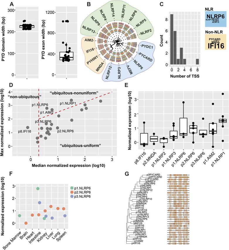
NFκB signaling following TLR stimulation, suggesting a direct               Here, we used publicly available FANTOM5 CAGE data to map
negative regulatory role (Anand et al, 2012). NLRP6 was also shown       promoters for all PYD-containing genes in various tissues and
to regulate a number of interferon-stimulated genes in response to       validated our findings using RNA-Seq. Most PYD genes are broadly
viral RNA through caspase-1–independent interactions (Wang et al,        expressed using more than one TSS. In human, we identified NLRP6
2015). NLRP6 is therefore emerging as a potential multifunctional        as a gene with multiple transcript variants, only one of which codes
PRR capable of eliciting diverse immune responses in various             for full-length translatable protein. Similarly, in mouse, one
cellular populations.                                                    prominent Nlrp6 species contains an expanded 59UTR that abol-
   NLRP6 protein is most highly expressed in the intestinal epi-         ishes translational efficiency both in vitro and in vivo, resulting in
thelium where it has been associated with regulating mucosal             nuclear RNA retention. Both untranslated isoforms represent the
host–microbiota interactions (Normand et al, 2011; Gremel et al,         dominant RNA species outside of the intestine, suggesting a conserved
2014; Wang et al, 2015). Despite restriction at the protein level, a     mechanism for translational gene silencing and tissue-specific
number of reports in mice have documented Nlrp6 RNA within               expression. We propose that alternative promoters represent a
broad tissue types including the kidney, liver, lung, lymphocytes,       powerful regulatory layer in determining the distribution of many
and bone marrow–derived cells (Elinav et al, 2011; Hara et al, 2018;     PYD-containing genes across tissue types.
Radulovic et al, 2019). Many PYD-containing genes show similarly
diverse expression profiles. For example, NLRP1 and NLRP3 protein
are expressed in PBMCs, macrophages, lymphocytes, and dendritic
cells (Kummer et al, 2007). cDNA profiling of various tissues has
                                                                         Results
revealed even more diverse patterns of expression though, with
                                                                         Genomic organization and primary structure of the human pyrin
many NLRs and regulatory genes showing almost uniform ex-
                                                                         domain
pression across multiple organ systems (Yin et al, 2009). As further
complexity, many PYD-containing genes are transcribed as sets of
                                                                         Given the central role of the PYD in initiating various innate immune
isoform variants that could be regulated through both alternative
                                                                         signaling cascades, we looked to profile the tissue distributions and
splicing and differential transcription start sites (TSSs). Few
                                                                         regulatory mechanisms governing the expression for all PYD-
studies however have sought to systematically evaluate the dif-
                                                                         containing genes. We retrieved transcript annotations on 21 hu-
ferential contributions of isoform variants to functional re-
                                                                         man PYD genes corresponding to 14 NLRs (NLRP1-14), 4 ALRs (AIM2,
sponses. It is established that isoforms can have dramatic
                                                                         PYHIN1, MNDA, and IFI16), and 3 regulators/adaptors (ASC/PYCARD,
functional differences in species- and tissue-specific manners, as
                                                                         PYDC1, and MEFV). The PYD is exclusively expressed at the amino (N)
human, but not mouse, NLRP3 was recently found to undergo
                                                                         terminus, and its sequence is encoded within a single exon of rank 1
splicing within exon 5 of the leucine-rich repeat that gives rise to a
                                                                         or 2. We first considered any superficial shared relationships in
nonfunctional isoform (Hoss et al, 2019). Conventional expression
                                                                         exonic organization and primary nucleotide sequence. PYD do-
profiling studies have used PCR-based techniques against
                                                                         mains have a median nucleotide width of 225 nt, though the lengths
amplicons that cover a range of possible RNA molecules, so our
                                                                         of the complete exons encoding the domains fall in two groups: one
knowledge of which isoforms are functionally relevant remains
                                                                         “long” group (NLRP3 and ASC transcripts) with median 1,029 nt and
limited. Moreover, techniques for high-dimensional single-cell
                                                                         the “short” group (all others) with median 320 nt (Fig 1A, right). We
RNA sequencing are still evolving the computational capability
                                                                         further analyzed amino acid sequences corresponding to the ac-
needed to resolve splicing variants, so isoforms are often ag-
                                                                         tual PYD domains using multiple sequence alignment and con-
gregated (Arzalluz-Luqueángeles & Conesa, 2018).
                                                                         structed a phylogenetic tree to determine whether there were
   Most genes contain multiple TSSs, each reflecting the integration
                                                                         higher order relationships. Similar to previous reports, three pat-
of complex regulatory elements acting in cis and trans to shape
                                                                         terns emerged: one cluster was formed by PYDC1, PYCARD, MEFV,
expression patterns (Lenhard et al, 2012). The functional annotation
                                                                         NLRP3, NLRP6, and NLRP12, with the remaining NLRs aligned sep-
of the mammalian genome 5 (FANTOM5) project mapped TSSs for
                                                                         arately, and the four ALRs formed a third group (Fig 1B) (Fairbrother
mammalian genes in various human and mouse cell types through
                                                                         et al, 2001).
cap analysis of gene expression (CAGE) and single-molecule cDNA
sequencing (FANTOM Consortium and the RIKEN PMI and CLST (DGT)
et al, 2014). It is now clear that alternative promoters exist for the   Characterization of the promoter landscape for all PYD-
majority of genes, defined as discrete TSS clusters with varying          containing genes
degrees of tissue-level specificity. Mechanistically, variation in TSS
use represents an added layer for tuning gene expression in tissue-      We leveraged publicly available FANTOM5 datasets to computa-
specific contexts. Downstream alternate promoters nested inter-           tionally explore 59 centered tissue expression patterns and build
nally within a transcript can yield truncated isoforms. Upstream TSS     promoter maps for human PYD-containing genes (FANTOM Consortium
utilization can produce variable leader sequences in the 59 UTR,         and the RIKEN PMI and CLST (DGT) et al, 2014). CAGE is a high-throughput
which contain upstream ORFs (uORFs) and unfavorable guanine-             transcriptome analytical tool that relies on selective retrieval of the
cytosine (GC) content that impact translation efficiency (Kozak,          7-methylguanosine–capped 59 end of Pol II RNA transcripts. The
1991). Context and tissue-selective 59UTR variation have been de-        resulting 59 ends are cleaved, amplified, and sequenced, giving rise
scribed for the related NOD2 sensor, though little is known re-          to a signal of peaks across the genome that corresponds to 59 TSSs
garding the PYD-containing gene family (Rosenstiel et al, 2007).         that can be used to define promoter regions (Kanamori-Katayama

Alternate promoters regulate NLRP6   Bracey et al.                       https://doi.org/10.26508/lsa.202000897   vol 4 | no 3 | e202000897   2 of 16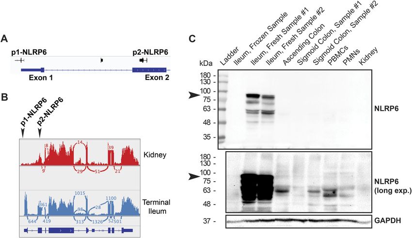
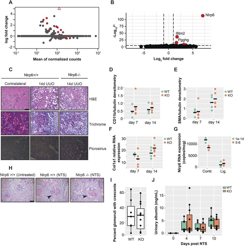
Figure 1. PYD-containing genes are transcribed by sets of promoters with diverse tissue distributions.
(A) Distribution of Pfam domain (left) and their corresponding exon (right) widths for human PYD-containing genes. (B) Phylogenetic tree for all PYD-containing genes
aligned by PYD domain. (C) Distribution of transcription start site (TSS) consensus cluster counts for all PYD genes in FANTOM5 cap analysis of gene expression data. Word
cloud highlights the PYD genes with the most TSS consensus clusters. (D) Distribution of normalized maximum and median expression values for PYD-containing gene TSS
clusters across tissues extracted from the FANTOM5 database. Dashed red lines indicate boundaries established from initial FANTOM5 analysis of all peak data; to the
left of vertical red line are TSS peaks detected with median expression
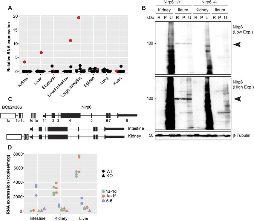
Table 1.    Characteristics of human NLRP6 transcription start site clusters.

 Transcription start site                Width (bp)               Exon                  Position                    GC content                   Distribution
 P1.NLRP6                               54                       Exon 1                 59UTR                      0.48                         Non-uniform
 P2.NLRP6                               60                       Exon 2                 PYD                        0.60                         Uniform
 P3.NLRP6                               155                      Intron 3               PYD-NACHT                  0.68                         Uniform

et al, 2011). We explored FANTOM5-pooled sample sets for eight                  given its broad distribution. We found three alternative NLRP6 TSSs:
diverse human tissue types: kidney, liver, lung, heart, spleen, in-             p1, p2, and p3 (Table 1). The p1.NLRP6 site is located within the 59UTR
testine, bone marrow, and brain. After normalization, all TSS peaks             of exon 1. Interestingly, the other two promoters are localized in-
were spatially clustered by distance into larger transcriptional units,         ternally. P2.NLRP6 is within exon 2 in the middle of the PYD se-
and units between tissue types aggregated to form sets of TSS                   quence, and p3.NLRP6 is within intron 3 between the PYD and
consensus clusters that reflect putative promoters. We then parsed               NACHT domains. These two transcripts may therefore be presumed
the data to select for regions corresponding to only PYD-containing             not to translate into functional PYD proteins. Despite reports for
genes. Many genes were not expressed under basal conditions in                  broad NLRP6 RNA expression, the use of the p1.NLRP6 site was
the tissues explored. Not surprisingly, several PYD genes contain               selective for the intestine. In the kidney, heart, lung, liver, spleen,
multiple promoters, with six genes containing two or more (Fig 1C               and brain, the p2.NLRP6 promoter was clearly dominant (Fig 1F).
and Table S1). NLRP1 and NLRP6 had the most TSSs of all NLRs with 4                 We verified the tissue-specific NLRP6 isoforms from the FANTOM5
and 3, respectively. For the non-NLRs, IFI16 was especially diverse             CAGE datasets experimentally using RNA-Seq on tissue biopsy samples
with eight possible promoters.                                                  of human kidney and small intestine. Using a deep sequencing count of
   Each promoter had a clear distribution of activity across samples            266M read pairs per sample, we were able to detect alternate splicing
that reflect tissue selective use (Fig 1D and E). To map the promoter            events and TSS use across samples. Indeed, NLRP6 in small intestine
landscape for all human PYD genes, we plotted maximum against                   (ileum) used a start site within exon 1 that aligned with the predicted
median scores and separated all promoters into three categories:                FANTOM5 data (Fig 2A and B). Surprisingly, in the kidney, we detected a
those where the median score was less than 0.2 tags per million as              truncated NLRP6 isoform lacking exon 1 corresponding to p2.NLRP6. We
nonubiquitous, those where the maximum score was greater than                   looked next at endogenous NLRP6 protein expression in various human
10× the median as ubiquitous and nonuniform, and those where the                tissue types. Similar to previous reports and in contrast to murine tissue,
maximum was less than 10× the median as ubiquitous and uniform.                 endogenous NLRP6 protein was highly detectable within the small
As in the initial FANTOM5 analysis for the human transcriptome,                 intestine, though not the large intestine. Thus we used small intestine
many PYD genes contain alternate promoters that fall in different               samples for human positive control tissue (Fagerberg et al, 2014). Ad-
expression categories (FANTOM Consortium and the RIKEN PMI and                  ditionally, a single freeze/thaw cycle disrupted NLRP6 protein signal in
CLST (DGT) et al, 2014). For example, P8.IFI16 was selective for bone           the small intestine, so we used only freshly obtained tissues (Fig 2C).
marrow as nonubiquitous, though the other seven IFI16 promoters                 Similar to the alternate promoter use, NLRP6 protein was only de-
were uniformly distributed across tissues.                                      tectable in small intestine samples (Fig 2C). Therefore, human samples
   To determine whether there were common sequence motifs that                  predicted to use p2.NLRP6, such as the kidney yielded no detectable
could give rise to shared expression profiles, we aligned the DNA                translated protein. The lack of protein signal was not the result of poor
sequences for all putative promoters, including +100 bp upstream                specificity or truncated protein variants, as we mapped the epitopes
(Fig 1G). The position of core TSS-adjacent promoter sequences was              recognized by commercially available human NLRP6 antibodies to the
largely uncorrelated, supporting the individualized tissue-selective            NACHT and PYD–NACHT interface (Fig S2). Together, these results suggest
distributions noted previously. We used maximum likelihood anal-                translational repression of human NLRP6 by alternative promoter use in
ysis and bootstrapping to further quantify the relationships (Fig S1).          a tissue-specific context outside of the intestinal epithelium.
Only two clusters emerged with meaningful alignments: p3.MNDA/
p8.IFI16 and p2.MNDA/p6.IFI16. Interestingly, although various pro-             Alternative promoters regulate the tissue distribution for mouse
moters for each gene may theoretically encode for the same protein              Nlrp6
product, they did not cluster together, and instead displayed sig-
nificantly different sequence profiles. These results reveal the di-              Much of our knowledge regarding PYD gene signaling comes from
versity in promoter use for PYD genes across tissue types. They                 mouse models with knockout/transgenic approaches. The pro-
highlight alternative promoters as a possible regulatory mechanism              moter complexity that we observed in human PYD genes could have
in determining heterogenous tissue-level expression profiles.                    species-specific patterns. We therefore went on to fully charac-
                                                                                terize protein and RNA expression profiles for the NLRs in mouse
Alternative promoters regulate the tissue distribution for human                tissues. In several mouse tissues, Nlrp6 was the most abundantly
NLRP6                                                                           expressed NLR at the RNA level under basal conditions, with exon
                                                                                5–6 amplicons readily detected in the kidney, liver, and intestinal
Because there was little correlation between various promoters                  tissue (Fig 3A). However, similar to the human samples, we only
even for the same PYD genes, we looked in greater detail at their               detected endogenous Nlrp6 protein in the intestine (both small and
locations and putative transcript products. We focused on NLRP6,                large intestines in mouse, Figs 3B and S3). Although Nlrp6 was readily

Alternate promoters regulate NLRP6   Bracey et al.                              https://doi.org/10.26508/lsa.202000897    vol 4 | no 3 | e202000897       4 of 16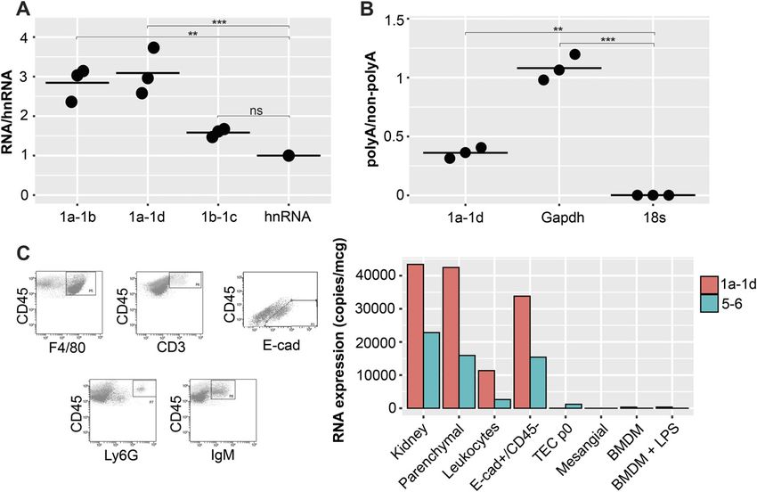
Figure 2. Human NLRP6 is regulated by tissue-selective alternate promoters.
(A) Gene-like representation of NLRP6 transcription start site clusters from FANTOM database. (B) Sashimi plot for NLRP6 showing alternative promoter use of p1.NLRP6
in the representative human small intestine (blue) and p2.NLRP6 in human kidney (red). (C) Immunoblot for NLRP6 protein in human fresh and frozen samples (ileum
only) for low and high exposures. Arrows indicate predicted NLRP6 size. Source data are available for this figure.
Source data are available for this figure.

detected in the small intestine control tissue, there was no signal                  kidney and liver, but not the colon (Figs 3D and S4A). Moreover,
evident in kidney lysates prepared with radioimmunoprecipitation                     Nlrp6Δ59UTR splicing was generalizable and not the result of ge-
assay (RIPA) or urea buffers (Fig 3B). Moreover, endogenous Nlrp6                    netic inbreeding, as it was also detected in the kidney and liver from
protein was not induced in kidneys or livers from mice treated                       mice across various strains (Fig S4B and C). We therefore annotated
systemically with the TLR3 ligand poly (I:C) to induce interferon-                   the proximal exons of BC024386 as part of the Nlrp6 genomic locus,
dependent gene expression, though it was readily detected in control                 representing an alternate promoter for Nlrp6.
large intestine colonic tissue (Fig S3). These results suggest that Nlrp6               The previous results suggest that Nlrp6Δ59UTR is a tissue-
is not regulated transcriptionally, but rather at the level of protein               selective variant outside of the intestine. Previous reports look-
translation to give rise to tissue-specific expression patterns.                      ing at Nlrp6 RNA expression have been limited by the use of relative
   We used RNA-Seq with a sequencing count of 67M single-end                         RNA expression against an arbitrary tissue/cell type. We measured
reads per sample to further explore the tissue-specific regulation of                 various Nlrp6 amplicons using absolute quantification against
Nlrp6. In the mouse intestine, Nlrp6 is encoded by eight exons with a                standard curves made from sequences of interest. Although the
59UTR of 185 bp in exon 1. Surprisingly, we found that Nlrp6 in mouse                common exon 5–6 region was present in the intestine, kidney, and
kidney underwent complex alternative splicing at the 59 end which                    liver, only the kidney and liver expressed exons 1a–1d and 1e–1f (Fig
gives rise to an expanded 59UTR leader sequence of 1,749 bp (Fig 3C).                3D). We examined Nlrp6 and BC024386 exon expression in Nlrp6−/−
Splicing occurred between exon 1 of Nlrp6, 2 novel upstream intergenic               mice generated by gene targeting and replacement of exons 1–2
exons and exon 1 of the adjacent upstream gene. BC024386 is located                  with an IRES-bgal-neomycin resistance cassette (Chen et al, 2011).
approximately 14.8 kb proximal to Nlrp6, contains three exons, and is                Whereas Nlrp6+/+ littermates expressed abundant Nlrp6Δ59UTR in
annotated as long non-coding RNA with only one 94-bp ORF (Fig 3C                     the kidney and liver, gene targeting of Nlrp6 exons 1 and 2 also
and Table 2). We annotated the putative splice sites for the novel Nlrp6             suppressed expression of BC024386 and the novel intergenic exons
variant, and they all followed the canonical GT/AG rule (Table 3).                   within those tissues (Fig 3D).
We amplified a PCR product using primers directed from exon 1 of                         Taken together, these results show that mouse Nlrp6 is regulated
BC024386 to exon 1 of Nlrp6. Sanger sequencing confirmed the                          by tissue-selective alternate promoters that give rise to at least two
presence of a splice variant containing exon 1 of BC024386 and 2 novel               distinct isoforms: one in the intestine containing the canonical 185-
intergenic exons leading to a variant Nlrp6 RNA with an expanded                     bp 59UTR and one expressed in the kidney and liver containing a
59 UTR: Nlrp6Δ59UTR. Interestingly, BC024386 was expressed in the                    large 1,749-bp 59UTR.

Alternate promoters regulate NLRP6    Bracey et al.                                  https://doi.org/10.26508/lsa.202000897    vol 4 | no 3 | e202000897      5 of 16
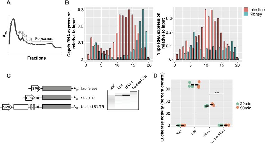
Figure 3. Murine Nlrp6 is regulated by tissue-selective alternate promoters.
(A) Tissue distribution of PYD-containing RNA transcripts in organs relative to the spleen (red = Nlrp6). (B) Immunoblot for Nlrp6 protein in mouse kidney and intestine (R, RIPA; P, insoluble
pellet; U, urea). Nlrp6 protein is only detectable in intestine. (C) (Top) Genomic organization and reannotation for mouse Nlrp6 locus. (Bottom) Transcript map for novel Nlrp6 exons and
splicing of tissue-selective 59UTR leaders in the kidney and intestine. (D) Absolute RNA expression of Nlrp6 amplicons corresponding to different 59UTR leaders in WT and KO mouse organs. n
= 3 biological replicates from littermate mice.
Source data are available for this figure.

Nlrp6Δ59UTR is regulated in epithelial cells                                                       expression of each exon–exon junction relative to heteronuclear,
                                                                                                   unspliced RNA (hnRNA). There was clear enrichment of amplicons
To better understand the cellular fate of Nlrp6Δ59UTR-containing                                   overlapping exons 1a–1b and 1a–1d relative to hnRNA (Fig 4A). In con-
transcripts, we went on to characterize its posttranscriptional regula-                            trast, amplicons for exons 1b–1c were not significantly different, sug-
tion. Both protein coding and noncoding RNAs (ncRNAs) can be spliced                               gesting that Nlrp6Δ59UTR was transcribed as one contiguous primary
and polyadenylated (Derrien et al, 2012). To confirm Nlrp6Δ59UTR                                    transcript with subsequent splicing to construct a leader sequence of
splicing, we isolated nuclei from Nlrp6+/+ mouse kidney and measured                               exons 1a-1d-1e-1f. Moreover, Nlrp6Δ59UTR was polyadenylated as there

Table 2.     Nlrp6 and BC024386 characteristics and predicted protein coding scores.

 RNA                          Size (bp)                  Longest ORF                      Homology to known ORF                           CPC score                    Predicted class
 BC024386                    1,673                       94                              No                                              −0.363511                    Noncoding (weak)
 Nlrp6                       4,438                       870                             Yes                                             8.483620                     Coding

Alternate promoters regulate NLRP6          Bracey et al.                                         https://doi.org/10.26508/lsa.202000897          vol 4 | no 3 | e202000897            6 of 16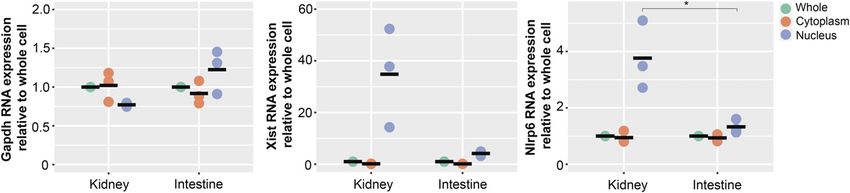
Table 3.   Nlrp6Δ59UTR splicing.                                                         consistent with publicly available single-cell RNA-Seq kidney atlases,
                                                                                         which support Nlrp6 RNA expression exclusively within epithelial cell
 Intron       Exon         59 donor            39 acceptor                Exon
                                                                                         populations (Wu et al, 2018). Crude leukocyte populations containing
 1           1 AAAG        GTTAGTGCTC          ATTTTTATCTTTCAG           2 CTTC          macrophages (CD45+/F4/80+), neutrophils (CD45+/Ly6G+), T lympho-
 2           2 TGAT        GTGAGACCTA          TCCCGGTGTCTGCAG           3 AGGC          cytes (CD45+/CD3+), and B lymphocytes (CD45+/IgM+) expressed very
 3           3 TTCT        GTGAGTGCGT          TATCCCTGCCCACAG           4 GCCC          little NLRP6Δ59UTR. Interestingly, mouse kidney tubular epithelial cells
                                                                                         in two-dimensional culture lost expression of Nlrp6Δ59UTR after a
                                                                                         single generation (Fig 4C, TECp0), and kidney mesangial cells were
was clear enrichment of Nlrp6Δ59UTR in samples prepared with cDNA
                                                                                         below the limit of detection. Some reports have suggested that mouse
primed from oligodT compared to random hexamers (Fig 4B).
                                                                                         BMDMs can form functional Nlrp6 inflammasomes, although others
   In the intestine, Nlrp6 RNA expression has been found primarily in
                                                                                         have found that FLAG-tagged Nlrp6 protein was restricted to the in-
enterocytes and colonic goblet cells (Chen et al, 2011; Elinav et al, 2011;
                                                                                         testinal tissue in mice (Wang et al, 2015; Hara et al, 2018). Consistent
Normand et al, 2011). It has also been described in various circulating
                                                                                         with the latter, we did not detect any Nlrp6 isoforms in BMDM either
immune cell populations including macrophages and lymphocytes
                                                                                         in resting states or following LPS stimulation (Fig 4C).
measured by relative RNA expression (Hara et al, 2018; Radulovic et al,
2019). To establish which cells within the kidney express Nlrp6Δ59UTR,
we fractionated fresh single-cell kidney preparations across a                           Nlrp6Δ59UTR is translationally silenced and retained in the
density gradient and sorted the samples by flow cytometry before                          nucleus in the kidney
absolute quantification by real-time PCR. The “parenchymal” layer
from density gradient separation alone retained high Nlrp6Δ59UTR                         Alternate promoters that give rise to variable leader sequences can
expression (Fig 4C). Further separation of this population revealed                      impact protein translation through several different mechanisms.
dominant expression within E-cadherin+/CD45− epithelial cells. This is                   First, splicing of new genetic material can simply disrupt the ORF.

Figure 4. Nlrp6Δ59UTR variant is spliced and polyadenylated in kidney epithelial cells.
(A) Nlrp6Δ59UTR RNA expression relative to Nlrp6 hnRNA in nuclei isolated from whole kidney. (B) Nlrp6Δ59UTR RNA expression in polyA versus non-polyA whole-cell
kidney RNA preparations. n = 3 biological replicates, P-values *0.05, **0.01, ***0.001, ****0.0001 by ANOVA with Tukey’s multiple comparison. (C) Density gradient separation
and flow sorting of kidney cells. (Left) Hierarchical gating for macrophages (CD45+ F4/80+), neutrophils (CD45+Ly6G+), T lymphocytes (CD45+ CD3+), B lymphocytes (CD45+
IgM+), and epithelial cells (CD45− E-cadherin+). (Right) Absolute RNA expression for Nlrp6 amplicons in various cell populations. TEC, tubular epithelial cells in 2D
culture; BMDM, bone marrow–derived macrophages in 2D culture; mesangial cells in 2D culture. Representative experiment from n = 6 pooled kidneys. hnRNA,
heteronuclear RNA.

Alternate promoters regulate NLRP6      Bracey et al.                                    https://doi.org/10.26508/lsa.202000897     vol 4 | no 3 | e202000897        7 of 16
Table 4. Nlrp6 UTR characteristics. 59UTR variant Length (bp) GC content ORF count uORF length (aa) Exon 1f 185 0.53 1 31 Exon 1a-d-e-f 1,749 0.51 5 101, 79, 62, 31, 31 Alternatively, UTRs can impact cap-dependent ribosomal binding (Fig 5B). In contrast, Nlrp6 detected by a shared exon 5–6 amplicon elements of translation through the primary structure with changes was only associated with polysomes in the intestine. In the kidney, in the GC content and insertion of uORFs, or the secondary structure Nlrp6 RNA was most abundant in fractions 1–8, reflecting free through hairpins and pseudoknots (Kozak, 1991; Sonenberg, 1993; untranslated RNA. Wang et al, 1999). We compared the UTR of Nlrp6Δ59UTR with the To directly compare the translational efficiency of the Nlrp6Δ59UTR canonical intestinal transcript (Table 4). Nlrp6Δ59UTR is 1,749 nt, and canonical Nlrp6 leader sequences, we in vitro transcribed chi- preserves the ORF, and contains five uORFs with only slightly lower meric RNAs containing each Nlrp6 UTR upstream of luciferase under GC content than the 1f-UTR. an SP6 promoter (Fig 5C). Each RNA chimera was 59 capped and polyA It is likely that the addition of ~1,500 nucleotides to a leader tailed. Translational efficiency was then assessed by measuring sequence would significantly impact protein translation even with luciferase activity relative to luciferase control RNA with no leader. As preservation of the ORF. To assess whether Nlrp6Δ59UTR is actively expected, the Nlrp6Δ59UTR leader completely abolished translational translated in vivo, we used polysome profiling. Polysome preparations efficiency, with no signal detected at both 30 and 90 min (Fig 5D). In were made from whole mouse kidney and intestine, size-fractionated comparison, the canonical Nlrp6 leader sequence was actively along a sucrose gradient, and the purified RNA retrieved from translated. eluted samples was used for cDNA synthesis and quantitative The cellular fate of untranslated RNAs is diverse—some mes- real-time PCR (Fig 5A). As expected, Gapdh RNA was enriched in sages are exported from the nucleus where they can interact with polysome-containing fractions in both kidney and intestine, cytoplasmic molecules, whereas others are retained in the nucleus reflecting an actively translated RNA species in both tissue types and participate in various signaling pathways (Quinn & Chang, Figure 5. Nlrp6Δ59UTR isoform has reduced translational efficiency. (A) Representative tracing for polyribosome profiling of mouse kidney tissue. (B) Nlrp6 RNA expression in polysome fractions from mouse kidney and intestine. Gapdh is comparison for an actively translating mRNA. (C) Chimeric RNA constructs and transcripts (right panel) used for in vitro translation. All were 59 capped and contained the SP6 promoter, designated leader sequences, luciferase reporter, and poly A tail. (D) In vitro translation of capped and tailed Nlrp6 59 leader RNA constructs. Xef RNA is negative control. Results are expressed as percent of luciferase control containing no leader sequence, n = 3 biological replicates translated in separate reactions. P- values *0.05, **0.01, ***0.001, and ****0.0001 by ANOVA with Tukey’s multiple comparisons test. Alternate promoters regulate NLRP6 Bracey et al. https://doi.org/10.26508/lsa.202000897 vol 4 | no 3 | e202000897 8 of 16
Figure 6. Nlrp6Δ59UTR is associated with tissue-selective Nlrp6 nuclear retention.
Nuclear/cytoplasmic fractionation and absolute Nlrp6 RNA expression in mouse kidney and intestine. Gapdh serves as control for cytoplasmic RNA, and Xist for nuclear
RNA. Note the scale for Xist as 10-fold greater than the others reflecting high nuclear concentration in both kidney and intestine. *P < 0.05 by ANOVA with Tukey’s multiple
comparison test, n = 3 biological replicates from littermate mice.

2016). Curiously, mouse Nlrp6 RNA was previously identified to be                         unilateral ureteric obstruction (UUO) on Nlrp6+/+ and Nlrp6−/− mice.
enriched in the nucleus in a study specifically exploring liver tissue,                   UUO is an epithelial-centered injury model that leads to kidney
although the mechanism and relevance remained unclear (Bahar                             tubulointerstitial inflammation, cell death, and fibrosis. Consistent
Halpern et al, 2015). To determine whether different isoforms of                         with the differential gene expression results, there was no differ-
mouse Nlrp6 are spatially distributed and contribute to nuclear                          ence in histological scoring of CD11b+ cellular infiltrate or markers
accumulation, we performed nuclear cytoplasmic fractionation of                          of fibrosis between Nlrp6+/+ and Nlrp6−/− mice at 7 and 14 d (Fig
mouse kidney and intestinal tissue. As expected, Gapdh was evenly                        7C–F). Furthermore, Nlrp6 RNA was not induced in response to the
distributed in the cytoplasm and nuclei, and the nuclear-specific                         injury. Amplicons directed against both exons 5–6 and 1a–1d de-
long non-coding RNA Xist was strictly nuclear in both kidney and                         creased substantially in ligated kidneys compared with contra-
intestine, reflecting pure fractions (Fig 6) (Clemson et al, 1996).                       lateral controls, suggestive of nonspecific loss of tubular epithelial
Interestingly, only the Nlrp6Δ59UTR RNA isoform from the kidney                          cell mass (Fig 7G). We also used a glomerular kidney injury model to
was enriched in the nucleus. In stark contrast, the canonical Nlrp6                      further assess the role of Nlrp6 within the kidney. Infusion of sheep-
isoform in the intestine was distributed in a similar profile to Gapdh                    derived anti-glomerular basement membrane (anti-GBM) serum
between the nucleus and cytoplasm, consistent with a translating                         results in a primary glomerular injury with crescent formation,
mRNA.                                                                                    secondary tubular cell injury, and albuminuria (Mesnard et al,
   Taken together, the aforementioned results confirm that only the                       2009). Both Nlrp6+/+ and Nlrp6−/− mice developed similar histo-
canonical intestine Nlrp6 mRNA isoform is actively exported to the                       logical injuries by 10 d (Fig 7H). As in UUO, there were no phenotypic
cytoplasm for translation. In contrast, the Nlrp6Δ59UTR variant                          differences. Both Nlrp6+/+ and Nlrp6−/− mice had similar degrees of
found in the kidney was associated with impaired translational                           albuminuria, and there were no differences in the number of
efficiency and nuclear accumulation.                                                      crescents found on histology (Fig 7I and J). Neither UUO nor NTS
                                                                                         resulted in any detectable Nlrp6 protein in the kidney (Fig S5).
                                                                                         Overall, these in vivo results suggest that Nlrp6 is dispensable
Nlrp6 is dispensable in the kidney
                                                                                         within the kidney and that alternative Nlrp6 promoter utilization
                                                                                         operates primarily as a means of tissue-selective translational gene
The observation that the Nlrp6Δ59UTR isoform silences protein
                                                                                         silencing.
expression outside of the intestine raises the question of whether
the RNA molecule itself still has a functional role as a ncRNA. To
address this, we performed RNA-Seq on the kidney tissue isolated
from Nlrp6+/+ and Nlrp6−/− littermate mice and explored whole                            Discussion
transcriptomes for differential gene expression under baseline
conditions. The transcriptomes were nearly identical between                             It has long been recognized that alternate promoters regulate gene
Nlrp6+/+ and Nlrp6−/− mice at baseline, with only two genes iden-                        expression. For example, the human dystrophin gene contains at
tified that were significantly up-regulated: Ifitm2 with log2fold                           least five promoters used in tissue- and development-specific
change of 1.6 and adjusted P-value 5.99 × 10⁻¹²; and Pgghg with                          patterns (Ahn & Kunkel, 1993). Human NOS1 is especially com-
log2fold change of 1.8 and adjusted P-value of 2.14 × 10⁻³³ (Fig 7A and                  plex with nine exon 1 leader isoforms in various tissues, each
B). Importantly, both Pgghg and Ifitm2 are located on chromosome                          imparting unique changes to translational efficiency (Wang et al,
7 immediately upstream of Nlrp6 by 12.4 and 19.7 kb, respectively.                       1999). Although the pathways by which variant promoters regulate
This suggests likely nonspecific cis-mediated changes secondary to                        protein expression are known, the magnitude of impact has only
Nlrp6 gene targeting and subsequent local chromatin alterations,                         recently become apparent with efforts to fully characterize
rather than biologically significant gene regulation.                                     mammalian transcriptomes across tissue types. Indeed, the vast
   We considered whether ncRNA signaling effects could be dis-                           majority of genes contain alternate promoters and display cell
ease- or injury-dependent. To this end, we performed experimental                        type–restricted expression profiles, with only a very small minority

Alternate promoters regulate NLRP6     Bracey et al.                                    https://doi.org/10.26508/lsa.202000897     vol 4 | no 3 | e202000897        9 of 16
Figure 7. Nlrp6 is dispensable in kidney epithelium. (A) MA plot showing differential gene expression in the kidney from Nlrp6+/+ and Nlrp6−/− littermates. Red signifies adjusted P-values < 0.1, triangle is Nlrp6. (B) Volcano plot highlighting similarities between Nlrp6+/+ and Nlrp6−/− kidney gene expression. All represent n = 3 littermate mice per group. (C) Representative histological sections from Nlrp6+/+ and Nlrp6−/− mice at 14 d showing H&E (top), Trichrome (middle), and Picrosirius Red for contralateral controls and unilateral ureteric obstruction (UUO) kidneys. Bar is 80 μm. (D, E) CD11b and ⍺SMA quantitative densitometry of protein expression by immunoblotting. (F) Col1a1 relative RNA expression in Nlrp6+/+ and Nlrp6−/− kidneys following UUO. (G) Absolute Nlrp6 RNA expression in contralateral control and ligated Nlrp6+/+ mouse kidneys at day 14 UUO. (H) Representative PAS-stained kidney sections from Nlrp6+/+ and Nlrp6−/− mice at 10 d following nephrotoxic serum (NTS). Black arrows point to glomeruli with crescents. Bar is 40 μm. (I) Percent of crescentic glomeruli for NTS mice at 10 d. (J) Urinary albumin from mice following NTS injury. All represent n = 3–7 mice per group for F1 littermate mice. truly aligned with “housekeeping” activities (FANTOM Consortium that a dominant component of the genome is dedicated to the and the RIKEN PMI and CLST (DGT) et al, 2014). When taken together, immune system. Different organs and tissues face very different co-expression clustering of all mammalian promoters has revealed threats. One might therefore expect heterogenous expression of Alternate promoters regulate NLRP6 Bracey et al. https://doi.org/10.26508/lsa.202000897 vol 4 | no 3 | e202000897 10 of 16
genes controlling polarizing events such as inflammatory cytokine           prevents viral entry to the cytoplasm and is expressed in BMDM
release and programmed cell death. It would be surprising if innate        (Wrensch et al, 2015). It remains to be explored whether the
immune sensors were evenly distributed without fine regulation;             phenotypes previously observed in BMDM and kidney injury at-
compartmentalization is critical for immune protection while, at the       tributed to Nlrp6 could in fact relate to off-target effects from gene
same time, preventing collateral damage.                                   targeting. Taken together, though, our results suggest that tissue-
    In contrast to prior observations that the PYD gene family             selective promoter utilization for NLRP6 functions solely as a means
members are broadly expressed, our analysis is more consistent             for translational silencing at baseline, a process that does not seem
with a mosaic pattern with different cell types expressing different       to be affected during organ injury or by exogenous stimuli used in
RNA isoforms that can give rise to different levels of protein products    this study. A regulatory role for noncoding NLRP6 RNA species in
under basal states. Such a complex system would arm tissues with           other tissues or disease contexts however cannot be entirely ruled
tunable way to regulate expression at the level of translation. Ar-        out.
guably, this could represent a more time- and energy-efficient way to          The characterization of the Nlrp6Δ59UTR isoform as dominant in the
express PYD genes strictly on an as-needed basis. For example,             kidney and liver reveals the mechanism for nuclear retention of Nlrp6
epithelial cells in different organ systems possess functional dif-        RNA that has been observed in previous studies (Bahar Halpern et al,
ferences while retaining a common cellular phenotype. The use of           2015). We have additionally uncovered a novel mechanism whereby
alternate promoters shifts the burden of managing genetic regula-          Nlrp6 is silenced through alternative splicing to generate a non-
tion from the level of DNA to RNA, allowing greater flexibility in          translatable isoform. Although it is clear that this process silences
determining which specialized transcripts are ultimately “on” or “off”     Nlrp6 expression, we did not extensively explore whether there were
in common cell types comprising different tissues.                         other physiological or pathophysiological circumstances leading to
    With regard to NLRP6, our analysis reveals broad translational         shifting promoter use within the same tissue leading to context-
silencing in various tissue types. Although BC024386 and the Nlrp6         dependent translational release. This work begins to define the PYD
proximal promoter region are not conserved between mice and                gene promoter landscape under baseline conditions. Future studies
humans, the functional paradigm of alternate promoters giving rise         should further examine basal and injury-induced regulation of pro-
to different isoforms in the intestine compared with other tissue          moter use at a systems level and their impact on isoform and protein
types is similar. The epithelial cells of the intestine face unique        expression during innate immune signaling.
environmental challenges that are not encountered by epithelial
cells of the kidney and liver, which are typically sterile (Peterson &
Artis, 2014). Therefore, our findings continue to support a se-
lective role for NLRP6 possibly in regulating or responding to the
                                                                           Materials and Methods
complex intestinal microbiota. These needs could have imparted
                                                                           PYD gene family domain and exon analysis
evolutionary selective pressure to continue suppressing NLRP6
protein expression outside of the intestine while still maintaining
                                                                           PYD-containing genes were identified using the ensembldb package
transcription.
                                                                           in Bioconductor (Rainer et al, 2019). All transcripts on UCSC GRCh38
    The absence of Nlrp6 protein in the kidney and liver does not
                                                                           were subset for domains annotated on the Pfam database with
entirely preclude a functional biological role. Prior animal studies
                                                                           protein domain ID PF02758 (PYRIN). Genomic coordinates for both
have found that Nlrp6 deletion results in more severe injury in a
                                                                           the PYD domains and their exons were extracted by transcript for
chemical-induced acute kidney injury mouse model, although
                                                                           analysis.
littermate controls were not included (Valiño-Rivas et al, 2020).
Previous studies for Nlrp6 in regulating the intestinal microbiota
and response to colitis models have shown significant heteroge-             FANTOM5 CAGE TSS clustering
neity depending on the use of littermate controls (Lemire et al, 2017;
Mamantopoulos et al, 2017). Moreover, the aforementioned Nlrp6             FANTOM5 CAGE data were accessed and analyzed using the CAGEr
expression in the kidney through immunoblotting and histo-                 package in Bioconductor (Haberle et al, 2015). The FANTOM5 da-
chemistry did not use knockout animals as negative controls,               tabase was queried, and we compiled CAGE data corresponding to
raising questions of specificity. Lastly, the discrepancy between our       human kidney, intestine, heart, lung, bone marrow, liver, brain, and
findings could relate to the difference in disease models—both              spleen samples. Raw peaks were normalized against a power law
UUO and NTS are chronic models of kidney injury, whereas the prior         distribution with α 1.14 and T = 107. TSSs were clustered in a 20-bp
role for Nlrp6 was proposed to regulate acute kidney injury. It is less    framework, and consensus clusters between all tissues were cal-
likely that strain-specific effects are at play, given the generaliz-       culated using a tags per million threshold of two and a maximum
ability of Nlrp6Δ59UTR expression across strains of mice. Aside from       distance of 100 bp for the 0.1–0.9 quantiles. We parsed the dataset
the two proximal genes (Ifitm2 and Pgghg), we did not detect any            to select only PYD-containing genes using ensembldb and analyzed
meaningful differentially expressed genes in kidneys from litter-          clusters overlapping PYD gene coordinates on UCSC GRCh37. We
mate Nlrp6+/+ and Nlrp6−/− animals at baseline nor were there              compiled each consensus cluster and the corresponding scores to
distinguishable phenotypes in either UUO or NTS disease models             visualize the distributions according to thresholds defined by
targeting the tubular epithelium and the glomerular compartment.           FANTOM5 analysis of all TSS cluster (FANTOM Consortium and the
Interestingly, IFITM2—one of the Nlrp6-proximal genes that was             RIKEN PMI and CLST (DGT) et al, 2014). Signal peaks were exported
differentially expressed—is an interferon-inducible protein that           and visualized on the Integrative Genomics Viewer (IGV) 2.4 browser.

Alternate promoters regulate NLRP6   Bracey et al.                        https://doi.org/10.26508/lsa.202000897   vol 4 | no 3 | e202000897   11 of 16
Multiple sequence alignment and phylogenetic tree analysis Quantitative real-time PCR
Annotated genomic sequences for both the PYD domains and TSS qRT-PCR was performed in 10 μL volumes using SYBR Green de-
clusters were translated to amino acid sequences and compiled tection (Bio-Rad) on a CFX96 Touch sequence detection system
into FASTA formats. These sequences were then aligned using (Bio-Rad Laboratories). For comparative Ct, fold change was nor-
MUSCLE, and the phangorn package dist.ml function was used to malized to Gapdh. For absolute quantification, standard curves
construct distance matrices and trees (Edgar, 2004; Schliep et al, were made by 10-fold dilution using gene fragments containing
2017). Parsimony scores for each model were compared, and we target sequences (Integrated DNA Technologies). Table 5 shows all
selected neighborhood joining clustering models. We separately oligosequences used for amplification.
used maximum likelihood methods and bootstrapping to verify
results and provide statistical analysis (Douady et al, 2003). Trees RNA sequencing
and alignments were visualized using the ggtree package in Bio-
conductor (Yu et al, 2018). Whole cell RNA from littermate Nlrp6+/+ and Nlrp6−/− mouse kidneys
in triplicate or human samples was used. RNA samples were
assessed using a Qubit fluorimeter with both double stranded DNA-
Human tissue samples
and RNA-specific fluorescent dyes. RNA integrity (RIN score) was
determined by using an Agilent TapeStation 2200 instrument. Ri-
Human intestinal tissues were obtained during colonoscopy
bosomal RNA depletion of the total RNA samples was performed
performed as part of colon cancer screening. A minimum of six
using the NEB ribosomal RNA depletion kit (E6350) as per the
biopsies taken for each site were assessed. Human kidney cortex
manufacturer’s protocols. RNA samples were converted into Illu-
tissue was obtained from the normal margins of kidney speci-
mina compatible cDNA sequencing libraries using NEB Ultra II
mens from patients undergoing a surgically indicated ne-
Directional RNA Library Prep kits for Illumina (E7760) and NEB Index
phrectomy. Fresh human tissues were immediately rinsed twice
primers as per the manufacturer’s protocols. Before pooling and
in cold PBS, cleaned of adventitia, and kept on ice for parallel
sequencing, each library was quantitated by qRT-PCR, in triplicate,
processing. RNA was immediately extracted from 100 mg frag-
using a Kapa Biosystems #KK4835 (#07960204001; Roche) Library
ments by Solution D method.
Quantification Kit for Illumina. qPCR was performed on an Applied
Biosystems StepOne Plus instrument. Equal amounts of each li-
Protein immunoblotting brary were combined into a single pool, denatured, and diluted as
per Illumina’s recommendations. The pool was then immediately
The tissue was rinsed in saline and processed in RIPA or urea sequenced on either an Illumina NextSeq 500 sequencer using a
buffers. Protein samples were separated on SDS–PAGE gels under high-output 2 × 75 cycle sequencing kit or on an Illumina NovaSeq
reducing conditions and transferred to nitrocellulose membranes, 6000 sequencer with a 2 × 50 bp sequencing kit using the manu-
blocked for 1 h in 5% BSA or milk proteins diluted in PBS or TBS facturer’s protocol.
containing 0.5% or 0.1% Tween 20, respectively. Following blocking, Spliced paired-end read genomic alignment was performed
membranes were incubated at 4°C overnight with the following using a Dragen v3.5.7 (Illumina Inc.) in two-pass RNA mapping mode
antibodies: rabbit anti-mouse Nlrp6 (E-20; Santa Cruz), mouse against GRCh38 for human and using minimap (v 2.17) in short
anti-human NLRP6 (Clint-1; Adipogen), rabbit anti-human GAPDH genomic paired-end reads mode against GRCm38 (Li, 2018).
(Cell Signaling), mouse anti-human tubulin (Sigma-Aldrich), mouse Differential gene expression was carried out using the DESeq2
anti-human NLRP6 (R&D Systems), rabbit anti-GFP (Thermo Fisher package in Bioconductor (Love et al, 2014). After genomic alignment,
Scientific), rabbit anti-mouse CD11b (Abcam), and mouse anti- unnormalized count matrixes were loaded to a DESeqDataSet and
mouse SMA (Clone 1A4; Sigma-Aldrich). Membranes were incu- differential expression was quantified using the DESeq command.
bated for 1 h at room temperature with the secondary antibody Shrunken log2fold changes were computed using the apeglm
at 1:5,000 in blocking buffer and visualized using ECL Western blotting package and the lfcShrink command (Zhu et al, 2019).
detection reagents (Amersham, GE Healthcare). Images were captured
using a ChemiDoc MP imaging device (Bio-Rad Laboratories). Flow cytometry and cell sorting
Whole mouse kidneys and intestine were perfused with cold saline,
RNA isolation and cDNA preparation rinsed, and resected. Single-cell suspensions were generated in
Multi-Tissue Dissociation Kit-2 on a gentleMACS Octo Dissociator
Whole cell RNA was extracted from 50 mg fresh tissue samples or with Heaters (Miltenyi Biotec) using the 37MTDK-2 setting. Sus-
cultured cells (10 cm plates) using guanidinium thiocyanate/ pensions were then filtered through 40-μm nylon strainers, rinsed
phenol–chloroform extraction by using the Solution D method in saline, and fractionated across a 9-ml 25–40–60% Percoll (GE
(Chomczynski & Sacchi, 2006). Samples were treated with DNase I Healthcare) density gradient. The lower layer corresponded to
(5U, 15 min at room temperature). cDNA synthesis was carried out “parenchymal” resident cells and the upper layer to immune cell
using Superscript II RT according to the manufacturers’ protocol populations and mesangial cells. Separate fractions were then
with 100 ng of whole cell RNA and random hexamers. Final cDNA incubated in Fc block at 1 μg/106 cells for 5 min at room tem-
products were diluted to 100 μl before use. perature, followed by primary antibody labeling with anti-CD45
Alternate promoters regulate NLRP6 Bracey et al. https://doi.org/10.26508/lsa.202000897 vol 4 | no 3 | e202000897 12 of 16Table 5. Oligonucleotide sequences.
Amplicon Forward (59-39) Reverse (59-39)
18s TCAGCCACCCGAGATTGAGC GTGCAGCCCCGGACATCTAA
Gapdh GGTGCTGAGTATGTCGTGGA GGCGGAGATGATGACCCTTT
Xist GCTTGAACTACTGCTCCTCCG GGCAATCCTTCTTCTTGAGGCA
Nlrp6 1a-1f ATCCTGAATGCCCAGCGGTGCC CTCATTCTGGGTGTGAGGGTGT
Nlrp6 1a-1d CGTGCATCAGACGCTCTCTA GGCAGGAAGGAATTTGAGGC
Nlrp6 exon 1e-1f CTGGAACTGGACTTACGGGT GGGCAGAAGGTTGGAGAGAT
Nlrp6 exon 5-6 GTGAGACAATGACTACCCCGAAAT GTCTCGGCAAACTGCATCAG
BC024386 exon 1 CGTGCATCAGACGCTCTCTA
BC024386 exon 1-2 TCGCACTCACTAAGCCATGG
BC024386 exon 1-intron 1 TGCCATTGTTTTGCAGATTTGG
BC024386 exon 1-intron 2-exon 3 (hnRNA) GCTTGGACACGCACAGAATC CCACTTCCTCAGCCCTGTATG
Nlrp1b GACTTTGTGGCTTGTTGAATG CATTTAGCTGCAGGTCTAGCTCTCT
Nlrp2 CCCTGCAAATGCTTAGATTGAA GGTCACTGCTGATTCTCAGTTG
Nlrp3 AGAGCCTACAGTTGGGTGAAATG CCACGCCTACCAGGAAATCTC
Nlrp4a TTGCTGCCCACTGCTTAAAAC CAGCCTTTCCATATAGCTGTGTTC
Nlrp5 GGCCAAAAATAGAGTGGGAGTAAAA GGCCACAGTTGTCCAGTATCAAC
Nlrp9a GTTATGGTTGCCTGGTTGCTATTT TTATTGTTGCCAAGTTTCAGGGTCTTT
Nlrp10 AACAGGGTCTCAGGCAGTCAAG ATCCACACCTGGGAGATGCA
Nlrp12 AGCGTGGTATATCCCTCGAAGA CCCTGAGCATCATGGAAAGAA
Nlrp14 GAGAGACTGGCCTTAGCAAGCT ACAAGCATAAATGTGTCAGCCTCTT
AIM2 ATCTAGGCTGATCCTGGGACTGT GTCCAGGCCGGTCAACAAC
(clone 30-F11; BioLegend), anti-E-cadherin (Cat. no. 147303; Bio- then precipitated overnight in 1:50 volume 5 M sodium chloride and
Legend), anti-CD3 (clone 145-2C11; Invitrogen), anti-IgM (clone 2.5 volumes 100% ethanol at −20°C, isolated by Solution D method,
EB121-15F9; Invitrogen), anti-Ly6G (clone HK1.4; BioLegend), and and further purified by precipitation with 7.5 M lithium chloride
anti-F4/80 (clone BM8; BioLegend). Flow cytometry was performed before heparinase treatment (0.4 U/ml, 1 h at room temperature)
using hierarchical gating on an FACSARIA III (BD Biosciences), and and cDNA synthesis.
RNA was isolated from sorted epithelial cells by Solution D.
Nuclear/cytoplasmic fractionation
Polysome profiling
Whole mouse kidneys and intestine were resected, rinsed in cold
Polysomes were prepared as previously described (Nagarajan & saline, and 20 mg distributed for homogenization in a dounce
Grewal, 2014). Briefly, the entire mouse kidney and intestine (small homogenizer in 500 μl lysis buffer containing 50 mM Tris pH 8.0, 140
and large) were rinsed with cold saline containing cycloheximide mM sodium chloride, 1.5 mM magnesium chloride, 0.5% (vol/vol)
100 μg/ml and homogenized with a mini-dounce homogenizer in NP-40, 1 mM DTT, 0.2 U/uL RNase OUT. Nuclei were pelleted at 300g
freshly prepared lysis buffer containing 1% (vol/vol) Triton X-100, for 2 min, and RNA from the supernatant and nuclear fractions was
0.5% (wt/vol) sodium deoxycholate, 0.5 mM DTT, 100 μg/ml cy- extracted by Solution D.
cloheximide, 1 mg/ml heparin, 1× Roche mini protease inhibitor tab,
2.5 μM PMSF, 2.5 mM sodium fluoride, 1 mM sodium orthovanadate, Cloning of NLRP6 leaders and luciferase chimeras
100 U/ml Riboblock (Fermentas) all in 25 mM Tris pH 7.4, 10 mM
magnesium chloride, and 250 mM sodium chloride. Samples were Chimeric 59UTR-luciferase constructs were cloned by FastCloning using
processed with 10 passes through a 25G needle, cleared at 12,000 g, Phusion HF polymerase according to manufacturer’s protocol (NEB) (Li
and the supernatant layered on a 12 ml 15–45% sucrose gradient for et al, 2011). pGEM-luc and pSP64 Poly(A) vectors were purchased from
ultracentrifugation at 37,000 rpm (SW41 Beckman rotor, Optima Promega. NotI and BglII sites were first cloned into pSP64 Poly(A) vector,
L-90-K; Beckman Coulter) for 4°C, 2 h 30 mins. Fractionation was and luciferase was cloned upstream of the poly(A) sequence. 59UTR from
carried out on the BR188 Density Gradient Fractionation System intestine and kidney/liver isoforms were cloned from GeneBlocks (In-
(Brandel) with 20 fractions collected per sample with continuous tegrated DNA Technologies) directly 59 to the luciferase sequence.
spectrophotometric absorbance measurements at 254 nM. RNA was Plasmid sequences were all verified by Sanger sequencing across inserts.
Alternate promoters regulate NLRP6 Bracey et al. https://doi.org/10.26508/lsa.202000897 vol 4 | no 3 | e202000897 13 of 16In vitro transcription and translation Supplementary Information
Each DNA construct was linearized by BglII digestion, and 1 μg of final Supplementary Information is available at https://doi.org/10.26508/lsa.
phenol–chloroform extracted/ethanol precipitated DNA was used 202000897.
for input to the mMessage mMachine SP6 transcription kit (Thermo
Fisher Scientific). Synthetic RNA was purified by phenol–chloroform
extraction, and sample integrity and concentration verified by an
Agilent Tapestation. RNA samples were aliquoted and maintained
Acknowledgements
at −80°C. For in vitro translation, rabbit reticulocyte IVT lysate
The authors thank Mona Chappellaz for her technical support. Infrastructure
(Thermo Fisher Scientific) was used according to the manufac-
and technical support were also provided by the Flow Cytometry Facility, the
turers’ protocol and input RNA concentration empirically deter- Biobank for the Molecular Classification of Kidney Disease, and the Centre
mined. Luciferase was then measured on a Monolight 3010 for Health Genomics and Informatics at the Snyder Institute for Chronic
Luminometer (BD Biosciences) using the Luciferase Assay Detec- Diseases and the University of Calgary. Grant Support: This work was by
tion System (Promega). supported by operating grants from the Canada Institutes for Health Re-
search and the Kidney Foundation of Canada. Support was also provided by
a team grant under the Canadian Institutes for Health Research (CIHR)
Mouse studies Inflammation in Chronic Disease Signature Initiative.
Nlrp6+/+ and Nlrp6−/− mice (Millenium Pharmaceuticals Inc.) on a Author Contributions
C57Bl/6 background were derived from Nlrp6−/+ breeding pairs
(Chen et al, 2011). Mice used in these studies were F1 littermates. The NA Bracey: conceptualization, data curation, formal analysis, in-
UUO model was performed as previously described (Vilaysane et al, vestigation, methodology, and writing—original draft, review, and
2010). Mice were sacrificed at 7 and 14 d for analysis. Anti-GBM editing.
glomerulonephritis was induced in mice using heat-inactivated JM Platnich: conceptualization, data curation, investigation, methodology,
sheep anti-GBM nephrotoxic serum (NTS, generously provided by and writing—original draft.
Dr L Mesnard) administered intravenously as previously described A Lau: conceptualization, data curation, investigation, and
(Mesnard et al, 2009). Urine samples were collected on days 0, 4, methodology.
and 9 following NTS injection and assayed for total protein H Chung: data curation, investigation, and methodology.
(Bradford assay), albumin (Bethyl Laboratories), and creatinine ME Hyndman: resources and writing—review and editing.
(Exocel) as per the manufacturer’s instruction. Mice were sacrificed JA MacDonald: resources, methodology, and writing—review and
at 9 d following NTS administration and kidneys collected for editing.
analysis. J Chun: conceptualization, supervision, methodology, and wri-
ting—review and editing.
Statistics PL Beck: resources.
SE Girardin: conceptualization and writing—review and editing.
Statistical analysis was done using both GraphPad Prism 8 and R PMK Gordon: conceptualization, data curation, formal analysis,
version 3.3.3 (R Core Team, 2020). Where appropriate, ANOVA with investigation, methodology, and writing—original draft, review, and
Tukey’s multiple comparison testing were used, with a P-value editing.
significance threshold of 0.05. DA Muruve: conceptualization, resources, data curation, formal
analysis, supervision, funding acquisition, validation, project ad-
ministration, and writing—original draft, review, and editing.
Ethics
Conflict of Interest Statement
Human intestinal biopsies and human nephrectomy sample col-
lection protocols were approved by the Conjoint Health Research
The authors declare that they have no conflict of interest.
Ethics Board at the University of Calgary and Alberta Health Ser-
vices. All mouse studies were approved and conducted in accor-
dance with guidelines set forth by the Animal Care Committee at the
University of Calgary and conform to the guidelines set by the References
Canadian Council of Animal Care.
Ahn AH, Kunkel LM (1993) The structural and functional diversity of protein
phosphatase 2A. Nat Genet 3: 283–291. doi:10.1038/ng0493-283
Akira S, Uematsu S, Takeuchi O (2006) Pathogen recognition and innate
Data Availability immunity. Cell 124: 783–801. doi:10.1016/j.cell.2006.02.015
Anand P, Subbarao Malireddi RK, Lukens J, Vogel P, Bertin J, Lamkanfi M,
Nlrp6+/+ and Nlrp6−/− RNA-Seq data are registered under NCBI Kanneganti TD (2012) NLRP6 negatively regulates innate immunity and
BioProject PRJNA684477 and can be accessed using the following host defense against bacterial pathogens. Nature 488: 389–393.
link: http://www.ncbi.nlm.nih.gov/bioproject/684477. doi:10.1038/nature11250
Alternate promoters regulate NLRP6 Bracey et al. https://doi.org/10.26508/lsa.202000897 vol 4 | no 3 | e202000897 14 of 16You can also read