Atypical structures of GAA/TTC trinucleotide repeats underlying Friedreich's ataxia: DNA triplexes and RNA/DNA hybrids - Oxford Academic ...
←
→
Page content transcription
If your browser does not render page correctly, please read the page content below
Published online 21 August 2020                                                  Nucleic Acids Research, 2020, Vol. 48, No. 17 9899–9917
                                                                                                                 doi: 10.1093/nar/gkaa665

Atypical structures of GAA/TTC trinucleotide repeats
underlying Friedreich’s ataxia: DNA triplexes and
RNA/DNA hybrids
Jiahui Zhang1 , Ashkan Fakharzadeh1 , Feng Pan1,2 , Christopher Roland1 and
Celeste Sagui 1,*
1
 Department of Physics, North Carolina State University, Raleigh, NC 27695-8202, USA and 2 Department of
Statistics, Florida State University, Tallahassee, FL 32306, USA

                                                                                                                                                              Downloaded from https://academic.oup.com/nar/article/48/17/9899/5895336 by guest on 20 October 2020
Received May 04, 2020; Revised July 27, 2020; Editorial Decision July 30, 2020; Accepted August 04, 2020

ABSTRACT                                                                        neurological disorders that exhibit a phenomenon known
                                                                                as ‘anticipation’, where the age of the onset of the disease
Expansion of the GAA/TTC repeats in the first in-                               typically decreases and the severity of the disease pheno-
tron of the FXN gene causes Friedreich’s ataxia. Non-                           type typically increases in each subsequent generation (4,8–
canonical structures are linked to this expansion.                              11). Longer repeat tracts become progressively more dele-
DNA triplexes and R-loops are believed to arrest tran-                          terious and constitute the major molecular determinant of
scription, which results in frataxin deficiency and                             anticipation in a significant number of diseases, with other
eventual neurodegeneration. We present a system-                                genetic modifiers and environmental factors accounting for
atic in silico characterization of the possible DNA                             the remainder of the effect (11,12). The expansion of mi-
triplexes that could be assembled with GAA and                                  crosatellite repeats is behind 50 neurodegenerative and neu-
TTC strands; the two hybrid duplexes [r(GAA):d(TTC)                             romuscular disorders (11,13–17). The repetitive, sequential
and d(GAA):r(UUC)] in an R-loop; and three hy-                                  structure of TRs causes slippage during DNA replication,
                                                                                repair, transcription and/or recombination (10–14,18–22)
brid triplexes that could form during bidirectional
                                                                                leading to expansion and high mutation rates.
transcription when the non-template DNA strand                                     Although the mechanisms underlying TREDs are under-
bonds with the hybrid duplex (collapsed R-loops,                                stood to be extremely complex, an important breakthrough
where the two DNA strands remain antiparallel). For                             has been the recognition that the critical step in all mod-
both Y·R:Y and R·R:Y DNA triplexes, the parallel                                els of repeat instability is the transient formation of atypi-
third strand orientation is more stable; both parallel                          cal, non-B DNA stable secondary structures in the expand-
and antiparallel protonated d(GA+ A)·d(GAA):d(TTC)                              able repeats (17,18,23–25). Indeed, expandable repeats have
triplexes are stable. Apparent contradictions in the                            been shown to display atypical structural characteristics, in-
literature about the R·R:Y triplex stability is proba-                          cluding single-stranded hairpins, Z-DNA, triple helices, G-
bly due to lack of molecular resolution, since shift-                           quartets, slipped-stranded duplexes and R-loops. Interest-
ing the third strand by a single nucleotide alters the                          ingly, the quest to understand the molecular mechanisms
                                                                                behind these diseases has also opened a window into the
stability ranking. In the collapsed R-loops, antiparal-
                                                                                understanding of the less known atypical secondary struc-
lel d(TTC+ )·d(GAA):r(UUC) is unstable, while parallel                          tures of nucleic acids, thus contributing to the basic field of
d(GAA)·r(GAA):d(TTC) and d(GA+ A)·r(GAA):d(TTC)                                 nucleic acids research.
are stable. In addition to providing new structural                                In this work, we present results related to the atypi-
perspectives for specific therapeutic aims, our re-                             cal structures associated with GAA/TTC TRs. Friedre-
sults contribute to a systematic structural basis for                           ich’s ataxia (FRDA) is caused by a GAA expansion in
the emerging field of quantitative R-loop biology.                              the first intron of the frataxin (FXN) gene (the conven-
                                                                                tion is that the repeat on the coding strand is consid-
INTRODUCTION                                                                    ered the disease-causing repeat for a specific locus). Ex-
                                                                                perimentally, GAA/TTC repeats have been found to form
Trinucleotide repeats (TRs) exhibit ‘dynamic mutations’                         either triplexes or R-loops. A DNA triplex (also known
that cause them to expand. After crossing a critical thresh-                    as H-DNA or triple-stranded DNA) was first reported in
old length, the expansion gives rise to trinucleotide repeat                    1957 (26). This is a non-canonical three-stranded structure
expansion disorders (TREDs) (1–7). These are inherited

* To   whom correspondence should be addressed. Tel: +1 919 515 3111; Email: sagui@ncsu.edu


C The Author(s) 2020. Published by Oxford University Press on behalf of Nucleic Acids Research.
This is an Open Access article distributed under the terms of the Creative Commons Attribution License (http://creativecommons.org/licenses/by/4.0/), which
permits unrestricted reuse, distribution, and reproduction in any medium, provided the original work is properly cited.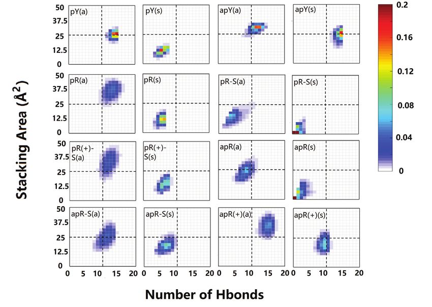
9900 Nucleic Acids Research, 2020, Vol. 48, No. 17

consisting of a Watson–Crick paired helical duplex and a        non-equivalent nucleotide arrangements (such as (GCC)n
third strand that binds to this duplex via Hoogsteen or         and (CCG)n homoduplexes, with CpG and GpC steps be-
reversed Hoogsteen hydrogen bonds. In the most general          tween the C–C mismatches); provide free energies, dynam-
case, DNA triplexes can occur in the cell naturally in the      ics of conformational transitions etc. These conformation
form of intramolecular triplexes formed at endogenous mir-      studies do not address the formation of the atypical struc-
ror repeats of oligopyrimidine/oligopurine sequences or by      tures: In the cell, in order for these atypical structures to be
applying an exogenous oligonucleotide, generally known          formed, it is necessary to cross a free energy barrier that is
as TFO––triplex-forming oligonucleotide (27,28). In ei-         sequence and repeat-length dependent. One of the advan-
ther case, these triplexes can induce transcriptional repres-   tages of MD is that simulations can start in any minimum
sion and site-specific mutagenesis or recombination (27). In    of the free energy. Thus, one can simply study the resulting
particular, TFOs can bind to sequence specific DNA du-          structure after its nucleation has taken place (similarly to
plexes and thus present enormous gene therapeutical po-         studying a folded protein after folding has taken place), and
tential (28). GAA/TTC sequences, representing pure purine       the length-dependence for the nucleation becomes irrele-

                                                                                                                                     Downloaded from https://academic.oup.com/nar/article/48/17/9899/5895336 by guest on 20 October 2020
and pure pyrimidine strands, are thus excellent candidates      vant. In this sense, MD studies are the same as the (relatively
for triplex formation. Using the standard notation (29)         scarce) X-ray and NMR studies (generally with very few re-
where R represents a pure purine strand (not to be confused     peats) that report on the already formed atypical structures
with the ‘R’ in an ‘R-loop’) and Y represents a pure pyrimi-    of TRs. In this work, we explore the structure and stabil-
dine strand, such that R:Y is the usual B-DNA duplex, then      ity of DNA triplexes by studying 16 geometrically different
both R·R:Y (purine third strand) and Y·R:Y (pyrimidine          GAA/TTC triplexes; we present results with respect to the
third strand) triplexes have been found in GAA/TTC se-          two hybrid RNA:DNA helical duplexes; and results related
quences (30–38).                                                to the three possible DNA·DNA:RNA hybrid triplexes that
   An R-loop is a three-stranded nucleic acid structures        can form a collapsed R-loop. Even though the formation
consisting of a hybrid RNA:DNA duplex formed by the             and stability of DNA triplexes has been reported experi-
template DNA and the RNA strands, along with the dis-           mentally (30–38), a search of the Protein Data Bank re-
placed, non-template, single-strand DNA. They were ini-         veals no molecular structure for the GAA/TTC triplexes.
tially observed in DNA replication, and are also observed       Our work provides a systematic characterization of these
during transcription. R-loops can regulate cellular pro-        structures and their relative stability, the particular hydro-
cesses such as gene expression, DNA replication and re-         gen bond patterns and the symmetry correlation between
pair, and immunoglobulin class-switch recombination, and        various triplexes. In particular, we show that apparent con-
as such they have been intensively studied (39,40). They        tradictions in the literature about the stability of R·R:Y
can also cause DNA damage and genome instability, and re-       triplexes is probably due to lack of molecular resolution,
cently, they have been linked to neurodegenerative diseases     since shifting the third strand by a single nucleotide alters
such as Friedreich’s Ataxia (41). While all sequences make      the stability ranking. We also probe collapsed R-loops, that
temporary R-loops behind RNA polymerase II (RNAPII),            have not been studied before, and rate their stability. Our
loops created in regions with asymmetric purine-rich and        work contributes to the understanding of the atypical struc-
pyrimidine-rich strands are susceptible to form stable, long-   tures related to GAA/TTC expansions. In addition, the def-
lived hybrids (42). The all-purine GAA and all-pyrimidine       inition of R-loops has recently been extended to include a
TTC strands are perfect candidates for R-loops (41,43,44),      large class of non-canonical structures suggesting a novel
and the hybrid duplexes can form in either of the two DNA       level of biological complexity (57). There is, however, scarce
strands due to bidirectional transcription (45–47). In par-     experimental data with molecular resolution for these struc-
ticular, increased expression of the FXN Antisense Tran-        tures. Our work contributes to widen the structural knowl-
script 1 (FAST-1) has been shown to cause heterochro-           edge of the R-loop repertoire. The fact that several of the
matin formation and transcriptional silencing of the FXN        structures presented here are stable strongly suggests that
gene (48). An intriguing possibility is the formation of        they may either coexist or compete in the transcriptional
a hybrid triple helix, where the ssDNA left behind by           R-loop.
RNAPII is no longer ‘loose’ but attaches through Hoog-
steen or reversed Hoogsteen hydrogen bonds to the hybrid
                                                                MATERIALS AND METHODS
DNA:RNA helix, thus forming a hybrid triplex (or ‘col-
lapsed R-loop’ (49)).                                           There are two parts to the work presented here: the ini-
   In this work, we report simulation results about atypi-      tial construction of the atypical secondary structures and
cal secondary structures associated with GAA/TTC TRs,           the subsequent MD simulations based on the initial model
obtained via classical atomistic molecular dynamics (MD)        triple helices. The initial structures for the triple helices were
simulations. Atomistic MD tools have proved extremely           constructed de novo using single strands with three repeats
valuable as they possess the ability to determine molecular     (nine nucleotides) each; the process involves both the care-
structures, dynamics and mechanisms at the atomic level,        ful determination of the relevant hydrogen bonds between
which are often beyond the resolution of experiments. Our       the third strand and the canonical duplex, and the assembly
recent characterization of homoduplexes, quadruplexes and       of the three strands. The modeling process and the resulting
hairpins conformations corresponding to the most com-           initial structures are described in the next section.
mon TRs and to several hexanucleotide repeats is an ex-            The MD simulations were carried out with the Amber18
ample of that (50–56), as these simulations sample both         package (58) with force field BSC1 (59) (DNA part) and
DNA and RNA sequences of different lengths, different           BSC0 (60) + OL3 (61) (RNA part) for different atypical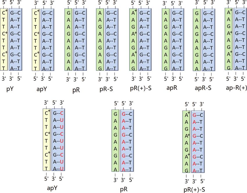
Nucleic Acids Research, 2020, Vol. 48, No. 17 9901
structures combined with the protonated AMBER force For the TTC·GAA:TTC triplex, we considered both pro-
field (62). The protonation was completed with tleap (58). tonated and unprotonated cytosines in the third strand.
The simulations for the pyrimidine third strand were carried We found that the unprotonated antiparallel case (apY
both with and without protonation for the cytosine. The in the notation described below) was completely unstable
TIP3P water model (63) was used for the explicit solvent (Supplementary Figure S6); and the parallel case (pY) was
simulations under periodic boundary conditions in trun- marginally stable (Supplementary Figure S7), certainly less
cated octahedron water boxes. The appropriate number of stable than its protonated case. Clearly, the protonation of
Na+ ions (parameters in (64)) was used for neutralization the cytosine allows for the formation of hydrogen bonds
of the nucleic acid charges. Additional simulations were run with the guanines of the GAA strand in the B-DNA duplex.
with Mg2 + ions (parameters in (65)) in a 0.20M concentra- From now on, we will only discuss the protonated cases. The
tion, with Cl− ions added for neutralization. One run, apR protonation of the cytosines is supported by experimental
was additionally run with 40 and 80 mM concentrations. evidence (35,69–72), both for a parallel or antiparallel cy-
Electrostatics were handled by the Particle Mesh Ewald tosine third basis. This results in two cases that can form
Downloaded from https://academic.oup.com/nar/article/48/17/9899/5895336 by guest on 20 October 2020
method (66), with a direct space cutoff of 9 Å. The cutoff for hydrogen bonds: the protonated pyrimidine third strand in
van der Waals interactions was set as 9 Å. We used Langevin either parallel or antiparallel direction (the latter shifted by
dynamics with a coupling parameter 1.0 ps−1 . The SHAKE one base). For the all-purine third strand GAA·GAA:TTC
algorithm (67) was applied to all bonds involving hydrogen triplex there are 6 cases. The GAA third strand can be par-
atoms. Hydrogen bonds were identified by cpptraj as sup- allel or antiparallel, and can be shifted as shown in Figure
plied by ambertools18 (58) with a distance cutoff of 3.5 Å 1. In addition, the adenine can be protonated, as it has been
and an angle cutoff of 140◦ . noticed that the protonated adenine can form good hydro-
Starting conformations for MD calculations were ob- gen bonding structure with the G:C base pair (73). Thus,
tained as follows. We first minimized the energy for the ini- we proposed two semi-protonated mismatched R·R:Y triple
tial conformations obtained by modeling: first, by keeping sequences (purine-parallel-protonated-shifted and purine-
the nucleic acid and ions fixed; then, by allowing them to antiparallel-protonated) to investigate the potential stabil-
move. Subsequently, the temperature was gradually raised ity and structure of the mismatched triplex sequences. This
using constant volume simulations from 0 to 300 K over gives a total of 8 different structures, as shown in Figure 1.
50 ps runs with a 1 fs time step. Then a 100 ps run at con- For each of the triplexes in Figure 1, we considered two con-
stant volume was used to gradually reduce the restraining formations for the third strand, which differ in the value of
harmonic constants for nucleic acids and ions. After we ob- the glycosidic angle, such that the nucleotides in the third
tained the starting conformations, we performed MD runs strand are all in an anti or all in a syn conformation. The
for 1 s with a 2 fs time step under a constant pressure of rationale behind this is that syn conformations are found in
1 atm. Conformations were saved every 20 ps. In the MD mismatches of trinucleotide repeats (50,52,54,74–78). Thus,
runs, weak constraints of 1kcal/mol on hydrogen bonds for we have a total of 16 different initial conformations. In order
the ending bases were added to the system in order to reduce to refer to them, we introduce the following notation. When
artificial fraying at the ends. the third strand is TTC or GAA we use the notation Y or
We also performed a test run with the same method and R. The C’s in the TTC third strand are always protonated,
procedure as mentioned above for an experimentally ob- as discussed above (so no need of extra notation). On the
tained structure of DNA triple helix with protonated cyto- other hand, one of the A’s in the GAA third strand can be
sine whose PDB ID is 1BWG (68). After a 1 s MD simu- neutral or protonated in order to form A+ ·G–C triple base
lation, we found that the triplex was stable and very close to planes with the G–C Watson–Crick pairs of the B-DNA du-
the experimental conformation. To further investigate the plex. In this case, we use the R(+) notation when A’s in the
stability of selected structures, we also performed MD runs GAA third strand are protonated. For parallel and antipar-
under higher temperatures with all other computational set- allel third strands, we precede the Y, R notation by ‘p’ or
tings being the same as above. ‘ap’, while if the strand is shifted, the notation ends with ‘-
S’. Finally, the anti or syn conformations of the glycosidic
RESULTS angle are denoted by (a) or (s). Thus, a triple helix where the
third strand GAA is parallel and shifted, and every first A in
Initial modeling of the DNA triple helices
GAA is protonated, with all the bases in anti conformation,
The triplexes consist of a regular B-DNA GAA/TTC he- is abbreviated as pR(+)-S(a).
lix with standard Watson–Crick basepairing, and a third all The 16 triplexes involve hydrogen bond patterns that
purine (R) GAA or all pyrimidine (Y) TTC strand, that is have traditionally been identified as Hoogsteen or reverse
placed in the major groove of the B-DNA helix, as steric Hoogsteen hydrogen bonds, and patterns that have not
clashes would prevent its placement in the minor groove. been reported previously which we identify as similar to
This allows for 8 possible triple helices as shown in Fig- those reported. Thus, we define ‘type H’ hydrogen bonds
ure 1, where the terms parallel or antiparallel refer to the as those bonds that represent traditional Hoogsteen bonds
orientation of the third strand with respect to the GAA or Hoogsteen-like hydrogen bonds, and ‘type RH’ hydro-
strand of the GAA:TTC B-DNA duplex. The assumption gen bonds as those bonds that represent traditional reverse
that the third strand bonds with the purines of the heli- Hoogsteen bonds or reverse-Hoogsteen-like bonds. These
cal duplex is based on the traditional Hoogsteen base tri- are displayed in Figure 2 and Table 1.
ads (69), where the middle base in the triad is always a To better understand the relation between these hydro-
purine, as this maximizes the number of hydrogen bonds. gen bond patterns, we can define pairs of triple helices that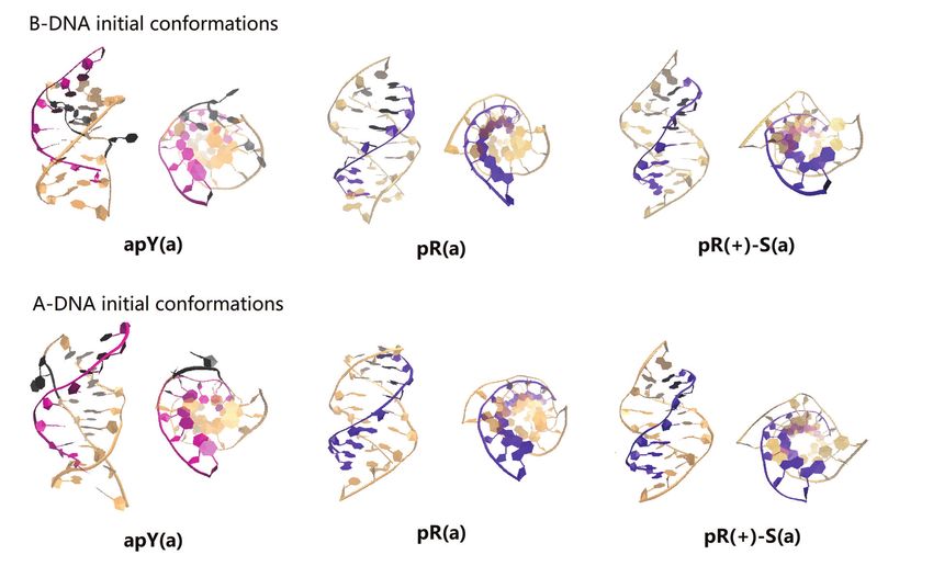
9902 Nucleic Acids Research, 2020, Vol. 48, No. 17

                                                                                                                                                                Downloaded from https://academic.oup.com/nar/article/48/17/9899/5895336 by guest on 20 October 2020
Figure 1. Scheme of the non-equivalent triplexes simulated in this work. Watson–Crick duplexes are highlighted in blue boxes, pyrimidine third strands in
yellow boxes, and purine third strands in green boxes. Top row: eight pure DNA triplexes (for each cartoon, two triplexes were studied, with the third strand
either in all anti or in all syn conformations). Bottom row: hybrid RNA/DNA triplex with the RNA strand (red) forming part of a hybrid Watson–Crick
duplex. The labeling notation is shown below each triplex.

are related by certain symmetries. We define two triplexes                       being directional counterparts when one of them is type H
as being conformational counterparts when they share the                         and the other is type RH. Thus, changing only the direction
same triple base steps but the third strand has the opposite                     of the third strand in the pY(a) destroys the T-O4 and A-N6
direction and the glycosidic angles of its bases are flipped,                    hydrogen bond, but a new T–O2 and A–N6 hydrogen bond
such as pY(a) and apY(s) triplexes (notice that the pyrim-                       (type RH) forms; pY(a) and apY(a) are directional coun-
idine antiparallel strands are shifted in order to hydrogen                      terparts.
bond with the B-DNA duplex). Such conformational coun-                              The initial triple helices were built starting with a stan-
terparts share the same initial hydrogen bond pattern. This                      dard B-DNA GAA/TTC duplex. For the third strand, we
can be understood by considering Figure 2. As an example,                        built another B-DNA duplex containing the sequence of the
consider the middle T·A:T base triplet in the case of pY(a).                     third strand, which in turn was isolated as a single strand as
Flipping the direction of the third strand without changing                      shown in Supplementary Figure S1. This third strand was
the glycosidic angle, destroys the T–O4 hydrogen bond be-                        then moved into the major groove of the GAA/TTC he-
cause the O4 atom in the third-strand T shifts away from the                     lix using the molecular editor Avogadro (79). Finally, for
N6 atom of the A basis that belongs to the B-DNA duplex.                         the syn conformations, we rotated the bases on the third
However, by rotating the basis by 180◦ toward the syn con-                       strand by 180◦ . These primitive models required adjust-
formation brings back the O4 atom and restores the hydro-                        ments that were achieved via MD with constraints through
gen bond. Thus, the simultaneous operations of inverting                         a 1 kcal/mol harmonic potential enforcing the hydrogen
the third strand direction and flipping the glycosidic angles                    bond patterns described above (Figure 2 and Table 1) dur-
of its bases by 180◦ leaves both the H-type and HR-type                          ing equilibration. These constraints were eliminated at the
bonds unchanged. In addition, we define two triplexes as                         start of the regular constant pressure MD simulations. A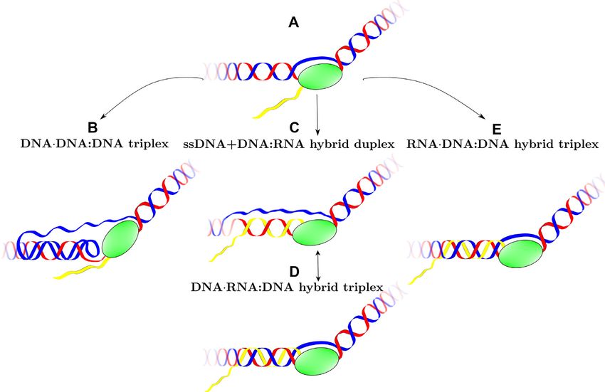
Nucleic Acids Research, 2020, Vol. 48, No. 17 9903

                                                                                                                                              Downloaded from https://academic.oup.com/nar/article/48/17/9899/5895336 by guest on 20 October 2020
Figure 2. Initial hydrogen bond patterns for all the triple helix sequences.

few hundred nanoseconds into the regular MD, some struc-                       TTC antiparallel (to the DNA GAA purine strand), or
tures maintained the hydrogen bond patterns, while oth-                        GAA parallel (to the RNA GAA purine strand). They
ers evolved to form different hydrogen bond patterns that                      can be denoted as d(TTC+ ) ·d(GAA):r(UUC) (apY) and
proved to be stable, and others just became unstable.                          d(GAA)·r(GAA):d(TTC) (pR). We consider the C’s in the
                                                                               third TTC DNA strand to be protonated. In addition, we
                                                                               consider a third case where one of the A’s of the GAA
Initial modeling of the hybrid triple helices
                                                                               third strand is protonated in order to form A+ –G pairs with
For the DNA·RNA:DNA hybrid triplexes, we only con-                             the RNA G’s in the hybrid duplex. This gives rise to the
sider the three most probable cases for the simplest ge-                       pR(+)-S case, according to our previous notation. Finally,
ometry of the R-loop (without folding back of the single                       we only consider anti conformations for the bases in the hy-
strands). First, notice that the third strand will still form                  brid triplex.
hydrogen bonds with the purine strand (DNA or RNA)                                The initial hydrogen bond patterns for the hybrid triple
of the hybrid duplex, for the same reasons as explained                        helices are exactly the same as the corresponding ones for
before. Second, we will assume that the two strands of                         pure DNA triple helices. For each triplex we consider two
DNA in the R-loop continue being antiparallel (i.e. dis-                       initial conformations: one where the hybrid RNA:DNA du-
card possible folding back events), and therefore the two                      plex starts from an ideal B-DNA conformation, and the
DNA strands in the DNA·RNA:DNA triplex are antipar-                            third DNA strand is also added in B-DNA conformation;
allel. That leaves only two cases: if the template DNA                         and one where both the hybrid duplex and third strand are
strand is GAA or TTC, then the third DNA strand is                             in A-DNA conformation.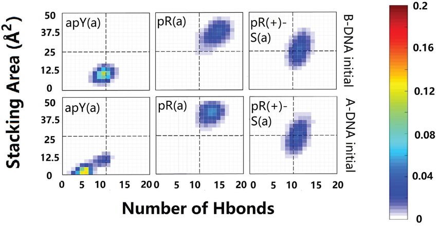
9904 Nucleic Acids Research, 2020, Vol. 48, No. 17
Table 1. Summary of nucleotide triplets and their corresponding confor- displaced stacking whose stacking interaction is influenced
mational counterparts by relative shift and twist (82,83). Also, it is noted that
Triple step type Corresponding triple sequence there exists an inter-strand stacking interaction, which is
as important as intra-strand although it needs no overlap
H-type C+ ·G:C pY(a) apY(s) area (84). As the stacking mechanism of DNA bases is
RH-type C+ ·G:C pY(s) apY(a)
H-type G·G:C pR(a) apR-S(s) quite complicated, we propose a mean-field approximation
RH-type G·G:C pR(s) apR-S(a) method to roughly quantify the – stacking interactions
H-type A·G:C pR-S(a) apR(s) of the third strand in a triplex. First, we calculate the effec-
RH-type A·G:C pR-S(s) apR(a) tive area of each step in the third strand:
H-type A+ ·G:C pR(+)-S(a) apR(+)(s)
RH-type A+ ·G:C pR(+)-S(s) apR(+)(a) Aieff = Ai0 (cos αi − sin αi ) (2)
H-type T·A:T pY(a) apY(s)
RH-type T·A:T pY(s) apY(a) where Ai0 is the area of the aromatic ring(s) of the ith third-
H-type G·A:T pR-S(a) pR(+)-S(a)
strand base in the triplex (based on their molecular struc-
Downloaded from https://academic.oup.com/nar/article/48/17/9899/5895336 by guest on 20 October 2020
apR(s) apR(+)(s)
RH-type G·A:T pR-S(s) pR(+)-S(s) ture, A0 is estimated to be 4.95 Å2 for T and C, and 8.29
apR(a) apR(+)(a) Å2 for A and G) and ␣i is the angle between the normal
H-type A·A:T pR(a) pR-S(a) vector of the third-strand base and the normal vector of
pR(+)-S(a) apR(s) the Watson–Crick base pair at the ith step. The cos ␣ rep-
apR-S(s) apR(+)(s)
RH-type A·A:T pR(s) pR-S(s) resents the effect of the parallel stacking as it projects the
apR(+)-S(s) apR(a) aromatic ring area onto the horizontal plane of the B-DNA
apR-S(a) apR(+)(a) Watson–Crick base pairs while the subtracting sin ␣ term
penalizes the T-shape stacking (strongly disliked by DNA),
as it projects the ring area onto the vertical plane. We set
New metrics for the stability analysis of the triple helices the cutoff value for ␣i to 45◦ , if ␣i > 45◦ , then Aieff is set to
be zero. In addition, because of the essential role of hydro-
During the simulations, we observed that the GAA/TTC gen bonds in the - stacking of base pairs (85), we force
DNA duplex part of the triplex is very stable at 300 K in all Aieff to be zero if Hieff is detected to be zero. This procedure
cases, and therefore measuring the triplex stability simply ensures that the base flipping observed in Supplementary
comes down to quantifying the stability of the third strand Figure S2.b does not contribute to the effective stacking in-
with respect to the helical duplex. We define new quantities teraction of our algorithm. After we get the values of the
to better characterize the presence of the third strand. From effective area of each step, the effective stacking interaction
the structural point of view, the two most important quan- between the ith and the (i + 1)th step is calculated as:
tities to stabilize the third strand are the hydrogen bonds
that it forms with the B-DNA duplex and the – stacking Sieff = Aieff · Aieff+1 (3)
interaction between the bases of the third strand. Simply
counting the total number of hydrogen bonds that the third The effective total stacking area is given by:
strand forms with the duplex does not result in an accu-
rate characterization of the triplex stability. As Supplemen- Seff = Sieff (4)
tary Figure S2.a shows, one can conceive of cases with a A schematic procedure of our algorithm is shown in the
high number of hydrogen bonds that link bases on different right panel of Supplementary Figure S2.c.
planes, which do not, however, result in an increased triplex
stability. We therefore define the hydrogen bond number for Molecular dynamics of the DNA triple helices
a third strand in a triple helix as:
Figure 3 shows a snapshot of the final structures obtained
Heff = Hieff (1) after 1 s MD runs for all the 16 triplex cases proposed.
We colored the Watson–Crick duplex part as light green
where Hieff is the number of hydrogen bonds that a base on while the third strand is colored either blue (pyrimidine)
the third strand forms with the bases of the duplex on the or red (purine). The side view allows us to see the attach-
same plane, as shown in Supplementary Figure S2.c. This ment (or lack thereof) of the third strand to the B-DNA du-
avoids counting inter-plane hydrogen bonds that destabilize plex through intra-step hydrogen bonds, while the top view
the triplex geometry. gives an idea of the base stacking. The conformations after
Compared to the simple definition of hydrogen bonds, 1s clearly show the structures that are unstable, as indi-
quantifying the – stacking interaction is far more com- cated by the detachment of the third strand. The first row
plex (80). One popular way to quantify it is to calculate in the figure shows that both the pY(a) triplex and its con-
the value of overlap area between the adjacent bases as ap- formational counterpart, the apY(s) triplex are stable, with
plied by 3DNA (81). However, for the third strand in the a well preserved triple helix structure after 1s MD simu-
triple helix this method may not work so well, as shown in lations. It is well known that in the syn conformation the
the extreme example of Supplementary Figure S2.b, where nucleotide bears a strong torsion because of the steric re-
all bases are perfectly stacked but not linked to the du- pulsion associated with the sugar ring. However, the struc-
plex. We notice that according to first principles calcula- ture of the apY(s) triplex is stable after 1 s due to the sta-
tions, the stacking of DNA bases is not the sandwich stack- bilization from hydrogen bonds and stacking interactions
ing presumed by the maximum overlap area, but a parallel- that overcome the destabilization caused by the torsionNucleic Acids Research, 2020, Vol. 48, No. 17 9905
Downloaded from https://academic.oup.com/nar/article/48/17/9899/5895336 by guest on 20 October 2020
Figure 3. Snapshot of the conformations of the 16 pure DNA triplexes at 1 s. The third strand of the triplex is colored either blue (pyrimidine) or red
(purine) while the duplex part is colored green.
at a temperature of 300 K. This is not the case for the pY(s) ing hydrogen bonds are displayed. Figures 5 and 6 show the
triplex, whose final structure is severely deformed. Its con- most commonly observed hydrogen-bond patterns that are
formational counterpart, the apY(a) triplex, looks stable different from the initial patterns for the inner base triplets
with some small deformation. in the most stable triplexes. The Y·R:Y (Figure 5) cases are
There are 12 cases with purine as the third strand (the simpler. For the conformational type H counterparts, pY(a)
R·R:Y triplexes). Figure 3 shows clearly unstable cases; the and apY(s) triplexes, the initial hydrogen bond patterns are
four stable cases seem to be pR(a), pR(+)-S(a), apR-S(a), preserved. The type RH apY(a) triplex shows fluctuations
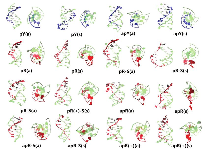
and apR(+)(a). To get a better statistical description that on the fourth plane (Supplementary Figure S3) with the ini-
goes beyond the final structures, we plot in Figure 4 the ef- tial pattern being more populated; the 5th plane remains the
fective stacking area versus the number of hydrogen bonds same, and the 6th plane loses one hydrogen bond.
as defined in the previous section. The distributions have For the stable R·R:Y triplexes we observe more variations
been computed over the last 800 ns. More stable triplexes in the hydrogen bond patterns (Figure 6). To describe the
have higher values in both functions, and therefore better change in hydrogen bonds, we define a notation such that
distributions tend to be located in the upper right quad- the first atom in the bond belongs to the third strand while
rant. This statistical analysis agrees with the qualitative de- the second atom belongs to the B-DNA duplex. First con-
scription provided by the final triplexes. For Y·R:Y, all but sider the pR(a) triplex: on the fourth plane G·G:C the sta-
the pY(s) triplex seem to have comparable stability. Re- ble hydrogen bond pattern is different from our initial guess.
sults for R·R:Y confirm what the final structures suggested: The initial N1(G)–O6(G) and N2(G)–N7(G) bonds are re-
the more stable cases are pR(a), pR(+)-S(a), apR-S(a) and placed by O6(G)–N4(C) and N1(G)–O6(G) which coexists
apR(+)(a). with a more stable bifurcated version [N1(G)–O6(G) and
As the triplexes evolve, initial hydrogen bond patterns N2(G)–O6(G)]. On the fifth and sixth planes, the most sta-
tend to change as shown for the inner steps in the triplexes in ble hydrogen bond pattern corresponds to the adenine in the
Supplementary Figures S3–S5, where dynamically coexist- third chain re-orienting itself so as to form hydrogen bonds9906 Nucleic Acids Research, 2020, Vol. 48, No. 17
Downloaded from https://academic.oup.com/nar/article/48/17/9899/5895336 by guest on 20 October 2020
Figure 4. Two-dimensional histograms of the effective stacking area versus the effective hydrogen bond number as obtained from the last 800 ns of the
MD simulations for the 16 DNA triplexes.
with both the A and T bases in the B-DNA duplex [N6(A)– that of the fifth plane is simply the vanishing of all hydro-
O4(T) and N1(A)–N6(A)]; this pattern coexists with slightly gen bonds. For the sixth plane, our initial guess is one of
fewer stable patterns where one of those bonds is broken the most common coexisting structures. The most stable hy-
(Supplementary Figure S4). drogen bonding structure displays only one hydrogen bond,
Now consider the pR(+)-S(a) case (type H). The stable N6(A)–O4(T), which may be due to a base stacking effect.
structure of the 4th plane is different from our initial guess, Similarly to the fifth plane, the loss of all hydrogen bonds
as the N1(A+)–N7(G) is replaced by N1(A+)–O6(G). How- also results in a coexisting pattern on the 6th plane.
ever, our initial guess is still present as one of the coexist- Finally, we consider the apR(+)(a) case (type RH, Fig-
ing patterns, while the other coexisting pattern loses the ure 6 and Supplementary Figure S5). For the 4th plane, our
N6(A+)–O6(G) initial bond (Supplementary Figure S4). initial guess is also the most stable structure, which coex-
For the fifth plane, the more stable structure differs from ists with two other patterns: one loses the N1(A+)–O6(G)
our initial guess as the N6(A)–N7(A) and N7(A)–N6(A) hydrogen bond, while the other forms the N1(A+)–N7(G)
bonds are replaced by an N1(A)–N6(A) bond; for the other bond in place of the N1(A+)–O6(G) bond. The fifth plane
important coexisting structure the hydrogen bonds link the preserves our initial guess. On the sixth plane, the most sta-
two bases in the B-DNA duplex: N1(A)–N6(A) and N6(A)– ble pattern augments the initial O6(G)–N6(A) bond with
O4(T) bonds. For the 6th plane, our initial guess still holds two additional hydrogen bonds, N7(G)–O4(T) and N2(G)–
as a coexisting structure; in the most stable structure the O4(T), which make the pattern more energetically favored.
original N1(G)–N7(A) bond becomes a more stable, bifur- In the most important coexisting structure, one of these
cated bond with the addition of an N2(G)–N7(A) bond. bonds is absent.
The next case to discuss is apR-S(a) (type RH, Figure 6 In order to determine the conformations that are proba-
and Supplementary Figure S5. Our guess structure is the fi- bilistically more stable, we consider again the possible can-
nal stable structure for its fourth and fifth planes. The most didates. Out of the three Y·R:Y cases that show stabil-
common alternative structure of fourth plane is given by ity, we believe that the case where the pyrimidines in the
the substitution of N2(G)–O6(G) by N2(G)–N7(G) while third strand are in syn conformation represents a long-livedNucleic Acids Research, 2020, Vol. 48, No. 17 9907
Downloaded from https://academic.oup.com/nar/article/48/17/9899/5895336 by guest on 20 October 2020
Figure 5. Dominant hydrogen bond patterns for the middle base planes of stable Y·R:Y triplexes.
Figure 6. Dominant hydrogen bond patterns for the middle base planes of stable R·R:Y triplexes.9908 Nucleic Acids Research, 2020, Vol. 48, No. 17
metastable state where the unfavorable torsion is stabi- ter is wider for the Y-third strand than for R-third strand).
lized by an initial configuration that maximizes hydrogen For the antiparallel third strand, the narrowest groove is be-
bonds and stacking interactions (in other words, if such tween the antiparallel third chain and the TTC strand of the
a state could be set up experimentally, the resulting con- duplex. The groove between the third strand and the GAA
formation would also prove to be long-lived). Discarding strand of the duplex and the original minor groove of the
such configuration leaves two candidates that show stabil- duplex are wider and approximately the same. With respect
ity in the 1s time scale: the pY(a) and the apY(a). None to ion distributions, the Na+ ion density in these simulations
of the R·R:Y cases shows stability for the syn conforma- is relatively small. Supplementary Figure S10 shows the ion
tions in the third strand. As a further test of stability, we distribution for the pY and pR triplexes for the last 200 ns.
carried out higher-temperature MD simulations. Final con- As it can be seen in the figures, most ions concentrate in
figurations at 300K were chosen as a start for these 1s the very electronegative, narrow groove between the third
simulations which entail 0-400 ns at 320 K, 401–1000 ns at strand and the GAA strand of the duplex. There are con-
340 K and 1001–1400 ns at 360 K. We obtained an aver- siderably fewer ions in the groove between the third strand
Downloaded from https://academic.oup.com/nar/article/48/17/9899/5895336 by guest on 20 October 2020
age structure based on the late conformations at 300 K and and the duplex TTC strand, and extremely few in the origi-
calculated the RMSD of the higher-temperature runs with nal minor groove of the B-DNA duplex. Typical parameters
respect to the average 300 K conformations, as shown in for DNA duplexes that form part of the triplexes pY and
Supplementary Figure S8. The difference between the two pR(+)-S are shown in Supplementary Figure S11. Average
pyrimidine directional counterparts is appreciable: the con- values for the last 200 ns of the simulations for twist, roll, he-
formations for the apY(a) triplex seem to undergo more lical rise, inclination, slide, and Zp for the Watson–Crick du-
fluctuations and perhaps some conformational instability plex part of the pR, pY, pR(+)-S and apR(+) triplexes are:
compared to pY(a) as the temperature increases. With re- twist, 31.3◦ , 30.9◦ , 31.1◦ , 33.2◦ ; roll, 1.24◦ , 1.49◦ , 2.52◦ and
spect to the purine case, the RMSD results for the higher- 2.89◦ ; helical rise, 3.35, 3.28, 3.24, 3.28 Å; inclination, 2.41◦ ,
temperature simulations indicate possible lower stability for 2.62◦ , 4.44◦ , 4.98◦ ; slide, –1.21, –1.18, –1.33, –0.90 Å; and
apR-S(a), and comparable stability for pR(a), and the two Zp, 0.32, 0.13, 0.43, 0.00 Å. We see in these values a mixture
protonated cases: pR(+)–S(a) and apR(+)(a). of B- and A-DNA features. Twist values show that attach-
Finally, these triplex simulations were run under the pres- ing a third strand to the GAA/TTC duplex slightly unwinds
ence of Mg2 + ions in the solution. In experiments, Mg2 + it, especially with the third strand in parallel position, mak-
ions play two roles: first, they lower the free energy barri- ing the duplex more A-DNA as far as twist goes. Average
ers for the assembly of secondary structures; and second, inclination values all assume small positive values, closer to
they stabilize the newly formed secondary structures. Since B-form. Helical rise and small values of roll put the duplex
our simulations start with triplexes already formed as initial closer to B-form, while the negative slide is more charac-
conditions, the first role of the ion is irrelevant, but the pres- teristic of the A-DNA. Finally, Zp values less than 0.5 put
ence of the Mg2 + ions could induce changes in stability. In these duplexes into the B-DNA camp.
these new simulations we used a concentration of 200 mM,
which is approximately 10 times or more the physiological
Molecular dynamics of the hybrid helical duplexes
concentration. We found that the presence of Mg2 + ions
preserves the relative stability of the triplexes as described There are two types of hybrid duplexes that are not equiva-
above with only Na+ ions (with minor variations in the sta- lent: RU=d(GAA):r(UUC) and DT=r(GAA):d(TTC). For
tistical graphs) except for the apR(a) triplex, that was un- each of these hybrids, we started the simulations with ideal
stable in absence of the Mg2 + ions but becomes stable in B-DNA and A-RNA conformations. Convergence of the
their presence (Figure S9 in Supplementary). So, this partic- simulations is confirmed by the convergence of these ini-
ular triplex was simulated again under 40 mM MgCl2 and tial duplexes to a final duplex that is independent of the
80 mM MgCl2 concentrations, which are still considerably initial conditions. This convergence can be appreciated in
larger than physiological concentrations. We found that un- the analysis of structural parameters presented in Figure 7,
der these reduced Mg2 + concentrations, the triplex is only which also make evident the periodicity of the three differ-
marginally stable (after careful equilibration, some runs re- ent steps of the sequence. Supplementary Figure S12 shows
main stable but the structure unravels in other runs). There- the RMSD of the four hybrid duplexes. Clearly these du-
fore, once the triplexes are formed, the presence of Mg2 + plexes are somewhere between A-RNA and B-DNA, but
ions does not affect the stability results obtained with only closer to A-RNA. In particular, the average RMSD over
Na+ ions, unless extremely high divalent concentration are the last 200 ns of the simulations are: for RU, about 1.97Å
used. with respect to A-RNA and 2.57 Å with respect to B-
Next, we briefly describe some other structural features of DNA; and for DT, about 1.56 Å with respect to A-RNA
the triplexes. A triple helix has three grooves whose widths and 1.92 Å with respect to B-DNA. Thus, DT is closer
are mainly determined by the nature and orientation of the to both A-RNA and B-DNA than RU is to either form.
third chain and the hydrogen bond patterns. Charged or Two main factors contributes to relative stability: hydro-
neutral bases do not alter the widths of the grooves. For the gen bonds and stacking area. With respect to the hydro-
parallel chains, the narrowest groove takes place between gen bonds, the most important difference is given by the
the parallel third strand and the GAA strand of the duplex. d(A):r(U) basepair that is extremely weak (86). With respect
This is followed in order of increasing width by the origi- to the stacking area of the base steps, we used 3DNA (87)
nal minor groove in the duplex, and last by the groove be- in order to compute the total stacking area of inner steps 2
tween the third chain and TTC chain in the duplex (the lat- to 8 and found this to be 48 Å2 for DT and 43 Å2 forNucleic Acids Research, 2020, Vol. 48, No. 17 9909
B
A
40 20
Twist Angle (°)
Inclination (°)
15
35
10
30
5
25 0
GA/XC AA/XX AG/CX GA/XC AA/XX AG/CX GA/XC AA/XX AG/CX GA/XC AA/XX A−X A−X G−C A−X A−X G−C A−X A−X G−C A−X A−X
C D
15 3.5
Helical Rise (Å)
Downloaded from https://academic.oup.com/nar/article/48/17/9899/5895336 by guest on 20 October 2020
Roll (°)
10
3
5
0 2.5
GA/XC AA/XX AG/CX GA/XC AA/XX AG/CX GA/XC AA/XX AG/CX GA/XC AA/XX GA/XC AA/XX AG/CX GA/XC AA/XX AG/CX GA/XC AA/XX AG/CX GA/XC AA/XX
E 1 F 2.5
0.5 2
Slide (Å)
0 1.5
Zp (Å)
−0.5 1
−1 0.5
−1.5 0
−2 −0.5
GA/XC AA/XX AG/CX GA/XC AA/XX AG/CX GA/XC AA/XX AG/CX GA/XC AA/XX GA/XC AA/XX AG/CX GA/XC AA/XX AG/CX GA/XC AA/XX AG/CX GA/XC AA/XX
Figure 7. Average basepair inclination, and basepair-step twist, roll, helical rise, slide and Zp of double helices. Red: d(GAA):d(TTC) (B-DNA); or-
ange: r(GAA):r(UUC) (A-RNA); blue and green: r(GAA):d(TTC) starting from ideal B-DNA (blue) or ideal A-RNA (green); yellow and purple:
d(GAA):r(UUC) starting from ideal B-DNA (yellow) or ideal A-RNA (purple). Data was averaged over the last 200 ns. ‘X’ in the x-axis labels stands
for either T or U, according to the sequence.
RU. Thus, DT displays better stability than its RU counter- unstable because the third strand detaches before the duplex
part. Interestingly DT=r(GAA):d(TTC) seems to achieve has time to equilibrate. Therefore, we ran an initial equili-
this stability by combining stabilizing features from A- and bration step where the third strand is constrained to remain
B-forms: rise, inclination, and roll, among other parame- attached to the duplex while it equilibrates, and this became
ters, are closer to B-DNA than A-RNA, while the weaker the new A-like initial conformation. Final conformations
RU=d(GAA):r(UUC) duplex is closer to A-RNA as mea- for the six cases are shown in Figure 8 (where bases that
sured by these parameters. Instead, the more discrimina- have flipped out are colored in black). The statistical anal-
tory parameters (88) slide and the Zp parameter, defined as ysis based on the effective hydrogen bonds and the effective
the mean z-coordinates of the backbone phosphorus atoms stacking areas is shown in Figure 9. The results shown in
with respect to individual dimer reference frames (89), are these figures indicate that when the DNA third strand is
closer to A-RNA, as shown in Figure 7. formed by the pyrimidines, the resulting apY triplexes are
unstable: bases are flipping out not only in the third strand
but also in the RNA strand that forms the hybrid duplex.
Molecular dynamics of the hybrid triple helices
On the other hand, the pR and pR(+)-S triplexes are sta-
As discussed in the Initial Modeling section, the sim- ble and their initial A- and B-forms converge to the same
plest collapsed R-loop (without back-folding of the single structure as the statistical analysis in Figure 9 clearly shows,
strands) can only form three hybrid triplexes. Our simula- with a strong distribution in the upper right quadrant
tions start with either an ideal B-DNA conformation or an for pR.
ideal A-DNA one. In real life, one expects the duplex part of Next, we examine the primary hydrogen bond patterns
the triplex, like the hybrid helical duplexes described above, for the three stable cases. These are shown in Supplemen-
to be somewhere in the spectrum between these two ideal tary Figures S13 and S14. For a given sequence, hydrogen
structures. In the ideal A-DNA triplex, the third strand ends bond patterns for the hybrid triplex are the same as those for
further away from the duplex than in the ideal B-DNA con- the pure DNA triplex except for the fifth plane of the pR(+)-
formation, and therefore the initial fully A-DNA confor- S triplex where a new bond between the N6 atom of DNA
mation in the pR triplex (but not in the pR(+)-S triplex) is adenine and the O4 atom of DNA thymine is the dominant9910 Nucleic Acids Research, 2020, Vol. 48, No. 17
Downloaded from https://academic.oup.com/nar/article/48/17/9899/5895336 by guest on 20 October 2020
Figure 8. Snapshot of the conformations of the 6 hybrid DNA/RNA triplexes at 1 s. The RNA strand of the triplex is colored either pink (pyrimidine) or
violet (purine) while the DNA strands are colored light orange. Bases that have lost all hydrogen bonds or are flipping out are indicated in black shadow.
Figure 9. Two-dimensional histograms of the effective stacking area versus the effective hydrogen bond number as obtained from the last 800 ns of the
MD simulations for the six hybrid DNA/RNA triplexes.
isomer in the hybrid triplex and a less populated isomer in third chain and the GAA strand of the duplex, with negli-
the pure DNA triplex. The statistical analysis shows that gible distribution in the original minor groove of the du-
the pR(+)-S conformations are less stable for the hybrid plex. The difference between the two triplexes is that the
triplexes than for the pure DNA triplexes: in the 2D his- pure DNA triplex still carries ions in the groove between the
tograms, the distributions in the RNA-containing triplexes third strand and the TTC strand of the duplexes, while for
are displaced downwards and towards the left compared to the hybrid triplex these ions almost disappear and most of
their counterpart in the pure DNA triplexes. While the pR the distribution is concentrated in the narrow groove. Pa-
triplexes settle quickly into their final hydrogen bond pat- rameters for the hybrid duplexes r(GAA):d(TTC) as they
terns, the pR(+)-S conformations show some coexisting iso- describe a free-standing hybrid duplex or a duplex part of a
mers after 200 ns, as shown in Supplementary Figure S14. triplex are shown in Supplementary Figure S15. These fig-
The Na+ ion distribution for the hybrid pR triplex for ures as well as the values of the Zp parameter calculated as
the last 200 ns is shown in Supplementary Figure S10. In 0.32Å for pR and 0.43Å for pR(+)-S show that the hybrid
both the pure DNA and hybrid pR triplexes, most of the duplexes that form part of a triplex are more B-like than
ion distribution sits in the very narrow groove between the A-like in comparison to their free-standing counterparts.Nucleic Acids Research, 2020, Vol. 48, No. 17 9911
DISCUSSION and the other is type RH, such as pY(a) and apY(a). In or-
der to characterize these triplexes and their stability we no-
One common characteristic underlying all TREDs is the
ticed that standard ways of characterizing hydrogen bonds
transient formation of atypical, non-B DNA stable sec-
and base stacking fail to give a true measure when applied to
ondary structures in the expandable repeats (17,18,23–25).
the third strand. Thus, we provided a new protocol to count
Although knowledge of these structures per se is not nearly
the hydrogen bonds and new definitions of base stacking
enough to understand these diseases, it certainly helps to
that explicitly consider the triplex geometry provided by the
understand the interaction of the TRs with the relevant pro-
third strand. Analysis of final conformations and statistical
teins as one tries to decipher the molecular mechanisms
two-dimensional histograms of the effective base stacking
behind the diseases. The GAA/TTC TRs associated with
versus the effective number of hydrogen bonds for the third
Friedreich’s ataxia are unable to form the hairpins that char-
strand allowed us to pick the triplexes that are more sta-
acterize other TRs: GAA/TTC repeats form DNA triplexes
ble. In all unstable triplexes but one, instability shows up
(30–38), traditional R-loops (41–44) or, possibly, collapsed
in the detachment of the third strand while the helical du-
R-loops, i.e. hybrid DNA·RNA:DNA triplexes, which have
Downloaded from https://academic.oup.com/nar/article/48/17/9899/5895336 by guest on 20 October 2020
plex remains stable. In the d(TTC+ )·d(GAA):r(UUC) hy-
not been directly measured. In the simplest triplex DNA
brid triplex, the detachment of the third strand also dis-
models, the strand that appears twice in a triplex does so
rupts the duplex. Plots of the effective base stacking versus
via a single turn, so that it ends antiparallel to itself. This
the effective number of hydrogen bonds for the third strand
would imply that Y·R:Y triplexes can only be parallel (pY)
capture the instability in both cases: the only circumstance
while R·R:Y triplexes can only be antiparallel (apR or apR-
where this description would fail corresponds to the highly
S). However, the other two types of triplexes, apY and pR
improbable case where the Watson–Crick base pairs break
(or pR-S), are important not only in the context of TFO
but the third strand forms a duplex with the purine strand
therapies but also as a natural possibility, as it is increas-
with H/HR hydrogen bonding.
ingly becoming clear that more complicated entities can
form at R-loops (57). Supplementary Figure S16 shows an
example of pR formation that could happen either intra- Sixteen non-equivalent DNA triple helices can be assembled
or inter-molecularly, as has been proposed to occur, for in- from GAA and TTC strands
stance, in plasmids (90). Since one of the driving forces of
We have provided physical arguments to figure out all pos-
non-B-DNA formation in the cell is negative supercoiling,
sible triplexes that can be formed with three DNA strands
these simulations, like FRET, gel electrophoresis, circular
composed of either GAA or TTC repeats. There are 8 re-
dichroism, UV melting, UV absorption and other experi-
sulting triplexes depicted in Figure 1, a number that doubles
ments that use short oligonucleotides, ultimately cannot re-
to 16 if one allows for the possibility of bases on the third
flect the structural constrains related to non-B-DNA for-
strand to be in syn conformation, a state of the glycosidic
mation. For instance, a number of experiments using GAA-
bond that has been observed in other TRs (50,52,54,74–
containing supercoiled plasmids report formation of Y·R:Y
78). For the TTC·GAA:TTC triplex, protonated cytosines
DNA triplexes only (32,91–94), but other plasmid experi-
(69–72) in the third strand allow the formation of hydro-
ments suggest the presence of R·R:Y triplexes, in particu-
gen bonds with the guanines of the GAA strand in the B-
lar as precursors to ‘sticky DNA’ (30,31,90,95–98). Inter-
DNA duplex. This results in only two cases that can form
estingly, R·R:Y triplexes have been proposed to act dur-
hydrogen bonds plus their syn counterparts. For the all-
ing DNA replication: Fork reversal could occur at paus-
purine third strand GAA·GAA:TTC triplex, there are six
ing forks when triplex formation occurs between two GAA
cases (plus the six syn counterparts): the GAA third strand
strands and one TTC strand (99,100) Here we discuss our
can be parallel or antiparallel, can be shifted or not, and
main results.
can have completely unprotonated adenines or up to one
protonated adenine (73) per repeat in order to form good
hydrogen bonding structure with the G:C base pair of the
Hydrogen bond patterns and their symmetry; measuring hy-
B-DNA duplex (Figure 1). The final conformations and the
drogen bonds and stacking for the third strand
statistical two-dimensional histograms of the effective base
We extended definitions of Hoogsteen and reverse Hoog- stacking versus the effective number of hydrogen bonds for
steen hydrogen bond pattern to encompass other similar the third strand leads to the stability ranking of the triplexes.
patterns classified as ‘H’ and ‘RH’ in Figure 2 and Table
1. These definitions facilitate the visualization of symmetry
DNA TTC·GAA:TTC triple helices: the TTC third strand in
properties of the triple helix: conformational counterparts
parallel alignment, pY(a), is most stable
share the same triple base steps but the third strand has
the opposite direction and the glycosidic angles of its bases For the TTC·GAA:TTC triplexes both parallel and an-
are flipped, such as pY(a) and apY(s) triplexes (notice that tiparallel conformations show good stability when the third
the pyrimidine antiparallel strands are shifted in order to strand bases are in anti conformation. Interestingly, the
hydrogen bond with the B-DNA duplex). Conformational apY(s) triplex also shows stability even though the pyrimi-
counterparts belong to the same ‘type’: the simultaneous dine bases of the third strand are in syn conformation (al-
operations of inverting the third strand direction and flip- though less common, pyrimidine bases can be found in syn
ping the glycosidic angles of its bases by 180◦ leaves both conformations (74)). In this case, the stabilization from hy-
the H-type and RH-type bonds unchanged. In directional drogen bonds and stacking interactions overcome the desta-
counterpart pairs, on the other hand, one triplex is type H bilization caused by the torsion at a temperature of 300 K.9912 Nucleic Acids Research, 2020, Vol. 48, No. 17
We believe that this is a metastable state facilitated by the the antiparallel third chain and the TTC strand of the du-
good stacking of the initial conformation. Under progres- plex, and the other two wider grooves are approximately
sive higher-temperature simulations, the parallel pyrimidine the same. The very narrow groove in the parallel triplexes
pY(a) triplex is seen as more stable than its directional coun- strongly attracts Na+ ions, with most (but not all) of the
terpart, apY(a), in agreement with experiments (33). cation density localized there (Supplementary Figure S10),
With respect to the hydrogen bonds, the original guesses and almost no ions in the original minor groove of the du-
in Figure 2 for the Y·R:Y triplexes are on cue; the most com- plex. Typical parameters for Watson–Crick duplexes that
mon variation is the formation of hydrogen bonds between form part of the triplexes display a mixture of B- and A-
bases belonging to the two pyrimidine strands. These cases, DNA features. Attaching a third strand to the GAA/TTC
however, are less populated, generally because the new pat- duplex slightly unwinds it, especially with the third strand
tern results in the same or fewer number of hydrogen bonds in parallel position, making the duplex more A-DNA as far
than the original pattern. as twist goes. Average inclination, helical rise and roll de-
Our results agree with experiments. Cytosines need to be part from ideal B-DNA values, but still are closer to B- than
Downloaded from https://academic.oup.com/nar/article/48/17/9899/5895336 by guest on 20 October 2020
protonated in order to form stable triplexes, which generally A-DNA. Negative average slides are more characteristic of
occurs under low pKa (28). However, cytosines buried in the A-DNA, but Zp valuesYou can also read