SURVEY AND SUMMARY The involvement of nucleotide excision repair proteins in the removal of oxidative DNA damage - Glen Research
←
→
Page content transcription
If your browser does not render page correctly, please read the page content below
Published online 3 October 2020 Nucleic Acids Research, 2020, Vol. 48, No. 20 11227–11243
doi: 10.1093/nar/gkaa777
SURVEY AND SUMMARY
The involvement of nucleotide excision repair proteins
in the removal of oxidative DNA damage
Namrata Kumar1,2,† , Sripriya Raja2,3,† and Bennett Van Houten 1,2,3,4,*
1
Molecular Genetics and Developmental Biology Graduate Program, School of Medicine, University of Pittsburgh,
Pittsburgh, PA 15213 USA, 2 UPMC Hillman Cancer Center, University of Pittsburgh, PA 15213, USA, 3 Molecular
Downloaded from https://academic.oup.com/nar/article/48/20/11227/5917651 by guest on 19 January 2021
Pharmacology Graduate Program, School of Medicine, University of Pittsburgh, Pittsburgh, PA 15213 USA and
4
Department of Pharmacology and Chemical Biology, School of Medicine, University of Pittsburgh, Pittsburgh, PA
15213, USA
Received June 27, 2020; Revised September 02, 2020; Editorial Decision September 05, 2020; Accepted September 07, 2020
ABSTRACT the DNA (4). Due to the extensive DNA damage caused by
oxidation, these lesions have been associated with a large
The six major mammalian DNA repair pathways were number of human maladies including neurodegeneration,
discovered as independent processes, each dedi- cancer and aging (5).
cated to remove specific types of lesions, but the past Some of the most widely studied DNA lesions result-
two decades have brought into focus the significant ing from oxidation are shown in Figure 1. One of the
interplay between these pathways. In particular, sev- best characterized oxidative lesions is 8-oxo-7,8-dihydro-
eral studies have demonstrated that certain proteins 2 -deoxyguanosine (8-oxoG), the major product produced
of the nucleotide excision repair (NER) and base ex- from the oxidation of guanine. Further oxidation of 8-oxoG
cision repair (BER) pathways work in a cooperative results in the formation of spiroiminodihydantoin (Sp) and
manner in the removal of oxidative lesions. This re- 5-guanidinohydantoin (Gh). Purine oxidation can also re-
view focuses on recent data showing how the NER sult in the formation of 5 ,8-cyclo-purine adducts. An im-
portant product of thymine oxidation is thymine glycol
proteins, XPA, XPC, XPG, CSA, CSB and UV-DDB,
(Tg). Cytosine is subject to methylation, resulting in the for-
work to stimulate known glycosylases involved in the mation of 5-methylcytosine (5mC). Oxidative removal of 5-
removal of certain forms of base damage resulting methylcytosine (5mC) occurs through an active enzymatic
from oxidative processes, and also discusses how process in which 5mC is oxidized in three steps by a family
some oxidative lesions are probably directly repaired of TET dioxygenases to form 5-hydroxymethyl-C (5hmC),
through NER. Finally, since many glycosylases are 5-formylC (5fC) and 5-carboxyC (5caC).
inhibited from working on damage in the context of Oxidative base lesions are commonly repaired via base
chromatin, we detail how we believe UV-DDB may be excision repair (BER) pathway (6). BER is initiated af-
the first responder in altering the structure of damage ter a lesion-specific DNA glycosylase cleaves the glycosidic
containing-nucleosomes, allowing access to BER en- bond, which frees the lesion, and creates an abasic site (1)
zymes. (Figure 2). Currently, there are 11 known mammalian DNA
glycosylases that can be categorized as monofunctional or
bifunctional. Monofunctional glycosylases only possess the
INTRODUCTION ability to break the glycosidic bond between the damaged
Reactive oxygen and nitrogen species (ROS/RNS), such base and the sugar moiety, resulting in an abasic site, which
as singlet oxygen, superoxide, hydrogen peroxide, hydroxyl is processed by AP endonuclease 1 (APE1) to form a 3 OH
radical, nitric oxide and peroxynitrite, can be generated en- and a deoxyribose-5 -phosphate (dRP). This dRP is re-
dogenously by normal cellular metabolism or inflamma- moved by the lyase activity of DNA polymerase  (pol
tion, or by exogenous sources such as ultraviolet (UV) or ). Bifunctional glycosylases have an additional AP lyase
ionizing radiation (IR) (1–3). Oxidation can either directly activity which allows for cleavage of the phosphate back-
or indirectly introduce a wide spectrum of base lesions in bone, creating a single strand break, leaving a free 5 phos-
* To whom correspondence should be addressed. Tel: +1 412 623 7762; Fax: +1 412 623 7761; Email: vanhoutenb@upmc.edu
†
The authors wish it to be known that, in their opinion, the first two authors should be regarded as Joint First Authors.
C The Author(s) 2020. Published by Oxford University Press on behalf of Nucleic Acids Research.
This is an Open Access article distributed under the terms of the Creative Commons Attribution Non-Commercial License
(http://creativecommons.org/licenses/by-nc/4.0/), which permits non-commercial re-use, distribution, and reproduction in any medium, provided the original work
is properly cited. For commercial re-use, please contact journals.permissions@oup.com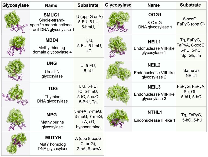
11228 Nucleic Acids Research, 2020, Vol. 48, No. 20
Downloaded from https://academic.oup.com/nar/article/48/20/11227/5917651 by guest on 19 January 2021
Figure 1. Chemical structures of oxidative lesions formed in DNA. (A) Various oxidation products of guanine. (B) Formation of cyclic guanosine by
oxidation. (C) Formation of cyclic adenosine by oxidation. (D) Enzymatic oxidative demethylation of 5-methylcytosine. (E) Oxidation of thymine to
thymine glycol. ROS produces over 100 different types of lesions in DNA, and this figure displays the structures of those damages that are discussed in this
review. ROS: Reactive oxygen species; DNMT: DNA methyltransferase; TET: Ten-eleven translocation enzymes.
phate and either a 3 -phospho-␣, -unsaturated aldehyde tional glycosylases Endonuclease VIII-like 1–3 (NEIL1–3),
(3 -PUA) (-elimination) or 3 phosphate (,␦-elimination). which will be discussed in detail in a later section. Tg is
APE1 acts on the -elimination product, while polynu- removed by the bifunctional glycosylase Endonuclease III-
cleotide kinase phosphate (PNKP) is required to process like 1 (NTHL1). 5fC and 5caC are removed by the action
the 3 phosphate after ,␦-elimination. The resulting 3 OH of thymine DNA glycosylase (TDG), which is a monofunc-
is bound by PARP1 which recruits the BER complex con- tional glycosylase. The structures of these glycosylases and
sisting of pol , XRCC1 and DNA ligase. The one base gap their substrates are given in Figure 3.
is then filled by pol  and the nick in the DNA is sealed by For more than a decade, studies have provided evidence
DNA ligase (7). The human 8-oxoG glycosylase (OGG1) suggesting a role for NER proteins in the repair of oxida-
is a bifunctional glycosylase responsible for the recognition tive damage through interactions with BER proteins, re-
and removal of 8-oxoG. Like several glycosylases, OGG1 is viewed (8–11). NER is the major pathway for the repair
product inhibited, binding avidly to abasic sites, and turns of bulky adducts and other helix-distorting lesions, such as
over slowly in the absence of other proteins such as APE1 UV-induced photoproducts, such as 6–4 photoproducts (6–
(7). Sp and Gh are removed by the actions of the bifunc- 4 PP) and cyclobutane pyrimidine dimers (CPD) (12). Un-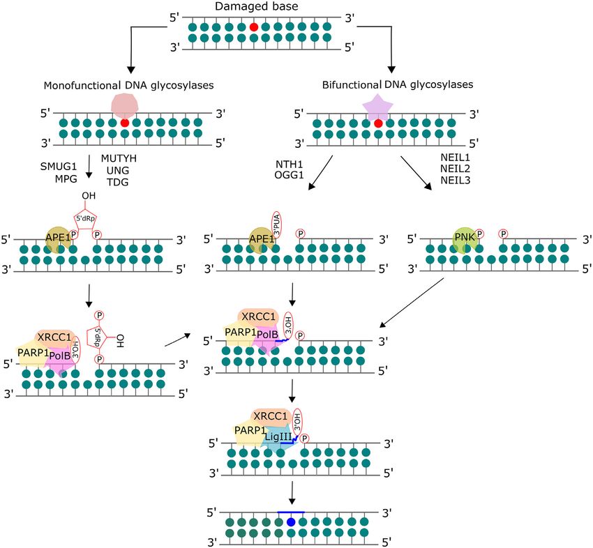
Nucleic Acids Research, 2020, Vol. 48, No. 20 11229
Downloaded from https://academic.oup.com/nar/article/48/20/11227/5917651 by guest on 19 January 2021
Figure 2. Mammalian base excision repair (BER) pathway. The base lesion is excised by a lesion-specific DNA glycosylase. Monofunctional glycosylases
break the glycosidic bond between the damaged base and the sugar moiety, resulting in an abasic site. AP endonuclease 1 (APE1) processes the abasic site
to form a 3 OH and a deoxyribose-5 -phosphate (dRP), which is removed by the lyase activity of DNA polymerase  (pol ). Bifunctional glycosylases
utilize their AP lyase activity to cleave the phosphate backbone, creating a single strand break, leaving a free 5 phosphate and either a 3´-phospho-␣,
-unsaturated aldehyde (3 -PUA) (-elimination) or a 3 phosphate (,␦-elimination). APE1 acts on the -elimination product while polynucleotide kinase
phosphate (PNKP) is required to process the 3 phosphate after ,␦-elimination. The resulting 3 OH is bound by PARP1 which recruits the BER complex
consisting of pol , XRCC1 and DNA ligase. The one base gap is then filled by pol  and the nick in the DNA is sealed by DNA ligase. Adapted from
Kumar et al. (8) with permission.
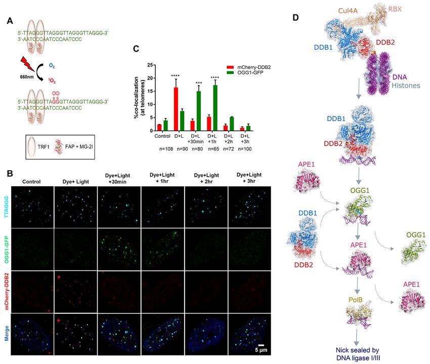
like BER that has a set of glycosylases each tuned to find the chromatin to ubiquitinate histones, making the lesion
and process specific altered bases (Figure 3), damage recog- more accessible to downstream repair proteins in the NER
nition proteins of NER are remarkable in that they have pathway, including XPC-RAD23B. XPC-RAD23B binds
a broad ability to dynamically detect many different struc- with high affinity to the strand opposite to the distorted
turally and chemically diverse lesions (13). There are two lesion, which begins the damage verification step of NER.
sub-pathways in NER: global-genome NER (GG-NER) XPC-RAD23B facilitates recruitment of the transcription
and transcription-coupled NER (TC-NER), reviewed in factor TFIIH. TFIIH is a multi-subunit protein, consist-
(12,14). These sub-pathways differ in the manner of le- ing of 10 proteins, including XPB and XPD, proteins that
sion recognition. In GG-NER, damage recognition pro- have DNA helicase folds. When XPD encounters the lesion,
teins scan the entire genome, including heterochromatic, its strand opening activity stalls and facilitates the recruit-
transcriptionally inactive regions, or the non-transcribed ment of XPA, RPA and XPG, collectively known as the pre-
strand for damage-induced structural distortions. In con- incision complex. XPB is believed to act as a translocase to
trast, TC-NER is initiated when RNA polymerase (RNAP) help reel the DNA into the pre-incision complex. XPA and
stalls at damaged site on the transcribed strand of active RPA recruit the heterodimeric endonuclease, XPF-ERCC1.
genes, in euchromatic regions of the genome. Defects in Recruitment of XPF-ERCC1 produces an endonucleolytic
NER are associated with two important human diseases, xe- incision 5 to the lesion. DNA polymerases ␦, ε or begin
roderma pigmentosum (XP) and Cockayne syndrome (CS). to fill in the repair patch, which stimulates the 3 endonu-
Damage recognition in GG-NER is initiated by two pro- cleolytic activity of XPG, leading to release of an oligonu-
teins, UV-DDB and XPC-RAD23B. In response to UV- cleotide of 22–27 nucleotides containing both the lesion and
induced DNA damage, UV-DDB in complex with CUL4 TFIIH. DNA ligase I seals the remaining nick in the repair
and RBX forms a ubiquitin E3 ligase complex and binds to patch. TC-NER damage recognition is initiated by the pres-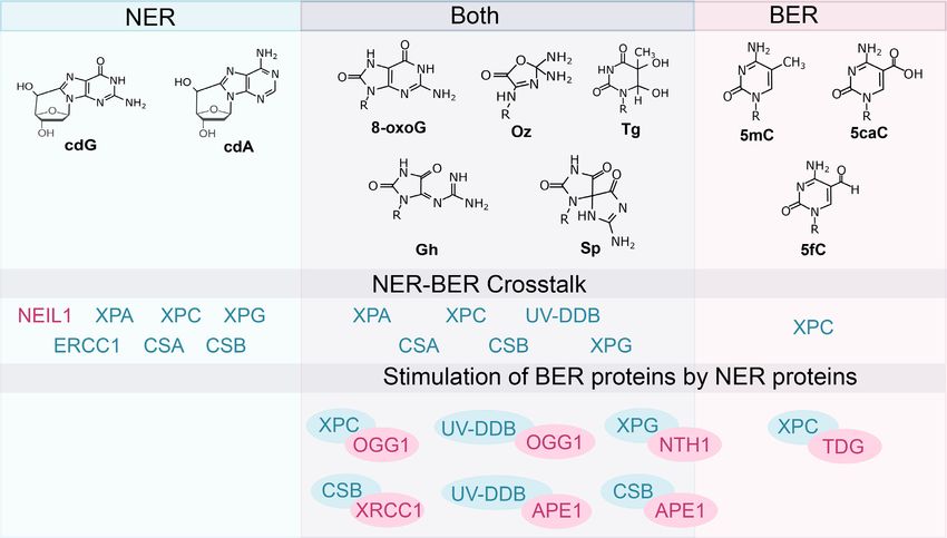
11230 Nucleic Acids Research, 2020, Vol. 48, No. 20
Downloaded from https://academic.oup.com/nar/article/48/20/11227/5917651 by guest on 19 January 2021
Figure 3. BER glycosylases, their structures and respective substrates. The glycosylases (green) are bound to DNA (purple) containing a lesion (pur-
ple, space-filled). All structures are human except SMUG1 (Xenopus laevis), MUTYH (Geobacillus stearothermophilus), NEIL2 (Monodelphis do-
mestica), NEIL3 (Mus musculus), NTHL1 (EndoIII, Geobacillus stearothermophilus). PDB: SMUG1 (1OE4), MBD4 (5CHZ), UNG (1EMH), TDG
(3UFJ), MPG (1BNK), MUTYH (4YOQ), OGG1 (1EBM), NEIL1 (5ITY), NEIL2 (6VJI), NEIL3 (3W0F), NTHL1 (1ORN). Abbreviations: U, uracil;
A, adenine; T, thymine; C, cytosine; G, guanine; 5-FU, 5-fluorouracil; 5-hmU, 5-hydroxymethyluracil; , etheno; FaPy, 2,6-diamino-4-hydroxy-5-N-
methylformamidopyrimidine; 8-oxoG, 8-oxoguanine; Gh, Guanidonohydantoin; Sp, Spiroiminodihydantonin; Im, iminoallantoin; 5fC, 5-formylcytosine;
5caC, 5-carboxycytosine; 5-BrU, 5-Bromouracil; Tg, Thymine Glycol; meA, 3-methyladenine; meG, 3-methylguanine; 5-hC, 5-hydroxycytosine; 5-hU,
5-hydroxyuracil; 2-hA, 2-hydroxyadenine
ence of a stalled RNAP at a lesion site, which facilitates re- tions to DNA and therefore, can act as good substrates for
cruitment of Cockayne syndrome proteins (CSB and CSA), nucleotide excision repair (NER) (11). Estimates of cyPu le-
and the accessory proteins (UVSSA, XAB2, and HMGN1) sions vary, but are generally considered to be less frequent
to the lesion site on the transcribed strand (15). XAB2 facil- than 8-oxoG, with cdG lesions about an order of magnitude
itates recruitment of XPA and subsequently TFIIH which more prevalent that cdA lesions (Table 1).
intertwines the two NER sub-pathways at the damage veri- As mentioned earlier, defects in NER genes can cause the
fication step. CS patients and a subset of XP patients display rare disorder xeroderma pigmentosum (XP). About 20%
signs of neurological degeneration and have been shown of XP patients exhibit neurological symptoms, which have
to accumulate unrepaired oxidative DNA lesions (16,17). been posited to be caused by endogenously accumulated ox-
Therefore, it is important to understand any crosstalk which idative DNA damage (16,17). Work by Lindahl’s group sup-
exists between the two repair pathways to gain a better un- ported this hypothesis by demonstrating that a subclass of
derstanding of disease progression. base lesions formed by ␥ -irradiation were repaired by nor-
mal cell extracts, but not by XP cell extracts (21). The first
direct evidence of NER involvement in the repair of cyPu
COULD CYCLOPURINE DEOXYNUCLEOSIDES EX-
lesions was provided by Kuraoka et al. Primer extension
PLAIN NEURODEGENRATION IN XERODERMA PIG-
assays using mammalian polymerase ␦ or T7 DNA poly-
MENTOSUM PATIENTS?
merase were performed to show that both 5 S- and 5 R-
5 ,8-Cyclopurine deoxynucleotides (cyPu) are endogenous cdA can block DNA synthesis by terminating product ex-
oxidative DNA lesions formed by the reaction of hydroxyl tension at the cdA site in a 5 -32 P-labeled DNA template-
radicals with DNA, and first identified after ionizing ra- primer substrate containing a site-specific 5 S- or 5 R-cdA
diation (18). These lesions contain damage to both the lesion. Therefore, if left unrepaired, cdA lesions can be
purine base and the 2 -deoxyribose sugar moiety by form- highly cytotoxic by blocking DNA replication (22). More-
ing a covalent bond between the C8 of the base and C5 of over, when HeLa cell extracts were incubated with 20 bp
2 -deoxyribose (19). Both cyclo-deoxyadenosine (cdA) and cdA substrates, no DNA glycosylase-mediated cleavage was
cyclo-deoxyguanosine (cdG) lesions exist as 5 R- and 5 S- observed suggesting that human DNA glycosylases can-
diaestereomers (20) (Figure 1) and cause significant distor- not act on cyPu residues. Finally, the authors used a dual-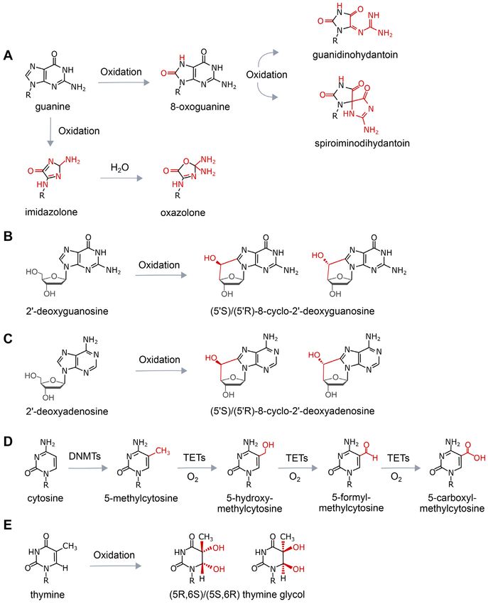
Table 1. Oxidative lesions and NER proteins

                      Frequency/106            Reference for                                                                                             Evidence for NER proteins
Lesion                   bases *             lesion frequency           Technique used                     Source of DNA              Repaired by           involved (reference)
8-oxoG                        1                   (113)                 COMET assay                          HeLa cells              BER/ TC-NER         XPA (67,69,73,75,76), XPC
                                                                                                                                                          (29,38,67,69,72,73), XPG
                                                                                                                                                        (67–69), UV-DDB (56), CSA
                                                                                                                                                                (75), CSB (75)
                              1.2                  (29)                     LC/MS                   Human primary keratinocytes
                              4.6                  (40)                 HPLC-ESI-MS                         Rat Liver
                            25**                  (114)                  LC-MS/MS                         Calf Thymus
                             0.35                 (115)            NaI extraction; HPLC-EC                 Mice Liver
Gh                       0.01–0.07                (116)                     LC/MS                     Mice Liver and Colon            BER/NER              XPA (83), XPC (83)
Sp                       0.01–0.07                (116)                    LC/MSfC                    Mice Liver and Colon            BER/NER              XPA (83), XPC (83)
8-cyclo-2 dG       2.8 (5 S); 0.7 (5 R)         (29)                   GC/MS***                  Human primary keratinocytes         NER                      XPC (29)
8-cyclo-2 dA            0.1–0.15                  (20)              LC/MS; GC/MS***                      Calf Thymus                   NER            XPA (23), XPC (29), CSA (30),
                                                                                                                                                                 CSB (30)
                        0.015–0.03                (117)             32 P-Postlabeling   Assay        Fetal and postnatal rat liver
                            0.2                    (29)                     LC/MS                   Human primary keratinocytes
5hmC                       247                    (118)         Tet-assisted bisulfite sequencing   Human embryonic stem cells           BER                     XPC (60)
                                                                           (TAB-Seq)
                            1300                  (119)              2D-TLC; LC/MS/MS                Mouse embryonic stem cells
5fC                          20                   (119)              2D-TLC; LC/MS/MS                Mouse embryonic stem cells          BER                     XPC (60)
5caC                          3                   (119)              2D-TLC; LC/MS/MS                Mouse embryonic stem cells          BER                     XPC (60)
Tg                          0.01                  (120)          Capillary electrophoresis and       human lung carcinoma cells          BER                     XPC (60)
                                                                   laser-induced fluorescence
                                                                            detection
Oxazolone                0.02–0.41               (40,114)                HPLC-ESI-MS                          Rat Liver                  BER                     XPC (80)

Abbreviations: 8-oxoG, 8-oxoguanine; Gh, Guanidonohydantoin; Sp, Spiroiminodihydantonin; 8-cyclo-2 dG, 8-cyclo-2 deoxyguanosine; 8-cyclo-2 dA, 8-cyclo-2 deoxyadenosine; 5hmC, 5-
hydroxymethylcytosine; 5fC, 5-formylcytosine; 5caC, 5-carboxycytosine; Tg, Thymine Glycol
* Steady state levels of damage found in purified DNA from various sources.
** Commercially available Calf thymus DNA probably contains higher than normal levels of 8-oxoG due to oxidation in purification and processing (114,121)
*** GC/MS – method of preparation contributes to high lesion frequency.
                                                                                                                                                                                        Nucleic Acids Research, 2020, Vol. 48, No. 20 11231

                                             Downloaded from https://academic.oup.com/nar/article/48/20/11227/5917651 by guest on 19 January 2021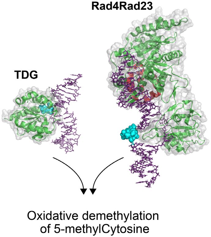
11232 Nucleic Acids Research, 2020, Vol. 48, No. 20

incision assay with cell extracts and closed circular DNA         suggesting that the binding was non-specific. Strikingly, an
substrates harboring a 5 S- or 5 R-cdA lesion to show that      earlier study had shown that both S- and R-cdA lesions
the cyPu residues are repaired by excision of a 26–28 bp          accumulate in Neil1−/− mice (32), suggesting the role of
DNA product. The excision was significantly suppressed            NEIL1 in cyPu repair, although the exact mechanism is still
in the presence of an XPA antibody, indicating the depen-         unclear. Furthermore, this study went on to demonstrate
dence of repair on the NER pathway. Interestingly, the R          that S-cdG was repaired slightly better than S-cdA by NER
form of cdA was repaired more efficiently (∼2-fold) than          in human HeLa cell extracts and that the base complemen-
the S form, however, both diastereoisomers were relatively        tary to the lesion affected the efficiency of repair. They spec-
poor (∼40–150-fold) NER substrates as compared to the 1–          ulated that both base pairing and base stacking are impor-
3-intrastrand d(GpTpG)-cisplatin crosslink substrate. This        tant for the recognition of cyPu lesions in the DNA and that
study clearly shows that cyPu lesions are removed by NER          an abnormal Watson-Crick base pairing (e.g. S-cdG:dT)
in vitro, and have the ability to cause local helix distortions   acts as a better substrate for NER. NMR combined with
and block polymerases. Brooks et al performed a host reac-        molecular dynamics have proven highly successful in under-

                                                                                                                                      Downloaded from https://academic.oup.com/nar/article/48/20/11227/5917651 by guest on 19 January 2021
tivation assay (HCR) in Chinese hamster ovary (CHO) cells         standing the alterations in the conformation of the DNA
using a plasmid expressing the luciferase (Luc) gene that         helix induced by DNA lesions. It would be of interest to use
contained a cdA lesion on the transcribed strand (23). They       these techniques to determine the distortions formed on the
showed that a single cdA lesion dramatically decreased the        DNA by the cyPu lesions and investigate the interactions of
Luc gene expression, suggesting that cdA can act as a strong      NEIL1 and other NER recognition proteins with cyPu le-
block to transcription. They also found that repair of cdA        sions (33,34).
lesion on the plasmid was significantly reduced in XPG and           Recently, for the first time, all four cyPu lesions (5 R-cdA,
ERCC1 mutant cells as compared to wildtype. They further          5 S-cdA, 5 R-cdG, 5 S-cdG) were examined in the same se-
confirmed this result by employing the HCR assay in SV-40         quence context by Shafirovich’s group and the NER effi-
transformed normal (GM00637) and XP-A (XP12BE) cell               ciencies were measured by excision assays using HeLa cell
lines, providing evidence for defective repair of cdA in the      extracts (35). In agreement with the study mentioned ear-
XP-A cells. The XP12BE cell line was derived from a XP-A          lier (22), 5 R-cyPu were repaired more efficiently than 5 S-
patient (XP2OS) who exhibited severe neurological symp-           cyPu. Molecular dynamics (100ns) revealed greater DNA
toms (24). Therefore, these studies provide a strong correla-     backbone distortions and diminished base stacking in the
tion between defective NER and neurodegeneration in XP            R form of cyPu as compared to the S form. In cells, DNA
patients. In this scenario, an obvious prediction would be        lesions are embedded in the nucleosome which can hin-
that NER deficient XP patients would have elevated levels         der the accessibility of some repair proteins (36–46). There-
of cyPu lesions in their DNA. Indeed, cyPu lesions have           fore, to explore the effect of CyPu on histone-DNA inter-
been shown to accumulate over age in the brain tissue of          actions, Shafirovich et al. embedded cyPu lesions in an ‘In’
Xpa−/− and Csb−/− mice (25,26), although unlike humans,           (fourth nucleotide on the 5 -side of dyad) and ‘Out’ (eighth
Xpa−/− mice do not display any neurological abnormali-            nucleotide facing the aqueous solution environment) ro-
ties (27,28). In another study, D’Errico et al used X-rays        tational setting near the dyad axis in nucleosomes recon-
to introduce base lesions in the DNA of normal and XP-C           stituted with either recombinant histones or histones ex-
keratinocytes and measured the accumulation of cyPu by            tracted from HeLa cells (47). In both cases, they made the
HPLC/MS. While both normal and XP-C cells accumu-                 surprising discovery that cyPu lesions were completely re-
lated equal numbers of cyPu lesions after 5Gy of X-rays,          sistant to excision by NER proteins in human cell extracts.
XP-C cells were inefficient in the removal of damage over         This suggests that even though cyPu lesions cause signifi-
time (29). Similar accumulation of cdA was also observed in       cant distortions to naked DNA duplex, they either do not
CSA deficient (CS-A) fibroblasts treated with X-rays (30).        significantly disturb the DNA-histone interactions at these
This is consistent with it being a strong transcription block-    specific positions or these lesions when embedded in a nu-
ing lesion as CSA and CSB both recognize DNA damage               cleosome escape detection by NER proteins. It also remains
in the context of transcription. Further studies are required     unknown whether these results are relevant in physiological
in more XP and CS patients with neurological disorders            conditions in vivo. It would be, therefore, of great interest
to establish a direct link between cyPu accumulation and          to extend these studies to living cells, although there are no
XP neurodegeneration. Despite the low prevalence of XP            tools available yet that could be used to specifically intro-
throughout the world, it would be of interest to set-up a         duce cyPu lesions in cells.
rapid autopsy program to measure cyPu lesions in brain tis-          As mentioned earlier, NER proteins recognize and repair
sues from deceased XP patients.                                   UV-induced photoproducts, 6–4 PP and CPD. While a 6–4
   Whether cdG lesions could be processed by DNA glyco-           PP causes a significant distortion in nucleosomal DNA (48),
sylases was investigated by Pande et al. (31). In this study,     a CPD causes less of a distortion (49). It is well-established
seven purified glycosylases (bacterial: Fpg, EndoIII, En-         that while XPC-RAD23B can recognize 6–4PP, this het-
doV, EndoVIII; human: NEIL1, NEIL2, and OGG1) failed              erodimer has limited ability to detect CPDs, (50) and in
to incise a 12 or 36 bp duplex containing a S-cdA or a S-         cells recruitment of XPC to sites of CPD in chromatin re-
cdG lesion. Even at high concentrations of the enzymes (200       quires UV-DDB. Furthermore, it is interesting to note that
fmol), no cleavage activity was observed. They also per-          XPC does not efficiently bind DNA configured on a nucleo-
formed binding assays with the glycosylases and found that        some (51). On the other hand, UV-DDB has been shown to
at very high concentrations (10–20 pmol), NEIL1 bound to          bind lesions directly in the nucleosome and even shift the
both cdA and cdG substrates as well as undamaged DNA,             nucleosomal register to provide access to more occluded
Nucleic Acids Research, 2020, Vol. 48, No. 20 11233
sites (49,52). Moreover, the binding of UV-DDB seems to
precede the activity of ATP-mediated chromatin remodel-
ers (53,54). More recently, as discussed in the last section,
our group has demonstrated that the oxidative base dam-
age 8-oxoG, which only causes a mild helix distortion, is
recognized by UV-DDB in naked duplex DNA, as well as
in living cells (55,56). The relatively mild nucleosome dis-
tortion caused by CPD and 8-oxoG is analogous to cyPu.
Therefore, it would be interesting to determine the full sub-
strate repertoire of UV-DDB and if UV-DDB is capable of
recognizing CyPu and other lesions in the context of nucle-
osomes.
Downloaded from https://academic.oup.com/nar/article/48/20/11227/5917651 by guest on 19 January 2021
OXIDATIVE REMOVAL OF 5MC MOIETIES IS STIMU-
LATED BY XPC
Figure 4. XPC and TDG in oxidative demethylation of 5-methylcytosine.
5-methylcytosine (5mC) is formed by the addition of a Based on the work by Ho et al. (60), XPC works to help turnover TDG,
methyl group to carbon 5 of cytosine through the action of which like other glycosylases, is product inhibited binding tightly to abasic
a DNA methyltransferase (57). As previously mentioned, sites. Shown here is the structure of the yeast, XPC homolog, Rad4 (green)
during an active enzymatic demethylation process, 5mC is Rad23 (red) bound to a DNA duplex (purple) containing a 6–4 photo-
oxidized by TET dioxygenases to 5hmC, 5fC, and 5caC. product (blue space-filled), PDB: 6CFI; Human TDG (green) bound to
a DNA duplex (purple) containing 2 -fluoro-2 -deoxyuridine (blue space-
The latter two lesions are removed by TDG. 5fC, and 5caC filled), PDB:3UFJ (122).
are some of the more common oxidative lesions with steady
state levels at 20 and 3 lesions per million bases, respectively
(Table 1). TDG has been shown to also remove deaminated the authors demonstrated longer retention of TDG on the
5mC (T:G) moieties from DNA (58). DNA. These data support the role of XPC in stimulating
TDG is a monofunctional glycosylase, which as previ- TDG activity by facilitating turnover of TDG from the
ously mentioned, binds avidly to abasic sites and thus be- abasic DNA product, (Figure 4). Lastly, the authors de-
comes product inhibited. Studies have demonstrated roles termined that TDG stimulation by XPC occurs through
for other BER proteins, including NEIL1 and APE1 in fa- interactions between the N-terminus of TDG and the C-
cilitating TDG turnover. However, the mechanism of DNA terminus of XPC. This study only looked at the role of XPC
demethylation by TDG in cells remains unclear (59). To this in TDG stimulation, it would be interesting to determine if
end, Ho et al investigated the role of XPC in epigenetic gene any other NER or BER proteins are recruited to the 5mC
regulation through stimulation of TDG. Using an ELISA moiety in response to XPC stimulation. Finally, it should
specific to 5mC, they were able to show XPC-dependent be pointed out that XP-C patients and Xpc-/- mice develop
DNA demethylation (60). While these authors reported that normally and do not appear to have large defect in epige-
XPC plays a role in DNA demethylation, they argued based netic programing during cellular differentiation. Thus, fur-
on a W690S variant of XPC that this activity was indepen- ther work is needed on the role of XPC, and NER proteins
dent of XPC’s role in NER. This W690S XPC variant, dis- in the oxidative removal of 5mC.
covered in an XP-C patient, XP13PV, was previously shown
to have reduced stability in cells, and cells expressing this
NER PROTEINS HELP MEDIATE THE REMOVAL OF
variant showed reduced rates of removal of UV-induced
THYMINE GLYCOL AND 8-OXOG
photoproducts (61). However, careful analysis of data pre-
sented in the Ho et al. study indicates that the W609S Guanine oxidation is a well-characterized DNA lesion.
XPC variant showed reduced stimulation, ∼20% increase of ROS acting on guanine results in the formation 8-oxoG,
TDG excision compared to TDG alone, of either 5fC:G or through two subtle modifications on guanine (Figure 1): the
5caC:G. In comparison, WT XPC fully doubled the activity addition of an oxo group on carbon 8 and the addition of
of TDG. These data suggest that reduced damage recogni- hydrogen to the seventh position nitrogen (62). These mod-
tion and/or DNA binding of this W690S XPC variant pre- ifications causes the base to rotate from the anti- to the
vented its stimulation of TDG. To provide further support syn-conformation with respect to the deoxyribose moiety
for the role of XPC in TDG stimulation, the authors per- around the glycosidic bond causing 8-oxoG to pair with A
formed a ChIP-seq analysis and determined co-enrichment during replication creating T:A transversions, if left unre-
of TDG and the XPC subunit, RAD23B at the promoter paired. The formation of 8-oxoG lesions in cells is estimated
region of embryonic stem cells. Additionally, using MeDIP- to occur up to 10 000 times per cell per day in humans,
seq to measure 5mC levels globally in the genome, the au- with the estimated steady-state levels of about 1–2 8-oxoG
thors were able to show reduced DNA methylation in cells lesions/106 guanines (63,64) (Table 1). While older litera-
overexpressing WT XPC. Single-particle tracking experi- ture has referred to this lesion as 8-hydroxy-guanine, this
ments utilizing Halo-tagged TDG and SNAP-tagged XPC, tautomeric form at physiological pH (7.4) is a minor prod-
revealed that overexpression of XPC led to a reduction uct (65). This relatively high lesion frequency of 8-oxoG
in the length of time Halo-tagged TDG remained bound (Table 1) coupled with the implications in genome instabil-
to DNA. Using shRNA to knockdown XPC expression ity emphasize the need for repair pathways dedicated to the11234 Nucleic Acids Research, 2020, Vol. 48, No. 20
removal of the 8-oxoG lesion. As mentioned earlier, 8-oxoG ence of XPG. In a later study of oxidative damage repair
is commonly removed through base excision repair (BER), in melanocytes by Wang et al. cells deficient in XPG pro-
through the actions of the DNA glycosylase OGG1. The tein were shown to have decreased repair of hydrogen per-
work by the Mitra laboratory in showing OGG1 is product oxide (H2 O2 )-mediated oxidative damage, when measured
inhibited and needs the actions of APE1 to facilitate OGG1 using a luciferase-based host cell reactivation assay (HCR)
turnover, imply the potential for other co-factors outside of (69). They were also able to show that cells with defective
BER to stimulate either OGG1 activity or processing of 8- XPA or XPC proteins showed reduced repair of oxidative
oxoG (7). damage. This study primarily focused on understanding ox-
The first implications of NER protein involvement in idative DNA damage repair capacity of melanocytes, as
oxidative DNA damage repair was shown using the Es- melanoma incidence is increased ∼1000-fold in XP patients
cherichia coli NER system consisting of the UvrABC com- (70). Wang et al. went on to further investigate the role of
plex (66). The authors used a DNA substrate containing a XPA in oxidative DNA damage repair and showed XPA
thymine glycol (Tg) lesion to show that UvrABC efficiently deficient cells had approximately a 4-fold reduction in ox-
Downloaded from https://academic.oup.com/nar/article/48/20/11227/5917651 by guest on 19 January 2021
recognizes and incises the lesion. This finding was recapit- idative damage repair capacity. While this study showed an
ulated in the mammalian system by Sancar and cowork- apparent involvement of NER proteins in the repair of ox-
ers (67). It has been estimated that the steady-state levels idative DNA damage, it remained unclear the specific roles
of Tg in mammalian cells are about two orders of magni- of XPA, XPC and XPG in this process. The authors hypoth-
tude lower than 8-oxoG adducts (Table 1). Using human esized in the case of melanocytes, the increased presence of
cell free extracts lacking any one of the XPA-XPG proteins, melanin which binds to DNA may inhibit the recognition of
they were able to show reduced excision of two common the damage by both BER and NER proteins. It would be in-
oxidative lesions, 8-oxoG and Tg, from damaged DNA sub- teresting to further investigate whether the levels of melanin
strate (67). The basis of this assay is that a DNA substrate is prevent recruitment of damage recognition proteins and in-
created by ligating a 5 32 -P end-labeled oligonucleotide con- hibit subsequent DNA repair.
taining an 8-oxoG or Tg moiety into a 139 bp DNA duplex. The Dogliotti lab provided the first evidence of an NER
Dual incisions by XPF/ERRC1 on the 5 side and XPG on protein, XPC, having a protective role against oxidative
the 3 side liberates excision products of ∼22–26 bases con- stress in human skin cells. Looking at keratinocytes and
taining the label. Additionally, they showed using a system fibroblasts with a nonsense mutation in the XPC protein,
of purified proteins the need for a complete NER system they were able to demonstrate an increased sensitivity to ox-
containing XPC-RAD23B, XPA, RPA, TFIIH (XPB and idizing damage, such as that from X-rays or potassium bro-
XPD), XPG and XPF-ERCC1, in order for proper exci- mate, through a colony formation assay (29). While X-rays
sion of 8-oxoG or Tg. This work from the Sancar labora- produce a wide spectrum of DNA lesions including vari-
tory clearly demonstrates the ability of purified NER pro- ous forms of base damage, single-strand and double-strand
teins to remove oxidative lesions, but did not assess whether breaks, potassium bromide produces primarily 8-oxoG and
there was any interaction between NER and BER proteins to a lesser extent other base damages (71). Additionally,
or whether NER is an important pathway for their removal D’Errico et al demonstrated using HPLC/MS an accumu-
in cells. The authors hypothesize the role of NER is to act as lation and subsequent delayed removal of 8-oxoG from
a slower alternative pathway for oxidative damage removal XPC-deficient skin cells. To strengthen support for the role
by BER, and the loss of this activity in XP patients, con- of XPC in oxidative DNA damage repair, they also showed
tributes to the accumulation of oxidative damage and sub- XPC-RAD23B-mediated stimulation of OGG1, the DNA
sequent neurodegeneration. It is important to point out the glycosylase responsible for 8-oxoG removal. They were un-
ability of the NER machinery to excise 8-oxoG had not been able to demonstrate XPA stimulation of OGG1, even at
confirmed in any other laboratory or through any other ap- high protein concentrations, even though previous studies
proaches, and it remains to be determined whether NER is alluded to a role for XPA in oxidative damage repair (67,69).
a back-up system for the removal of 8-oxoG in the absence Furthermore, through far western blot analysis the authors
of BER. demonstrated a direct binding between OGG1 and XPC-
Following this pioneering work by the Sancar laboratory, RAD23B, showing XPC enhances the ability of OGG1 to
which established an in vitro role of NER proteins in oxida- recognize 8-oxoG lesions. The authors did not show a direct
tive DNA damage repair, attention shifted to understand- interaction between XPC and damaged DNA, indicating its
ing the roles of specific NER proteins in the repair of 8- role is possibly to facilitate the turnover of OGG1. In a later
oxoG and other lesions induced by oxidative DNA dam- study by the Rainbow lab, XPC deficient fibroblasts were
age. Klungland et al began by characterizing the role of shown to have reduced removal of 8-oxoG (72). 8-oxoG was
XPG in BER of the oxidative lesions, thymine glycol and introduced onto the -galactosidase reporter gene via gen-
dihydrouracil. These lesions are excised by the bifunctional eration of a singlet oxygen by methylene blue plus visible
DNA glycosylase, NTH1. Using a reconstituted BER sys- light. Using this reporter gene, they conducted a host cell
tem containing hNTH1, APE1, pol , and XRCC1-DNA reactivation assay (HCR) to investigate the effect of XPC
ligase III, the Lindahl laboratory was able to show stimu- on DNA damage repair. Additionally, while the authors
lation of NTH1 by XPG (68). Specifically, they were able demonstrated pre-treatment of cells with UVC does not
to show enhanced binding of NTH1 to damaged DNA in change the relative repair rate of 8-oxoG in XPC-deficient
the presence of XPG. The authors also looked at the abil- cells, pre-treatment with UVC resulted in an approximate
ity of XPG to stimulate OGG1 excision of 8-oxoG but were 1.5-fold increase in overall repair of 8-oxoG. These data im-
unable to detect any enhanced OGG1 activity in the pres- ply a potential role of other proteins induced by UV damageNucleic Acids Research, 2020, Vol. 48, No. 20 11235
mediated through p53 stabilization induced gene expres- displayed a repair rate similar to that of the wild-type non-
sion, in the repair of oxidative damage. One such protein transcribed strand. Finally, they showed that in the absence
may be DDB2. of OGG1, this transcriptional effect of XPA and CSB was
Taken together these data suggested BER may not work lost, suggesting that RNAP is not inhibited by 8-oxoG, but
in isolation to remove oxidative damage. However, the spe- instead by the resulting SSB created by the actions of OGG1
cific molecular roles NER proteins may play in the repair and APE1. The protective role of XPA was further investi-
of oxidative damage remained unclear. To this end, Par- gated by the Yasui lab, which utilized the TATAM (tracing
lanti et al provided significant insights on the roles of NER DNA adducts in the targeted mutagenesis) system to study
proteins, specifically XPA, XPC, CSA, and CSB, on 8- 8-oxoG lesions into XPA knockout cells (76). Introduction
oxoG repair (73). First, using fibroblasts derived from NER of a single 8-oxoG lesion in cells deficient of XPA had no
deficient mice, they showed reduced repair and increased effect on mutagenesis. Tracks of ionizing radiation can cre-
accumulation of 8-oxoG in XPA−/− , XPC−/− , CSA−/− , ate closely spaced multiple lesions and it was interesting to
CSB−/− and OGG1−/− , knockout (KO) cells. Unsurpris- note that this group was able to show increased mutagenesis
Downloaded from https://academic.oup.com/nar/article/48/20/11227/5917651 by guest on 19 January 2021
ingly, the OGG1−/− KO cells showed the greatest reduction in XPA deficient cells with the introduction of multiple 8-
in repair and subsequent accumulation of 8-oxoG. How- oxoG lesions, specifically when the lesions were introduced
ever, all of the NER protein MEF KO cell lines also showed on the actively transcribed strand.
a noticeable reduction in the rates of 8-oxoG repair com- The Vermeulen lab developed an imaging system to study
pared to the WT cells with sufficient NER proteins. They the roles of NER proteins in 8-oxoG repair. They locally in-
also compared repair capacity in both the single and dou- duced 8-oxoG lesions via singlet oxygen by using a photo-
ble KO cell lines and were able to demonstrate a more sensitizer and a 405nm laser (38). Using this highly inno-
pronounced reduction in repair in the XPA−/− / CSB−/− , vative approach, the authors were able to show a difference
XPC−/− /CSB−/− , double KO cell lines, which resembled in the recruitment kinetics of CSB and XPC to damaged
the repair capacity of the OGG1 −/− cell line. Alternatively, sites, with CSB showing slightly enhanced recruitment over
the repair capacities of the XPA−/− and XPC−/− double XPC (t1/2 = ∼9 s for CSB versus ∼13 s for XPC). CSB and
KO cells resembled that of the single KO cells. These data XPC are involved in different sub-pathways of NER, offer-
suggest CSB and OGG1 are involved in the same repair ing an explanation for their different rates of recruitment to
pathway, one that may be different from that of XPC and damage sites. In support of a role of CSB in transcription-
XPA. However, the exact molecular details of these path- coupled repair of 8-oxoG, they were able to show enhanced
ways remain unresolved. This group were also able to reca- CSB recruitment to the transcriptionally active nucleolus,
pitulate the results from the mouse experiments in human while XPC was seen more in the heterochromatic nucleo-
XPA fibroblasts, by showing decreased repair of 8-oxoG in plasm, supporting its role in GG-NER. This study while
addition to increased sensitivity to oxidizing agents. Using demonstrating the recruitment of CSB and XPC to sites
siRNA targeting OGG1 in the XP12SV40 cell line, an XP- of oxidative damage, did not address whether these pro-
A deficient cell line they were able to show impaired repair teins are directly involved in the removal of 8-oxoG, either
of 8-oxoG, when compared to XP-A cells or cells with de- through DNA glycosylase stimulation or in some other step
ficient XPC. It remains unclear how the authors were able in BER. In a later study, these same authors looked at the
to demonstrate impaired repair of 8-oxoG in XPA deficient role of CSB in 8-oxoG repair by looking at OGG1 recruit-
cells in one study, but failed to show XPA-mediated stim- ment (77). Using the previously described photosensitizer
ulation of OGG1, the glycosylase mediating the repair of and laser strategy, they induced 8-oxoG lesions and mon-
the lesion, in another study (29). These conflicting results itored recruitment of CSB and OGG1 to the damage site
further reiterates the ambiguity of the role of XPA in 8- and demonstrated that CSB recruitment to damage sites
oxoG removal, and other published studies show contrast- is independent of OGG1. These data seem at odds with
ing results (74). The Spivak and Hanawalt laboratory de- the previous work by Spivak and Hanawalt who suggested
veloped a cutting-edge strategy to investigate the role of TCR of 8-oxoG can only occur after processing of the le-
XPA in repair of relatively low levels of oxidative dam- sion to a strand break. Furthermore, the Menoni et al.
age. Using a comet-FISH (combining single-cell gel elec- study (77) demonstrated CSB is able to stimulate XRCC1
trophoresis with a fluorescence in situ hybridization assay), recruitment to 8-oxoG, in transcriptionally active regions.
they were able to demonstrate roles for XPA and CSB in The authors hypothesize the role of CSB is to aid in the
the transcription-coupled repair of 8-oxoG (75). Using the recruitment of XRCC1 and other BER proteins by facil-
ATM gene, they fluorescently labeled the 5 and 3 ends us- itating chromatin remodeling to aid accessing lesions, es-
ing different probes and tracked the increasing distance be- pecially single-strand breaks. Additionally, it is important
tween the probes after treatment with potassium bromate, to note that APE1 has been shown to interact directly with
as a measure of BER-mediated single-strand break forma- CSB and is stimulated up to 6-fold in an ATP-independent
tion. In this way the authors were able to show the repair manner (78). Thus, CSB may provide an important role dur-
rates of CSB and XPA deficient cells were comparable to the ing transcription-coupled BER by orchestrating both back-
repair rates of the wild-type non-transcribed strand show- tracking of stalled RNAP, as well as coordinating down-
ing the repair of 8-oxoG is coupled to transcription. To fur- stream BER proteins. Finally, it was shown that CSB de-
ther support the idea of 8-oxoG processing to be coupled ficient cells are hypersensitive to killing by hydroxymethyl-
to transcription, the authors performed the comet-FISH deoxyuridine (hmdU) treatment, suggesting a direct role of
assay on cells deficient in UVSSA and RNAPII, key pro- CSB in SMUG1-mediated removal of this oxidized base
teins in TC-NER, and were able to show that these cells from DNA (78).11236 Nucleic Acids Research, 2020, Vol. 48, No. 20
NER AND BER WORK COOPERATIVELY TO REPAIR lesions, the same group studied relative binding affinities of
OXIDIZED 8-OXOGUANINE LESIONS XPC-RAD23 and NEIL for 147 bp duplexes containing Sp
or Gh lesions using EMSA analysis (84). They found that
The oxidation of guanine can create 2,2-diamino-4-[(2-
XPC-RAD23 bound to Sp containing substrate with high
deoxy--D-ery-pentofuranosyl) amino)-5(2H)-oxazolone
affinity (low nM range) and that XPC could effectively com-
(Oz), and 8-oxoG. The Oz lesion is normally processed by
pete away NEIL1 binding to these substrates at equal molar
BER in mammalian cells by the activities of NEIL1 and
concentrations. They then followed NEIL1 incision burst
NTH1 (79). However, using a defined system, Hanaoka
kinetics under single turnover conditions on both substrates
and coworkers showed that Oz is also a poor substrate for
in the absence and presence of XPC-RAD23 and found that
NER, and that the overall affinity of XPC-RAD23B to a
equal molar concentrations of added XPC-RAD23 greatly
DNA duplex containing Oz was significantly lower than a
reduced the amplitude of the burst phase of NEIL1 cleav-
6–4 PP containing duplex (80). The 8-oxoG lesion is several
age. Together these data strongly suggest that XPC-RAD23
orders of magnitude more sensitive to oxidation than the
and NEIL1 can directly compete for Sp and Gh lesions. Ad-
Downloaded from https://academic.oup.com/nar/article/48/20/11227/5917651 by guest on 19 January 2021
parent guanine moiety resulting in two oxidation products,
ditionally, it has been demonstrated NEIL2 and NEIL3 can
Sp and Gh, which are less common oxidative lesions,
act to remove Sp and Gh lesions (85,86). Further cellular
having steady state levels in the range of 0.01–0.07 lesions
work such as transfecting plasmids carrying these defined
per million bases (Table 1). These lesions can be removed
lesions into cells and using immunofluorescence to measure
by BER proteins, however work from several laboratories
the binding kinetics of these proteins will be necessary to
suggest that NER can also process these lesions. Early
better understand if XPC or other NER proteins can bind
studies with the bacterial NER UvrABC system indicated
to these lesions in the cell nucleus.
that this enzyme incised a duplex containing several lesions
to different extents, 8-oxoG:A the worst (10% completion)
A NEW ROLE OF UV-DDB IN THE REMOVAL OF 8-
with Gh (23%) and Sp (32%) lesions being moderate
OXOG
and amine modified Sp to 62% completion (81). These
results suggest that a larger and more distorting lesion is As previously mentioned, OGG1 is product-inhibited and
recognized and incised the most efficiently by the bacterial needs the activity of APE1 to turnover and work on other 8-
NER proteins. oxoG lesions (7). Another factor which can impair the activ-
Work from the Shavirovich and Geacintov laboratories ity of BER proteins is the inaccessibility of oxidative lesions
indicated that human NER proteins from cell extracts can in the context of chromatin. DNA glycosylases have been
perform dual incisions on Sp or Gh lesions embedded in shown to have impaired activity on damage when the lesion
a 135 bp duplex. XPA depletion studies with antibodies is contained within a nucleosome (42–46,87–90). It is im-
to XPA or extracts from XPC−/− fibroblasts failed to pro- portant to note, while certain glycosylases such as SMUG1
duce dual incisions (82). Adding back purified XPC to the are completely inhibited, others such as OGG1, AAG or
latter extract was able to restore NER activity. The same UDG can recognize outward facing lesions and can read-
study showed that NEIL1 can also process these lesions in ily initiate BER (88,90–92). In addition, both NEIL1 and
an extract and it was not clear if these two pathways work NTH1 have been shown to show reduced activity on Tg
synergistically or in an antagonistic manner. This question substrates embedded in nucleosomes (91,93,94). The issue
was elegantly explored in living cells by the same group in of lesion accessibility in the context of chromatin is an im-
which they transfected internally 32 P-labeled DNA hairpin portant factor in NER (36,37). UV-damaged DNA bind-
in which the label was placed near the lesion (83). By trans- ing protein (UV-DDB), a heterodimeric protein consisting
fecting this DNA duplex into human cells, they were able to of DDB1 and DDB2, has a demonstrated role in NER for
follow the excision of the oligonucleotide containing the le- damage recognition in the context of chromatin (95). In re-
sion via a NER pathway or direct incision by the activity of sponse to UV-induced damage, UV-DDB as part of a ubiq-
NEIL1 initiating BER. They found that compared to a sub- uitin E3 ligase complex with Cul4A and RBX, ubiquiti-
strate containing a benzo[a]pyrene-dG lesion, Sp or Gh le- nates histone H2A to facilitate chromatin remodeling to in-
sions were processed by NER 8- or 6-fold, respectively, less crease lesion accessibility (55). UV-DDB is known to recog-
efficiently. Transfection of the Gh substrate into XPA−/− nize and bind sites of UV damage, but has also been shown
cells failed to produce the characteristic excision product to bind more strongly to a short DNA duplex containing
by NER, whereas transfection into NEIL1−/− cells reduced an abasic site, as compared to a DNA substrate contain-
but did not eliminate the BER incision product. Taken to- ing a CPD, suggesting a possible role for UV-DDB in BER
gether these data suggested to the authors that the amount (96,97). In order to directly test this hypothesis, we recently
of XPC and XPA and perhaps their relatively low affinity investigated whether UV-DDB plays an important role in
for Sp and Gh substrates versus 5–10-fold higher levels of initiating the repair of 8-oxoG lesions (56). It should be
NEIL1 with high catalytic efficiency in the cell, probably noted that previous studies missed that UV-DDB was able
pushes repair of these substrates into a BER pathway. How- to discriminate between non-damaged DNA and DNA con-
ever, it is not clear whether these transfected DNA hairpins taining 8-oxoG (96). We have found that the presence of
were associated with nucleosomes and what role chromatin magnesium helps increase damage specificity by greatly de-
structure may play on the processing of Sp and Gh lesions creasing binding to non-damaged DNA duplexes (55,56).
by NER and BER pathways. Finally, to better explore the EMSA assays conducted in the presence of 5 mM Mg2+
question of relative affinities of XPC-RAD23B for Sp and showed that UV-DDB preferentially bound abasic sites,
Gh lesions and its ability to compete with NEIL1 for these CPD and 8-oxoG, with equilibrium dissociation constants,Nucleic Acids Research, 2020, Vol. 48, No. 20 11237
Kd , of 3.9, 30 and 160 nM, respectively, with high speci- the chromatin architecture at sites of damage, particularly
ficity as compared to undamaged DNA (Kd = 1108 nM) in inaccessible heterochromatic regions, and then stimulates
(55,56). Using an incision assay, with 8-oxoG and THF as the down-stream processing of the lesion. Future structural
substrates for OGG1 and APE1 specifically, we were able to and real-time imaging studies should focus on the mecha-
show that UV-DDB increased the incision activity of both nism of 8-oxoG recognition by DDB2 in reconstituted nu-
OGG1 and APE1 by 3- and 8-fold, respectively. We then uti- cleosomes and in mammalian cells. Future experiments also
lized single-molecule fluorescence microscopy of quantum need to address whether the DDB1-Cul4A E3 ligase is in-
dot (Qdot) labeled proteins and a unique DNA tightrope volved in 8-oxoG recognition.
optical platform to elucidate that UV-DDB increases the
turnover and thus the incision activity of both OGG1 and
CONCLUSION AND OUTLOOK
APE1. In this assay DNA containing an abasic site every 2
kb was suspended from poly-Lysine coated 5 micron beads Here we have illustrated the cooperativity, and in the case
(34) and Qdot labeled OGG1 or APE1 binding to DNA of Sp and Gh lesions, antagonism, between NER and BER
Downloaded from https://academic.oup.com/nar/article/48/20/11227/5917651 by guest on 19 January 2021
was followed over time in the absence and presence of UV- proteins during oxidative DNA damage repair, as well as
DDB. We demonstrated that UV-DDB helped dissociate instances where the oxidative lesions are repaired by NER
these proteins from the DNA in a concentration dependent proteins directly. The structure of the oxidative lesion in-
manner. Furthermore, by orthogonally labeling UV-DDB fluences the role of NER proteins in the oxidative damage
and OGG1 or APE1 with different colored Qdots, we were repair response. A comprehensive summary of the involve-
able to show UV-DDB can form transient complexes with ment of UV-DDB, XPC, CSA, CSB and XPG in the re-
OGG1 and APE1 to facilitate their removal from the le- moval of oxidative lesions, specifically 8-oxoG, is presented
sion site. Using an in vitro BER reaction, the activity of pol in Figure 6. Our lab was recently able to demonstrate early
 was measured though the incorporation of radiolabeled recruitment of UV-DDB to sites of 8-oxoG damage, preced-
dCTP into the gap created by the dual action of APE1 in- ing OGG1 recruitment (56), strengthening the argument for
cision and pol  dRPase activity. This assay revealed that interaction between NER and BER pathways. There are 11
addition of UV-DDB increased BER product formation by mammalian glycosylases which each recognize a range of
30-fold. Additionally, we were able to show that the newly lesions induced by oxidative damage (6). To this end, cur-
incorporated dCTP could be ligated into full length prod- rent work in our lab is focused on understanding the role of
uct, indicating that UV-DDB does not have an inhibitory UV-DDB in stimulation of the other mammalian glycosy-
effect on downstream steps in BER. Lastly, we used a highly lases beyond OGG1. We have preliminary data suggesting
innovative chemoptogenetic system to generate site specific MUTYH activity is stimulated by UV-DDB (56). Addition-
8-oxoG lesions at telomeres (Figure 5). This system consists ally, we are examining the role of UV-DDB in the recruit-
of a fluorogen activating peptide (FAP) that can bind to ment of downstream NER or BER proteins at oxidative le-
a malachite green dye (MG-2I) with high affinity and be sion sites to elucidate a role for UV-DDB in facilitating the
excited at a far-red light wavelength (660 nm). When ex- repair process. The evidence of interplay between the two
cited, the FAP-MG-2I combination generates singlet oxy- repair pathways could suggest direct interactions between
gen, which is highly reactive and short-lived and can oxi- BER and NER proteins, both on and off DNA, which
dize nearby macromolecules including proteins and DNA. needs further study by yeast-2-hybrid screens, co-IP and/or
When fused to a telomere binding protein (TRF1), the FAP- biochemical/biophysical methods, such as single-molecule
MG-2I plus light system generates targeted singlet oxygen in vitro techniques with a purified protein system or cell ex-
and oxidizes telomeric DNA to form almost exclusively 8- tracts to examine protein-protein and protein–DNA inter-
oxoG lesions. Using this FAP-TRF1 targeting system, we actions at a defined lesion site (99). Moreover, these tech-
found that UV-DDB was recruited to telomeric 8-oxoG le- niques can be used to examine, in real-time, how proteins
sions prior to OGG1, indicating a direct role for UV-DDB act in unison to faithfully process DNA lesions. With the
as an early damage sensor in chromatin, acting before the advancement of single-particle tracking in living cells and
initiation of BER (Figure 5). in situ labeling of proteins (39–41), and introduction of site-
Two independent studies have highlighted the role of the specific lesions (56), we are currently developing methods
DDB2 subunit in chromatin decompaction (53,54) and may to monitor the behavior of DNA repair proteins at damage
give insight into how DDB2 could help facilitate BER. In sites in different chromatin states in real time.
these studies, the authors used cell lines containing hete- Most glycosylases cannot efficiently process lesions em-
rochromatic lac operator (lacO) regions and found that the bedded in a nucleosome (42–46,87–90). Therefore, addi-
lacO array size significantly increased when bound with lac tional studies need to be conducted to further understand
repressor (lacR) tethered to DDB2 (DDB2-LacR) as com- the molecular mechanisms of lesion recognition, specifi-
pared to being bound with lacR alone. The increase in lacO cally in the context of nucleosomes, chromatin remodeling,
array size is consistent with significant chromatin unfolding. and glycosylase activity. Work from the Thoma laboratory,
They further showed a similar change in chromatin state strengthened the argument for a role for UV-DDB in nu-
triggered by DDB2 at sites of UV damage. Additionally, re- cleosome remodeling by demonstrating the ability of UV-
cruitment of DDB2 was independent of its E3 ligase activity DDB to modify the register of the DNA on the nucleo-
and ATP-driven chromatin remodeling. Based on these data some, increasing lesion accessibility (100). The direct role of
and the inefficiency of OGG1 to act on lesions embedded DDB2 in chromatin decompaction also provides a mecha-
in nucleosomes (98), we postulate UV-DDB facilitates the nism of how DDB2 could help facilitate BER (53,54). Thus,
recognition of 8-oxoG by BER proteins by first modifying DDB2 either acting alone or in conjunction with its asso-You can also read