COVID-19: Mechanisms of Vaccination and Immunity - MDPI
←
→
Page content transcription
If your browser does not render page correctly, please read the page content below
Review
COVID-19: Mechanisms of Vaccination
and Immunity
Daniel E. Speiser 1, *        and Martin F. Bachmann 2,3,4, *
 1    Department of Oncology, University Hospital and University of Lausanne, 1066 Lausanne, Switzerland
 2    International Immunology Centre, Anhui Agricultural University, Hefei 230036, China
 3    Department of Rheumatology, Immunology and Allergology, Inselspital, University of Bern,
      3010 Bern, Switzerland
 4    Department of BioMedical Research, University of Bern, 3008 Bern, Switzerland
 *    Correspondence: daniel.speiser@unil.ch (D.E.S.); martin.bachmann@me.com (M.F.B.)

 Received: 2 July 2020; Accepted: 20 July 2020; Published: 22 July 2020

 Abstract: Vaccines are needed to protect from SARS-CoV-2, the virus causing COVID-19. Vaccines
 that induce large quantities of high affinity virus-neutralizing antibodies may optimally prevent
 infection and avoid unfavorable effects. Vaccination trials require precise clinical management,
 complemented with detailed evaluation of safety and immune responses. Here, we review the pros
 and cons of available vaccine platforms and options to accelerate vaccine development towards
 the safe immunization of the world’s population against SARS-CoV-2. Favorable vaccines, used in
 well-designed vaccination strategies, may be critical for limiting harm and promoting trust and a
 long-term return to normal public life and economy.

 Keywords: SARS-CoV-2; COVID-19; nucleic acid tests; serology; vaccination; immunity

1. Introduction
      The COVID-19 pandemic holds great challenges for which the world is only partially prepared [1].
SARS-CoV-2 combines serious pathogenicity with high infectivity. The latter is enhanced by the fact
that asymptomatic and pre-symptomatic individuals can transmit the virus, in contrast to SARS-CoV-1
and MERS-CoV, which were transmitted by symptomatic patients and could be contained more
efficiently [2,3]. To limit the damage of COVID-19, primary efforts focus on confinement, with physical
distancing and multiple further measures preventing infection [4,5]. At present, many investigations
aim at defining optimal strategies to limit viral transmission while simultaneously permitting business
and social activities [6]. Scientific insights and understanding of the biological mechanisms of the
virus and its capability to spread are primordial. Built on this knowledge, the practical strategies may
have at least three priorities: firstly, to continue hygiene measures and physical distancing; secondly, to
maximize the viral monitoring, both geographically and in time, to focus viral containment locally and
limit transmission wherever and whenever possible; thirdly, to rapidly increase the immunity of the
world’s population.
      The diagnosis of SARS-CoV-2 infection is achieved through detection of viral RNA from a nasal
pharyngeal swab or saliva, by nucleic acid tests (NATs) or tests that detect viral protein antigens [5,7].
In infected individuals, the results are only positive for a relatively short time window, on average until
the 14th day after symptom onset [8]. Furthermore, a positive NAT result does not allow scientists
to conclude whether the affected person is or will become immune. Therefore, serological tests are
needed, as they can detect the various types of antibodies in the blood that persist for months or
even years.

Vaccines 2020, 8, 404; doi:10.3390/vaccines8030404                             www.mdpi.com/journal/vaccines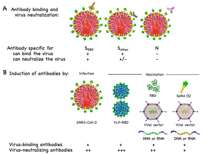
increase of new infections. Spontaneous disappearance of the virus is unlikely. Additional outbreaks
are expected when the safety measures are abandoned (Figure 1A). It may take significantly longer
than one year until the majority of the population acquire immunity through infection. As discussed
further below, it will be important to determine to which degree natural infection induces immunity
and for how long it can protect from re-infection.
The immune response to the SARS-CoV-2 involves innate immune activation and antigen-
Vaccines 2020, 8, 404 2 of 19
specific responses of B and T cells [10]. Protection from viral infection is mainly achieved by virus-
neutralizing antibodies, a principle that applies to the vast majority of viral infections to which
humans acquire robust immune protection due to infection or vaccination. It is urgent to develop
The world can probably not afford to let the majority of citizens undergo SARS-CoV-2 infection,
vaccines aiming at the induction of protective immune responses, primarily through virus-
as the overall burden would be
neutralizing antibodies enormous.
specific Current Although
for SARS-CoV-2. data indicate
at leastthat
1–2 ayears
pandemic COVID-19
are required to makeoutbreak
infects only low percentages
effective (usually
vaccines available in vaccination
globally, the singlemay digit range)
still of the
be the most population,
rapid at least
and economical in countries
strategy
to achievemeasures
that take effective widespreadagainst
immuneviral protection (Figure
spread [9]. 1A). So-called
To avoid “herd immunity”
pandemic is reached
propagation, the when
reproduction
a critical percentage of the population has become immune, leaving the virus only limited local
number Ro (viral transmission) must remain below 1, meaning that each infected person transmits on
chances to circulate. This may be the case when >90% of persons are immune. However, broad
1
simple meansagainst
measures a highly
viral unfavorable
spread will thenexponential
be sufficient toincrease of virus.
contain the new In infections.
case of future outbreaks of newly emerging microbes belonging to well-studied
Spontaneous disappearance of the virus is unlikely. Additional outbreaks are expected when the safety pathogen families,
measures previously
are abandonedestablished experience will accelerate the development and use of vaccines, allowing us
(Figure 1A). It may take significantly longer than one year until the majority
to reach herd immunity more rapidly (Figure 1B). Several organizations, including WHO, the
of the population acquire immunity through infection. As discussed further below, it will be important
Coalition for Epidemic Preparedness Innovations (CEPI) and the global Vaccine Alliance GAVI, are
to determine to which
increasing degreeefforts
preparation natural infection
for future induces
pandemics. Theimmunity and from
lessons learned for how long it will
SARS-CoV-2 can protect
from re-infection.
certainly support this development which, however, still remains very challenging [1].
Vaccines 2020, 8, x 3 of 21
Figure of
Figure 1. Model 1. immunity
Model of immunity
inducedinduced by infection
by infection and vaccination.
and vaccination. (A) The
(A) The world world
waswas poorly
poorly prepared
for the firstprepared
wave of for the first wave of SARS-CoV-2 spread (blue parts of the curve) [9]. Installed measures for
SARS-CoV-2 spread (blue parts of the curve) [9]. Installed measures for limiting
limiting viral spread (*) largely halted further infections (black). Subsequent relaxing of these
viral spread (*) largely halted further infections (black). Subsequent relaxing of these measures may
measures may lead to new waves of viral spreading. Even if this happens several times, it may take
lead to new waves ofmore
significantly viral
thanspreading. Even
one year until if this of
the majority happens several
individuals becometimes,
infecteditand
may take significantly
immune. Once
more thanavailable,
one yearvaccination
until the (green)
majoritywillofmore
individuals become
rapidly induce infected
immunity in aand immune.
critical Once
percentage available,
of the
vaccinationpopulation,
(green) willwhich
moreis necessary
rapidly for herd immunity.
induce immunityThe in model scenarios
a critical shown are
percentage based
of the on current which
population,
evidence and the assumption that infection and proper vaccines induce immune responses that often
is necessary for herd immunity. The model scenarios shown are based on current evidence and the
protect from (re-) infection. However, there is increasing evidence that immunity to SARS-CoV-2 may
assumption that infection and proper vaccines induce immune responses that often protect from (re-)
only be temporary. This notion is not considered for this Figure because the quantitative importance
infection. However, there is increasing
of waning immunity evidence
remains still unknown. that
(B)immunity to SARS-CoV-2
In case of the emergence of may only
a novel be temporary.
pathogen,
This notionvaccination
is not considered
may start for
muchthis Figure
earlier because
provided one the quantitative
is prepared, importance
i.e., has of waning
ample experience immunity
with this
remains stillfamily of pathogens,
unknown. (B) Inenabling
case ofrapid vaccine development
the emergence of a novelandpathogen,
production. vaccination
Unfortunately,maythis was
start much
not the case for SARS-CoV-2. Not shown is the most favored scenario in which the population is
earlier provided one is prepared, i.e., has ample experience with this family of pathogens, enabling
previously vaccinated against a given pathogen, precluding viral spread, which is fortunately the case
rapid vaccine development and production. Unfortunately, this was not the case for SARS-CoV-2. Not
for immunity to many childhood diseases. Additionally, not shown is the in-between scenario in
shown is the most
which favored
a vaccine scenario
is available butin which
larger partsthe population
of the population isarepreviously vaccinated
not (yet) vaccinated, whichagainst
can then a given
pathogen, be precluding viral spread, which is fortunately the case for immunity to many childhood
readily achieved.
diseases. Additionally, not shown is the in-between scenario in which a vaccine is available but larger
Here, we review the mechanistic understanding of immunity and vaccination against SARS-
parts of the population are not (yet) vaccinated, which can then be readily achieved.
CoV-2. We discuss vaccine target antigens and current vaccine candidates in preclinical and clinical
testing. We also focus on the development and validation of precision serology to support preclinical
and clinical evaluation of vaccine candidates and research on immune responses induced by natural
infection. The basic and applied knowledge acquired during the last few decades is highly useful to
design promising vaccines with increased likelihood of inducing protective immune responses and
avoiding adverse effects and harmful disease enhancement. Now, probably more than ever, the
world depends on rational effective strategies built on robust scientific evidence.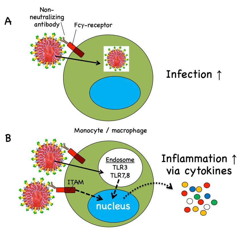
Vaccines 2020, 8, 404                                                                                3 of 19

     The immune response to the SARS-CoV-2 involves innate immune activation and antigen-specific
responses of B and T cells [10]. Protection from viral infection is mainly achieved by virus-neutralizing
antibodies, a principle that applies to the vast majority of viral infections to which humans acquire
robust immune protection due to infection or vaccination. It is urgent to develop vaccines aiming
at the induction of protective immune responses, primarily through virus-neutralizing antibodies
specific for SARS-CoV-2. Although at least 1–2 years are required to make effective vaccines available
globally, vaccination may still be the most rapid and economical strategy to achieve widespread
immune protection (Figure 1A). So-called “herd immunity” is reached when a critical percentage of
the population has become immune, leaving the virus only limited local chances to circulate. This may
be the case when >90% of persons are immune. However, broad immunization is already very helpful
as soon as “only” approximately 60–70% have become immune, because relatively simple measures
against viral spread will then be sufficient to contain the virus. In case of future outbreaks of newly
emerging microbes belonging to well-studied pathogen families, previously established experience
will accelerate the development and use of vaccines, allowing us to reach herd immunity more
rapidly (Figure 1B). Several organizations, including WHO, the Coalition for Epidemic Preparedness
Innovations (CEPI) and the global Vaccine Alliance GAVI, are increasing preparation efforts for future
pandemics. The lessons learned from SARS-CoV-2 will certainly support this development which,
however, still remains very challenging [1].
     Here, we review the mechanistic understanding of immunity and vaccination against SARS-CoV-2.
We discuss vaccine target antigens and current vaccine candidates in preclinical and clinical testing. We
also focus on the development and validation of precision serology to support preclinical and clinical
evaluation of vaccine candidates and research on immune responses induced by natural infection.
The basic and applied knowledge acquired during the last few decades is highly useful to design
promising vaccines with increased likelihood of inducing protective immune responses and avoiding
adverse effects and harmful disease enhancement. Now, probably more than ever, the world depends
on rational effective strategies built on robust scientific evidence.

2. Vaccine Development
      Vaccine development has started at a strongly accelerated pace already, shortly after the beginning
of the SARS-CoV-2 outbreak [11–13]. At the time of finalizing this review, there are already more than
twenty vaccines being tested in clinical trials. The WHO is publishing a regularly updated list of the
vaccines in development [14]. As a specialist in the advance of epidemic vaccines, CEPI has organized a
global consultation committee which helped to launch the COVID-19 Vaccine Development Taskforce,
focusing on vaccine manufacturing and financing, in collaboration with GAVI and the World Bank [15].
Very useful comments on COVID-19 vaccines are regularly published in the scientific literature [16–19].
The knowledge gained through previous coronavirus outbreaks provides a favorable scientific basis for
vaccine design (Box 1)—for example, by helping to identify potentially protective epitopes, the Achilles
heels of a virus. However, the fact that SARS-CoV-1 was spontaneously eliminated from the human
population, and MERS-CoV was largely controlled without the need for large-scale pharmaceutical
interventions, led to a drastic reduction in research funding during the last decade, severely limiting
further research and vaccine development. Therefore, there is currently only limited experience on
coronavirus vaccination; the first human coronavirus vaccine has yet to be approved.
      In view of the urgency of making vaccines available for billions of people, one must primarily
focus on vaccines that can be produced in massive amounts and for which the production knowhow
and facilities are available or can be built rapidly [20].
      Vaccines can be based on whole viruses (live-attenuated or inactivated), viral vectors, nanoparticles
or virus-like particles, subunit components, proteins/peptides, RNA, DNA or live cells. The first
vaccine trial against COVID-19 was started in China on February 15, 2020 (Table 1), using dendritic
cells that are genetically modified with structural and enzymatic proteins of SARS-CoV-2. A second
trial, also in China, was done with a similar vaccine, complemented by the infusion of antigen-specific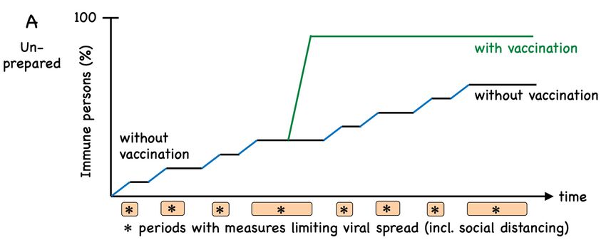
Vaccines 2020, 8, 404 4 of 19
T cells. While both of these vaccines are tested therapeutically in COVID-19 patients, most other
vaccines are tested in healthy volunteers. In the US, the first trial was launched in March 2020, using
lipid nanoparticle encapsulated mRNA encoding the spike (S) protein, sponsored by Moderna and the
National Institute of Health [21]. In early April 2020, a DNA vaccine trial was initiated with a plasmid
encoding the S protein, sponsored by Inovio Pharmaceuticals and CEPI. Since mid-April 2020, several
vaccines consisting of inactivated SARS-CoV-2 virus have been tested in China [22]. The first viral
vector COVID-19 vaccine was developed at the University of Oxford, UK. It is based on a chimpanzee
adenovirus and encodes the S protein [23], and it is now in phase 2/3 testing. A similar vaccine is based
on adenovirus-5. After promising results in phase 1 in Wuhan, China [24], this vaccine has also moved
forward, to a phase 2 trial (Table 1).
Table 1. SARS-CoV-2 vaccine candidates in clinical trials.
Platform, Route of Target Trial Phase, Registry Number, Study Start,
Vaccine Candidate Developer
Administration (SARS-Cov-2) Link
Synthetic minigene Artificial antigen presenting Selected conserved Shenzhen
Phase 1/2, NCT04299724, 15 February 2020
transfected APCs cells (APCs) modified with structural and protease Geno-immune Medical
http://szgimi.org/en/news.php
Covid-19/aAPC lentiviral vector, s.c.. protein domains Institute, China
Synthetic minigene Dendritic cells modified
Viral structural Shenzhen
transfected APCs + with lentiviral vector, s.c., Phase 1/2, NCT04276896, 24 March 2020
proteins and a Geno-immune Medical
cytotoxic T cells plus i.v. infusion of http://szgimi.org/en/news.php
polyprotein protease Institute, China
LV-SMENP-DC cytotoxic T cells
Phase 2, NCT04341389, 12 April 2020
Recombinant adenovirus, Viral vector, Adenovirus 5, CanSino Biologics,
Spike protein http://www.cansinotech.com/homes/article/
Ad5-nCoV i.m. China
plist/56.html
Viral vector
Phase 2b/3, 2020-001228-32, 4 May 2020
Recombinant adenovirus, (non-replicating) University of Oxford,
Spike protein https://www.ox.ac.uk/news-and-events/
AZD1222 Chimpanzee Adenovirus, UK, & AstraZeneca
for-journalists
i.m.
Recombinant adenovirus, Viral vector, Adenoviruses Gamaleya Research Phase 1, NCT04436471, 17 June 2020
Spike protein
Gam-COVID-Vac (Lyo) 5 and 26, i.m. Institute, Russia http://gamaleya.org/
Inovio Phase 1, NCT04336410, 3 April 2020, and
Plasmid, DNA, i.d., followed by
Spike protein Pharmaceuticals USA, Phase 2, https://www.inovio.com/our-focus-
INO-4800 electroporation
& CEPI serving-patients/covid-19/
Plasmid + adjuvant, AnGes and Osaka Phase 1/2, NCT04463472, 29 June 2020
DNA, i.m. Spike protein
AG0301-COVID19 University, Japan https://www.anges.co.jp/en/
Plasmid, Phase 1/2, NCT04445389, 17 June 2020
DNA, i.m. Spike protein Genexin Inc., Korea
GX-19 http://www.genexine.com/m62.php?cate=1
Phase 2, NCT04405076, 25 May 2020
Lipid nanoparticle Moderna and Natl Inst
https:
encapsulated RNA, mRNA, i.m. Spike protein Allergy & Infectious
//www.niaid.nih.gov/clinical-trials/safety-
mRNA 1273 Diseases (NIAID), USA
immunogenicity-study-vaccine-covid-19
Lipid nanoparticle
Various viral ags (4 BioNTech, Germany, & Phase 1/2, NCT04368728, 29 April 2020
encapsulated RNA, mRNA, i.m.
vaccine candidates) Pfizer, USA https://investors.biontech.de/press-releases
BNT162
Lipid nanoparticle
Phase 1, NCT04449276, 18 June 2020
encapsulated RNA. mRNA, i.m. Spike protein CureVac, Germany
https://www.curevac.com/covid-19
CVnCoV
COVAC1 mRNA in lipid Imperial College Phase 1, ISRCTN17072692, 1 April 2020
Spike protein
(LNP-nCoVsaRNA) nanoparticle, i.m. London, UK http://www.imperial.ac.uk/news
Protein + adjuvant, Protein subunit vaccine, Spike protein and Phase 1/2, NCT04368988, 25 May 2020
Novavax, USA
NVX-CoV2373 i.m. Matrix-M adjuvant http://ir.novavax.com/press-releases
Clover Biopharma,
Protein + adjuvant, Protein trimeric subunit Spike protein, AS03, Phase 1, NCT04405908, 19 June 2020
Australia, GSK,
SCB-2019 vaccine, i.m. CpG, alum adjuvant http://www.cloverbiopharma.com/
Dynavax
Sinovac Research and Phase 1/2, 16 April 2020, and Phase 3
SARS-CoV-2 inactivated Inactivated virus + alum
Entire virus Development Co, http://www.sinovacbio.com/?optionid=
virus, PiCoVacc adjuvant
China 754&auto_id=904
SARS-CoV-2 inactivated Chinese Academy of Phase 1/2, NCT04412538, 15 May 2020
Inactivated virus Entire virus
virus Medical Sciences http://english.cas.cn/newsroom/news/
SARS-CoV-2 inactivated Phase 1/2, ChiCTR2000031809, 11 April 2020
Inactivated virus Entire virus Sinopharm
virus http://www.chinacdc.cn/en/
Vaccines 2020, 8, 404                                                                                         5 of 19

                              Box 1. Vaccines against SARS-CoV-1 and MERS-CoV.

     Currently there is no vaccine available against these two viral threats [25,26]. Nevertheless, several vaccine
 candidates against SARS-CoV-1 have been developed based on VLPs, DNA, proteins and viruses (inactivated,
 live-attenuated, recombinant vectors) [27,28]. While the majority were characterized preclinically, only a few were
 tested in phase 1 clinical studies [29,30]. Against MERS-CoV, vaccines have been developed based on inactivated
 virus, DNA and protein, generating preclinical data [31] and phase 1 trial results [32]. For both diseases, larger
 studies to determine whether the vaccines could protect from natural infection have not been performed.

      A study of non-human primates vaccinated with an inactivated virus showed high levels of
neutralizing antibodies, high levels of protection and no unfavorable disease enhancement [33].
In the past, some inactivated coronavirus vaccines have been shown to increase disease severity in
animals [34], as outlined below in the section on disease enhancement. Attention must be paid to the
high mutation rate (e.g., in the S1/S2 junction) which occurs in vitro during the production process
of inactivated viruses, requiring careful selection of appropriate vaccine strains. Attenuated viruses
may be promising, based on the long history of delivering successful vaccines [35,36] and novel
genetic techniques increasing the likelihood to create better vaccine strains [37]. Nevertheless, it takes
considerable time to identify strains with the right balance between sufficient attenuation and induction
of adequate immune responses. Furthermore, it will be highly demanding to produce sufficient
amounts of biosafety level-3 viruses to meet the high global needs. An attractive possibility may be to
use inactivated attenuated viruses, as the latter may be more easily grown than the wild-type virus.
      Nanoparticles and virus-like particles (VLPs) have delivered successful vaccines. They can be
engineered to display epitopes of foreign viruses on their surface, rendering those epitopes highly
immunogenic. Molecules that stimulate innate immunity can be encapsulated within VLPs to enhance
immune responses and trigger favorable T helper type 1 (Th1) polarized immune responses (type 1
immunity) rather than potentially disease enhancing Th2 polarization [38].
      The clinical trials that are now urgently needed to evaluate the COVID-19 vaccine candidates
serve to determine optimal vaccine dose and scheduling and whether multiple booster vaccinations
are required. Usually, more robust and long-term immunity can be induced by sequential vaccinations,
possibly necessary for those with expected weak immune responses, such as in elderly or immune
deficient individuals [39]. The clinical studies should also reveal whether the vaccine candidates induce
unwanted adverse effects such as local skin toxicity or fever and flu-like symptoms. Autoimmune
reactions may also occur, possibly with hematological or neurological manifestations [40]. Fortunately,
for most vaccines, severe unwanted adverse effects are very rare. Nevertheless, all safety issues must
be carefully studied before a vaccine is used widely.
      Table 1. Anti-SARS-CoV-2 vaccines in clinical evaluation, registered at clinicaltrials.gov,
clinicaltrialsregister.eu and/or chictr.org.cn. At present, 24 and 142 vaccine candidates are in clinical
and preclinical evaluation, respectively [14].
      The time required for vaccines reaching the wider public also highly depends on regulatory
authorities and their flexibility to accelerate the process and vaccine approval, as compared to standard
procedures established for less urgent health threats. The WHO roadmap [41] provides corresponding
guidelines and support for regulation and ethics and the use of platforms for developing vaccines and
therapeutics in the most efficient ways. Furthermore, properly designed clinical trials may greatly
reduce the time for clinical development—for example, by testing several vaccines simultaneously
in adaptive trials with a low number of shared control groups [11], requiring unusual cooperativity.
Ultimately, however, the most efficient and least toxic vaccines will succeed, even if their development
and production take longer. For more details on the production, distribution and safety/efficacy
assessment of the different vaccine platforms, we refer to the links in Table 1 and to the existing
literature [20,42–45].
Vaccines 2020, 8, 404 6 of 19
3. Vaccine Antigens
3.1. B Cell/Antibody Targets
Protection induced by currently available vaccines against viruses is primarily based on
virus-neutralizing antibodies. Such antibodies usually block the interaction of the virus with its
cellular receptor or prevent conformational changes required for fusion of the virus with the cell
membrane. The SARS-CoV-1 virus has been studied in substantial detail (Box 2). Recent investigations
have shown that the new SARS-CoV-2 virus uses a similar strategy for cell entry [46]. Attachment
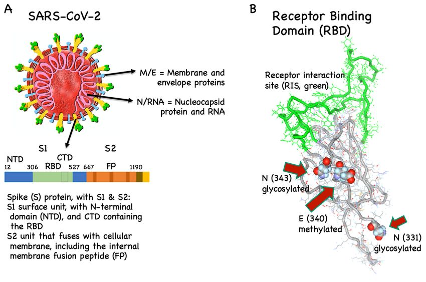
to host cells takes place via binding of the viral S protein (Figure 2A) to the angiotensin-converting
enzyme 2 (ACE2), the viral receptor on host cells. Subsequently, the S protein is primed by host cell
proteases, by furin and the serine proteases TMPRSS2 and TMPRSS4, enabling the fusion of viral and
cellular membranes and the consequent entry of viral RNA into the host cell [47].
Vaccines 2020, 8, x 8 of 21
2. SARS-CoV-2,
FigureFigure the spike
2. SARS-CoV-2, the (S) protein
spike and its receptor
(S) protein and its binding
receptor domain
binding (RBD).
domain(A) Coronaviruses
(RBD). (A)
have their name because
Coronaviruses theyname
have their are decorated
because theyby prominent
are decorated S by
proteins (yellow/green).
prominent It is the only viral
S proteins (yellow/green).
proteinIt that
is theinteracts
only viral protein
with hostthat
cellsinteracts withmost
and is the host diverging
cells and isprotein
the most diverging
between proteincoronaviruses,
different between
different
particularly coronaviruses,
in its particularly
receptor binding domain in (RBD,
its receptor
green).binding domain
RBD binds (RBD, green).converting
to angiotensin RBD bindsenzyme
to 2
angiotensin converting enzyme 2 (ACE2, not shown) on the host’s cell surface. The
(ACE2, not shown) on the host’s cell surface. The fusion peptide (FP) fuses with the host cell membrane. fusion peptide
(FP) fuses with the host cell membrane. Specific antibodies against RBD and FP can neutralize SARS-
Specific antibodies against RBD and FP can neutralize SARS-CoV-2 NTD/CTD, N-/C-terminal domains.
CoV-2 NTD/CTD, N-/C-terminal domains. (B) RBD is glycosylated and methylated, which may
(B) RBD is glycosylated and methylated, which may hinder the induction of neutralizing antibodies.
hinder the induction of neutralizing antibodies. In contrast, the receptor interaction site (RIS, green)
In contrast, the receptor interaction site (RIS, green) is not glycosylated.
is not glycosylated.
3.2. T Cell Targets
Box 2. The complexity of the SARS-CoV-1 family and antibody specificities.
CD4 and CD8 T cells recognize and react to SARS-CoV-2 antigens [64], contributing to immune
The family ofparticularly
protection, SARS-CoV-1byviruses
reducing consists ofseverity
disease several groups of strains
[65,66]. To (comparable
some extent, this may toalso
manybe other viruses)
the case
hostedfor
bycross-reactive
animals, humans or both [25]. The antibodies are specific for the spike (S), membrane
T cells induced by seasonal coronaviruses [67]. For disease prevention, T cells alone
(M), envelope
(E), nucleocapsid (N) and further viral proteins. Many of them are strain- or group-specific and thus recognize
are probably less potent than neutralizing antibodies [68]. Preventive anti-viral vaccines are
only some but not all SARS-CoV-1 viruses. Antibodies may be neutralizing, although the majority are not. The
successful because they induce antibodies that neutralize viral particles in the extracellular space,
neutralizing antibodies are mainly specific for S protein [48–50] and, to a minor extent, also 3a protein [51].
immediately
Neutralizing afterepitopes
antibody body entry
have and before
been viruses
found to be infect
highlythe host’s cells.
conserved Importantly,
in several B cell responses
viral strains, indicating that
and antibody production are strongly promoted by CD4 T helper cells. Therefore, vaccines
vaccines that elicit such antibodies are protective against multiple strains. These epitopes map primarily should to the
simultaneously
receptor binding domaininduce bothofBthe
(RBD) cells and T cells.
S protein.
T cell antigens must be presented by HLA molecules on the surface of antigen-presenting cells
and infected cells. As HLA molecules differ in most people due to the huge genetic HLA
polymorphism, viral recognition by T cells is based on a very large diversity of antigenic
peptide/HLA complexes. Each person has her/his own T cell specificities for those antigenic peptides
that bind to her/his HLA molecules. Vaccines containing short antigenic peptides or mini-genes will
not work for most people, i.e., for those whose HLA molecules cannot present the respective antigenic
peptides. In contrast, vaccination with long peptides or full-length viral proteins or corresponding
DNA/RNA are potentially useful for many or all individuals.Vaccines 2020, 8, 404 7 of 19
S protein interaction with ACE2 is well described for both SARS-CoV-1 and -2 and relies on a
particular domain within the S protein, the so-called receptor binding domain (RBD). Indeed, most
antibodies capable of neutralizing coronaviruses are directed against RBD [50,52] (Box 2). Hence,
the primary immune mechanism of avoiding infection is through blocking viral attachment to ACE2.
Therefore, generating a vaccine inducing antibodies against RBD is the strategy used by the majority of
COVID-19 vaccine candidates [53]. It has recently been shown that RBD is glycosylated and methylated.
Generally, such posttranslational modifications are difficult to reproduce in vaccines, meaning that
vaccines may display (slightly) different epitopes than the virus. Consequently, the antibodies induced
by the vaccines may potentially be cross-reactive and non-protective. Interestingly, however, the
receptor interaction site (RIS) directly binding to ACE2 is not glycosylated, indicating that this RIS may
potentially be an ideal vaccine candidate [54,55] (Figure 2B).
The second most frequent choice is to use the whole S1 subunit. Other vaccine manufacturers use
the full-length S protein [32] and/or the fusion peptide (FP), which is part of the S2 unit and fuses with
the cell membrane and therefore also has neutralizing epitopes [56,57]. The latter is not the case for the
N-terminal domain (NTD) of the S protein and the membrane (M), envelope (E) and nucleocapsid (N)
proteins, all of which are not directly targeted by the current vaccine candidates [58], also because of
the risk of inducing disease enhancing antibodies.
Vaccine antigens may be used in the form of protein or peptides. A recent study has shown that a
SARS-CoV-2 S1-Fc fusion protein readily induced neutralizing antibodies in non-human primates [59].
Proteins and peptides may be rendered more immunogenic by formulating them with strong adjuvants.
Another strategy is to display vaccine antigens on VLPs, which are often highly immunogenic. Zha et
al. have shown that RBD-VLPs efficiently induced SARS-CoV-2-neutralizing antibodies in mice [60].
Further options are to insert RBD into viral vectors or DNA or RNA. A potential challenge is that the
induction of neutralizing antibodies depends on antigen display in the correct conformation, which is
not guaranteed when a protein or peptide is expressed and displayed in isolation at the site of injection.
This may be easier to achieve with the full S protein. However, S protein vaccination may induce
non-wanted antibodies in addition to the neutralizing ones directed against RBD [61]. Therefore,
provided that one succeeds in constructing a vaccine displaying RBD or even only the RIS in the proper
conformation, RBD or RIS may be preferable to the whole S protein.
The most obvious isotype to be induced by a COVID-19 vaccine is IgG, preferably the more
protective IgG1 and IgG3 subclasses. However, IgA may also be of importance to reduce infection of
mucosa and epithelial cells in the respiratory tract, as well as endothelial cells, which may be widely
targeted by the virus. While mucosal immunization at a large scale in a rapid fashion might be difficult,
the use of an adjuvant that triggers the production of IgA might be an important consideration. TLR7/8
and TLR9 ligands are good candidates as they potently promote IgA responses [62,63].
3.2. T Cell Targets
CD4 and CD8 T cells recognize and react to SARS-CoV-2 antigens [64], contributing to immune
protection, particularly by reducing disease severity [65,66]. To some extent, this may also be the case
for cross-reactive T cells induced by seasonal coronaviruses [67]. For disease prevention, T cells alone
are probably less potent than neutralizing antibodies [68]. Preventive anti-viral vaccines are successful
because they induce antibodies that neutralize viral particles in the extracellular space, immediately
after body entry and before viruses infect the host’s cells. Importantly, B cell responses and antibody
production are strongly promoted by CD4 T helper cells. Therefore, vaccines should simultaneously
induce both B cells and T cells.
T cell antigens must be presented by HLA molecules on the surface of antigen-presenting cells and
infected cells. As HLA molecules differ in most people due to the huge genetic HLA polymorphism,
viral recognition by T cells is based on a very large diversity of antigenic peptide/HLA complexes.
Each person has her/his own T cell specificities for those antigenic peptides that bind to her/his HLA
molecules. Vaccines containing short antigenic peptides or mini-genes will not work for most people,Vaccines 2020, 8, 404 8 of 19
i.e., for those whose HLA molecules cannot present the respective antigenic peptides. In contrast,
vaccination with long peptides or full-length viral proteins or corresponding DNA/RNA are potentially
useful for many or all individuals.
CD8 cytotoxic T cells primarily recognize viral peptides that are synthesized within the infected
cell. In contrast, protein antigens that are picked up from the extracellular space are poorly presented
to CD8 T cells. The one exception to this rule is in small fractions of dendritic cells that are capable
of so-called cross-presentation, i.e., the cellular uptake of extracellular protein (especially particulate
antigens) and the presentation of processed peptides on HLA class I molecules to CD8 T cells [69].
Cross-presentation is rather slow and often rate limiting, which is a major reason why full-length
protein vaccines are inefficient for inducing CD8 T cell responses.
As compared to antibody induction, it is generally more challenging to induce long-term T
cell responses through vaccination. Most vaccines are either attenuated pathogens or more often
dead/synthetic vaccines which do not replicate in vivo. Yet, substantial microbial replication in vivo is
usually required for the induction of strong T cell responses [70]. It is therefore particularly difficult to
induce durable CD8 T cells by currently available vaccine technologies. For preventive vaccination,
this might not be a major problem, since CD8 T cells are not specialized to prevent infections. Rather,
CD8 T cells are important once host cells are infected. Therefore, these cells have their primary role in
individuals with established infection.
The induction of CD4 T cell help is often not rate limiting in vaccination, probably because
low numbers of these cells are already sufficient for supporting antibody production. Nevertheless,
vaccination may fail due to CD4 T cell non-responsiveness. Since T cell help can be provided by CD4
T cells with other specificities by intermolecular help, a smart approach is to supplement vaccines
by inserting microbial antigens to which most humans are already immunized [71]. The consequent
immune response will be stronger because boosting previously primed and established CD4 T cells is
more efficient than priming.
4. Disease Enhancement
4.1. T Cell-Dependent Disease Enhancement
It is not recommended to vaccinate for T cell responses without also efficiently inducing neutralizing
antibodies, because the latter are likely the crucial key effectors, and also because T cells, particularly
CD8 T cells, can cause extended tissue damage through their cytotoxicity against infected cells,
which is likely increased in the absence of antibodies that neutralize the viruses in the extracellular
space. Indeed, pure CD8 T cell responses induced by vaccination may enhance potentially lethal
immunopathology [72].
A concrete safety concern is the potential activation of Th2 cells. This was first observed in
a clinically tested respiratory syncytial virus (RSV) vaccine, which consisted of inactivated virus
and worsened clinical symptoms, with the deaths of two children upon infection with RSV. Disease
enhancement was caused by Th2 cell-mediated eosinophilia, a problem associated with vaccines
for respiratory vaccines. It is therefore essential to skew the response by vaccination towards Th1
polarization. Indeed, combining inactivated RSV with TLR-agonists reduces lung pathology upon
viral challenge in murine models [73]. Experience from SARS and MERS vaccine candidates indicates
that this risk exists also for coronavirus vaccines. Immunization with inactivated SARS-CoV-1 [34]
caused eosinophilic infiltration in murine models upon viral challenge. While immunization with
the viral nucleoprotein may be co-responsible [74], immunization with whole S protein and S
protein-based VLPs also triggered the induction of Th2 cells and eosinophilic inflammation upon viral
challenge [22]. In contrast, RBD based vaccines induced neutralizing antibodies in the absence of
disease enhancement [75]. It should be noted that it may also be advisable to avoid Th17 cells secreting
IL-17 upon stimulation, as this may lead to the pulmonary recruitment of neutrophils, contributing to
lung damage [76].Vaccines 2020, 8, 404 9 of 19
4.2. Antibody-Dependent Disease Enhancement
Antibodies can either protect from infection and/or disease severity or be inefficient in doing so.
In addition, some antibodies can be harmful. Antibody-mediated enhanced disease may be caused by
two different mechanisms. The first is called antibody-dependent enhancement (ADE) of infection,
which occurs when antibodies promote viral uptake via Fcγ receptors (Figure 3A), thereby increasing
viral infection and pathogenicity [77,78]. ADE of infection is well known for flaviviruses, particularly
Dengue virus. The pathogenicity of these viruses occurs via their tropism to macrophages, which can
be enhanced by IgG antibodies enhancing viral uptake via Fcγ receptors, increasing cellular infection.
Particularly important are antibodies that are cross-reactive to different flavivirus family members.
Previous exposure to vaccines or infection-displaying antigens from a different Dengue virus serotype
may result in more severe Dengue fever, while infection with the same virus results in protection [79,80].
ADE of infection is preferentially mediated by low-affinity, cross-reactive antibodies and requires the
expression of Fcγ receptors by target cells, i.e., the cells that allow viral replication and thus give rise to
higher viral burden.
Vaccines 2020, 8, x 11 of 21
Antibody-dependent
Figure 3.Figure enhancement (ADE) of infection and ADE of inflammation. (A) ADE
3. Antibody-dependent enhancement (ADE) of infection and ADE of inflammation. (A) ADE
of infection occurs through antibodies
of infection occurs through antibodiesthat
thatmediate
mediate Fcγ receptor-mediated
Fcγ receptor-mediated viralviral
uptake,uptake,
leadingleading
to to
increasedincreased
cellularcellular
infection. This mechanism may not apply to human SARS coronaviruses
infection. This mechanism may not apply to human SARS coronaviruses since Fcγ since
receptor-expressingcells
Fcγ receptor-expressing cellsunlikely
unlikely propagate
propagate those viruses
those in patients.
viruses (B) ADE (B)
in patients. of inflammation may
ADE of inflammation
occur via Fcγ receptor-mediated virus transfer into endosomes, where viral
may occur via Fcγ receptor-mediated virus transfer into endosomes, where viral RNA binds to RNA RNA binds to RNA
receptors, triggering inflammatory responses. Alternatively, activatory Fcγ receptors may signal via
receptors, triggering inflammatory responses. Alternatively, activatory Fcγ receptors may signal via
their ITAM leading to the production of pro-inflammatory cytokines. ADE of inflammation may
their ITAM leading
occur to harboring
in patients the production
very highof pro-inflammatory
viral cytokines.
load in their lungs. ITAM, ADE oftyrosine-based
immunoreceptor inflammation may
occur in patients
activationharboring
motif. very high viral load in their lungs. ITAM, immunoreceptor tyrosine-based
activation motif.
Together, these data suggest that efficient and safe strategies of vaccination may be achieved by
the preferential usage of antigens that display neutralizing epitopes and the relative avoidance of
other epitopes to limit the risk through disease enhancing antibodies. Hence, RBD or RIS alone,
perhaps combined with the fusion peptide, could be optimal, as other parts of the S protein and the
other SARS-CoV-2 surface proteins could be potentially involved in ADE. Furthermore, vaccines that
promote Th1 cell responses are preferable, which can be achieved by using viruses/viral vectors or
innate immune stimulators with type 1 polarization capabilities [87–89].Vaccines 2020, 8, 404 10 of 19
There is no evidence in humans that ADE of infection occurs with SARS-CoV-1 [78]. However,
this has been demonstrated in feline infectious peritonitis [81]. Furthermore, the enhancement of
hepatitis was observed in ferrets challenged after vaccination with recombinant modified vaccinia
Ankara virus expressing the SARS-CoV-1 S protein [82]. Interestingly, similar to Dengue viruses, feline
coronavirus infects macrophages, making it more likely that disease severity may increase through
ADE-mediated viral uptake via Fcγ receptors. In contrast, human SARS coronaviruses appear to have
different tissue tropisms. SARS-CoV-1 infects pneumocytes in the lungs and surface enterocytes in the
small bowel [83], both of which do not express Fc receptors. Occasionally, SARS-CoV-1 was also found
in lung macrophages; localization was, however, restricted to phagosomes, suggesting that the virus is
degraded rather than infecting macrophages [84,85]. Since the two SARS viruses seem to have similar
cellular uptake mechanisms [46], COVID-19 is probably not worsened through ADE of infection.
The second mechanism is ADE of inflammation (Figure 3B). Activatory Fcγ receptors have special
signaling motifs (immunoreceptor tyrosine-based activation motifs; ITAMs) that may directly mediate
immune cell activation. Alternatively, Fcγ receptor-mediated viral uptake into immune cells may
promote the production of inflammatory molecules by triggering RNA sensors [86]. These pathways
lead to up-regulation of inflammatory cytokines and chemokines such as TNF, IL-6, CCL2 and CCL3
and reduced production of anti-inflammatory factors like IL-10 and TGFβ [74,86]. However, this
mechanism is unlikely to be induced by efficient vaccines, because it requires a high viral load, as
found in severely ill patients, a situation prevented by vaccine-induced neutralizing IgG antibodies.
Together, these data suggest that efficient and safe strategies of vaccination may be achieved
by the preferential usage of antigens that display neutralizing epitopes and the relative avoidance
of other epitopes to limit the risk through disease enhancing antibodies. Hence, RBD or RIS alone,
perhaps combined with the fusion peptide, could be optimal, as other parts of the S protein and the
other SARS-CoV-2 surface proteins could be potentially involved in ADE. Furthermore, vaccines that
promote Th1 cell responses are preferable, which can be achieved by using viruses/viral vectors or
innate immune stimulators with type 1 polarization capabilities [87–89].
5. Assays for Measuring Coronavirus-Specific Immune Responses
5.1. Serology
Most respiratory viruses induce IgM, followed by IgG and IgA antibody responses. Seroconversion
to SARS-CoV-2 infection occurred after 7 days in about half of the patients and by day 14 in nearly all
patients [90]. On average, the IgM response may peak 7 to 10 days after infection and the IgG response
at about 3 weeks [91]. Already, many serology assays are available for detecting SARS-CoV-2-specific
antibodies, some of which have reached sufficient reliability to be suitable for mass testing [7,92].
It is important to validate these assays such that results can be pooled and compared; centralized
laboratories may greatly contribute to this. Serological methods may be used to detect (prior) infection
or to evaluate possible protection from infection. Tests that indicate prior infection have to be highly
specific for SARA-CoV-2 and may include several viral proteins such as nucleoprotein and spike
protein to increase sensitivity. A test that discovers IgM antibodies may indicate ongoing infection,
while IgG in the absence of IgM may indicate clearance of the virus. IgA antibodies are potentially
useful if saliva is to be tested. The major issue of an IgA test is false negativity as IgA levels are often
low. Therefore, results may need to be verified by additional serological testing and/or NATs.
Unfortunately, most serology tests cannot directly determine whether the detected antibodies
neutralize the virus, a mandatory parameter if protection is to be predicted. Upscaling of neutralizing
antibody testing is limited because assays that directly demonstrate neutralization of SARS-CoV-2
must be done with the virus itself, which is only safe in highly specialized biosafety level-3 laboratories.
Many groups have developed neutralization assays with retroviruses or vesicular stomatitis virus (VSV)
pseudotyped with the SARS-CoV-2 S protein, allowing neutralization assays to be done in biosafety
level-2 facilities [78,93–96]. A surrogate for neutralization is antibodies specific for RBD, as most SARSVaccines 2020, 8, 404 11 of 19
Vaccines 2020, 8, x 13 of 21
virus-neutralizing antibodies are binding to RBD [52,96,97] (Figure 4A), although a small number of
T cells are infrequently analyzed, and only in specialized laboratories, because viable cells
antibodies may neutralize SARS without binding RBD [98] (Box 2). In contrast to tests indicating
require special and labor intensive laboratory handling [104]. The great variability of peptide/HLA
infection, where false negative responses should be minimized, in assays predicting protection, false
antigens further complicates matters. In general, the presence of CD4 T helper cell responses can be
positive results should be minimized. As RBD substantially differs between different coronaviruses
deducted by the demonstration of T cell help-dependent antibody responses which switch to IgG
(binding to different receptors), this domain may also be ideal in terms of virus specificity.
isoforms.
Figure
Figure 4. 4.Different
Differenttypes
types of
of antibodies
antibodiesand andinduction
induction of antibodies by infection
of antibodies and vaccination.
by infection (A)
and vaccination.
Antibodies (orange or brown) specific for viral surface proteins can bind to SARS-CoV-2,
(A) Antibodies (orange or brown) specific for viral surface proteins can bind to SARS-CoV-2, in contrast in contrast
to antibodies
to antibodies (pink)
(pink) specificfor
specific forthe
theviral
viralnucleoprotein
nucleoprotein (N), (N),which
whichisisnot
notaccessible
accessiblein in
viable viruses.
viable viruses.
Antibodies
Antibodies (orange)
(orange) that
that bindtotoRBD
bind RBDare
arelikely
likely neutralizing,
neutralizing, asasthey
theyblock
blockthe
theattachment
attachment of of
thethe
virus
virus
to its receptor (ACE2) on the surface of host cells (not shown). Most antibodies
to its receptor (ACE2) on the surface of host cells (not shown). Most antibodies (brown) binding (brown) binding to to
other moieties of the spike (S) protein (and antibodies binding to envelope or membrane proteins of
other moieties of the spike (S) protein (and antibodies binding to envelope or membrane proteins of
SARS-CoV-2; not shown) may not neutralize the virus. +, yes; +/- eventually; - no. (B) Virus-binding
SARS-CoV-2; not shown) may not neutralize the virus. +, yes; +/- eventually; - no. (B) Virus-binding
antibodies may be induced by infection or vaccine candidates. Virus-like particles displaying RBD
antibodies may be induced by infection or vaccine candidates. Virus-like particles displaying RBD
(VLP-RBD) have a high likelihood of inducing neutralizing antibodies, provided that they display
(VLP-RBD) have a high likelihood of inducing neutralizing antibodies, provided that they display
RBD (green) in a repetitive and thus highly immunogenic manner. Alternatively, RBD-based vaccines
RBD (green) in a repetitive and thus highly immunogenic manner. Alternatively, RBD-based vaccines
may be produced with RBD peptide, or viral vectors, DNA or RNA encoding RBD. The same vaccine
may be produced with RBD peptide, or viral vectors, DNA or RNA encoding RBD. The same vaccine
types may incorporate alternative antigens such as the full S protein (yellow), which may differ in the
types mayofincorporate
degree alternative
immunogenicity but may antigens
also besuch
more aslikely
the full
to S protein
trigger (yellow), which
virus-binding may differ in
non-neutralizing
theantibodies,
degree of immunogenicity but may also be more likely to trigger virus-binding
possibly increasing the risk for antibody-dependent enhancement (ADE). Inactivated and non-neutralizing
antibodies, possibly
live-attenuated increasing
viruses the risk
(not shown) for antibody-dependent
are expected enhancement
to have relatively similar antigenic(ADE).
profilesInactivated
to wild-
andtype
live-attenuated viruses
virus. +++, strong; (not shown) +are
++ intermediate; expected to have relatively similar antigenic profiles to
weak.
wild-type virus. +++, strong; ++ intermediate; + weak.
6. Immune Responses to Natural Infection Versus Vaccination
SARS-CoV-2 is known to mutate, bearing the risk that existing neutralizing antibodies may lose
There is currently still only little understanding of the relationships between SARS-CoV-2
their protective power [78]. Even though this possibility cannot be excluded, coronaviruses appear to
infection, antibody responses and protection. A central issue is to determine whether vaccine
change relatively slowly [64], in contrast to others such as influenza viruses or HIV-1. Coronaviruses
candidates induce immunity. There are serious ethical and technical limitations to challenging
have an RNA proofreading capability [99], supporting the view that members of this virus family
vaccinated volunteers with live wild-type viruses with the aim of determining vaccine effectiveness.
depend on a relatively high degree of genetic stability. Indeed, common S protein mutations ofVaccines 2020, 8, 404 12 of 19
SARS-CoV-2 are unlikely to affect antibody epitopes [50]. Thus, antibodies may keep their protective
power for a time period long enough to justify vaccine approaches. The major threat of coronavirus
evolution may not be the slow adaptation to existing immunity, as known from influenza as genetic
drift (requiring yearly new vaccines). Rather, the high danger comes from genetic recombination events
in animals infected with multiple viruses, as known from influenza as genetic shift, the typical origin
of influenza pandemics [100]. In any case, vaccine development must be accelerated to cope with
future challenges, since new coronaviruses or other microbes may threaten the world’s population.
5.2. Cytokine Measurements and T Cell Analysis
Assessment of Th2 cytokines such as IL-4/IL-5, or IL-17 as a marker for Th17 cells, may help to
evaluate the risk of disease enhancement. Furthermore, measurements of cytokines and inflammatory
parameters are important for monitoring patients with (risk of) severe SARS, in which strong cytokine
production and inflammatory reactions may be associated with lung damage [101,102]. Increased
levels of IL-6, ferritin, D-dimer, LDH and cardiac troponin I were found to be associated with increased
disease severity [103]. Since novel therapies must closely consider and target pathogenic mechanisms,
novel approaches are aiming at their early detection, hopefully permitting treatment early upon
infection so as to prevent progression to severe disease and a lethal outcome.
T cells are infrequently analyzed, and only in specialized laboratories, because viable cells require
special and labor intensive laboratory handling [104]. The great variability of peptide/HLA antigens
further complicates matters. In general, the presence of CD4 T helper cell responses can be deducted
by the demonstration of T cell help-dependent antibody responses which switch to IgG isoforms.
6. Immune Responses to Natural Infection Versus Vaccination
There is currently still only little understanding of the relationships between SARS-CoV-2 infection,
antibody responses and protection. A central issue is to determine whether vaccine candidates induce
immunity. There are serious ethical and technical limitations to challenging vaccinated volunteers with
live wild-type viruses with the aim of determining vaccine effectiveness. Alternatively, challenging
with seasonal coronaviruses or attenuated viruses could be considered for this purpose, substantially
reducing the risk for trial participants. In any case, trials should include volunteers with a high risk of
natural infection, such as populations in highly affected regions and healthcare workers.
Based on the current evidence, SARS-CoV-2 infection induces at least some degree of immunity,
perhaps even in the majority of individuals [105]. However, immunity might be less sound as compared
to immunity induced by SARS-CoV-1 infection, after which about 90% of patients had antibodies
still detectable after two years [106]. At three years post infection, IgG antibodies and neutralizing
antibodies were detectable in about 80% of patients [107]. SARS-CoV-2 induces milder disease, on
average, and is often only limited to the upper respiratory tract, in contrast to SARS-CoV-1, which
nearly always affected the lower respiratory tract as well. Re-infection with SARS-CoV-2 has been
reported but these cases could also reflect false negative results from viral RNA testing, potential
inadequate timing of sampling or reactivation of the virus [108,109]. For MERS, it is unknown whether
it induces protective immunity [110].
Interesting insights are offered from studying seasonal coronavirus common cold infections, which
occur typically in winter to spring and are usually of mild nature [111]. Indeed, the experimental
induction of relatively mild infection with seasonal coronaviruses were followed by short-lived
protection in the range of one year [112] or less [113]. More encouragingly, however, both studies
demonstrated that protection against experimental infection correlated with increased levels of IgA
and IgG antibodies present at the time of inoculation, both for homologous and heterologous viruses.
Furthermore, these seasonal coronavirus infections come in waves that may be self-limiting, as in
the years 1977–1979, when 90% of individuals had neutralizing antibodies against the 229E strain,
presumably reducing infection rates [112].Vaccines 2020, 8, 404 13 of 19
A reason for the short duration of protection may be that coronaviruses have an interesting
strategy to evade neutralizing antibody induction. As discussed previously for adenoviruses [114],
SARS viruses may simply dilute the S protein in a sea of other proteins on the viral surface. In this
way, the spacing of S proteins becomes too large for optimal B cell activation, which is at a distance
of 5–10 nm [115], resulting in suboptimal generation of anti-S neutralizing antibodies. The fact that
this protein is long and embedded in the membrane of a large virus may reduce its highly repetitive
and rigid display, further reducing its immunogenicity. For these reasons, immune responses against
SARS viruses may be dominated by non-protective antibody responses. If these considerations apply,
SARS-CoV-2 may not be able to rapidly adapt to strong neutralizing antibody responses as it has
evolutionarily never been confronted with that challenge. Hence, COVID-19 vaccines designed to
optimally expose the RBD to the immune system for the efficient induction of neutralizing antibody
responses could potentially exert un unprecedented pressure on the virus, resulting in a halt of viral
spread (Figure 4B).
7. Conclusions and Perspectives
A major hurdle is the very limited pre-existing clinical experience with any coronavirus vaccine,
increasing the failure risk of COVID-19 vaccine trials and consequent delay. Fortunately, the ongoing
multitude of parallel vaccine development may compensate for the experience deficit. Furthermore,
there are many basic, translational and preclinical data in coronavirus research, which together with
the massive ongoing scientific effort forms a favorable basis for rapid progress.
We suggest that COVID-19 vaccines are promising when they induce large quantities of high
affinity neutralizing antibodies and only relatively low amounts of other antibodies and immune
responses bearing the risk of disease enhancement. Targeted by most neutralizing antibodies, RBD
may be the virus’ Achilles heel. However, it is still only partially possible to predict vaccine efficacy
and safety [116]. It remains justified to pursue the development of multiple different vaccine types,
also against other target antigens, possibly increasing the likelihood of success.
The large scale use of vaccines inducing neutralizing antibodies is the best option to maximize the
percentage of the population with immunity to SARS-CoV-2. It is realistic to achieve herd immunity
through vaccination, whereas broad natural infection appears too risky for humans and the economy,
unless viral spread induces immunity in much larger fractions of the world’s population than currently
known and expected, possibly in countries with less rigorous measures for avoiding viral spread. Due
to the urgency, COVID-19 vaccination is given high priority.
Once proven efficient and safe, vaccines should undergo registration to ensure that the world is
prepared for current and possible future SARS-CoV-2 outbreaks. At the same time, measures should
be put in place, as is the case for influenza virus vaccines, that allow the rapid adaptation of existing
vaccine platforms to newly emerging coronaviruses.
Author Contributions: D.E.S. and M.F.B. performed literature research, gave substantial scientific input, wrote
the first draft and all subsequent versions and finalized the manuscript. Both authors have read and agreed to the
published version of the manuscript.
Funding: The work was supported by the Swiss National Science Foundation (SNF grants 31003A 149925 and
310030-179459), the Universities of Lausanne and Bern, Switzerland, and the International Immunology Centre,
Anhui Agricultural University, Hefei, China.
Acknowledgments: We thank Mona Mohsen for the figure design, Andris Zeltins for Figure 2B and advice and
all members of the Bachmann lab for their numerous contributions.
Conflicts of Interest: M.F.B. owns shares of Saiba GmbH, which is involved in the development of a vaccine
against COVID-19. D.E.S. declares no competing interests.You can also read