Histone-Mutant Glioma: Molecular Mechanisms, Preclinical Models, and Implications for Therapy - MDPI
←
→
Page content transcription
If your browser does not render page correctly, please read the page content below
International Journal of
            Molecular Sciences

Review
Histone-Mutant Glioma: Molecular Mechanisms,
Preclinical Models, and Implications for Therapy
Maya S. Graham 1            and Ingo K. Mellinghoff 1,2, *
 1    Department of Neurology, Memorial Sloan Kettering Cancer Center, New York, NY 10065, USA;
      grahamm3@mskcc.org
 2    Human Oncology and Pathogenesis Program, Memorial Sloan Kettering Cancer Center,
      New York, NY 10065, USA
 *    Correspondence: mellingi@mskcc.org

 Received: 1 September 2020; Accepted: 25 September 2020; Published: 29 September 2020

 Abstract: Pediatric high-grade glioma (pHGG) is the leading cause of cancer death in children.
 Despite histologic similarities, it has recently become apparent that this disease is molecularly
 distinct from its adult counterpart. Specific hallmark oncogenic histone mutations within pediatric
 malignant gliomas divide these tumors into subgroups with different neuroanatomic and chronologic
 predilections. In this review, we will summarize the characteristic molecular alterations of pediatric
 high-grade gliomas, with a focus on how preclinical models of these alterations have furthered our
 understanding of their oncogenicity as well as their potential impact on developing targeted therapies
 for this devastating disease.

 Keywords: pediatric high-grade glioma; diffuse midline glioma; oncohistone; H3K27M

1. Introduction
     Pediatric high-grade glioma (pHGG) is the leading cause of cancer death in children, with a
median overall survival of less than one year [1]. This dismal prognosis has remained stagnant for
decades despite remarkable progress in other tumor types. The more recent exponential increase in
our ability to characterize human tumor biopsies at a molecular level has spurred further investigation
of the biological underpinnings of this disease.
     While adult high-grade glioma is typified by a combination of mutations and gene copy alterations
in core signaling pathways, epigenetic dysregulation has emerged as a prominent feature in pediatric
tumors [2,3]. The field of cancer epigenetics is expanding, and mutations in chromatin regulators
such as readers, writers, and erasers of histone modifications have now been catalogued in a variety
of different human cancers [4]. However, the first cancer-associated mutations in histone genes
themselves—so-called “oncohistones”—were discovered in pHGG. Two groups simultaneously
described mutually exclusive recurrent somatic missense mutations in the amino tail of histone
H3 genes: a lysine-to-methionine substitution at position 27 of histone 3.1 or 3.3 (H3K27M) and a
glycine-to-arginine (or valine) substitution at position 34 of histone 3.3 (H3.3G34R/V) [5,6]. Subsequent
characterization of a larger number of tumors revealed that oncohistones delineate subgroups of pHGG
with distinct ages of onset, anatomical locations, and coincident mutations [7]. H3K27M mutations
are found in the vast majority of midline pHGG, including over 80% of those in the pons (previously
known as diffuse intrinsic pontine glioma or DIPG) and 60% of non-brainstem midline structures
such as thalamus [8] and spinal cord [9]. By contrast, H3.3G34R/V mutations are found in 16–20% of
supratentorial pHGG of the cerebral hemispheres and are exemplified by an older age of onset [9,10].
H3K27M mutations have come to be seen as a defining characteristic of midline pHGG, as underscored
by their inclusion as a new molecularly-based diagnostic subgroup termed diffuse midline glioma

Int. J. Mol. Sci. 2020, 21, 7193; doi:10.3390/ijms21197193                       www.mdpi.com/journal/ijms
Int. J. Mol. Sci. 2020, 21, 7193 2 of 23
Int. J. Mol. Sci. 2020, 21, x FOR PEER REVIEW 2 of 23
(DMG) midline
diffuse in the 2016
glioma update
(DMG) of in
thethe
World
2016 Health
update of Organization (WHO)Organization
the World Health criteria [11].(WHO)
Despite their
criteria
combined grouping in this diagnostic classification, H3.3K27M tumors and
[11]. Despite their combined grouping in this diagnostic classification, H3.3K27M tumors and H3.1K27M tumors do seem
to typify distinct
H3.1K27M tumors clinical
do seem categories,
to typifywith H3.1K27M
distinct clinicaltumors restricted
categories, withalmost exclusively
H3.1K27M tumorstorestricted
the pons
and demonstrating a slightly younger age of onset [8].
almost exclusively to the pons and demonstrating a slightly younger age of onset [8].
In addition
In addition to to the
the distinct
distinct clinical
clinical presentation,
presentation, tumors
tumors with
with specific
specific histone
histone mutations
mutations havehave aa
distinct set
distinct setofofcooccurring
cooccurring genetic alterations
genetic that that
alterations appear to contribute
appear to theirto
to contribute oncogenicity. For example,
their oncogenicity. For
H3.3K27M mutant pHGG are enriched for PDGFRA amplification,
example, H3.3K27M mutant pHGG are enriched for PDGFRA amplification, while H3.1K27M while H3.1K27M tumors
tumorsare
associated
are associated with ACVR1
with ACVR1 mutations
mutations [10,12,13].
[10,12,13].H3.3G34R/V
H3.3G34R/Vtumors,tumors, meanwhile,
meanwhile, are are enriched
enriched for for
mutations in the histone H3.3 chaperone complex protein ATRX [5,9,10]. TP53
mutations in the histone H3.3 chaperone complex protein ATRX [5,9,10]. TP53 mutation is common mutation is common
in all
in all histone-mutant
histone-mutant gliomaglioma subsets
subsets (Figure
(Figure 1).
1). Serial
Serial tumor
tumor biopsies,
biopsies, autopsy
autopsy sampling,
sampling, andand clonal
clonal
evolution analyses
evolution analyses suggest
suggest that
that oncohistones
oncohistones (and
(and their
their obligate
obligate partner
partner mutations)
mutations) remain
remain detectable
detectable
throughout the course of disease, with oncohistone mutation as an initial event
throughout the course of disease, with oncohistone mutation as an initial event followed closely followed closely
by p53by
p53 pathway alteration and, subsequently, subclonal growth factor signaling
pathway alteration and, subsequently, subclonal growth factor signaling mutation [14,15]. mutation [14,15].
Figure 1. Oncoprint
Figure 1. Oncoprintdepicting
depictingtumor
tumor locations
locations andand selected
selected typical
typical genetic
genetic alterations
alterations in histone-
in histone-mutant
mutant pHGG. Original data publicly available in PedcBioPortal.
pHGG. Original data publicly available in PedcBioPortal.
Despite thiscomprehensive
Despite this comprehensive characterization
characterization of pediatric
of pediatric histone-mutant
histone-mutant glioma,mysteries
glioma, clinical clinical
mysteries remain. Oncohistone
remain. Oncohistone mutation
mutation occurs occurspatients
in adult in adultaspatients as well,
well, albeit much albeit
moremuch more
rarely. rarely.
Small case
Small
series case series
suggest thatsuggest
H3K27M thatcontinues
H3K27Mtocontinues
be found to be found
primarily inprimarily in midline
midline tumors tumors
in adults, in adults,
though with
though with
a shift in a shift in predilection
predilection toward thalamic toward
andthalamic and spinal
spinal cord cord involvement
involvement [16–18], and[16–18], and that
that H3G34R/V
H3G34R/V
continues tocontinues
be foundto be foundinprimarily
primarily in cortical
cortical tumors [19].tumors [19].
However, However,
our our understanding
understanding of
of their clinical
their clinical
trajectory, astrajectory, as well they
well as whether as whether
harborthey
the harbor the same
same obligate obligate
partner partner mutations
mutations as their
as their pediatric
pediatric counterparts,
counterparts, remains toremains to be determined.
be determined. In aggregate,
In aggregate, the discovery
the discovery of driver oncohistone
of driver oncohistone mutations
mutations has
has led to an led to anofexplosion
explosion of research
research into into their
their molecular molecular consequences,
consequences, with the hopewith the hopethese
that defining that
defining these will
consequences consequences
ultimatelywill
leadultimately
to improvedleadtargeted
to improved targeted therapies.
therapies.
2. Experimental
2. Experimental Models
Models
Prior to
Prior to the
the discovery
discovery of
of the
the molecular
molecular hallmarks
hallmarks described
described above,
above, preclinical
preclinical models
models of of pHGG
pHGG
were restricted
restrictedtotopatient-derived
patient-derivedxenografts, which
xenografts, werewere
which in and
in ofand
themselves challenging
of themselves to procure
challenging to
given the often-precarious neuroanatomical location of these tumors. They are also limited
procure given the often-precarious neuroanatomical location of these tumors. They are also limited in their ability
to serve
in their as preclinical
ability subjects
to serve for immunotherapy
as preclinical studies
subjects for given the need
immunotherapy for immunocompromised
studies given the need for
host mice. The subsequent
immunocompromised advances
host mice. in our understanding
The subsequent advances in ofour
pHGG biology have
understanding of allowed for the
pHGG biology
development of increasingly faithful and diverse preclinical models. Several groups have
have allowed for the development of increasingly faithful and diverse preclinical models. Several capitalized on
the elucidation
groups of defining concurrent
have capitalized mutationsof
on the elucidation by defining
establishing genetically
concurrent engineered
mutations bymouse models
establishing
(GEMMs), either
genetically by conditional
engineered genetic manipulation
mouse models (GEMMs), either or xenografts of engineered
by conditional genetic transformed
manipulationcells.or
xenografts of engineered transformed cells. In keeping with the relative abundance of research on
H3.3K27M molecular mechanisms, the majority of preclinical models to date have focused on this
Int. J. Mol. Sci. 2020, 21, 7193                                                                               3 of 23

In keeping with the relative abundance of research on H3.3K27M molecular mechanisms, the majority
of preclinical models to date have focused on this particular oncohistone [20,21] (Table 1). Interestingly,
H3.3K27M as an isolated driver has failed to be oncogenic in multiple models [22–24], perhaps in
keeping with a predominant effect of “setting the stage” for subsequent oncogenic insults in a uniquely
vulnerable cell-of-origin. Recent elaboration of H3.1K27M-specific partner mutations and molecular
effects has allowed for the successful development of models for this oncohistone as well. There are no
published preclinical models of H3.3G34R/V cortical tumors as of yet, though preliminary data have
been presented [25].

      Table 1. Experimental models of histone-mutant pHGG. DNp53: dominant negative p53; ESC:
      embryonic stem cell; GFAP: glial fibrillary acidic protein; [i]NPC: [induced] neural progenitor cell;
      OPC: oligodendrocyte precursor cell; Pax3: paired box gene 3. See text for other abbreviations common
      to multiple tables and figures.

      Category         Technique    Genotype         Cell Type          Age          Phenotype      Reference
       Ex vivo                      H3.3K27M,       NPCs derived                     high grade
       altered       human iNPCs     p53 loss,      from human      neonatal (P6)   glioma (grade     [26]
        cells                      PDGFRAD842V         ESCs                              III)
                                                    NPCs isolated
                                                        from
                                    H3.3K27M,                        adolescent      high grade
                      mouse NPCs                     embryonic                                        [27]
                                     PDGF-B                         (6–10 weeks)       glioma
                                                       mouse
                                                      forebrain
                                                    NPCs isolated
                                                        from
                                    H3.3K27M,                        adolescent
                                                     embryonic                       no tumors        [27]
                                     p53 loss                       (6–10 weeks)
                                                       mouse
                                                      forebrain
                                                      nestin+
                                   PDGF-B, p16                        neonatal       high grade
       GEMM            RCAS/tv-a                     hindbrain                                        [28]
                                      loss                             (P0–3)          glioma
                                                       NPCs
                                    H3.3K27M,         nestin+
                                                                      neonatal       high grade
                                     p53 loss,       hindbrain                                       [22,29]
                                                                       (P3–4)          glioma
                                     PDGF-B            NPCs
                                                      nestin+
                                    H3.3K27M,                         neonatal      proliferating
                                                     hindbrain                                        [29]
                                     p53 loss                          (P3–4)         clusters
                                                       NPCs
                                    H3.3K27M,          Pax3+
                                                                                     high grade
                                     p53 loss,       hindbrain      neonatal (P3)                     [30]
                                                                                       glioma
                                     PDGF-B            NPCs
                                    H3.1K27M,
                                                      nestin+
                                     p53 loss,                        neonatal       high grade
                                                     hindbrain                                        [31]
                                     PDGF-A,                           (P3–5)          glioma
                                                       NPCs
                                   ACVR1R206H
                                    H3.1K27M,         nestin+
                                                                      neonatal      proliferating
                                     p53 loss,       hindbrain                                        [31]
                                                                       (P3–5)         clusters
                                   ACVR1R206H          NPCs
                                    H3.1K27M,
                                                      nestin+
                                   p53 loss, pten                     neonatal      proliferating
                                                     hindbrain                                        [31]
                                        loss                           (P3–5)         clusters
                                                       NPCs
                                   ACVR1R206H
                                    H3.1K27M,         nestin+
                                                                      neonatal      proliferating
                                     p53 loss,       hindbrain                                        [31]
                                                                       (P3–5)         clusters
                                   ACVR1G328V          NPCs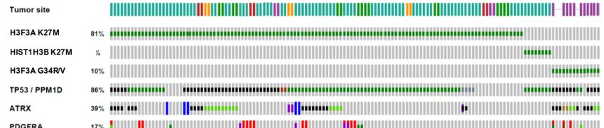
Int. J. Mol. Sci. 2020, 21, 7193 4 of 23
Table 1. Cont.
Category Technique Genotype Cell Type Age Phenotype Reference
H3.1K27M,
nestin+
p53 loss, pten neonatal proliferating
hindbrain [31]
loss (P3–5) clusters
NPCs
ACVR1G328V
H3.3K27M, forebrain or
In utero p53 loss ± hindbrain embryonic high grade
[24]
electroporation PDGFRA, periventricular (E12.5–13.5) glioma
ATRX loss NPCs
forebrain or
H3.3K27M, hindbrain neonatal proliferating
[24]
p53 loss periventricular (P0–2) clusters
NPCs
forebrain or
H3.3K27M,
hindbrain neonatal proliferating
p53 loss, ATRX [24]
periventricular (P0–2) clusters
loss
NPCs
H3.3K27M,
p53 loss + hindbrain
embryonic high grade
PDGF-B, periventricular [32]
(E13.5) glioma
PDGFRAWT or NPCs
PDGFRAD842V
H3.3K27M, forebrain
embryonic high grade
DNp53, periventricular [33]
(E13.5) glioma
PDGFRAD842V NPCs
NRASV12 , p53
loss, ATRX high grade [25] SNO
unknown “postnatal”
loss ± glioma 2018
H3.3G34R
H3.3K27M,
neonatal high grade
Transgenic p53 loss, nestin+ NPCs [23]
(P0–1) glioma
PDGFRAV544ins
medulloblastoma,
H3.3K27M, neonatal
nestin+ NPCs high grade [23]
p53 loss (P0–1)
glioma
p53 loss, neonatal high grade
nestin+ NPCs [23]
PDGFRAV544ins (P0–1) glioma
H3.3K27M,
nestin+ NPCs n/a no tumors [24]
p53 loss
H3.3K27M,
GFAP+ NPCs n/a no tumors [24]
p53 loss
H3.1K27M,
high grade
ACVR1G328V , Olig2+ OPCs n/a [34]
glioma
PIK3CAH1047R
H3.1K27M, proliferating
Olig2+ OPCs n/a [34]
ACVR1G328V clusters
H3.3WT,
H3.3K27M neonatal (P2); [35–40];
orthotopic high grade
PDX and n/a adolescent catalogued
xenograft glioma
H3.1K27M (4–6 weeks) in [41]
tumors
2.1. Ex Vivo Somatically Altered NPCs
Genetic hallmarks of pHGG have been leveraged in the development of xenograft models
using engineered cells. The first such model used lentiviral transduction of H3.3K27M,
activated PDGFRAD842V , and shRNA targeting p53 in neural progenitor cells (NPCs) derived from
human embryonic stem cells, which were then orthotopically transplanted into early postnatal
immunocompromised mice [26]. The resultant tumors diffusely infiltrated the pons within months
and showed elevated proliferation consistent with malignant glioma (Ki67 10%), though they did not
show the necrosis or microvascular proliferation that are pathognomonic for glioblastoma. By contrast,
tumors failed to form when H3.3 wild-type (WT) was transduced in place of the K27M mutant.
An analogous approach was used to combine H3.3K27M with PDGF-B overexpression in
embryonic mouse forebrain NPCs, which were then transplanted into the pons of adolescent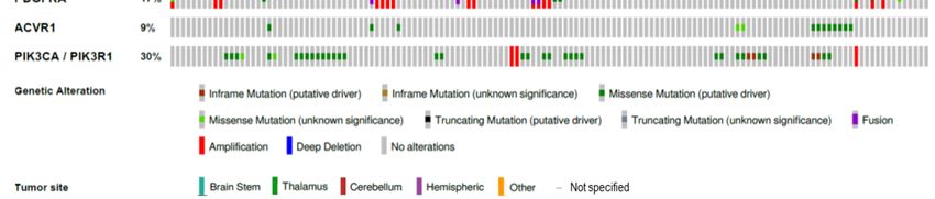
Int. J. Mol. Sci. 2020, 21, 7193                                                                     5 of 23

immunodeficient mice [27]. This led to accelerated formation of pontine HGG as compared with H3.3WT.
Notably, the introduction of H3.3K27M in p53-knockout NPCs did not result in tumor formation,
contrary to findings in an in utero electroporation model described below [24]. The combination of all
three hits (H3.3K27M, p53 loss, and activated PDGFR signaling) was not assessed in this system.

2.2. RCAS/tv-a System
      The first GEMM of DMG predates the discovery of oncohistones and utilized the RCAS/tv-a
gene delivery system, which targets ectopic expression of the avian virus receptor tv-a to specific
cells, thus allowing for selected expression of oncogenes delivered via injection of avian-based
RCAS virus-producing cells [42]. Becher and colleagues successfully generated brainstem high-grade
gliomas by targeting PDGF-B combined with Cre-mediated p16INK4A/ARF loss to nestin-positive neural
progenitors around the fourth ventricle in neonatal mice [28].
      This model was modified once H3K27M oncohistones were found, using RCAS vectors for
PDGF-B, H3.3K27M, and Cre-mediated p53 loss in the same hindbrain nestin-positive progenitors [29].
The combination of all three genetic insults was required for pHGG formation, though H3.3K27M +
p53 loss was sufficient to produce proliferating clusters of cells that were not seen in H3.3WT mice.
Further analysis of this model confirmed that high-grade tumors were only formed in the presence
of H3.3K27M mutant (and not WT) virus, which also decreased tumor latency [22]. Targeting of
these RCAS viruses to Pax3-positive brainstem cells was also capable of driving glioma development,
though notably there was no difference in tumor incidence, latency, or grade between H3.3K27M and
H3.3WT mice [30].
      More recently, the RCAS/tv-a system has been successfully applied to H3.1K27M, with targeting of
H3.1K27M, multiple clinically relevant ACVR1 mutants and Cre-mediated p53 loss in nestin-positive
brainstem progenitors [31]. While the combination of these three genetic hits resulted in premalignant
glioma-like lesions, they were insufficient for frank tumor formation despite multiple different
combinations and the addition of pten loss. Brainstem gliomas formed only when PDGF-A was added
as a fourth insult; in this setting, the presence of combined H3.1K27M and mutant ACVR1 served to
increase tumor incidence and grade while decreasing tumor latency. It should be noted that mutations
or amplifications activating PDGF signaling are relatively rare in the subset of H3.1K27M + ACVR1
mutant pHGG [10], though it is possible that PDGF signaling is indirectly activated in these tumors by
other means.

2.3. In Utero Electroporation (IUE)
      While the above models recapitulated many of the histological and molecular features of DMG,
they were not entirely faithful to the relevant genetic alterations of this tumor as they utilize exogenous
expression of PDGF ligand as opposed to cell-intrinsic activation of PDGF receptor. This is largely
due to the restricted size of transcripts amenable to RCAS incorporation. As such, these models may
erroneously incorporate paracrine effects on cells other than the putative cell-of-origin and will fail to
capture any non-canonical effects that may be mediated by mutant receptors.
      Preclinical models incorporating PDGF receptor perturbation have been established harnessing in
utero electroporation of oncogenic transposon vectors injected into the ventricles of the developing
mouse, allowing genomic incorporation of the transposon only in subventricular neural progenitor
cells [43,44]. Strikingly, no tumors formed when combining H3.3K27M and p53 loss driven by either
nestin or GFAP promoters, nor by electroporation of Sleeping Beauty-based transposons in the neonatal
mouse [24].
      In contrast, introduction of H3.3K27M via a piggyBac transposon system combined with
CRISPR/Cas9-mediated loss of p53 in the embryonic mouse brain was sufficient to cause tumorigenesis
with 100% penetrance in both forebrain and hindbrain. Notably, no tumors formed with the introduction
of H3.3WT or H3.3G34R in this system. This was the first GEMM to show that H3.3K27M and p53
loss alone were competent to drive pHGG formation, possibly due to the embryonic (as opposed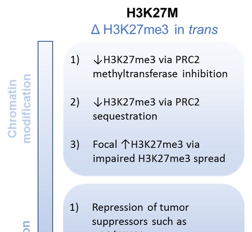
Int. J. Mol. Sci. 2020, 21, 7193                                                                  6 of 23

to postnatal) cell of mutation. Whether this indicates an embryonic cell-of-origin remains unsettled,
as prior work in adult gliomas suggests the possibility of distinct cell of mutation and cell-of-origin
populations [45]. Further fine-tuning of this model with the addition of ATRX loss and PDGFRA
overexpression significantly shortened tumor latency.
     Other IUE-based models have directly compared the effects of exogenous PDGF ligand versus
receptor. The combination of piggyBac transposon-mediated embryonic IUE of H3.3K27M, p53 loss,
and either PDGF-B, PDGFRAWT , or constitutive mutant PDGFRAD842V all resulted in fully penetrant
glioma formation, but with distinct differences in histological characterization [32]. Exogenous PDGF
ligand led to cell-extrinsic perivascular changes that contributed to the shortest latency, while WT
PDGFRA led to less aggressive tumors with longer latency as compared with PDGFRAD842V .
     Finally, transposon-mediated somatic alteration in neonatal mice has been explored to develop a
model of H3.3G34R pHGG. In an oral presentation at a recent Society for Neuro-Oncology meeting,
the combination of H3.3G34R, NRASV12 , and shRNAs targeting p53 and ATRX was reported to form
tumors, though with increased latency when compared with H3.3WT in the same context [25]. As NRAS
mutation is not classic of pHGG, it remains to be seen if the underlying mechanisms of tumorigenesis in
this model faithfully recapitulate those of the corresponding human tumors. Further characterization
of this and other models incorporating H3.3G34R will be essential to deepening our understanding of
this particular pHGG subset.

2.4. Transgenic Mice
     The above-mentioned GEMMs all rely on oncogene expression from exogenous promotors and
dictate the geography of transformation by choosing the location of virus injection or electroporation.
Recent advances in conditional germline knockin mouse models have eliminated those constraints.
H3.3K27M was knocked into the endogenous H3F3A locus and combined with p53 loss and the
constitutively active PDGFRAV544ins mutant driven by a tamoxifen-inducible Cre recombinase in
neonatal nestin-positive cells throughout the developing brain [23]. This model led to spontaneous
malignant brain tumor formation, with H3.3K27M driving hindbrain specificity of tumorigenesis and
PDGFRA signaling driving pHGG identity.
     A similar approach was used to knock H3.1K27M and ACVR1G328V into their respective
endogenous loci driven by Cre recombinase in Olig2-positive oligodendrocyte precursor cells
(OPCs) [34]. These two genetic insults served to arrest glial differentiation and promote proliferation,
though they were insufficient to drive tumorigenesis. The addition of endogenous PIK3CAH1047R
knockin gave rise to spontaneous midbrain and thalamus HGG, albeit with a protracted latency of
over one year. Tumorigenesis in the absence of H3.1K27M was even more protracted, suggesting this
oncohistone played a role in accelerating tumorigenesis. In contrast to other GEMMs, combination with
p53 loss was not explored, though p53 mutations are common in H3.1K27M tumors, which may have
contributed to the prolonged tumor latency.

2.5. Patient-Derived Xenografts (PDXs)
     Orthotopic patient-derived xenografts consist of dissociated patient tumor cells, usually passed
briefly through cell culture and then implanted stereotactically into the brainstem of an
immunocompromised mouse. The first PDX model of DMG was developed by Monje and colleagues
in 2011 using short term neurosphere culture of early postmortem tissue from a patient with diffuse
intrinsic pontine glioma [35]. While the histone-mutant status of the tumor was not known at the
time and was later revealed to be H3 wild type, this protocol for transient neurosphere culture
of DMG tissue followed by xenografting has subsequently been successfully applied to multiple
H3K27M-mutant tumors [36,37]. Tumor latency in these models ranges from 3 to 6 months from
implantation, considerably longer than in analogous PDX models of adult glioblastoma.
     More recently, systematic characterization of pHGG PDX models from both early biopsies as well
as autopsy tissue has been pursued and biobanks have been established [38–40]. Comparison of these
Int. J. Mol. Sci. 2020, 21, 7193                                                                  7 of 23

models may help elucidate and distinguish tumorigenic mechanisms present at diagnosis as opposed
to at terminal disease, complementing tumor evolution studies done on primary human tumors [14,46].
      Of note, while several groups have established PDXs from cortically based pediatric
tumors [38,47,48], none of these have harbored H3.3G34R/V mutations. All published PDX models of
histone-mutant pHGG are too numerous to specify here; however, they have been comprehensively
catalogued in a recent review [41].

3. Molecular Mechanisms of Oncogenicity
     Since the discovery of oncohistones in pHGG, substantial effort has been devoted to uncovering
the molecular mechanisms by which they promote tumor formation, often leveraging the experimental
models delineated above. Even initial studies showed disparate effects of different histone H3 genes
and in different cell types, highlighting the nuanced consequences of chromatin dysregulation in
oncogenesis. Below, we summarize the evolution of our current understanding of the downstream
ramifications of oncohistone mutation in pediatric malignant glioma [49–51].

3.1. H3.3K27M
      The consistent presence of a heterozygous K27M mutation in an individual histone H3 gene
(of which there are 16 total in humans) suggests a dominant negative mechanism of action. Indeed,
quantitative mass spectrometry studies have shown that mutant K27M histone H3 comprises only 3–17%
of total H3 protein in human DMG samples [29]. Early chromatin studies in these tumors uncovered a
stark global loss of di- and tri-methylation at H3K27 (H3K27me2, me3) [29,52–54]. These epigenetic
marks, which are associated with gene silencing, are catalyzed by the Polycomb repressive complex
2 (PRC2), a Polycomb group protein comprised of several subunits: the catalytic enhancer of zeste
homolog 1 (EZH1) or EZH2, embryonic ectoderm development (EED), and suppressor of zeste 12
homolog (SUZ12). Subsequent evaluation of PRC2 components in the context of H3.3K27M mutation
led to several hypothesized mechanisms underlying this H3K27me3 loss (Figure 2).
      PRC2 enzymatic inhibition: In vitro studies showed that synthetic H3.3K27M peptide is able
to impair PRC2 methyltransferase activity on both mutant and WT H3.3-containing nucleosomes in
HEK293T cells [29]. Crosslinking assays demonstrated that the K27M peptide interacts with the EZH2
active site [29], further substantiated by the solution of the crystal structure of human PRC2 bound to
H3.3K27M, which showed the mutant moiety in the active pocket of the SET domain of EZH2 [55].
In vitro histone methyltransferase assays demonstrated a 40–70% decrease in EZH2 activity in multiple
cell lines with exogenous H3.3K27M, amplified to an 85% decrease in K27M mono-nucleosomes [52].
Additional in-depth biochemical analyses confirmed this inhibition of EZH2 methyltransferase activity
as well as mapped the binding of EZH2 to the histone H3 tail [56]. Altogether, these studies suggest
that H3.3K27M can directly inhibit the catalytic activity of the PRC2 methyltransferase complex.
      PRC2 sequestration: Further analysis in these studies also demonstrated preferential binding
of H3.3K27M to PRC2 components. Coimmunoprecipitation experiments in multiple cell types
showed enrichment of EZH2 and SUZ12 at K27M mono-nucleosomes as compared with WT
mono-nucleosomes [52]. H3.3K27M peptide bound to EZH2 16-fold more tightly than WT H3.3
in vitro [55], suggesting trapping and sequestration of PRC2 may play a role in the inhibitory effect of
H3.3K27M. Studies in mouse embryonic stem cells found that EZH2 was redistributed and sequestered
at poised enhancers (defined as chromatin regions marked by H3K27me3 and H3K4me1) that also
contained H3.3K27M [57]. However, proteomic studies using quantitative mass spectrometry in
Drosophila models failed to identify an enrichment of any PRC2 subunits with H3.3K27M-containing
nucleosomes [58]. Furthermore, epigenomic profiling using ChIP-seq in DMG cells showed the
PRC2 components EHZ2 and SUZ12 are largely excluded from K27M-containing chromatin [59],
countering the sequestration theory.
uncovered a stark global loss of di- and tri-methylation at H3K27 (H3K27me2, me3) [29,52–54]. These
epigenetic marks, which are associated with gene silencing, are catalyzed by the Polycomb repressive
complex 2 (PRC2), a Polycomb group protein comprised of several subunits: the catalytic enhancer
of zeste homolog 1 (EZH1) or EZH2, embryonic ectoderm development (EED), and suppressor of
zeste 12 homolog
Int. J. Mol. (SUZ12).
Sci. 2020, 21, 7193 Subsequent evaluation of PRC2 components in the context of H3.3K27M
8 of 23
mutation led to several hypothesized mechanisms underlying this H3K27me3 loss (Figure 2).
Figure 2.
Figure Putativeoncogenic
2. Putative oncogenicmechanisms
mechanisms of
of H3K27M
H3K27M in
in pHGG.
pHGG.
Impaired
PRC2 H3K27me3
enzymatic spread:In
inhibition: A vitro
morestudies
recent study leveraging
showed CRISPR-Cas9
that synthetic H3.3K27Mtechnology
peptidetoisgenerate
able to
isogenicPRC2
impair DMGmethyltransferase
cell lines also found that PRC2
activity on bothrecruitment
mutant andto its
WT high-affinity sites at
H3.3-containing unmethylated
nucleosomes in
CpG islands is not altered in the presence of H3.3K27M [60], adding to the opposition
HEK293T cells [29]. Crosslinking assays demonstrated that the K27M peptide interacts with the EZH2 of a simple
sequestration
active site [29],model.
furtherInstead, they found
substantiated that
by the H3K27me3
solution of thedeposition was restricted
crystal structure of humanto narrow peaks
PRC2 bound
surrounding these high affinity sites, with slightly broader deposition of H3K27me2 away from these
sites. This impaired spread was reversible, as removal of the mutant K27M allele restored the wild
type pattern of H3K27me2/me3 deposition. An independent investigation of H3.3K27M mutant
and WT DMG cells confirmed this impaired spread of H3K27me3 from PRC2 high affinity sites and
suggested enhanced allosteric inhibition of PRC2 by H3K27me3 in the presence of the K27M mutant as
a possible mechanism [55,61]. These studies suggest more nuanced regulation of PRC2 localization in
the presence of mutant K27M beyond direct inhibition of activity or direct binding and sequestration.
Additional epigenetic alterations: Such a nuanced model is supported by our broader
understanding of chromatin modification mediated by H3.3K27M beyond loss of H3K27 trimethylation.
For instance, several of the studies that initially described a global loss of H3K27me3 in K27M
mutant cells also described a somewhat paradoxical gain of H3K27me3 at certain loci [52,53].
Further characterization of these loci revealed them to be strong Polycomb targets associated with CpG
islands [27], as determined by density of H3K27me3 in H3WT cells and retention of H3K27me3 after
treatment with EZH2 inhibitors. This is in keeping with the impaired spread of H3K27me3 from such
sites in other studies [60]. As Polycomb target genes are intimately involved with development andInt. J. Mol. Sci. 2020, 21, 7193 9 of 23
differentiation, it has been suggested that these sites of retained or enhanced H3K27me3 may play just
as important a role in tumorigenesis as the loss of H3K27me3. This theory is supported by impaired
growth of DMG cells in the presence of an EZH2 inhibitor [27,59,60] as well as prolonged survival in
a DMG preclinical model [27]. However, these findings were not replicated by other groups [22,62],
and the role of H3.3K27M mutant status for this growth inhibitory effect remains unclear [27,60].
H3.3K27M also impacts activating chromatin marks such as H3K27 acetylation (H3K27ac), which is
enriched in K27M mutant cells [29,58,63]. H3K27ac is a marker of super-enhancers that is bound by
bromodomain and extra terminal (BET) proteins such as BRD2 and BRD4 to facilitate transcription
initiation and elongation. These BET proteins are also found to be enriched at K27M-containing
nucleosomes [58,59], and shRNA-mediated loss of BRD4 extended survival in a DMG xenograft
model [64].
Yet another layer of complexity is added by considering the temporal evolution of these epigenetic
effects. The use of an inducible H3.3K27M model showed that PRC2 initially colocalizes with K27M
but that this colocalization decreases with time [61]. Despite no longer being physically associated,
the methyltransferase activity of PRC2 from K27M mutant cells remains impaired, suggesting that
H3.3K27M may induce a lasting perturbation in PRC2 such as a conformational change. Additional work
has shown that H3.3K27M decreases EZH2 automethylation [65], which preferentially impairs
PRC2-mediated conversion of H3K27me2 to me3, potentially explaining the more potent impact on
H3K27me3 than H3K27me2 [60].
Changes in gene expression: Given the striking global loss of H3K27me3, one might have
expected an accompanying global increase in gene expression due to the loss of silencing. However,
RNA sequencing studies have not borne this out. In fact, comparison of H3K27 WT and H3K27M tumors
show relatively modest differences in gene expression [23,52,53,60]. Nevertheless, these selective
changes are broadly consistent, with multiple groups able to reliably separate H3.3K27M tumors
using unsupervised clustering of gene signatures [5,9,10,66]. There is some variation in the exact
set of differentially expressed genes between studies, which may in part be due to the multiple
different modalities used to generate these signatures (tumors versus cell lines, primary samples versus
genetically engineered models, human versus mouse origin). However, a persistent theme is the
relative upregulation of PRC2 target genes by H3.3K27M as assessed by gene set enrichment analysis.
Genes involved in neural development and differentiation are also upregulated in the presence of
H3.3K27M [24,26,60], such as the inhibitor of differentiation (ID) gene family. A potential mechanism
underlying these changes is the derepression of so-called “poised” or “bivalent” promoters, marked by
the repressive H3K27me3 as well as the activating H3K4me3, by the loss of H3K27 trimethylation [23,67],
as PRC2 targets and neural developmental genes are highly enriched in the population of bivalent
promoters. An H3.3K27M-dependent redistribution of EZH2 to analogous poised enhancers has
also been described [57]. This release of bivalent promoters parallels a process seen during normal
developmental transitions [68] and is consistent with the framing of pediatric gliomagenesis as a
corruption of normal developmental pathways and systems. Other studies have highlighted an
oncogenic role for downregulation of tumor suppressor genes in the H3.3K27M context. For instance,
the cell cycle gene CDKN2A isoform p16/INK4A locus often retains H3K27me3 in K27M-mutant cells,
associated with decreased expression and accelerated tumor formation [22,27], though this finding is
variably present [59].
Growth and transformation: The integrated impact of H3.3K27M mutation on relevant cell biology
has also been extensively explored. Studies using both mouse and human neural progenitor cells
engineered to express H3.3K27M in the presence of concurrent oncogenic mutations have demonstrated
enhanced soft agar colony forming ability in the presence of K27M [26,27], suggesting this mutation
promotes self-renewal capacity. This is supported by increased serial clonogenicity in neural stem
cells derived from an H3.3K27M-based genetically engineered mouse model [23]. H3.3K27M mutation
also confers a modest proliferative advantage in neural stem cells [23,26]; this effect appears to be
narrowly restricted by cellular context, as no proliferative advantage was seen in embryonic stem cellsInt. J. Mol. Sci. 2020, 21, 7193 10 of 23
or astrocytes [26]. This enhanced self-renewal and proliferation is coupled with altered differentiation
potential. As referenced above, multiple gene ontology analyses of differentially expressed genes
in the H3.3K27M context are enriched for neural lineage differentiation gene sets [22,23,59,60,67].
Impaired differentiation in H3.3K27M tumors has also be substantiated at the single cell level, where an
OPC-like subset comprises the majority of cells across multiple different patient tumors [69]. This single
cell RNA sequencing-based study also found an increased proportion of dividing and undifferentiated
cells in K27M mutant tumors as compared with other canonical subgroups (such as IDH-mutant
tumors). Functional assays of differentiation capacity showed reduced ability for H3.3K27M cells
to generate astrocytes and, to a lesser extent, oligodendrocytes [26]. Of note, many of the studies
investigating self-renewal and differentiation capacity were carried out in the context of H3.3K27M in
combination with its frequent concurrent PDGFRA activation and p53 loss, while addition of K27M
alone did not recapitulate these features. However, a study directly evaluating the role of H3.3K27M in
these processes via shRNA-mediated knockdown in DMG xenografts confirmed an intrinsic promotion
of stemness, proliferation, and impaired differentiation [67].
The exceptional spatio-temporal specificity of mutant tumors suggests a narrow developmental
window in which a relevant cell type is susceptible to transformation by this mutation [70]. Profiling of
these tumors suggests they recapitulate some aspects of the normal developmental hierarchy in neural
and glial progenitor cells [69]. Indeed, many of the preclinical models described below revealed
remarkable restrictions in the precursor cell populations competent to drive H3.3K27M-mediated
gliomagenesis [24,26]. While both neural stem/progenitor cells and early oligodendrocyte precursor
cells have been capable of tumor formation in different experimental models, elegant work comparing
the resultant epigenomic remodeling has revealed disparate chromatin patterning in these two contexts,
with the active chromatin landscape of transformed OPCs more closely resembling that of DMG
tumors [71]. The geography of these transformed cells also plays a role, as hindbrain precursor cells
seem preferentially susceptible to transformation by H3.3K27M [23,72]. This geographic predilection is
supported by dysregulation of H3K27 methylation in other pediatric hindbrain tumors such as subsets
of medulloblastoma [73] and posterior fossa ependymoma [74–76].
3.2. H3.1K27M
While the K27M mutation most commonly occurs at the histone H3.3 variant locus H3F3A,
a minority of DMG harbor this mutation at histone H3.1 loci [12,13,77]. Initial characterization of
these tumors grouped them with their H3.3 counterparts, as both demonstrate global reductions
in H3K27 trimethylation [29]. However, more focused evaluations of these specific histone variant
mutations have begun to uncover subtle differences in the dysregulation mediated by the two isoforms.
Transcriptional and methylation-based profiling can reliably distinguish between H3.3 and H3.1
mutant tumors [8,78], with H3.3K27M correlating with proneural and oligodendroglial signatures,
while H3.1K27M correlated with mesenchymal and astrocytic signatures. It is difficult to distinguish
putative unique impacts of oncohistone isoforms from residual signatures derived from potentially
distinct cells-of-origin or divergent concomitant mutations in bulk studies. Indeed, the H3.3K27M
partner mutation PDGFRA and the H3.1K27M partner mutation ACVR1 have established roles in
oligodendrocytic and astrocytic development, respectively. Evaluation of H3.3K27M and H3.1K27M
in isogenic early OPCs does substantiate disparate chromatin localization of the histone variants as
well as distinct patterns of active enhancers and gene expression even in the absence of additional
mutations [71].
3.3. H3.3G34R/V
The molecular mechanisms underlying H3.3G34R/V-mediated oncogenesis are relatively
uncharted by comparison, though they are clearly fundamentally distinct. While H3G34 itself is not
subject to posttranslational modification, H3.3G34R/V affects trimethylation at the neighboring H3K36,
an activating mark associated with transcriptional elongation [79]. In contrast to the dominant effect ofInt. J. Mol. Sci. 2020, 21, 7193 11 of 23
H3K27M, this alteration occurs only in cis at mutant-containing nucleosomes [29,53]. Initial in vitro
methylation assays demonstrated decreased H3K36 trimethylation at H3.3G34 mutant nucleosomes,
possibly via impaired binding of the H3K36 trimethyltransferase SETD2 [29,53]. Subsequent structural
modeling suggested steric hindrance of SETD2 binding by the large side chains in arginine or valine
substitutions as the underlying mechanism [80]. The putative oncogenic role of H3K36me3 loss
is bolstered by the discovery of mutually exclusive SETD2 missense and truncating mutations in
cortical pHGG [81]. However, chromatin landscape profiling studies have shown a modest increase
in H3K6me3 at certain genes such as MYCN, particularly those enriched in variant histone H3.3
deposition [66,82]. In vitro binding assays have shown preferential binding and inhibition of the
H3K36 demethylase KDM4 by H3.3G34R/V [82], suggesting a dynamic interplay between H3K36
methylating and demethylating enzymes across specific genomic loci.
Gene ontology analysis of ChIP-seq datasets between H3.3G34V and H3.3WT glioma cell lines
showed enrichment in forebrain and cortical development gene lists [66], though it is difficult to ascribe
these differences to the direct effect of H3.3G34V alone as opposed to potentially confounding variables
such as the age, location, cell-of-origin, and concurrent mutations of the tumors of origin. In addition
to its role in transcriptional elongation, H3K36me3 is also implicated in DNA mismatch repair [83].
Accordingly, H3.3G34R/V has been shown to confer defective homologous recombination-mediated
mismatch repair [84] and an associated mild hypermutator phenotype [80]. Finally, very recent work
has shown thatInt.H3.3G34R isx preferentially
J. Mol. Sci. 2020, 21, FOR PEER REVIEW bound by the chromatin reader RACK7 11 of 23in pHGG cells,
which leads to suppressed MHC class II protein expression and vesicular
repair [83]. Accordingly, H3.3G34R/V has been shown to confer defective homologous transport [85], alluding to a
possible role in impaired antitumor immunity.
recombination-mediated mismatch repair [84] and an associated mild hypermutator phenotype [80].
Finally, very recent work has shown that H3.3G34R is preferentially bound by the chromatin reader
RACK7 in pHGG cells, which leads to suppressed MHC class II protein expression and vesicular
4. Developmenttransport
of Novel Therapies
[85], alluding to a possible role in impaired antitumor immunity.
Steady progress in elucidating
4. Development the molecular vulnerabilities of histone-mutant pHGG in conjunction
of Novel Therapies
with the development of biologically
Steady relevantthemouse
progress in elucidating models
molecular has culminated
vulnerabilities in several
of histone-mutant pHGGpreclinical
in studies
of therapeutics tailored towith
conjunction the the
unique susceptibilities
development of biologicallyof thesemouse
relevant tumors models[86],
hasexploiting
culminated inboth
severalthe downstream
preclinical studies of therapeutics tailored to the unique susceptibilities of these tumors [86],
consequences ofexploiting
oncohistone mutation as well as their obligate partner mutations (Figure 3). Several of
both the downstream consequences of oncohistone mutation as well as their obligate
these approaches nowmutations
partner serve as the basis
(Figure forofongoing
3). Several clinical
these approaches nowtrials
serve as(Table 2).for ongoing clinical
the basis
trials (Table 2).
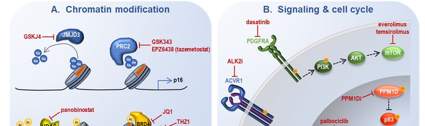
Figure 3. Strategies for molecular intervention in oncohistone-driven pHGG. Ac: acetylation; ER:
Figure 3. Strategies for molecular intervention in oncohistone-driven pHGG. Ac: acetylation;
endoplasmic reticulum; Me: methylation; RNA pol II: RNA polymerase II. See text for other
ER: endoplasmic reticulum;
abbreviations commonMe: methylation;
to multiple RNA pol II: RNA polymerase II. See text for other
tables and figures.
abbreviations common to multiple tables and figures.Int. J. Mol. Sci. 2020, 21, 7193 12 of 23
Table 2. Ongoing clinical trials for histone-mutant pHGG. BMI1: B cell-specific Moloney murine
leukemia virus integration site 1; CED: convection enhanced delivery; IDO: indoleamine 2,3-dioxygenase;
IL12: interleukin 12; IV: intravenous; PO: oral; RT: radiotherapy; TMZ: temozolomide. See text for other
abbreviations common to multiple tables and figures.
Category Intervention Administration Trial Identifier Tumor Eligibility Phase
HDACi &
NCT02717455 nonprogressive;
chromatin panobinostat PO I
(PBTC-047) recurrent/refractory
modifiers
entinostat PO NCT02780804 recurrent/refractory I
valproic acid +
PO NCT03243461 newly diagnosed III
RT/TMZ
panobinostat
NCT03566199
nanoparticles CED newly diagnosed I/II
(PNOC015)
(MTX110)
vorinostat +
newly diagnosed;
temosirolimus PO NCT02420613 I
recurrent/refractory
± RT
Fimepinostat
newly diagnosed;
[dual PO PNOC016 I
recurrent/refractory
HDACi/PI3Ki]
marizomib ±
PO NCT04341311 newly diagnosed I/II
panobinostat
PTC596 [BMI1i]
PO NCT03605550 newly diagnosed Ib
+ RT
cemiplimab
NCT03690869 newly diagnosed;
Immunotherapy (REGN2810) + IV I
(PNOC013) recurrent/refractory
RT
H3K27M
NCT02960230
vaccine + IV newly diagnosed I
(PNOC007)
nivolumab
NCT02359565
pembrolizumab IV recurrent/refractory I
(PBTC-045)
APX005M
NCT03389802 newly diagnosed;
[CD40 agonistic IV I
(PBTC-051) recurrent/refractory
Ab]
indoximod +
PO NCT04049669 newly diagnosed II
RT/TMZ
IL12
intratumoral NCT03330197 newly diagnosed I/II
adenovirus
B7-H3 CAR-T intratumoral; newly diagnosed;
NCT04185038 I
cells intraventricular recurrent/refractory
GD2 CAR-T
IV NCT04099797 newly diagnosed I
cells
nanoliposomal
Cytotoxic CED PNOC009 newly diagnosed I/II
irinotecan
gemcitabine IV NCT02992015 newly diagnosed I
abemaciclib ±
Kinase Inhibitor PO NCT02644460 recurrent/refractory I
RTInt. J. Mol. Sci. 2020, 21, 7193 13 of 23
Table 2. Cont.
Category Intervention Administration Trial Identifier Tumor Eligibility Phase
ribociclib + NCT03387020
PO recurrent/refractory I
everolimus (PBTC-050)
ribociclib + newly diagnosed
PO NCT03355794 I
everolimus (s/p RT)
palbociclib +
TMZ + PO + IV NCT03709680 recurrent/refractory I
irinotecan
dasatinib + newly diagnosed;
PO NCT03352427 II
everolimus recurrent/refractory
paxalisib
(GDC-0084) newly diagnosed
PO NCT03696355 I
[dual (s/p RT)
PI3K/mTORi]
NCT03598244
Savolitinib PO recurrent/refractory I
(PBTC-049)
Adavosertib
PO NCT01922076 newly diagnosed I
(MK-1775) + RT
ONC201 newly diagnosed;
Other PO NCT03416530 I
[DRD2i] recurrent/refractory
124 I-8H9
CED NCT01502917 nonprogressive I
(omburtamab)
INCB7839 NCT04295759
PO recurrent/refractory I
[ADAM10i] (PBTC-056)
4.1. Reversal of Epigenetic Alterations
Given the profound epigenetic ramifications of oncohistone mutation, it is perhaps not surprising
that several approaches aim to reverse chromatin aberrations associated with these tumors.
H3K27me3 demethylase inhibition: As global loss of H3K27me3 is one of the molecular hallmarks
of H3K27M pHGG, one of the first therapeutic studies evaluated pharmacologic inhibition of the K27
demethylase JMJD3 using GSKJ4 [87]. This led to restoration of H3K27me3 and improved survival in
H3K27M orthotopic xenografts, with no effect in H3WT xenografts.
EZH2 inhibition: Highlighting the nuanced regulation of H3K27me3 in these tumors,
residual PRC2 methyltransferase activity remains required for effective tumor growth, as genetic
EZH2 loss impaired H3.3K27M-engineered mouse NPC xenograft growth and EZH2 inhibition with
two different compounds (GSK343, EPZ6438/tazemetostat) led to growth arrest in human pHGG
cells [27,59], though there have been conflicting reports regarding this in vitro finding [22,62]. In vivo
effects of small molecule EZH2 inhibition have not yet been decisively characterized.
BET inhibitors: Increased H3K27ac is another mainstay of H3K27M pHGG, and several strategies
to either modify this aberrant acetylation or mitigate its effect have also proven fruitful. Pharmacologic
inhibition of bromodomain proteins involved in H3K27ac recognition and subsequent transcription
preferentially inhibited growth in H3K27M pHGG cells and extended survival in H3K27M PDX
models [59]. Evaluation of a broader range of pHGG cell lines and BET inhibitors has confirmed the
potential utility of this approach but highlighted the importance of potency and brain penetrance for
further investigations [64].
CDK7 inhibition: Alternate means of attenuating the anomalous transcription present in
H3K27M pHGG cells has also been explored via inhibition of CDK7, a cyclin-dependent kinase
that phosphorylates RNA polymerase II to regulate consequent transcription. The CDK7 inhibitor
THZ1 also potently inhibited H3K27M pHGG cell viability and led to a modest but significant
improvement in xenograft survival [64]. In combination with the BET inhibitor studies, this suggests
abrogation of the transcriptional consequences of altered H3K27ac as a promising strategy for H3K27M
pHGG treatment.Int. J. Mol. Sci. 2020, 21, 7193 14 of 23
HDAC inhibition: Interestingly, additional studies have intimated that increased H3K27ac may
not contribute to pHGG pathogenesis and may in fact be exploitable as a vulnerability particular to
H3K27M-mutant tumors. For instance, genome-wide mapping in H3K27M-mutant cells revealed
pervasive increases in H3K27ac deposition throughout the genome that induced expression of repeat
elements such as endogenous retroviral elements, with implications for antitumor immunity [63].
Polyacetylation at adjacent residues has also been shown to block the interaction of PRC2 with H3K27M
and thus disrupt the PRC2 inhibition [56]. These findings potentially explain the therapeutic benefit
of histone deacetylase (HDAC) inhibitors such as panobinostat. In a screen of dozens of promising
agents using a panel of patient-derived DMG cell lines, HDAC inhibitors emerged as the leading
category of interest, with panobinostat demonstrating marked impairment in cell viability as well
as prolonged survival in PDX models [37]. This therapeutic effect has been substantiated by others,
though the selective vulnerability of H3K27M-mutant tumors remains debated [63]. Importantly,
pHGG cells that survived panobinostat treatment developed resistance to panobinostat re-challenge [37],
underscoring the likely need for synergistic treatment strategies in this disease.
Epigenetic Combination Therapies: Synergistic treatments have been validated in vitro with
some of the epigenetic disrupters explored as monotherapy above, including BET inhibitors [37,64]
and CDK7 inhibitors [64] combined with panobinostat. A more expansive in vitro followed by
in vivo screen of the combinatorial druggable DMG landscape, with clinically relevant factors such as
potency and blood–brain-barrier penetrance taken into account, revealed the proteasome inhibitor
marizomib combined with panobinostat as the most promising pairing [88]. Additional in vivo studies
have suggested inhibition of lysine demethylase [89,90], DNA methylation [63], AXL kinase [91],
and PI3K [92] as promising combination strategies with panobinostat, several of which are now in
clinical trials.
4.2. Blocking Oncogenic Signaling Pathways
Many of the canonical cooperating mutations in histone-mutant pHGG involve growth factor
signaling or cell cycle regulation, emphasizing the importance of these pathways in promoting
oncohistone-mediated gliomagenesis and prompting therapeutic trials of their blockade.
ALK2 inhibition: Small molecule inhibition of ALK2 (ACVR1) with two different compounds
demonstrated efficacy in ACVR1-mutant PDX models [93].
PPM1D inhibition: Inhibition of mutant PPM1D prolonged survival in a PDX model, and in vitro
studies suggest synergy with ionizing radiation [94] and PARP inhibition [95] in PPM1D-mutant models.
Growth factor signaling inhibitors: Growth factor signaling inhibitors have typically been explored
in combination with additional treatments given concerns regarding the subclonal nature of relevant
mutations as well as redundancy in the downstream signaling networks. PDGFRA signaling inhibition
with dasatinib prolonged survival in an IUE-based H3.3K27M pHGG model, and combination with
the mTOR inhibitor everolimus demonstrated synergy [33].
CDK4/6 inhibition: Blockade of cell cycle progression via CDK4/6 inhibition has also been pursued,
in part given the role of upstream p16 downregulation in H3K27M pHGG. Palbociclib treatment
improved survival in PDX models derived from untreated DMG biopsies [96], with in vitro evidence
of synergy when combined with mTOR inhibition [97].
4.3. Immunotherapy
The largely immunosuppressive microenvironment of gliomas has hampered the application of
immunotherapies that have shown efficacy in other cancer types. There are some suggestions that the
microenvironment of pHGG is less immunosuppressive than that of its adult counterpart [98] and as
such may be more amenable to immunomodulatory treatments [99].
Immunomodulatory and oncolytic viruses: An adenoviral-based TK/Flt3L immunostimulatory
gene therapy approach showed efficacy in an ACVR1-mutant mouse model with histologic evidence
of anti-tumor cytotoxic response [100]. Antitumor immunity has also been triggered using theInt. J. Mol. Sci. 2020, 21, 7193 15 of 23
oncolytic adenovirus Delta-24-RGD, with efficacy demonstrated in several immunodeficient and
immunocompromised pHGG models [101]. Analogous to the concept of checkpoint inhibition,
tumor immune evasion inhibition via disruption of CD47 binding to SIRPα prolonged survival in
multiple pediatric brain tumor models including DMG [102].
Vaccines and adoptive cell therapies: Leveraging the tumor-specific nature of oncohistones,
immune targeting of H3K27M has been pursued, with both peptide vaccine [103] and adoptive T cell
receptor [104] paradigms showing preclinical promise. Other adoptive cell therapies such as chimeric
antigen receptor (CAR) T cells targeting pHGG-associated antigens have demonstrated substantial
efficacy in vivo, with anti-B7-H3 CAR-T [105] and anti-GD2 CAR-T [106] progressing to clinical trials.
Of note, a minority of mice in the anti-GD2 CAR-T study developed on-target pontine inflammation
that resulted in ventricular compression and hydrocephalus, a finding that raises caution for all
immunotherapies targeting tumors with such a perilous neuroanatomical location.
4.4. Tumor Microenvironment and Metabolism
Emerging unconventional approaches to histone-mutant pHGG are galvanizing new areas of
therapeutic research, including modifying input from neighboring neurons as well as capitalizing on
particular metabolic susceptibilities in these tumors.
ADAM10 inhibition: Neuronal activity can promote pHGG proliferation via release of postsynaptic
neuroligin-3 cleaved by the metalloproteinase ADAM10, inhibition of which impairs PDX growth [107].
ONC201: Dopamine receptor D2 is highly expressed on some pHGG, and the dopamine receptor
D2/D3 antagonist ONC201 has shown some suggestion of clinical activity in preliminary trials [108,109].
MI-2: Screening of a chemical library for inhibitors of H3K27M pHGG growth uncovered MI-2 as
a lead candidate [26]. While this drug was initially characterized as a menin inhibitor, inhibition of
lanosterol synthase and resultant disruption of cholesterol homeostasis appear to be its relevant
mechanism of action [110].
Mitochondrial targeting: Tumor profiling indicated notable decrease in mitochondrial DNA
quantity in pHGG, and shifting glucose metabolism from glycolysis to mitochondrial oxidation in
combination with metformin to further target mitochondrial function and radiation to potentiate
apoptosis was efficacious in in vivo models [111]. Recent work also suggests unique susceptibility of
PPM1D-mutant pHGG to NAMPT inhibition in vivo [112], supporting the concept of pHGG-specific
metabolic vulnerabilities.
5. Conclusions
Histone-mutant pHGG is an intricate and devastating disease with a paucity of clinically proven
interventions. The unprecedented proliferation of relevant molecular studies in the past decade has
deepened our understanding of its underlying oncogenic mechanisms and uncovered novel targets
for therapy. Central to this ongoing effort has been the development of preclinical models in which
to explore molecular mechanisms and screen potential therapeutics. Further work is needed to
deconvolute the sometimes contradictory observations emerging from different models, which are
likely due to the remarkable contextual specificity of these effects. Different cell types at different
developmental ages from different neuroanatomic locations with different species of origin may show
altered consequences of oncohistone mutation, highlighting the importance of developing model
systems that accurately reflect the biology of the human disease. We are just beginning to unravel
the mechanistic foundations of this idiosyncrasy, such as relative levels of H3K27M and PRC2 [61].
Incisive investigations leveraging our current complement of genomic editing, epigenomic profiling,
and single cell sequencing tools is sure to further propel this accelerated progress, with meaningful
therapeutic strategies on the horizon.
Author Contributions: M.S.G. and I.K.M. contributed to the conceptualization, investigation, writing, and revision
of the manuscript. All authors have read and agreed to the published version of the manuscript.You can also read