Hippocampal granule cell dispersion: a non-specific finding in pediatric patients with no history of seizures
←
→
Page content transcription
If your browser does not render page correctly, please read the page content below
Roy et al. Acta Neuropathologica Communications                  (2020) 8:54
https://doi.org/10.1186/s40478-020-00928-3

 RESEARCH                                                                                                                                             Open Access

Hippocampal granule cell dispersion: a
non-specific finding in pediatric patients
with no history of seizures
Achira Roy1, Kathleen J. Millen1,2 and Raj P. Kapur1,3,4*

  Abstract
  Chronic epilepsy has been associated with hippocampal abnormalities like neuronal loss, gliosis and granule cell
  dispersion. The granule cell layer of a normal human hippocampal dentate gyrus is traditionally regarded as a
  compact neuron-dense layer. Histopathological studies of surgically resected or autopsied hippocampal samples
  primarily from temporal lobe epilepsy patients, as well as animal models of epilepsy, describe variable patterns of
  granule cell dispersion including focal cell clusters, broader thick segments, and bilamination or “tram-tracking”.
  Although most studies have implicated granule cell dispersion as a specific feature of chronic epilepsy, very few
  “non-seizure” controls were included in these published investigations. Our retrospective survey of 147 cadaveric
  pediatric human hippocampi identified identical morphological spectra of granule cell dispersion in both normal
  and seizure-affected brains. Moreover, sections across the entire antero-posterior axis of a control cadaveric
  hippocampus revealed repetitive occurrence of different morphologies of the granule cell layer – compact, focally
  disaggregated and bilaminar. The results indicate that granule cell dispersion is within the spectrum of normal
  variation and not unique to patients with epilepsy. We speculate that sampling bias has been responsible for an
  erroneous dogma, which we hope to rectify with this investigation.
  Keywords: Hippocampus, Granule cell dispersion, Human, Epilepsy, Dentate gyrus, Temporal lobe epilepsy, Gliosis

Introduction                                                                             putative hippocampal abnormalities are the cause or ef-
Chronic epilepsy is often associated with several patho-                                 fect of epilepsy [14, 25].
logical hippocampal abnormalities [44]. These include                                      GCD has been reported commonly in the dentate
sclerosis, characterized by neuronal loss in both dentate                                gyrus (DG) of seizure patients and considered a hallmark
gyrus and Ammon’s horn, gliosis, mossy fiber sprouting;                                  of chronic epilepsy, especially TLE [3, 21, 30, 35, 55, 56].
as well as granule cell dispersion (GCD), which is ob-                                   GCD was also reported to be pronounced in sudden un-
served in the presence or absence of sclerosis [9, 10, 15,                               explained deaths in infants [26, 31]. Many studies using
30, 55, 56]. These studies were mostly performed in sur-                                 animal models have similarly concluded that hippocam-
gical resections or autopsied brain samples obtained                                     pal GCD is a specific feature of chronic epilepsy [40, 43,
from patients suffering from febrile seizures and tem-                                   60]. The granule cell (GC) layer is generally observed as
poral lobe epilepsy (TLE); it is unclear whether the                                     a highly compact layer of neuronal cell bodies in the
                                                                                         dentate gyrus. GCD refers to broadening with or without
                                                                                         bilamination of the GC layer, often blurring the bound-
* Correspondence: raj.kapur@seattlechildrens.org
1
 Center for Integrative Brain Research, Seattle Children’s Research Institute,
                                                                                         ary between the GC layer and molecular layer of the
Seattle, Washington, USA                                                                 dentate gyrus [8, 17]. Additionally, some surgical cases
3
 Department of Pathology, Seattle Children’s Hospital, Seattle, Washington, USA          also demonstrated the existence of focal clusters of
Full list of author information is available at the end of the article

                                          © The Author(s). 2020 Open Access This article is licensed under a Creative Commons Attribution 4.0 International License,
                                          which permits use, sharing, adaptation, distribution and reproduction in any medium or format, as long as you give
                                          appropriate credit to the original author(s) and the source, provide a link to the Creative Commons licence, and indicate if
                                          changes were made. The images or other third party material in this article are included in the article's Creative Commons
                                          licence, unless indicated otherwise in a credit line to the material. If material is not included in the article's Creative Commons
                                          licence and your intended use is not permitted by statutory regulation or exceeds the permitted use, you will need to obtain
                                          permission directly from the copyright holder. To view a copy of this licence, visit http://creativecommons.org/licenses/by/4.0/.
                                          The Creative Commons Public Domain Dedication waiver (http://creativecommons.org/publicdomain/zero/1.0/) applies to the
                                          data made available in this article, unless otherwise stated in a credit line to the data.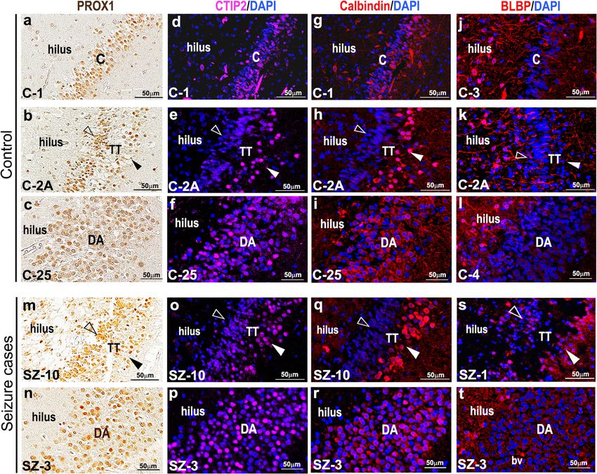
Roy et al. Acta Neuropathologica Communications   (2020) 8:54                                                  Page 2 of 20

granule cells outside the regular GC layer [54]. GCD has        related criteria were abstracted from the archived re-
been most consistently correlated with a history of se-         cords: age, gender, post-mortem interval (i.e. duration
vere febrile or infection-induced seizures during early         between time of death and that of autopsy), presence or
life (< 4 years age), as well as with duration and fre-         absence of seizure history (and seizure interval from on-
quency of epileptic events [30, 36, 54].                        set until death for epileptic patients) and major clinical
   Criteria for determining dispersion vary and depend,         or pathological diagnoses. Corrected age was calculated
in part, on the mode of measurement [8]. A variety of           considering 40 gestational weeks (GW) as day 0. Among
diagnostic criteria for GCD has been suggested, varying         the 147 autopsy cases studied, 80 were males and 67
from complex morphometric analyses [24, 37] to sub-             were females, with the corrected age range spanning
jective assessment of dentate gyrus histology [1, 8, 9, 35,     from 19GW (i.e. −21 weeks) to 20.4 years (i.e. +1060
36]. Some reports have suggested abnormal neuronal              weeks). Post-mortem interval (PMI) ranged from 2 to
migration, loss of hilar cells and genetic defects to be re-    336 h.
sponsible for GCD, all influenced by seizure occurrence
[10, 30, 54]. However, a substantial cohort of controls,        Histological assessment of GCD
with no history of seizures, was seldom included in the         Presence or absence of GCD (disaggregated, tram-track)
clinical studies; many lacked even 1 control specimen.          was determined by analyzing hematoxylin-and-eosin
   Although most reports claim GCD to be a unique fea-          (H&E)-stained coronal human hippocampal sections.
ture of epileptic brains, occasional contradictions in ei-      Archived coronal sections of hippocampus, which were
ther the identification or interpretation of GCD have           originally prepared as part of routine autopsy examin-
been published. The first such report identified GCD in         ation, were screened retrospectively by a single observer
two neurologically normal (non-seizure) patients [25].          (AR). All available sections (298 sections from 147 pa-
They suggested that the pathological changes in the DG          tients) were reviewed. For most of the patients, this con-
are not exclusively related to epilepsy, and speculated         sisted of a single section (Control: 79/126, 62.70%;
that GCD may be a separate developmental disorder, in-          Seizure: 12/21, 57.14%); two sections were available for
dependent of TLE [8, 25]. Another study suggested that          26 controls (20.63%) and 2 seizure cases (9.52%), and
GCD is not a common result of recurring seizure occur-          three or more sections for the remainder (Control: 21/
rences originating at an early age, but is more dependent       126, 16.67%; Seizure: 7/21, 33.33%). Criteria for diagno-
on the type of epileptic syndrome [37]. Finally, there are      sis of GCD were the following, irrespective of whether
conflicting data on whether GCD in epileptic patients is        the interfaces between the DG and the hilus or molecu-
due to ectopic GC neurogenesis [6, 20]. At present, there       lar layer were sharp versus diffuse:
is no consensus over the clinical relevance of GCD or its
role in surgical prognosis [8, 9].                               (a) “Disaggregated” GCD: focal broadening of the GC
   We began to question the association between seizures             layer (> 120 μm thickness) without any gaps,
and GCD in the hippocampus, after the phenomenon                 (b) “Tram-track” GCD: focal bilamination of the GC
was observed in cadaveric hippocampi of non-seizure                  layer with a distinct cell-sparse zone between the
patients. In order to better understand GCD and its clin-            inner and outer layers.
ical correlates, we conducted a retrospective assessment
of 147 cadaveric pediatric hippocampi (21 epileptic               In our study, we encountered a few cases with small
cases, 126 controls with no history of seizure), with a         clusters of “ectopic” granule cells in the hilar region (2
focus exclusively on the DG. We based our study on the          seizure cases, 2 controls) or in the DG molecular layer
hypothesis that GCD is not specifically associated with         (1 control, 1 seizure case). These ectopic clusters were
epilepsy and may be a variant of normal or a non-               always associated with TT or DA.
specific alteration associated with a wide variety of brain       Any cases deemed initially as definitive or equivocal
insults.                                                        GCD were re-examined by two observers (AR and RPK)
                                                                to arrive at a consensus diagnosis. No hippocampal sec-
Materials and methods                                           tion was excluded from our analysis. Presence versus ab-
Human hippocampal samples and clinical records                  sence of GCD, along with GCD subtyping, was based on
After appropriate approval from the Institutional Review        pooled data obtained from all hippocampal sections of a
Board, 147 archived H&E-stained, coronal sections of            single patient, including sections from both hippocampi
hippocampus and associated clinical history were ob-            when available. For one control (C-20), the entire hippo-
tained from pediatric patients autopsied at Seattle Chil-       campus was divided coronally into serial tissue slabs
dren’s Hospital between (2014–2019). 126 controls (C,           (each ~ 5 mm thick) along the antero-posterior axis.
no history of seizures) and 21 seizure (SZ) cases were in-      Hematoxylin-eosin (H&E)-stained sections from each
cluded in this retrospective study. The following patient-      slab were examined.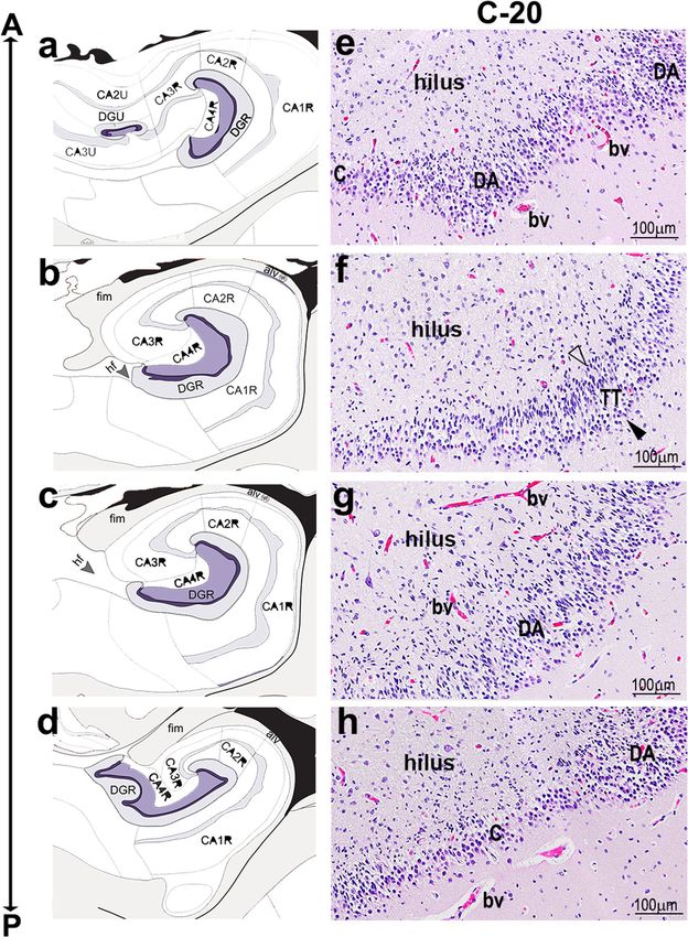
Roy et al. Acta Neuropathologica Communications   (2020) 8:54                                                 Page 3 of 20

Immunohistochemistry                                            2629482) to visualize nuclei. Sections were cover-slipped
Immunohistochemistry and immunofluorescence were                using Fluorogel mounting medium (Electron Microscopy
performed on ~ 10 control brains and 6 epileptic                Sciences, USA, Cat# 50–247-04). Primary antibodies
brains, chosen based on preliminary tissue histology            used: rabbit anti-Calbindin (Swant, Switzerland, RRID:
and sufficient tissue availability. The chosen cases            AB_2721225), rat anti-CTIP2 (Abcam, UK, RRID:AB_
represent a wide range of overlapping ages in the pa-           2064130), rabbit anti-PAX6 (Biolegend, USA, RRID:AB_
tient and control cohorts (Supplementary Table 1,               2565003), rat anti-TBR2 (anti-EOMES, eBioscience,
Additional File 1). Paraffin-embedded, formalin-fixed,          USA, RRID:AB_11042577), rabbit anti-BLBP (Abcam,
hippocampal tissue blocks (~ 4-5 mm thick) were ob-             UK; RRID:AB_2100476), rabbit anti-IBA1 (FUJIFILM
tained and sectioned at average thickness of 5 μm.              Wako Chemicals, USA; RRID:AB_839504). Immuno-
Sections were then deparaffinized, as mentioned in              stained sections were imaged in Olympus VS-120 slide-
the next section, and immunolabelled with mouse                 scanner microscope using Olympus VS-Desktop 2.9
anti-CD163 (Leica, Germany; RRID:AB_2756375),                   software, and later processed in ImageJ 1.51j8 and Ima-
rabbit anti-SOX2 (Invitrogen, USA; RRID:AB_                     geJ2 (NIH, Bethesda, Maryland, USA) and Olympus VS-
2539862), goat anti-PROX1 (R&D systems, USA;                    Olyvia 2.9 software programs respectively.
RRID:AB_2170716), mouse anti-human CD68 (Dako,                     Each antibody was validated for immunofluorescence/
Denmark; RRID:AB_2074844) and mouse anti-GFAP                   immunohistochemistry by the correspondent manufac-
(Dako, Denmark; RRID:AB_2109952) using a Ventana                turer, and these data are available publicly on the com-
Benchmark II automated immunostainer. PROX1                     pany websites with indicated catalog numbers. This was
immunolabeling needed extra stringent conditions:               also validated by us in our experiments, replicating pub-
treatment with citrate buffer (20 mins, steamer) and            lished/expected expression in appropriate control tissue.
overnight antibody incubation at 4 °C. Other anti-              No outliers were encountered.
bodies were subjected to mild to standard condition-
ing and a short 30 mins of antibody incubation at               Quantitative analysis
37 °C. All sections were then treated with biotinylated         Data was collected from H&E-stained coronal sections.
secondary antibodies (Vector Laboratories, USA,                 Thickness and length measurements of human hippo-
RRIDs: AB_2336123, AB_2313606; Jackson ImmunoR-                 campal GC layer were made using Olympus VS-Olyvia
esearch Labs, RRID:AB_2338586), and later stained               2.9, Olympus VS-Desktop 2.9 (Olympus Corporation,
using the DAB method (Vectastain ABC-Peroxidase                 Tokyo, Japan) and ImageJ2 software programs (NIH, Be-
kits, Vector Laboratories, USA, RRID:AB_2336818).               thesda, Maryland, USA). Maximum GC layer thickness
The sections were finally counterstained with                   in each GCD sample was measured at the region where
hematoxylin.                                                    the layer was the thickest. For the ratio measurement of
                                                                bilaminar GC layers, the entire thickness of the DG was
Immunofluorescence                                              measured – from the edge proximal to the hilus to the
Paraffin-embedded human hippocampal sections were               edge proximal to the molecular layer. The individual
warmed on a hot-plate at approximately 45 °C for 15–            thicknesses of the inner and outer layers were obtained
20 min, to melt the paraffin. Slides were then immedi-          as well. To correlate maximum GC layer thickness ver-
ately transferred to fresh 100% xylene and processed            sus age, cadaveric sections showing compact, DA and
through an ethanol hydration gradient (100, 90, 70, 50%         TT phenotypes were compared with corresponding cor-
ethanol solutions for 5 min each), before immersion in          rected age of death. To plot GCD length as a proportion
distilled water. After deparaffinization, immunofluores-        of total DG length, the inner aspect of the DG was mea-
cence was performed as previously described [48].               sured for 12 controls and 12 seizure cases, using one
Briefly, sections were washed thrice in phosphate buffer        coronal hippocampal section with GCD per sample.
saline (PBS), boiled in 10 mM sodium citrate solution           Statistical significance was assessed using Welch-
for antigen retrieval, blocked in 5% serum in PBS with          corrected t-test (for GCD length/Total length plot) and
0.1% Triton X-100 (0.1% PBX solution) and then incu-            two-way ANOVA followed by Tukey post-hoc test (for
bated overnight at 4 °C with primary antibodies. Sections       GCD proportion plots across groups, age and PMI, GC
were then washed thrice in 0.1% PBX solution, incu-             layer thickness comparison plots, GCD occurrence with
bated with appropriate species-specific secondary anti-         clinical diagnoses). Linear regression was used for the
bodies conjugated with Alexa 488, 568 or 647                    maximum GCD thickness versus age of death plots.
fluorophores (Invitrogen, USA; RRIDs: AB_2534088,               These analyses were performed in GraphPad Prism v7.0
AB_10563566, AB_141778) for 2 h at room temperature             (GraphPad Software Inc., San Diego, USA) and in
and then counterstained with DAPI (4′,6-Diamidino-2-            Microsoft Excel. Differences were considered significant
Phenylindole, Dihydrochloride; Invitrogen, RRID:AB_             at p < 0.05. Data are represented as stacked and regular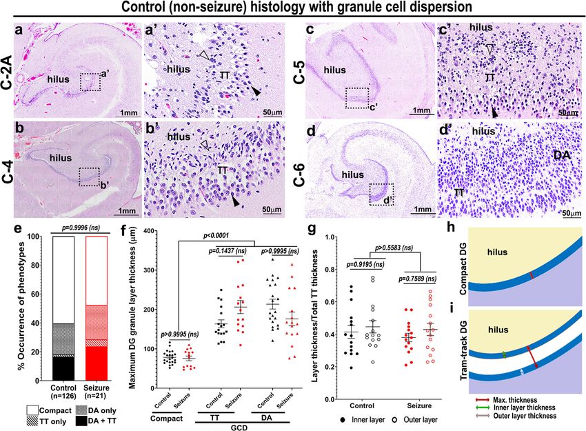
Roy et al. Acta Neuropathologica Communications          (2020) 8:54                                                                    Page 4 of 20

bar graphs and scatter plots with mean ± SEM in Fig. 2                      refer to this as “disaggregated” GCD (DA). The second
and Supplementary Fig. 2 (Additional File 1). Measure-                      subtype has a bilaminar appearance of the GC layer with
ments for Supplementary Fig. 6 were obtained with                           cell-sparse zone in the center [3, 30]; we refer to this as
Nikon Elements BR v3–2 (Nikon Corporation, Tokyo,                           “tram-track” GCD (TT). By studying the H&E-stained
Japan) and final data are represented as mean ± SD.                         hippocampal specimens from patients with seizure his-
                                                                            tory (n = 21), we categorized the morphology of GC layer
Results                                                                     for each as compact (10/21; 47.62%, subtype equivalent
Defining types of GCD in seizure-affected human                             to non-GCP of [8]), only DA (5/21; 23.81%), only TT (1/
hippocampi                                                                  21; 4.76%), or both DA and TT (5/21; 23.81%) (Table 1).
Stereotypically, the human hippocampal GC layer ap-                         Focal granule cell loss was observed in only 1 seizure
pears to have a compact, neuron-dense histology, with a                     case (rigorous morphometric analysis of neuronal dens-
sharp boundary separating the molecular layer (Fig. 1b’,                    ity was not performed). As previously reported [30], we
c,c’). While studying the cadaveric hippocampal samples                     also encountered dispersion and variation in the pattern
from patients with history of epilepsy or seizure, we ob-                   and thickness of the GC layer at the angles and enfolded
served co-existence of compact GC layer and a range of                      regions of both control and seizure-affected hippocampi.
GCD subtypes in the DG (Fig. 1d-d”, e), as originally re-                   However, these areas were excluded from our consider-
ported in hippocampal samples of TLE patients [30].                         ation of “GCD” and also from our quantitative analyses.
These were later classified by Blumcke et al. into differ-
ent categories of DG granule cell pathology (GCP) [8].                      GCD is evident in brains irrespective of the history of
This classification offered 3 major categories – 1) entire                  seizures
DG appears normal, or non-GCP; 2) Type 1 GCP, char-                         While evaluating age-matched cadaveric controls for
acterized by severe cell loss; and 3) Type 2 GCP, that in-                  seizure-affected brain specimens, we serendipitously
cludes at least one DG focus of broadening, clustering or                   identified GCD in some samples. This led us to retro-
duplication of GC layer [8]. In our study, we further sub-                  spectively investigate the presence of GCD in a large set
divided Type 2 GCP category broadly into two subtypes.                      of 126 control human brains, with no history of seizures,
The first is marked by broad, less dense GC layer, often                    and compare the findings with those observed in the 21
with poorly defined borders with the molecular layer; we                    seizure cases (Additional File 2). We identified both DA

 Fig. 1 Cadaveric hippocampi reveal a spectrum of GCD in patients with epilepsy. (a, b, b’) Schematics explaining the coronal sectioning and
 structure of human hippocampus. (c-e) H&E-stained coronal sections of dentate gyrus (DG) from human cadavers, obtained from SCH archives:
 representative images demonstrate a typical structure (c) and compactness of the GC layer (c’) in a control human dentate gyrus (with no history
 of epilepsy/seizure), and GCD in certain seizure cases, ranging from focal “tram-track” (TT) phenotype (d-d”) to more diffused “disaggregated” (DA)
 form (e). Arrowhead indicates outer granular zone distal to hilus; open arrowhead indicates inner granular zone proximal to hilus; bv, blood
 vessel. Scalebars: 1 mm (c, d); 50 μm (c’, d’, d”, e)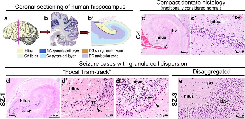
Roy et al. Acta Neuropathologica Communications (2020) 8:54 Page 5 of 20
Table 1 List of cadaveric epilepsy cases with recorded clinical features. 21 cadaveric epilepsy cases with history of single or multiple
events of seizures, accompanied with a table of clinical history obtained from the archives of Seattle Children’s Hospital Department
of Pathology (2014–2019), are tabulated. The features analyzed included age of death, gender, post-mortem interval (PMI), presence
or absence of GCD subtypes, clinical diagnoses like seizure interval (from seizure onset until death), evidence of malformation/
anomaly in the central nervous system (CNS), and/or of other CNS (acquired forms of CNS injury such as hypoxic-ischemic
encephalopathy (HIE), cerebral edema) and non-CNS conditions. GW, gestational weeks; TT, tram-track; DA, disaggregated; mo,
months
Code Age of Corrected Gender PMI Seizure Seizure GCD Diagnoses
death age of (Y/N) interval (Y/N)
(GW) death
TT DA CNS malformation/ Other (CNS) Non-CNS conditions
anomaly
SZ-1a 7 days 7 days M 12 h Y < 24 h Y Y encephalopathy, – Proteus mirabilis septic
seizures shock, coagulopathy
and acidosis, hypoxic
respiratory failure
SZ-2a 6 years 6 years F 10 h Y 6 years; N N global developmental hypoplastic recurrent pulmonary
intractable delay, chronic seizure cerebellum infections
disorder with static
encephalopathy
SZ-3a 16 years 16 years F 22 h Y 13 years N Y severe congenital severe gliosis acute
neuromuscular and neuronal bronchopneumonia,
disorder, complex loss cardiac arrest
status epilepticus
SZ-4b 6 years 6 years M not Y > 5 years; multiple Y Y brain overgrowth gliosis, mild congenital
known episodes till with polymicrogyria, ventriculomegaly diaphragmatic hernia,
death, intractable diffused cortical coagulopathy, defects
dysplasia, AKT3 in liver, spleen and
R465W mutation, kidney
possibly SUDEP
SZ-5a 3 years 3 years M 2h Y 7 weeks; multiple Y N subclinical status cerebral edema, pulmonary arrest
episodes till epilepticus HIE, anoxic brain
death, intractable injury secondary
to pulmonary
arrest
SZ-6a 2 days 2 days F 21.5 h Y 2 days (possible Y Y – cerebral edema, acute chorioamnionitis
seizures) HIE with funisitis and
three vessel umbilical
vasculitis
SZ-7a 4 days 4 days M 38 h Y < 24 h N Y – HIE severe acidosis,
cardiorespiratory
failure, visceral
anomalies
SZ-8a 18mo 18mo M 26 h Y 1–3 months of Y Y developmental delay focal neuronal abdominal distension,
age; no further and seizure disorder loss and gliosis brady-cardiac arrest,
seizures (2q21.1 duplication) in hippocampus acute pan-lobar
post-treatment pneumonia
of phenobarbital
SZ-9a 2 years 2 years M 72 h Y 4 months; multiple N N retinoblastoma with – pulmonary edema,
episodes between diffuse congestive
first seizure and leptomeningeal hepatomegaly
death spread and direct
infiltration of brain
and spinal cord
SZ-10a 5 weeks 33GW F 17.5 h Y < 24 h Y Y multiple seizure hemorrhage and necrotizing
(28GW) events necrosis enterocolitis
secondary to
dural venous
thrombosis.
SZ-11a 12 years 12 years F 96 h Y 16 months; N N – cerebral edema, appendicitis
intractable HIE and brain
death
SZ-12a 19 years 19 years M 216 h Y 14 years; N N seizure disorder – Trisomy 16p/monosomy
intractable 9p, respiratory distress
syndrome, Pseudomonas
pneumonia, cardiomegalyRoy et al. Acta Neuropathologica Communications (2020) 8:54 Page 6 of 20
Table 1 List of cadaveric epilepsy cases with recorded clinical features. 21 cadaveric epilepsy cases with history of single or multiple
events of seizures, accompanied with a table of clinical history obtained from the archives of Seattle Children’s Hospital Department
of Pathology (2014–2019), are tabulated. The features analyzed included age of death, gender, post-mortem interval (PMI), presence
or absence of GCD subtypes, clinical diagnoses like seizure interval (from seizure onset until death), evidence of malformation/
anomaly in the central nervous system (CNS), and/or of other CNS (acquired forms of CNS injury such as hypoxic-ischemic
encephalopathy (HIE), cerebral edema) and non-CNS conditions. GW, gestational weeks; TT, tram-track; DA, disaggregated; mo,
months (Continued)
Code Age of Corrected Gender PMI Seizure Seizure GCD Diagnoses
death age of (Y/N) interval (Y/N)
(GW) death
TT DA CNS malformation/ Other (CNS) Non-CNS conditions
anomaly
SZ-13a 7 weeks 39 4/7 F 14 h Y 6 weeks (possible N Y epilepsy (focal – severe pulmonary
(32 4/7 GW) GW seizures) apoptotic neurons hypertension,
within DG) veno-occlusive disease
SZ-14a 8 years 8 years F 65 h Y 2 years (sleep N N encephalomalacia, – unbalanced
EEG done) hydrocephalus, chromosomal
seizures, translocation,
developmental delay congenital mitral valve
stenosis, heart failure
SZ-15a 7 weeks 7 weeks F 3h Y 6 weeks N Y hypotonia and elevated CSF –
episodic breathing and plasma
progressing to glycine levels
seizures
SZ-16a 17 years 17 years F 96 h Y 5 days N N generalized tonic- acute Type 1 diabetes,
clonic seizure, brain hemorrhage, oligoarticular juvenile
herniation edema arthritis, celiac disease
SZ-17a 17 years 17 years M 64 h Y ~ 17 years; N Y spastic quadriplegia, white matter acute kidney injury,
intractable epilepsy, static gliosis obstructive apnea,
leukoencephalopathy, hypotonia
ventriculomegaly
SZ-18a 8 days 8 days M 42 h Y 8 days N N seizures (treated with brain herniation, ornithine
(onset at birth) antiepileptics) diffuse cerebral transcarbamylase
edema deficiency,
hyperammonemia,
hepatosplenomegaly
SZ-19a 3 years 3 years F 15.6 h Y 2.5 years N N severe craniofacial neurological –
malformations, seizure injury, meningitis
history
SZ-20a 4 years 4 years M 15.5 h Y 16 months N N seizures (no subacute diffuse multiple chromosomal
recurrence post- CNS abnormalities and
treatment with medi- hemorrhagic associated chronic
cations), global devel- necrosis with health problems,
opmental delay massive atypical lymphoid
intraventricular hyperplasia, concerning
blood clot primary or secondary
immunodeficiency
SZ-21 9 years 9 years M 2h Y 2 months N N refractory status diffuse severe –
a
epilepticus secondary gliosis, patchy
to febrile infection- neuronal loss,
related status epilepti- dramatic loss of
cus, multiple events CA1 neurons
a
based on microscopic evaluation of archived hippocampal sections
b
case published in [3, 46]; unused right hemisphere was obtained from SCH morgue and pathological studies were done by RPK on the right
hippocampus for the first time for this study
and TT subtypes of GCD in control DG, similar to that hoc test, confirmed no significant difference across these
seen in cadaveric epileptic brains (Fig. 2a-d’, Table 2; 4 groups (p > 0.9996; Fig. 2e). Furthermore, we often ob-
Supplementary Fig. 1, Additional File 1). The proportion served the existence of more than one morphological
of each subtype observed in controls was as follows: type of GC layer (compact, DA, TT) in the same hippo-
compact (76/126; 60.32%), only DA (27/126; 21.43%), campal section, irrespective of the seizure history, con-
only TT (2/126; 1.58%), both DA and TT (21/126; sistent with previous reports [8, 21].
16.67%). Statistical analysis between control and seizure
cases, using two-way ANOVA, followed by Tukey post-Roy et al. Acta Neuropathologica Communications (2020) 8:54 Page 7 of 20 Fig. 2 Cadaveric hippocampi reveal the GCD spectrum in controls with no history of seizures. (a-d’) H&E staining of coronal hippocampal sections of control human cadavers, with no history of epilepsy or seizure, revealed the entire spectrum of GCD ranging from focal tram-track (TT) to disaggregated (DA) forms, as observed in some seizure cases. Often, TT and DA forms are seen at the same plane of section. Open arrowhead indicates inner granular zone proximal to hilus; arrowhead indicates outer granular zone distal to hilus. (e) Frequencies of GCD subtypes in seizure cases (11/21) were not significantly different from that in controls (50/126), as determined by two-way ANOVA, followed by Tukey post- hoc test (p > 0.9996). (f) Comparison of the maximum thickness of GC layer between control and seizure hippocampal samples, measured as shown in (h,i), revealed no significant differences in all DG subtypes – compact, TT, DA. But the maximum GC thicknesses of both TT and DA sets were significantly higher than the compact ones (p < 0.0001). (g) Ratios of inner or outer layer thickness to the total GC thickness, as demonstrated in (i), showed no significant difference between control and mutant, as well as within each group. Data are represented as 100% stacked columns (e), mean ± SEM (f) or mean ratio ± SEM (g) in scatter plots; two-way ANOVA followed by Tukey post-hoc test were performed. Differences were considered significant at p < 0.05; ns, not significant. Scalebars: 1 mm (a, b, c, d); 50 μm (a’, b’, c’, d’) Morphometric measurements of GCD are similar in GC layer occurs during the first 8 postnatal months, seizure patients and controls when immature cells gradually disappear from the sub- Measuring the maximum thickness of compact, TT and granular zone near the hilus [49, 50]. We categorized DA sub-zones of GC layer demonstrated that both types the age of death into bins and calculated the proportion of dispersed DG were significantly thicker than compact of control and seizure cases per bin that demonstrated DG (p < 0.0001) in control and seizure-affected brains; GCD. Statistical analyses showed no significant correl- however, no significant difference was observed between ation between GCD occurrence and the age of death, in control and seizure cases in thicknesses of either the either control or seizure groups (p > 0.2; Supplemen- compact or dispersed areas (Fig. 2f,h,i). Moreover, for tary Fig. 2a, Additional File 1). The PMI for the en- foci of tram-track GCD, the ratios of the inner or outer tire cohort largely varied between 2 h and 336 h. layer thicknesses to the total (maximum) GC layer thick- Comparison of GCD and non-GCD numbers across ness were not significantly different for both control and different PMIs showed no significant correlation be- seizure-affected brains (Fig. 2g,i). Cell migration to the tween development of GCD and PMI in both control
Roy et al. Acta Neuropathologica Communications (2020) 8:54 Page 8 of 20
Table 2 List of cadaveric controls with recorded clinical features, demonstrating GCD. 50 controls with no history of seizures, that
demonstrated presence of at least one type of GCD in the studied hippocampal section obtained from the archives of Seattle
Children’s Hospital Department of Pathology (2014–2019), are tabulated. Other related clinical features obtained/analyzed were age
of death, gender, post-mortem interval (PMI), presence or absence of GCD subtypes, clinical diagnoses like evidence of
malformation/anomaly in the central nervous system (CNS), and/or of other CNS (acquired forms of CNS injury such as hypoxic-
ischemic encephalopathy (HIE), cerebral edema) and non-CNS conditions. GW, gestational weeks; TT, tram-track; DA, disaggregated;
mo, months
Code Age of Corrected Gender PMI Seizure GCD Diagnoses
death age of (Y/N) (Y/N)
(GW) death
TT DA CNS malformation/ Other (CNS) Non-CNS conditions
anomaly
C-1a 1 day 37GW F 14 h N N Y large occipital neuronal multiple congenital
(37GW) (focal DA) encephalocele, disorganization, HIE abnormalities
focal dysplasia
C-2A, 4 weeks 31GW M 32 h N Y Y – - RH-isoimmunization
C-2Ba (27GW) hydrops fetalis, liver
failure, respiratory
distress
C-3a 3 weeks 29GW M 23 h N Y Y – intraventricular necrotizing enterocolitis
(26GW) hemorrhage, and pneumatosis, sepsis
frontoparietal
periventricular
leukomalacia
C-4a 3 weeks 3 weeks F 57.5 h N Y Y – diffuse edema congenital
diaphragmatic hernia,
coagulopathy, defects
in liver, spleen and
kidney
C-5a 7 weeks 28GW M 12 h N Y Y – HIE with multi-organ hypoxic
(27GW) periventricular ischemic injury
leukomalacia
C-6a 2.5mo 10 weeks F 12 h N Y Y – cerebral atrophy immunodeficiency
(term) disorder of undefined
etiology, massive
hepatomegaly, active
bronchopneumonia,
cardiomegaly
C-7a 15 days 30GW F 6h N Y Y – – congenital heart disease,
(28GW) acute multifocal
pneumonia, congestion
and hemorrhage
C-8a 3mo 7 weeks F 17 h N Y Y – cerebral atrophy with Trisomy 21, severe
(35GW) HIE, edema hepatic fibrosis with
cholestasis, pneumonia,
cardiac defects, liver
and kidney injury
C-9a 2mo 8 weeks M 20 h N Y Y – remote HIE without neonatal gastroschisis
acute changes, focal repair, cardiovascular
cystic periventricular defects
leukomalacia
C-10a 3mo 12 weeks F 16 h N Y Y – subacute HIE with congenital cardiovascular
uncal herniation defects, 15q26-qter
deletion, multi-organ
hypoxia
C-11a 7 days 33GW F 15 h N Y Y – subdural hematoma, cystic necrosis of liver,
(32GW) diffuse HIE with respiratory failure,
widespread gliosis and congested spleen
early mineralization,
periventricular
leukomalacia,
hemorrhage, few
pyknotic and
karyorrhectic cells noted
in hippocampusRoy et al. Acta Neuropathologica Communications (2020) 8:54 Page 9 of 20
Table 2 List of cadaveric controls with recorded clinical features, demonstrating GCD. 50 controls with no history of seizures, that
demonstrated presence of at least one type of GCD in the studied hippocampal section obtained from the archives of Seattle
Children’s Hospital Department of Pathology (2014–2019), are tabulated. Other related clinical features obtained/analyzed were age
of death, gender, post-mortem interval (PMI), presence or absence of GCD subtypes, clinical diagnoses like evidence of
malformation/anomaly in the central nervous system (CNS), and/or of other CNS (acquired forms of CNS injury such as hypoxic-
ischemic encephalopathy (HIE), cerebral edema) and non-CNS conditions. GW, gestational weeks; TT, tram-track; DA, disaggregated;
mo, months (Continued)
Code Age of Corrected Gender PMI Seizure GCD Diagnoses
death age of (Y/N) (Y/N)
(GW) death
TT DA CNS malformation/ Other (CNS) Non-CNS conditions
anomaly
C-12a 2mo 5 weeks F 8h N Y Y – diffuse mild complex congenital
(37GW) cerebral WM gliosis heart disease,
cardiomegaly, aspiration
pneumonitis
C-13a 8 years 8 years F 7h N N Y – hemorrhagic methylmalonic acidemia,
infarction, mild WM chronic liver failure,
atrophy, DG coagulopathy, severe
hypoplasia and diffuse
neuronal loss, gliosis bronchopneumonia,
recurrent fevers
C-14a 2mo 8 weeks F 20 h N Y Y – remote HIE without neonatal gastroschisis
(term) acute changes, focal repair, cardiovascular
cystic periventricular defects, Clostridium
leukomalacia infection
C-15a 18 days 18 days M 13.5 h N Y Y – kernicterus involving Beckwith-Wiedemann
hippocampi, diffuse Syndrome, respiratory
gliosis with failure, acute kidney
periventricular injury, thymic cortical
eukomalacia stress
C-16a 10 weeks 2 weeks M 11 h N N Y – – liver dysfunction of
(32GW) uncertain etiology,
cytomegalovirus
infection
C-17a 23 days 28GW M 16.25 h N Y Y – severe intracranial necrotizing enterocolitis,
(25GW) hemorrhage severe pneumonia,
pulmonary hemorrhage
C-18a 6 years 6 years F 68 h N Y Y – craniosynostosis GLIS3 mutation, hepatic
surgery fibrosis
C-19a 6 years 6 years M 15 h N Y Y diffuse infiltrating – –
pontine glioma,
mild ventriculomegaly
C- 12 h 38GW M 38.5 h N Y Y – HIE asystole at birth,
20b (38GW) bilaterallydilated ureters
and bladder, increased
extramedullary
hematopoiesis
C-21a 3 days 38GW M 15.75 h N N Y – mild HIE and edema hemorrhagic and
(38GW) necrotic small bowel,
anomalies in alimentary
tract, liver failure
C-22a 8 weeks 2 weeks M 8h N Y Y mild mild diffuse gliosis of Pentalogy of Cantrell,
(34GW) ventriculomegaly white matter left pulmonary artery
stenosis
C-23a 10 years 10 years M 11 h N Y Y – immunodeficiency,
Pseudomonas and
Aspergillus infection
C-24a 3 days 38GW M 15.75 h N N Y – mild edema and HIE hemorrhagic and
(38GW) necrotic small bowel,
anomalies in alimentary
tract, liver failure
C-25a 17 years 17 years M 59 h N N Y – – recurrent B cell
lymphoblastic leukemia
and aspergillosisRoy et al. Acta Neuropathologica Communications (2020) 8:54 Page 10 of 20
Table 2 List of cadaveric controls with recorded clinical features, demonstrating GCD. 50 controls with no history of seizures, that
demonstrated presence of at least one type of GCD in the studied hippocampal section obtained from the archives of Seattle
Children’s Hospital Department of Pathology (2014–2019), are tabulated. Other related clinical features obtained/analyzed were age
of death, gender, post-mortem interval (PMI), presence or absence of GCD subtypes, clinical diagnoses like evidence of
malformation/anomaly in the central nervous system (CNS), and/or of other CNS (acquired forms of CNS injury such as hypoxic-
ischemic encephalopathy (HIE), cerebral edema) and non-CNS conditions. GW, gestational weeks; TT, tram-track; DA, disaggregated;
mo, months (Continued)
Code Age of Corrected Gender PMI Seizure GCD Diagnoses
death age of (Y/N) (Y/N)
(GW) death
TT DA CNS malformation/ Other (CNS) Non-CNS conditions
anomaly
C-26a 4mo 4mo M 55 h N N Y axonal mixed deafness growth delay, respiratory
sensory/ motor distress
neuropathy
C-27a 2mo term F 58 h N N Y – periventricular cardiopulmonary
(32GW) (40GW) leukomalacia with abnormalities, congenital
acute HIE cardiac anomalies,
renomegaly
C-28a 1 week 1 week M 45 h N N Y – HIE, periventricular congestion and
leukomalacia with hemorrhage
prominent gliosis
and neuronal loss
C-29a 10mo 10mo F 46.5 h N N Y – global chronic HIE, heterotaxy syndrome,
hippocampus shows complex congenital
mild loss of neurons heart disease
in CA1 region
C-30a 3 years 3 years M 16.5 h N N Y – HIE post cardiac arrest, asthma, acute sepsis,
early necrosis of cardiac arrest, stress
hippocampus atrophy
C-31a 3mo 101 days F 13 h N N Y – mild HIE respiratory distress,
(42GW) pulmonary vein stenosis
C-32a 14 days 30GW M 16 h N N Y – mild HIE, diffuse gliosis massive subacute
(28 week) in WM hepatic necrosis with
iron overload,
coagulopathy, chronic
neonatal lung disease,
multiple organ defects
C-33a 3 years 3 years M 20.5 h N N Y global developmental HIE with edema myopathy, cardiac
delay failure, respiratory
failure, infectious
diseases, respiratory
distress, sepsis
C-34a 5weeks 39GW F 84 h N N Y – – necrotizing enterocolitis
(33GW)
C-35a 18 h 18 h M 14 h N N Y – – complex congenital
heart disease, total
anomalous pulmonary
venous return,
lymphatic distention
C-36a 17 years 17 years F 69.5 h N Y Y medulloblastoma, widespread brain death pulmonary thrombi
brain injury related and congestion and
to Aspergillus hepatosplenomegaly
encephalo-meningitis,
lateral ventriculomegaly
C-37a 9 days 9 days F 31 h N N Y – HIE, brain injury liver steatosis
C-38a 6 h 1.5 weeks F 78 h N N Y – HIE cardiac respiratory
(41 5/7 GW) failure, coagulopathy,
anemia, severe
metabolic acidosis,
in-utero feto-maternal
hemorrhage
C-39a 16mo 16mo M 15 h N N Y – multifocal brain diffuse adherent bowel,
infarction with global necrotizing soft tissue
HIE (CA1 dispersed) infections, cardiac
arrest historyRoy et al. Acta Neuropathologica Communications (2020) 8:54 Page 11 of 20
Table 2 List of cadaveric controls with recorded clinical features, demonstrating GCD. 50 controls with no history of seizures, that
demonstrated presence of at least one type of GCD in the studied hippocampal section obtained from the archives of Seattle
Children’s Hospital Department of Pathology (2014–2019), are tabulated. Other related clinical features obtained/analyzed were age
of death, gender, post-mortem interval (PMI), presence or absence of GCD subtypes, clinical diagnoses like evidence of
malformation/anomaly in the central nervous system (CNS), and/or of other CNS (acquired forms of CNS injury such as hypoxic-
ischemic encephalopathy (HIE), cerebral edema) and non-CNS conditions. GW, gestational weeks; TT, tram-track; DA, disaggregated;
mo, months (Continued)
Code Age of Corrected Gender PMI Seizure GCD Diagnoses
death age of (Y/N) (Y/N)
(GW) death
TT DA CNS malformation/ Other (CNS) Non-CNS conditions
anomaly
C-40a 4 weeks 4 weeks F 24 h N Y Y – mild HIE with mild truncus arteriosus
gliosis
C-41a 4 days 27GW F 144 h N N Y – widespread HIE splenic congestion
(27GW)
C-42a 8 years 8 years F 15 h N N Y – subdural hematoma B-cell acute
lymphoblastic
leukemia, sepsis,
acute kidney injury,
cardiac instability
C-43a 6mo 6mo M 41.5 h N N Y – global remote HIE Denys-Drash Syndrome,
chronic kidney disease,
Pseudomonas abscess,
multiple cardiac arrests
C-44a 6 years 6 years F 13 h N N Y diffuse intrinsic – –
6mo 6mo pontine glioma
C-45a 3 days 40GW F 40.5 h N N Y – – profound hypoxemic
(40 1/7GW) respiratory failure,
lung developmental
arrest
C-46a 3 weeks 38GW M 19 h N N Y – acute HIE congenital heart
(35GW) disease, kidney
hemorrhage
C-47a 16 days 16 days M 10 h N N Y – HIE, diffuse WM gliosis, complex congenital
periventricular leukomalacia, heart disease, status
subarachnoid hemorrhage post-surgical repair
C-48a 7 weeks 7 weeks M 50 h N N Y – – necrotizing enterocolitis
C-49a 35GW 35GW M 63 h N Y N – – congenital pulmonary
dysplasia, interstitial
chromosomal deletion
ch17
C-50a 6 days 6 days M 69 h N Y N – subicular necrosis, acute HIE 22q11.2 chromosomal
deletion, DiGeorge
syndrome
a
based on microscopic evaluation of archived hippocampal sections
b
step sections as described in text
Table 3 Summary of study parameters. Summary table of variables used in this retrospective study to compare between the control
(no seizure history) and seizure groups. Variables shown are corrected age of death, post-mortem interval (PMI), gender and seizure
interval (from seizure onset until death). Comparison demonstrated broad overlap especially in the range of age of death and PMI
between control and seizure sets
Group Number of cases Range of corrected age of death Range of PMI Gender (% of total) Range of seizure interval
Control 126 -21 to + 1060 weeks 2 to 336 h Male: 69 (54.76%); Female: 57 (45.24%) –
Seizure 21 -7 to + 990.7 weeks 2 to 216 h Male: 11 (52.38%); Female: 10 (47.62%) < 24 h to 17 yearsRoy et al. Acta Neuropathologica Communications (2020) 8:54 Page 12 of 20
and seizure groups (Supplementary Fig. 2b, Additional Calbindin is noted to be prominent in outer granule
File 1). Comparison of maximum GC layer thickness cells and absent or sparse in the inner layer/half of the
in compact, DA and TT forms showed that both dis- GC layer; however no overt cell loss were observed in
aggregated and “tram-track” GC layer thicknesses most of these brains [1]. Similarly in our study, Calbin-
were wider than the “compact” thickness for both din and CTIP2 expression was stronger in the outer
control and seizure brains, across ages of the cadav- layer of the compact as well as “tram-track” control and
eric samples (Supplementary Fig. 2c, c', Additional epileptic DG, compared to the inner GC layer (Fig. 3d,e,
File 1). While other groups showed no correlation g,h,o,q). On the other hand, CTIP2 and Calbindin were
with the age of death, the “tram-track”-ed seizure expressed in the entire “disaggregated” GC layer, in both
cases (Seizure TT) demonstrated a slightly positive controls and seizure cases (Fig. 3f,i,p,r). BLBP was
correlation of GC layer thickness with increasing age expressed in the hilus and the molecular layer of the
beyond 60 weeks (R2 = 0.4517, p = 0.0167; Supplemen- dentate in both control and seizure-affected brains
tary Fig. 2c, Additional File 1). Table 3 summarizes (Fig. 3j-l, s,t), as expected [38, 53].
the overlapping range of clinical parameters (age of To determine whether the inner layer of tram-track
death, PMI, gender proportion) between control and DG corresponds to the sub-granular zone harboring
seizure groups used in this study. progenitors and immature neurons, we examined the
Further, we segregated the patient history into differ- distributions of putative neural progenitor markers
ent diagnostic categories and analyzed whether putative (SOX2, PAX6, TBR2) in control and seizure-affected
GCD occurrence is dependent on any particular clinical hippocampi (Supplementary Fig. 3a-o, Additional File 1)
diagnoses (Supplementary Fig. 2d, Additional File 1). [12, 61]. No overt differences were observed in the ex-
Two-way ANOVA revealed no statistically significant pression of these markers within the dispersed (DA or
correlation between the different diagnostic subgroups TT) dentate gyri of control or seizure hippocampi. Our
of patients and the presence of GCD in either control or data are thus consistent with the study that identified
seizure cases. We observed higher incidence of diagnoses disassociation of proliferation with GCD in the brains of
related to the central nervous system for seizure cases TLE patients [20].
than controls; however due to relatively low sample size
of the seizure cases, this was also not considered statisti- GCD is not dependent on increased gliosis or injury-
cally significant. Finally, we calculated the proportion of driven mechanisms irrespective of seizure occurrence
GC layer affected by GCD per plane of section in con- To determine whether tissue injury or macrophage-
trols and epileptic brains and found no significant differ- related inflammation is associated with the GCD
ences between them (p = 0.801; Supplementary Fig. 2e, phenotypes, we examined the hippocampal expres-
Additional File 1). Hence, GCD was found to occur at a sion of injury markers GFAP and CD163 in 6 seizure
similar proportion in both control and seizure-affected patients and 10 controls (Fig. 4) [18, 19]. Enhance-
brains and this occurrence was largely independent of ment of strongly expressing GFAP+ and CD163+
age, gender, PMI or clinical diagnoses. cells denotes activation of astrocytes (gliosis) and
inflammation-related M2 macrophages respectively,
Molecular expression of cells is identical in both control both indicative of tissue injury [19, 57]. We observed
and seizure-affected dentate gyri with GCD increased gliosis in some of the cadaveric seizure-
To investigate molecular differences between control affected hippocampi, independent of GCD (Fig. 4b,f,
and seizure-affected brains, we performed immunohisto- h), although the DG in every epilepsy case did not
chemistry and immunofluorescence on the cadaveric have increased GFAP+ astrocytes (Fig. 4d). Enhanced
hippocampal sections. PROX1, marking cells in the adult GFAP expression or gliosis was generally not ob-
GC layer and sub-granular zone [34], was expressed served in the control sections, irrespective of pres-
similarly in both control and seizure-affected DG ence of GCD (Fig. 4j,l,n,p); among the 10 controls
(Fig. 3a-c,m,n). CTIP2 is typically expressed in the hip- evaluated immunohistochemically, we encountered
pocampal CA1 and post-mitotic dentate granule cells only 2 control brains that exhibited mild gliosis (as
[47], predominantly in the superficial GC layer; its ex- represented in Fig. 4r). We also did not observe any
pression in human DG gets attenuated gradually after significant differences in the number of CD163+ cells
mid-gestation [12]. In normal human DG, Calbindin im- in the studied cadaveric control or epileptic hippo-
munostaining is observed in hilus, molecular and GC campi (Fig. 4c,e,g,i,k,m,o,q,s). Further, we studied the
layers, with strongest expression in the early-born post- expression of CD68 and IBA1, generic markers for
mitotic neurons, located in the outer part GC layer distal macrophages and microglia respectively, which con-
to the hilar region [2]. In human brains affected by dif- gregate in response to tissue injury and cellular acti-
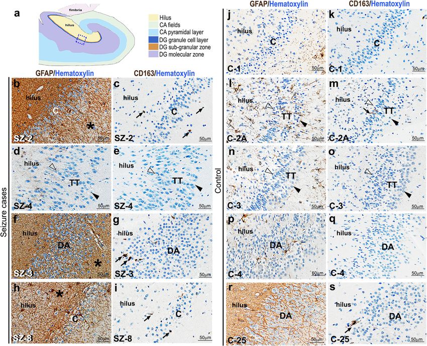
ferent types of epilepsy and/or hippocampal sclerosis, vation [28, 57]. Similar to CD163, CD68+ cells wereRoy et al. Acta Neuropathologica Communications (2020) 8:54 Page 13 of 20 Fig. 3 Analysis of cell types in cadaveric control and seizure-affected dentate gyri demonstrating GCD. (a-t) Immunohistochemistry studies were performed on coronal paraffin sections of control and seizure-affected cadaveric hippocampi using GC markers, namely PROX1, CTIP2, Calbindin, BLBP. Representative images of compact, tram-track (TT) and disaggregated (DA) DG from both control and seizure cases are demonstrated. No difference was observed between control and seizure brains with respect to molecular expression of PROX1, CTIP2, Calbindin and BLBP across groups. PROX1 was expressed in both inner and outer GC layers of the tram-track DG (a-c,m,n). CTIP2 and Calbindin expression is more prominent in the outer layer, compared to the respective inner layers, as seen in compact and tram-track DG (d,e,g,h,o,p); but was expressed in the entire DA zone both in control and seizure cases (f,i,p,r). BLBP marked the inner granular layer and the hilar region more densely, as expected (j-l,s,t). BLBP staining in SZ-1 showed some non-specific background due to presumed cell autolysis (s). TT, tram-track; arrowhead, outer granular zone distal to hilus; open arrowhead, inner granular zone proximal to hilus; bv, blood vessel. Scalebars: 50 μm (a-t) minimal and expressed non-differentially between Extent of GCD changes across antero-posterior levels of control and epileptic brains (Supplementary Fig. 4a- human hippocampus e, k-o; Additional File 1). We did encounter a few While analyzing multiple sections from the cadaveric IBA+ activated microglia, identified by rounded, hippocampal paraffin blocks, we observed that GCD bushy cellular morphology; but there was no correl- was present inconsistently and to variable extents in ation between their presence and GCD or seizure different sectioning planes of both control and occurrence (Supplementary Fig. 4f-j, p-t; Additional seizure-affected hippocampi (Supplementary Fig. 5a- File 1). Thus, our results indicate that occurrence of d", Additional File 1). This suggested that the putative dispersed GC layer in both controls and seizure seizure-related nature of “disaggregated” and/or cases is not directly associated with either injury or “tram-track” GC layer in the literature might merely inflammation, although gliosis appeared to be more be a consequence of sampling bias. As mentioned in common in epileptic hippocampi. the Methods, our designation of controls and seizure
Roy et al. Acta Neuropathologica Communications           (2020) 8:54                                                                     Page 14 of 20

 Fig. 4 GCD occurrence does not correlate with increased hypoxia/ischemia or gliosis in both control and seizure cases. (a) Schematic of coronal
 human hippocampus; box showed the region of interest represented in (b-o). (b-s) Representative images of control and seizure hippocampal
 samples, with compact (C), tram-track (TT) and disaggregated (DA) DG, studied for injury markers GFAP and CD163. Enhancement of GFAP
 expression and increase in CD163+ cells mark gliosis and M2 macrophages respectively, both indicative of tissue injury. Although there was
 observable gliosis in most epilepsy brains (asterisk; b, f, h), it did not correlate with the occurrence of GCD (d-g). SZ-8 demonstrated focal granule
 cell loss as well as gliosis (h,i). Enhanced gliosis was never observed in the studied control sections (j, l, n, p), except mildly in one case (r).
 Although a few CD163+ cells were observed in epilepsy and control sections (black arrows; c, g, i, m, s), the number of M2 macrophages were
 not significantly different between control and seizure sections (c, e, g, i, k, m, o, q, s). Arrowhead, outer granular zone distal to hilus; open
 arrowhead, inner granular zone proximal to hilus. Scalebars: 50 μm (b-s)

cases having or not having GCD was largely based on                           prominent TT, then a combination of TT and DA
archived cadaveric histological slides of the hippocam-                       forms and back to compact forms. Although no for-
pus. To verify the possibility of sampling bias, we cor-                      mal morphometric analysis was performed, no correl-
onally sectioned one control hippocampus (C-20) at                            ation was suggested between anterior-posterior
different levels across the antero-posterior axis, each                       location or DG contour and any specific pattern of
section a maximum of ~ 5 mm apart from the next.                              GCD. Instead, GCD appeared to be a sporadic and
Contours of the hippocampus, including DG, nor-                               somewhat randomly distributed variation in DG
mally change along the antero-posterior axis (Fig. 5a-                        histology.
d). At different points along the same axis we also                             Collectively, the data suggest that GCD is a more com-
observed each of the morphological subtypes of the                            monly occurring phenomenon than previously appreci-
GC layer (Fig. 5e-h). The morphology of GC layer                              ated, and that over-representation of seizure patients in
varied from overlapping compact and DA forms to                               the hippocampal studies may relate to a more thorough
Roy et al. Acta Neuropathologica Communications         (2020) 8:54                                                                   Page 15 of 20

 Fig. 5 GCD is not consistent across sectioning planes. (a-d) Schematics of the human hippocampus, modified from the Allen Human Brain Atlas,
 at different levels along the antero-posterior (A-P) axis. CA marks subdivisions of cornus ammonis (CA1–4); DG marks the dentate gyrus. The
 hippocampal morphology changes along the A-P axis. (e-h) Representative images of H&E-stained coronal hippocampal sections of C-20,
 specifically depicting the GC layer. Sectioning plane of each section roughly corresponds to that of the adjacent schematic. The control GC layer
 demonstrated the entire spectrum of GCD categories: compact (C), disaggregated (DA) and tram-track (TT), often showing co-existence and
 repetition along the A-P axis. Arrowhead, outer granular zone distal to hilus; open arrowhead, inner granular zone proximal to hilus; bv, blood
 vessel. Scalebars: 100 μm (e-h)

sampling in these cohorts. Most importantly, GCD is                         controls without seizure history, we identified certain
definitely not an exclusive or characteristic feature of                    histopathological characteristics, known to be unique
brains affected by epilepsy.                                                to epileptic brains, in both groups. We found no sig-
                                                                            nificant differences in neuronal morphology, molecu-
Discussion                                                                  lar expression in different cell types or extent of cell
In this retrospective study of 147 cadaveric hippo-                         loss between control and seizure-affected hippocampi.
campi from pediatric patients with seizures and                             Our study challenges the existing dogma in
Roy et al. Acta Neuropathologica Communications (2020) 8:54 Page 16 of 20
neuropathology that GCD is a specific feature of Not surprisingly, some variation of the Blumcke et al.
chronic epilepsy, such as TLE. (2009) definition has been used in many published stud-
ies of GCD and its putative specific association with epi-
Defining granule cell dispersion (GCD) lepsy; however, most of these either lacked or included
The GC layer is a prominent neuronal layer typically only a few non-seizure controls for comparison (Supple-
composed of a densely packed C-shaped band of cells mentary Table 2, Additional File 1). Further, we noted
in a normal hippocampus. A granule cell consists of that alternative methods of GCD assessment exist. Since
clusters of apical dendrites, extending through the dispersed GC layer is generally thicker than “normal”,
dentate molecular layer, and basal dendrites, which some studies assessed GCD based on a morphometric
extend into the hilus and molecular layer in human method of averaging measurements of GC layer thick-
hippocampus [4, 51]. Granule cell axons (or mossy fi- ness across “straight” parts of DG and then re-averaging
bers) project into the hilus to synapse with hilar in- values obtained from several sections per hippocampus
terneurons, which in turn continue to synapse onto [24, 37, 52]. In these studies, GCD was defined as an
pyramidal neurons in area CA3, thus establishing part average DG thickness whose value measured greater
of the hippocampal circuitry. The structure and syn- than a range of average thicknesses obtained from non-
aptic connectivity of granule cells are vital for brain seizure controls. As illustrated in Supplementary Fig. 6
functions, especially learning and memory consolida- (in Additional File 1), this approach, though quantitative,
tion. Ontologically, granule cells localized proximal to is likely to exclude focal dispersion, which is specifically
the outer molecular layer are born earlier than the associated with epilepsy in many publications. Although
ones found at the inner side, adjacent to the hilus [7, the morphometric approach may seem less subjective
45, 50]. Pathologically, this cell layer is found to be than our study design, determination of what constitutes
susceptible to hippocampal insults, injury, hypoxia, a “straight” segment of the DG is also highly subjective
brain malformations and epilepsy [15, 23, 29]. These and may bias against inclusion of focal GCD in these
conditions often lead to hippocampal sclerosis charac- analyses.
terized by neuronal cell loss and reactive gliosis,
microglial infiltration and, according to prior studies, GCD is not exclusively present in seizure-affected brains
GCD [10, 54]. Although many reports have accepted the correlative
GCD is a histological phenotype reported in the af- dogma of presence of GCD with seizure history, contra-
fected dentate gyri of patients suffering from chronic dictions exist in the field. One study suggests that GCD
epilepsy [3, 5, 10, 15, 30, 55, 56]. GCD has been de- is more correlated with learning and memory changes
scribed in ~ 40% of the hippocampi of TLE patients [9, than to seizures, hippocampal sclerosis or neuronal loss
56, 59]. Similar to the human epilepsy-affected brains, [8]. GCD has also been observed in many children, with
animal models of epilepsy demonstrated different kinds no history of seizures, who died suddenly and unexpect-
of dispersed GC layer, thought to be replicative of hu- edly [26, 31, 32]. Although the hippocampal findings in
man patient pathophysiology [40, 43]. Variable overlap- the latter research were interpreted as potentially signifi-
ping patterns of GCD have been described. These cant malformations, the possibility of normal variation
include focal dispersed clusters of granule cells ectopi- was not excluded. Another retrospective study of 68
cally present in either hilus or molecular DG layer, seg- SUDEP (sudden unexpected death in epilepsy) cases
mental broadening and duplication or “tram-tracking” of showed no evidence of significant differences in hippo-
the GC layer [8, 21, 30]. Some reports categorized bila- campal position or shape or GC abnormalities compared
mination separately from GCD [56]. In our study, both to 53 age-matched non-epilepsy controls, although nei-
focal broadening and bilamination of GC layer were con- ther DA nor TT were specifically evaluated [52]. Simi-
sidered as subtypes of GCD. larly, a recent study concerning sudden unexplained
Different approaches also exist with regard to the as- death in childhood (SUDC) cases also could not clearly
sessment of what should be considered as “GCD”. In our relate hippocampal abnormalities to either cause or ef-
study, we have adapted and modified the GCD classifica- fect of seizures [39]. We are aware of only one report
tion as proposed by Blumcke et al. [8], where the pres- mentioning the existence of GCD in human patients
ence of different grades of GCD was subjectively with no history of epilepsy or seizures [25]. This study
assessed, primarily based on differential GC density in reported bilateral GCD in 3 post-mortem pediatric cases,
the DG (focal broadening), blurring of the boundary or of which only 1 had epilepsy. GCD in the non-seizure
ectopic presence of clusters or bilayer of granule cells. brain samples included both DA and TT subtypes, as de-
Definition of GCD in this manner can be easily trans- fined in our study. We also observed GCD in the pub-
lated to routine surgical pathology practice, as it requires lished images of developing normal human
no special stains, image analysis or laborious cell counts. hippocampus, at GW20 and GW 23–25 [12]. However,You can also read