Transposon Insertion Mutagenesis in Mice for Modeling Human Cancers: Critical Insights Gained and New Opportunities - MDPI
←
→
Page content transcription
If your browser does not render page correctly, please read the page content below
International Journal of
            Molecular Sciences

Review
Transposon Insertion Mutagenesis in Mice for
Modeling Human Cancers: Critical Insights Gained
and New Opportunities
Pauline J. Beckmann 1            and David A. Largaespada 1,2,3,4, *
 1    Department of Pediatrics, University of Minnesota, Minneapolis, MN 55455, USA; pjjackso@umn.edu
 2    Masonic Cancer Center, University of Minnesota, Minneapolis, MN 55455, USA
 3    Department of Genetics, Cell Biology and Development, University of Minnesota, Minneapolis,
      MN 55455, USA
 4    Center for Genome Engineering, University of Minnesota, Minneapolis, MN 55455, USA
 *    Correspondence: larga002@umn.edu; Tel.: +1-612-626-4979; Fax: +1-612-624-3869

 Received: 3 January 2020; Accepted: 3 February 2020; Published: 10 February 2020

 Abstract: Transposon mutagenesis has been used to model many types of human cancer in mice,
 leading to the discovery of novel cancer genes and insights into the mechanism of tumorigenesis.
 For this review, we identified over twenty types of human cancer that have been modeled in the
 mouse using Sleeping Beauty and piggyBac transposon insertion mutagenesis. We examine several
 specific biological insights that have been gained and describe opportunities for continued research.
 Specifically, we review studies with a focus on understanding metastasis, therapy resistance, and
 tumor cell of origin. Additionally, we propose further uses of transposon-based models to identify
 rarely mutated driver genes across many cancers, understand additional mechanisms of drug
 resistance and metastasis, and define personalized therapies for cancer patients with obesity as
 a comorbidity.

 Keywords: animal modeling; cancer; transposon screen

1. Transposon Basics
     Until the mid of 1900’s, DNA was widely considered to be a highly stable, orderly macromolecule
neatly organized into chromosomes. Barbara McClintock challenged this paradigm in 1950 when she
published her studies on the first transposable elements, Ac and Ds, which she discovered in maize [1].
She found that these transposable elements, or transposons, could cause large genetic changes and
reversibly alter gene expression. Transposons have been classified based on their mechanism of
movement throughout the genome (transposition). Class I is made up of retrotransposons which
mobilize through an RNA intermediate-based “copy-and-paste” mechanism. This review will focus
on class II elements, which use a DNA-mediated “cut-and-paste” mode of transposition. In nature,
transposons encode an enzyme to direct their transposition called a transposase, and transposase
recognition sequences on both ends (terminal inverted repeats (TIRs)), which direct transposase binding
and mobilization of the transposon. For use in a laboratory setting, the transposon and transposase can
be physically separated, with the transposase supplied in trans. This allows the transposon to encode
alternative DNA sequences and for the system to be more intricately regulated. Transposon insertion
is a mutagenic process and can result in both gain and loss of function mutations.
     Transposition technology can be used in both “forward” and “reverse” genetic studies. Reverse
genetics involves targeting a specific gene of interest to facilitate gain or loss of function studies.
For example, knocking out or overexpressing a putative oncogene in a relevant cell line and analyzing
the resultant phenotypic changes. These studies are quite useful for validation and functional analysis

Int. J. Mol. Sci. 2020, 21, 1172; doi:10.3390/ijms21031172                          www.mdpi.com/journal/ijms
Int. J. Mol. Sci. 2020, 21, 1172 2 of 19
of single genes but are limited in their scope. Forward genetic studies obtain a phenotype through
mutagenesis on a genome-wide scale, allowing the study of many genes and pathways simultaneously.
For example, chemical mutagens, ionizing radiation, or transposition can be used to create a desired
phenotype (i.e., change in leaf structure or tumor formation), and then mapping of the associated genetic
changes will give insight into what genes or gene sets are involved in the phenotype under study.
Transposons have been used to study gene function successfully in many organisms, including
yeast, plants, invertebrates, and vertebrates. For example, the prokaryotic bacteriophage Mu
transposition complex has been used to disrupt gene expression in yeast, mouse, and human cells [2].
The maize DNA transposons Ac/Ds, En/Spm, and Mu have been used in maize, rice, tomato, and
Arabidopsis [3–7]. The Drosophila mauritiana transposon Mos1 has been used successfully in several
forward genetic screens in Caenorhabditis elegans to identify important genes in a variety of biological
processes [8–11]. P element transposons and transposable elements with diverse insertional specificities
including Tol2, piggyBac (PB), and Minos have been instrumental to our current understanding of the
Drosophila melanogaster genome [12–15]. Tol2 (isolated from medaka fish) and insect-derived PB and
Minos have also been used in mutagenesis in vertebrates such as the mouse and zebrafish [16–18].
Sleeping Beauty (SB) is derived from elements cloned from salmonid fish and has been widely used in
insertional mutagenesis screens in mice [19–29] and shown to be active in other vertebrates including
cultured cell lines, rats, zebrafish, and Xenopus [19,30–32].
The main practical differences between transposable elements include cargo capacity, integration
site preference, and the rate of “local hopping.” Cargo capacity varies greatly among transposable
elements; this is an important factor to consider, particularly for delivery of complex genetic cargos
or longer genes. Transposition frequency of Tc1/mariner family members, including SB and Minos,
decreases with increasing transposon length [33–35], although SB has shown to be able to deliver
very large BAC constructs (>60 kb) [36] and has been modified to handle large sequences with more
efficiency (>10 kb) [37]. PB and Tol2 are more tolerant of increasing transposon size, making them a
preferred choice for larger sequences [16,38]. Integration site preference is also important to consider
when choosing the appropriate transposon vector. For use in mutagenesis, it is preferable to use a
transposon system with a propensity to land within genes, like PB, to increase the chance of changing
gene expression [39]. On the other hand, a nearly random mutagenesis system is likely to have less bias
for a subset of genes. For use in a gene therapy setting, systems without a proclivity for transcriptional
units like SB, are superior [40]. Some transposons display a sequence preference for integration, with
Tc1/mariner elements (SB, Frog Prince, Minos, and Hsmar1) integrating into a TA dinucleotide sequence
and PB targeting a “TTAA” sequence. In the case of SB, DNA structure and bendability are the primary
predictive factor for integration and compared to other transposon systems, SB integration is affected
little by gene content or other genomic features, making it an ideal tool for random mutagenesis [41].
Finally, local hopping, or a preference for transposons to land into cis-linked sites in close proximity of
the donor locus, plays a significant role in the saturation efficiency during a mutagenesis experiment.
PB and SB both exhibit local hopping, although PB-mediated local hopping is less pronounced [39,42].
Local hopping may be advantageous for a particular experiment, for example if saturation of a specific
chromosome is of interest. If not, it can be circumvented by use of multiple transposon locations and/or
taken into account during the analysis of the mutation data generated.
In comparison to other methods of identifying genetic drivers of cancer such as CRISPR/Cas9
or retroviruses, transposon insertion mutagenesis has its advantages and disadvantages (Table 1).
While both CRISPR/Cas9 and transposon systems can cover the entire genome, transposon screens
carry a slight bias related to local hopping and insertion preference that can be eliminated with careful
guide RNA library design. However, in the context of in vivo models of cancer, CRISPR/Cas9 is hardly
comparable to the utility of transposon mutagenesis. While CRISPR/Cas9 can be used to create loss and
gain of function mutations, genome wide screens are done in such a way that each cell suffers a single
mutation. Transposon mutagenesis in vivo allows for the accumulation of multiple, independent
mutations that can cooperate to cause a phenotype. Therefore, transposon mutagenesis more accuratelyInt. J. Mol. Sci. 2020, 21, 1172 3 of 19
reflects the complexity of human cancer, which evolves in a stepwise manner. More recently these
technologies have been combined by using a transposase (either PB or SB) to deliver single guide
RNAs (sgRNAs) and Cas9 into mice in a reverse genetic approach [43,44]. Weber et al. delivered SB
transposase and a transposon containing many sgRNA and Cas9 sequences flanked by SB recognition
sequences by tail vein injection resulting in the formation of hepatocellular carcinoma and intrahepatic
cholangiocarcinoma [43]. This combination allowed delivery of multiple sgRNAs simultaneously and
more high-throughput screening. Slow transforming retroviruses have been used to identify important
drivers of mouse lymphoma (MuLV) and mammary tumors (MMTV) [45,46], however the application
of these viruses is limited due to their cellular tropisms. The main advantage of transposon-based
mutagenesis systems to retroviral screens is their tissue flexibility and the modifiable nature of the
components, allowing tumorigenesis in nonlymphoid and non-mammary tissues.
Table 1. Systems for Cancer Functional Genomics.
Mutagenesis System Advantages Disadvantages
• Genome wide • Difficult to employ in primary cells, useful in
• Useful in loss and gain of only established cell lines
CRISPR/Cas9 function studies • Difficult to employ in vivo
• Bias can be eliminated by careful • Difficult to select for phenotypes requiring
guide RNA library design multiple cooperating genetic alterations
• Bias for or against parts of the genome due to
• Genome wide local hopping and insertion site preference
• Useful in loss and gain of • Some genes are unlikely to be activated by
function studies transposon insertion if first ATG is in exon 1
• Allows screens to be done in cell • Some genes are unlikely to be inactivated due to
lines or primary cells in vivo their small size (e.g., microRNAs)
Transposon
• Useful for selection of traits • Does not induce the full spectrum of mutations
requiring multiple found in human cancers (e.g., point mutations
cooperating mutations and translocations)
• Non-coding or regulatory regions • Transposon mutagenesis can create mutations
of the genome can be identified not tagged by the transposon due
to re-mobilization
• Tend to not induce loss of function mutations,
relatively few tumor suppressor genes
• Many have been isolated with identified in screens
various tissue tropisms • Systems generally found and not created,
• Can activate endogenous meaning there are no retroviruses useful for
Retroviruses promoters by modeling many important types of cancer
enhancement mechanisms • Tissue tropisms limit usefulness and types of
• Do not require generation of new cancer that can be modeled
transgenic lines of mice • Generally, cells must be dividing for infection
• Many retroviruses have severe strain-specific
effects and limitations
2. Transposons to Model Human Cancer in Mice
Transposase systems, mainly SB, have been used to model and identify genetic drivers in
many types of human cancer (Table 2). For use in forward genetic screens, the SB transposon and
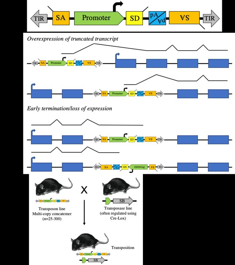
transposase have been modified to achieve sufficient mutagenesis to drive tumor formation (Figure 1A).
The first transposons used, T2/Onc and T2/Onc2, use the murine stem cell virus long terminal repeat
(MSCV-LTR) promoter followed by a splice donor (SD) sequence to drive gene expression and
bidirectional splice acceptors (SA) and polyadenylation signal (pA) to terminate gene transcription
and ablate expression [20,21]. This allows SB-mediated transposon insertion mutagenesis to identify
both oncogene and tumor suppressor gene candidates (Figure 1B). An optimized SB transposase
sequence (SB11) was knocked into the Gt(ROSA)26Sor locus, facilitating ubiquitous expression [21,34].
By crossing the R26-SB11 mouse with mice carrying either T2/Onc or T2/Onc2, researchers were able
to induce leukemia in mice (Figure 1C) [22]. Subsequently, a conditional SB mouse was created
(R26-lsl-SB11), allowing tissue and temporal-specific transposition and modeling of very specificInt. J. Mol. Sci. 2020, 21, 1172 4 of 19
cancers [23]. For example, we used Nestin-driven Cre recombinase to drive SB expression solely in the
developing central nervous system and to identify novel genetic drivers of childhood brain tumors [28].
The expression profiles of many of the Cre strains described in Table 1 have been characterized by The
Jackson Laboratory [47]. While transposon-mediated mutagenesis screens have taught us a great deal
about cancer development over the last two decades, we would like to focus on a few studies and
overall
Int. J. Mol. lessons
Sci. 2020, learned.
21, 1172 6 of 20
Figure
Figure1.1.Sleeping
SleepingBeauty
Beauty(SB)
(SB)transposons
transposonscancanbebedesigned
designedtotorandomly
randomlyinduce
inducesomatic
somaticcell
cellgain
gainand
and
loss
lossofoffunction
functionmutations.
mutations.(A) (A)Structure
Structureofofaaproto-typical
proto-typicaltransposon
transposonvector
vectorforforsomatic
somaticcell
cellor
orcell
cell
line
linemutagenesis
mutagenesis studies.
studies. AA strong
strong promoter followed
followed bybyan
anexon
exonwith
withaasplice
splicedonor
donor(SD)
(SD)isispresent
presentto
to activate transcription of downstream exons. Splice acceptors (SA) and a bi-directional
activate transcription of downstream exons. Splice acceptors (SA) and a bi-directional polyadenylation
polyadenylation site (pA)
site (pA) are included are included
to disrupt to disrupt
gene expression. gene
(B) In expression.cells,
mutagenized (B) transposons
In mutagenized cells,
can activate
transposons
endogenouscan activate endogenous
proto-oncogenes proto-oncogenes
or disrupt endogenous tumor or disrupt endogenous
suppressor tumor suppressor
genes depending on where
genes depending
insertion occurs on
andwhere
in whatinsertion occurs(C)
orientation. andTransposon
in what orientation.
transgenic (C)mice
Transposon transgenic
are usually produced mice
by
are usually
standard produced
pronuclear by standard
injection pronuclear
resulting injection
in the generation resulting
of lines in the generation
with multicopy of lines
concatomers. Thesewith
are
crossed to concatomers.
multicopy mice expressing the are
These transposase
crossed totomice
generate mice with
expressing the somatic cell transposition.
transposase to generate mice with
somatic cell transposition.
3. Cell of Origin
SB mutagenesis has been used to test the impact of the cell of origin and stage of differentiation
on transformation potential. Berquam-Vrieze et al. initiated transposition at increasinglyInt. J. Mol. Sci. 2020, 21, 1172 5 of 19
Table 2. Published Sleeping Beauty and piggyBac Cancer Screens in Mice.
Tumor Type Transposase Transposon Cre Sensitizing Mutations Refs
Sarcomas
Fibrosarcoma CGS-SB10 T2/Onc - p19arf [20]
Osteosarcoma R26-lsl-SB11 T2/Onc Osx-Cre Trp53 [48]
Cnp-Cre, Trp53, EGFR
Peripheral nerve sheath tumor R26-lsl-SB11 T2/Onc, T2/Onc15 [29,49]
Dhh-Cre Nf1
Histiocytic sarcoma R26-lsl-SB11 T2/Onc, T2/Onc2 Lyz2-Cre - [50]
Carcinomas
Skin K5-SB11 T2/Onc2 - Hras [25]
K5-SB11, K5-Cre, Trp53, β-catenin, Cdh1,
Mammary T2/Onc2, T2/Onc3 [51–55]
R26-lsl-SB11 Wap-Cre FGFR, Pten
R26-lsl-SB11,
T2/Onc, T2/Onc2,
Pancreatic R26-lsl-SB13, Pdx1-Cre Kras [24,56,57]
T2Onc3, ATP1
R26-lsl-PB
Gastric adenoma R26-lsl-SB11 T2/Onc3 β-actin-Cre Smad4 [58]
Vil-CreERT2, Apc, Kras, Smad4, Trp53,
Intestinal tract R26-lsl-SB11 T2/Onc, T2/Onc2 [59–63]
Vil-Cre, Ah-Cre Tgfbr2
T2/Onc, T2/Onc2, HBsAg, Trp53, Myc,
Liver R26-lsl-SB11 Alb-Cre [27,64–69]
T2/Onc3 Steatosis, Pten, Sav1, Met
Lung R26-lsl-SB11 T2/Onc Spc-Cre Trp53, p19arf, Pten [70]
CGS-SB10,
Prostate R26-SB11, T2/Onc, T2/Onc3 PB-Cre p19arf, Pten [71,72]
R26-lsl-SB11
Thyroid R26-lsl-SB11 T2/Onc2 Tpo-Cre Hras [73]
R26-lsl-SB11, T2/Onc, T2/Onc2,
Melanoma R26-lsl-SB13, T2/Onc3 Tyr-Cre-ERT2 Braf [74–77]
Act-PBase Luc-PB[mut]7
Hematopoietic
Vav-iCre,
R26-lsl-SB11,
T cell leukemia T2/Onc2 Lck-Cre, - [21,78]
R26-SB11
CD4-Cre
R26-SB11,
T cell lymphoma T2/Onc, ATP2 CD4-Cre Trp53, ITK-SYK, Pdc1 [79,80]
R26-lsl-PB
R26-lsl-SB11, Stat5b, Etv6-RUNX1
B cell leukemia T2/Onc Cd79a-Cre [81,82]
Etv6-RUNX1-HSB5 fusion
R26-lsl-SB11, T2/Onc, T2/Onc2, Aid-Cre, Eµ-TCL1, Pax5,
B cell lymphoma Etv6-RUNX1-HSB5, T2/Onc15, ITP1, CD19-Cre, Etv6-RUNX1 fusion, [23,83–86]
R26-PB ITP2 Cnp-Cre Trp53, Pten, Blm
β-actin-Cre,
Trp53, Jak2, Npm1c,
Acute myeloid leukemia R26-lsl-SB11 T2/Onc2, GrOnc Vav-Cre, [87–90]
BCR-ABL
Mx1-Cre
Mixture of T cell and B cell
R26-SB11 T2/Onc - Rassf1a, Cadm1 [91,92]
lymphoma, myeloid leukemia
Erythroleukemia R26-lsl-SB11 T2/Onc2 Mx1-Cre Cyclin E [93]
Myeloid and lymphoid
R26-lsl-SB11 T2/Onc2 Vec-Cre - [94]
malignancies, thymus, spleen
Brain tumors
R26-lsl-SB11,
T2/Onc, T2/Onc2, β-actin-Cre,
Medulloblastoma/CNS-ET * R26-SB11, Ptch1, Trp53, Pten [28,95–99]
T2/Onc3 Nestin-Cre
Math1-SB11
T2/Onc, T2/Onc2,
R26-lsl-SB11,
Glioma T2/Onc3, Nestin-Cre Trp53, p19Arf, Blm, Csf1 [26,100,101]
R26-SB11
T2OncATGInt. J. Mol. Sci. 2020, 21, 1172 6 of 19
Table 2. Cont.
Tumor Type Transposase Transposon Cre Sensitizing Mutations Refs
Multiple tumor types
Skin, brain, airway, liver,
leukemia, lymphoma, R26-SB11 T2/Onc3 - Rag2 [102]
intestine
Leukemia, medulloblastoma, CGS-SB10,
T2/Onc - p19Arf [22]
glioma R26-SB11
Skin, liver, lung, brain,
lymphoma, sarcoma, R26-SB11 T2/Onc3 - - [23]
mammary, colon, etc.
T cell and B cell leukemia,
ATP1, ATP2,
lymphoma, skin, sarcoma, R26-PB - - [103]
ATP3
intestinal tract, lung, liver, etc.
Prostate, mammary and skin
R26-SB11 ITP2m - Pten, Blm [104]
carcinomas
Sarcoma, carcinoma, leukemia,
R26-PB ATP2 - p19Arf [105]
resistance to MDM2 inhibition
Liver, lung carcinoma, skin
R26-PB ATP1 - - [106]
carcinoma, lymphoma
* CNS-ET—Embryonal tumor of the central nervous system.
3. Cell of Origin
SB mutagenesis has been used to test the impact of the cell of origin and stage of differentiation
on transformation potential. Berquam-Vrieze et al. initiated transposition at increasingly differentiated
stages in T-cell development using Cre-inducible SB and 3 different Cre transgenes [78]. Vav-iCre,
Lck-Cre, and CD4-Cre induce Cre expression in hematopoietic stem cells, immature T-cells without CD4
or CD8 expression, or late-stage T-cells expressing both CD4 and CD8, respectively. The authors found
that Vav-iCre mice had a significantly shorter survival time, indicating hematopoietic stem cells are
a more permissive cell for leukemia induction than more differentiated populations. In agreement
with this, they found that there was an increased average number of driver insertion mutations
per leukemia clone with increased differentiation. In other words, it took more “genetic hits”
to transform a more differentiated cell of origin. This concept that transformation potential
is lost with differentiation has been shown in other models, including intestinal cancer and
medulloblastoma [107,108]. When Berquam-Vrieze et al. compared genetic drivers in tumors generated
with the three Cre transgenes, they found significantly different gene profiles for each differentiation
stage, suggesting that the biology of each cell of origin greatly affects the genetics of tumor development.
Interestingly, Berquam-Vrieze and colleagues compared subsets of SB-induced mouse lymphoma and
found that the CD4-Cre (most differentiated cell of origin) lymphoma matched the expression patterns
of human ETP-ALL, a subtype of T-ALL defined by expression patterns of early T-cell precursors.
This was unexpected, as this was the most differentiated cell of origin in the study. Therefore, this study
sheds light on the potential cell of origin for human ETP-ALL, suggesting it may be a more differentiated
T cell that regains expression patterns of earlier T-cell progenitors, rather than an undifferentiated more
stem-like cell.
4. Identification of Rare Events
One challenge in human cancer genetics has been identifying rarely mutated driver mutations,
including both tumor suppressors and proto-oncogenes. This has sometimes been referred to as the
“long tail” problem, reflecting the large number of genes, that are altered in a relatively small percentage
of cancer cases. Many such genes exist in a “gray area” and it cannot easily be determined if their
alteration is selected for in human cancer development. Sequencing of many human cancer cases will
be required to determine if their alteration is statistically significant [109]. Transposon-based forward
genetic screens can provide contributing circumstantial data that such candidates may be driverInt. J. Mol. Sci. 2020, 21, 1172 7 of 19
alterations in cancer. Each screen that is completed reports a list of a few frequently mutated candidate
genes and many more infrequently mutated candidates. When multiple screens are combined and
analyzed together, infrequently altered drivers become more visible across many cancers. For example,
Rreb1 is a tumor suppressor gene that has been identified as a candidate driver in a low number of
many tumor types, including intestinal and pancreatic cancer and B-cell lymphoma [24,59,85]. Another
example is Foxr2. Transposon mutagenesis studies identified Foxr2 as a strong candidate driver of
malignant peripheral nerve sheath tumors (MPNST), osteosarcoma, and medulloblastoma [28,29,48,96].
Interestingly, human FOXR2 is amplified and overexpressed in a subset of human MPNST and activated
by translocation or amplification in a subset of human embryonal tumors of the central nervous
system [29,110]. Various other studies indicate that FOXR2 high level expression is a feature of a subset
of many tumor types, where it is likely a driver mechanism [111–113].
Based on the nature of how transposon-based screens work, they are uniquely able to identify
proto-oncogenes activated by creation of fusion transcripts. We queried the Candidate Cancer Gene
Database of SB screen-derived cancer gene candidates for those with recurrent fusion transcripts among
the TCGA [114]. Indeed, many of the SB-predicted oncogenes are activated by translocations similar to
those described for FOXR2. This includes known proto-oncogenes like ERG and RAF1, but many more
novel proto-oncogene candidates including AMBRA1 and RALY. AMBRA1 is a regulator of autophagy
and has been shown to affect drug resistance in several cancers [115–117]. RALY is an RNA-binding
protein implicated in metastasis and associated with poor prognosis in breast and colorectal cancer
and hepatocellular carcinoma [118–120]. More analysis to identify novel fusion transcripts at the RNA
level could identify more novel, poorly understood drivers that have been missed through traditional
analysis but may have meaningful implications.
Thus, transposon-based screens pooled together can be used to identify and prioritize novel
human oncogenes activated in a rare subset of many cancers. This is relevant for new clinical trial
designs, called “basket trials,” in which a single drug is tested in a variety of tumor types with a specific
genetic alteration. For example, chromosomal fusion events involving the carboxy-terminal kinase
domain of TRK (tropomysosin receptor kinase) have been identified in many cancers and shown to
drive constitutively active, ligand-independent signaling which results in tumorigenesis regardless of
tissue origin [121–126]. Larotrectinib is a potent and selective inhibitor of TRK proteins and has shown
a durable antitumor effect in patients with TRK fusions regardless of patient age or tumor type [127].
5. Drivers of Metastasis
Two screens, one in medulloblastoma and one in osteosarcoma, have sought to address the
questions of clonality and drivers of metastasis [48,99]. These studies found that when drivers of
primary and metastatic tumors were compared, there was varying degrees of overlap. In the case of
osteosarcoma, there were rare instances where multiple metastatic tumors within the same mouse
were significantly different from each other [48]. In the case of medulloblastoma, dissemination from
the primary tumor likely occurred early in the tumor development, potentially in multiple “seeding”
events or in an on-going fashion [99]. These findings indicate that metastatic cancer develops in a
rare subclone, perhaps early after tumor development, and that using targeted therapies based on
the drivers in the primary tumor will not be enough to eliminate metastatic tumors as other driver
alterations may have taken over a primary role in tumor maintenance. Additionally, several strong
candidate drivers of metastasis were identified in these papers, including alterations in Pten, Gsk3b,
Snap23, and Raf1 in osteosarcoma [48]. Loss of PTEN has since been identified as a marker of poor
clinical prognosis and lung metastasis in osteosarcoma [128].
We believe that new transposon-based screens could be designed to better facilitate identification
of metastatic drivers (Figure 2A). For practical purposes, a small screen can be done to produce a
small number of primary tumors in mice expressing transposase, harboring a mutagenic transposon
array, and any predisposing background mutations of interest. Primary tumors can then be removed
and transplanted as allografts into multiple recipient mice. Ideally the tumors would be implantedInt. J. Mol. Sci. 2020, 21, 1172 8 of 19
orthotopically, and the primary tumor would be removed at a pre-defined size, allowing the metastases
to expand. The timing of primary tumor removal will need to be optimized for every cancer type
and experimental condition to balance death caused by the primary tumor and leaving the tumor in
long enough for metastasis to occur. The metastatic clones can then be harvested and their genetic
drivers identified in a much more expedited fashion compared to undergoing a full screen. The drivers
identified in metastases can be compared to each other and the primary tumor to identify genes
involved in metastasis and to provide knowledge on the clonality of the metastases in the cancer being
studied. It may also be possible to provide adjuvant or neoadjuvant chemotherapy to better mimic the
selective pressures
Int. J. Mol. that
Sci. 2020, 21, 1172human metastases have undergone upon disease recurrence. 9 of 20
Figure 2. Future
Figure experiments
2. Future experimentsfor transposon
for transposoninsertional mutagenesis
insertional screens.
mutagenesis (A,B)(A,B)
screens. To identify drivers,
To identify
induce tumor
drivers, formation
induce with insertional
tumor formation mutagenesis
with insertional and implant
mutagenesis andtumors
implantorthotopically into syngeneic
tumors orthotopically into
recipients.
syngeneic (A) The primary
recipients. tumor
(A) The is expanded
primary tumor is and removed
expanded andbefore
removedendpoint, allowing allowing
before endpoint, the metastatic
the
lesions to expand
metastatic andtobe
lesions harvested
expand and for driver analysis.
be harvested (B) Implanted
for driver tumors
analysis. (B) are treated
Implanted tumorswith
areatreated
targeted
with a or
inhibitor targeted inhibitor
left untreated or their
and left untreated and their(C)
drivers compared. drivers
Mice compared.
undergoing(C) Mice undergoing
mutagenesis are fed a
mutagenesis
normal or high are
fat fed
diet.a normal or high
At endpoint allfat diet. At
tumors areendpoint
harvestedall and
tumors aredrivers
their harvested and their drivers
compared.
compared.
6. Therapy Resistance
Transposon-based mutagenesis in the presence of a targeted therapy offers a powerful tool for
understanding genetic pathways to therapy resistance in cancer, which is a major problem in the
quest for durable cures. For example, the BRAFV600E mutation is present in approximately half ofInt. J. Mol. Sci. 2020, 21, 1172 9 of 19
6. Therapy Resistance
Transposon-based mutagenesis in the presence of a targeted therapy offers a powerful tool for
understanding genetic pathways to therapy resistance in cancer, which is a major problem in the quest
for durable cures. For example, the BRAFV600E mutation is present in approximately half of human
melanoma, resulting in hyperactivation of the MAPK pathway [129]. While targeted therapy using
vemurafenib was initially promising, tumors eventually recurred showing re-activation of the MAPK
pathway [130]. Mann et al. performed SB mutagenesis in a BRAFV600E -driven mouse melanoma and
identified many candidate cooperating genetic alterations [75]. Using a similar screening strategy,
but including vemurafenib treatment of one cohort, Perna et al. were able to compare drivers in
vemurafenib-resistant and treatment-naïve tumors, and these authors identified novel mediators of
vemurafenib-resistance including Eras [74]. ERAS is an activator of PI3K/AKT signaling, which the
authors show fosters resistance to vemurafenib through inactivation of the pro-apoptotic protein BAD.
Therefore, dual treatment with a PI3K inhibitor along with vemurafenib may be a promising treatment
in the clinic if observed therapy-related toxicities can be overcome (NCT01512251).
In another example, Kas et al. studied resistance to AZD4547, a selective FGFR inhibitor,
by orthotopically implanting an SB-accelerated mammary tumor with FGFR2 activation into syngeneic
FVB mice and treating with AZD4547 [54]. FGFR is upstream of both the MAPK-ERK and PI3K-AKT
pathways and is frequently hyperactivated in human cancers [131,132]. Clinical trials of several
FGFR inhibitors have shown success in a subset of patients, but mechanisms of FGFR-inhibitor
resistance are still being understood [133]. Treatment resistant versus naïve tumors were compared by
RNA-sequencing and analysis of transposon insertion mutations. The authors identified a diverse
spectrum of resistance mechanisms to FGFR inhibition. Reactivation of the MAPK-ERK pathway was
the dominant form of resistance, suggesting that combining FGFR and MEK/ERK pathway inhibitors
may be the most effective strategy for patients with FGFR activation. In addition, the authors found
that Abcg2, a drug efflux pump, expression was upregulated in some AZD4547 treated tumors, while
inactivation of Rasa1 was found in other AZD4547 resistant tumors. These results provide guidance
that future drug design of FGFR inhibitors should be specifically made to be poor substrates for
drug efflux pumps. More pre-clinical models of SB-mediated accelerated tumor evolution could be
used to predict drug resistance mechanisms in the clinic to give additional options to patients and
facilitate more intelligent drug design (Figure 2B). Similarly, to the metastasis experiments described
above, these experiments could be done using tumors derived from a small screen allografted into
several recipient mice. Such studies would ideally be done in immunoproficient mice in the context
of recurrent metastatic disease, to best approximate the clinical situation for patients. Drivers in
therapy-resistant tumors would then be compared to untreated tumor drivers to find pathways
involved in drug resistance.
In addition, a similar in vivo screen has been used to gain insight into resistance to the MET
inhibitor Fortinib in a model of SB-accelerated medulloblastoma [98]. The PB system was used to
identify mechanisms of resistance to an Mdm2 inhibitor in PB-accelerated tumors of various types
passaged as allografts [134]. Although the following studies were not carried out in vivo, but rather
in cell lines, it is worth noting that transposon mutagenesis has been successfully used to screen for
cancer cell drug resistance in several reports [134,135]. Taken together, these studies suggest that
cancer evolution in response to the selective pressures of therapy can be usefully explored using
transposon mutagenesis.
7. Obesity and Tumor Development
Worldwide, the incidence of obesity has nearly tripled since 1975 [136]. Of particular concern,
the prevalence of overweight and obese children ages 5-19 has risen from just 4% in 1975 to over 18%
in 2016 [136]. The fundamental cause for this increase in obese and overweight people is an increase
in energy-rich food intake and a reduction in activity. Excess adipose tissue predisposes individuals
to develop type 2 diabetes mellitus, cardiovascular disease, and several types of cancer [137–139].Int. J. Mol. Sci. 2020, 21, 1172 10 of 19
Obesity has been associated with increased cancer risk in colorectal, kidney, pancreatic, gallbladder,
thyroid, breast, ovarian, esophageal, liver, and endometrial cancer [140–142]. Increased BMI (body
mass index) has been associated with reduced cancer survival and increased recurrence after radio- or
chemo-therapy [143–145].
However, despite strong clinical, preclinical, and epidemiological evidence linking obesity to
increased cancer risk [138,139,146,147], the mechanisms behind this are still not completely understood.
Local dysregulation in adipose tissues of obese individuals results in systemic metabolic changes
including insulin resistance, chronic inflammation, and hyperglycemia [148,149]. Dysregulated
paracrine signaling from adipocytes shapes a permissive microenvironment to tumor development
and progression through secretion of signaling molecules (including proinflammatory cytokines,
proangiogenic factors, and adipokines) and by acting as an energy reservoir [139,150–152]. For example,
chronic inflammation brought on by obesity results in increased expression of signal transducer and
activator of transcription 3 (STAT3) and nuclear factor-κB (NF-κB), which increase cellular proliferation
and pro-survival gene expression [153–155]. Adipose tissue also hosts many immune cells which are
significantly altered in the context of obesity [156,157].
Given the complex differences in the biology surrounding a cancer developing in the context of
obesity, it is likely that the genetic drivers differ in these cancers. Transposon mutagenesis offers an
opportunity to reflect these changes in how we model cancer in the mouse. For example, Tschida
et al. used SB insertion mutagenesis to model hepatocellular carcinoma in the context of steatosis
or accumulation of fat in the liver [27]. By comparing steatosis-associated drivers to drivers found
in another screen with normal diet [65], the authors were able to identify steatosis-specific drivers.
Many published screens should be repeated with the addition of diet-induced obesity in the mice
and compared to available normal diet studies to identify targets specific to obesity (Figure 2C).
While metastatic driver and therapy resistance studies can be done faithfully with orthotopically
implanted tumors, we recommend diet-induced obesity screens be done with the full transposon
mutagenesis process. While this is a larger undertaking, it will identify drivers of initiation and
early progression in tumor formation, rather than just later drivers of progression and metastasis.
These obesity-specific therapies are necessary to address the clearly unmet and increasing need
for patients.
8. Conclusions and Future Directions
Transposon insertion mutagenesis is a powerful tool to facilitate accelerated evolution and further
our understanding of the many dimensions of cancer development, progression, and response to
therapy. In this review, we have covered some of the contributions made to the field of cancer biology.
These have included models of many types of human cancers, providing insight into the genetic
drivers of these cancers as well as powerful pre-clinical model systems. In these studies, the effects
of increasing differentiation/linage commitment on cancer development have been studied, as have
mechanisms of therapy resistance and metastasis. It was determined that the differentiation status
of the tumor cell of origin affects the number of mutations required for tumor formation and the
end-tumor expression patterns in surprising ways. Combination therapies have been proposed based
on SB screens done in the context of a targeted therapy, such as dual treatment with a PI3K inhibitor
and vemurafenib in melanoma and FGFR and MEK/ERK pathway inhibitors for breast cancer. In both
osteosarcoma and medulloblastoma, drivers found in metastatic clones varied from those in other
metastases as well as the primary tumor, indicating that targeted therapies based on the genetics of the
primary tumor or even a single metastatic clone are unlikely to eliminate all metastases present.
In the future, we predict transposon mutagenesis will be used to mirror changes in our population
by incorporating changes in nutrition in mouse models. This will allow more precise and appropriate
therapy to be delivered to patients suffering from obesity in addition to cancer. In addition,
transposon mutagenesis studies may help to suggest changes in cancer therapy by identifying
resistance mechanisms to targeted therapies as described above and novel ideas for better drug design,Int. J. Mol. Sci. 2020, 21, 1172 11 of 19
including making drugs poor substrates for specific efflux pumps. We predict that transposon screens
will be used to identify metastasis-specific drivers in additional tumor types providing additional
treatment options for patients with high-risk disease. Lastly, we propose future screens to study the
effects of aging on tumor development. For example, it would be interesting to compare screens with
mutagenesis initiated in younger versus older mice (>1-year-old) through the use of tamoxifen-induced
Cre. These studies may more accurately reflect the development of some tumor types that mainly
occur in adult tissues but are poorly modeled by transposon-mutagenesis timed during embryogenesis
or early development.
Author Contributions: P.J.B. and D.A.L. wrote the manuscript. All authors have read and agreed to the published
version of the manuscript.
Acknowledgments: Support for this research was provided by The American Cancer Society (Research Professor
Award #123939 to DAL).
Conflicts of Interest: Largaespada is the co-founder and co-owner of several biotechnology companies including
NeoClone Biotechnologies, Inc., Discovery Genomics, Inc. (recently acquired by Immunsoft, Inc.), and B-MoGen
Biotechnologies, Inc. (recently acquired by biotechne corporation). He consults for Genentech, Inc., which is
funding some of his research. Largaespada holds equity in and serves as the Chief Scientific Officer of Surrogen,
a subsidiary of Recombine-tics, a genome-editing company. The business of all these companies is unrelated to
the contents of this manuscript. P.J.B. has no conflicts of interest to disclose.
References
1. Mc, C.B. The origin and behavior of mutable loci in maize. Proc. Natl. Acad. Sci. USA 1950, 36, 344–355.
2. Paatero, A.O.; Turakainen, H.; Happonen, L.J.; Olsson, C.; Palomaki, T.; Pajunen, M.I.; Meng, X.; Otonkoski, T.;
Tuuri, T.; Berry, C.; et al. Bacteriophage Mu integration in yeast and mammalian genomes. Nucleic Acids Res.
2008, 36. [CrossRef] [PubMed]
3. Wisman, E.; Cardon, G.H.; Fransz, P.; Saedler, H. The behaviour of the autonomous maize transposable
element En/Spm in Arabidopsis thaliana allows efficient mutagenesis. Plant Mol. Biol. 1998, 37, 989–999.
[CrossRef] [PubMed]
4. Enoki, H.; Izawa, T.; Kawahara, M.; Komatsu, M.; Koh, S.; Kyozuka, J.; Shimamoto, K. Ac as a tool for the
functional genomics of rice. Plant J. 1999, 19, 605–613. [CrossRef] [PubMed]
5. Meissner, R.; Chague, V.; Zhu, Q.; Emmanuel, E.; Elkind, Y.; Levy, A.A. Technical advance: A high throughput
system for transposon tagging and promoter trapping in tomato. Plant J. 2000, 22, 265–274. [CrossRef]
[PubMed]
6. Kuromori, T.; Hirayama, T.; Kiyosue, Y.; Takabe, H.; Mizukado, S.; Sakurai, T.; Akiyama, K.; Kamiya, A.;
Ito, T.; Shinozaki, K. A collection of 11 800 single-copy Ds transposon insertion lines in Arabidopsis. Plant J.
2004, 37, 897–905. [CrossRef] [PubMed]
7. Greco, R.; Ouwerkerk, P.B.; Sallaud, C.; Kohli, A.; Colombo, L.; Puigdomenech, P.; Guiderdoni, E.; Christou, P.;
Hoge, J.H.; Pereira, A. Transposon insertional mutagenesis in rice. Plant Physiol. 2001, 125, 1175–1177.
[CrossRef]
8. Ruaud, A.F.; Bessereau, J.L. Activation of nicotinic receptors uncouples a developmental timer from the
molting timer in C. elegans. Development 2006, 133, 2211–2222. [CrossRef]
9. Ruaud, A.F.; Bessereau, J.L. The P-type ATPase CATP-1 is a novel regulator of C. elegans developmental
timing that acts independently of its predicted pump function. Development 2007, 134, 867–879. [CrossRef]
10. Gally, C.; Eimer, S.; Richmond, J.E.; Bessereau, J.L. A transmembrane protein required for acetylcholine
receptor clustering in Caenorhabditis elegans. Nature 2004, 431, 578–582. [CrossRef]
11. Yook, K.; Hodgkin, J. Mos1 mutagenesis reveals a diversity of mechanisms affecting response of
Caenorhabditis elegans to the bacterial pathogen Microbacterium nematophilum. Genetics 2007, 175, 681–697.
[CrossRef] [PubMed]
12. Hummel, T.; Klambt, C. P-element mutagenesis. Methods Mol. Biol. 2008, 420, 97–117. [PubMed]
13. Handler, A.M.; Harrell, R.A., 2nd. Germline transformation of Drosophila melanogaster with the piggyBac
transposon vector. Insect Mol. Biol. 1999, 8, 449–457. [CrossRef] [PubMed]
14. Loukeris, T.G.; Arca, B.; Livadaras, I.; Dialektaki, G.; Savakis, C. Introduction of the transposable element Minos
into the germ line of Drosophila melanogaster. Proc. Natl. Acad. Sci. USA 1995, 92, 9485–9489. [CrossRef]Int. J. Mol. Sci. 2020, 21, 1172 12 of 19
15. Urasaki, A.; Mito, T.; Noji, S.; Ueda, R.; Kawakami, K. Transposition of the vertebrate Tol2 transposable
element in Drosophila melanogaster. Gene 2008, 425, 64–68. [CrossRef]
16. Ding, S.; Wu, X.; Li, G.; Han, M.; Zhuang, Y.; Xu, T. Efficient transposition of the piggyBac (PB) transposon in
mammalian cells and mice. Cell 2005, 122, 473–483. [CrossRef]
17. Drabek, D.; Zagoraiou, L.; deWit, T.; Langeveld, A.; Roumpaki, C.; Mamalaki, C.; Savakis, C.; Grosveld, F.
Transposition of the Drosophila hydei Minos transposon in the mouse germ line. Genomics 2003, 81, 108–111.
[CrossRef]
18. Kawakami, K.; Shima, A.; Kawakami, N. Identification of a functional transposase of the Tol2 element,
an Ac-like element from the Japanese medaka fish, and its transposition in the zebrafish germ lineage.
Proc. Natl. Acad. Sci. USA 2000, 97, 11403–11408. [CrossRef]
19. Ivics, Z.; Hackett, P.B.; Plasterk, R.H.; Izsvak, Z. Molecular reconstruction of Sleeping Beauty, a Tc1-like
transposon from fish, and its transposition in human cells. Cell 1997, 91, 501–510. [CrossRef]
20. Collier, L.S.; Carlson, C.M.; Ravimohan, S.; Dupuy, A.J.; Largaespada, D.A. Cancer gene discovery in solid
tumours using transposon-based somatic mutagenesis in the mouse. Nature 2005, 436, 272–276. [CrossRef]
21. Dupuy, A.J.; Akagi, K.; Largaespada, D.A.; Copeland, N.G.; Jenkins, N.A. Mammalian mutagenesis using a
highly mobile somatic Sleeping Beauty transposon system. Nature 2005, 436, 221–226. [CrossRef] [PubMed]
22. Collier, L.S.; Adams, D.J.; Hackett, C.S.; Bendzick, L.E.; Akagi, K.; Davies, M.N.; Diers, M.D.;
Rodriguez, F.J.; Bender, A.M.; Tieu, C.; et al. Whole-body sleeping beauty mutagenesis can cause penetrant
leukemia/lymphoma and rare high-grade glioma without associated embryonic lethality. Cancer Res. 2009,
69, 8429–8437. [CrossRef] [PubMed]
23. Dupuy, A.J.; Rogers, L.M.; Kim, J.; Nannapaneni, K.; Starr, T.K.; Liu, P.; Largaespada, D.A.; Scheetz, T.E.;
Jenkins, N.A.; Copeland, N.G. A modified sleeping beauty transposon system that can be used to model a
wide variety of human cancers in mice. Cancer Res. 2009, 69, 8150–8156. [CrossRef] [PubMed]
24. Perez-Mancera, P.A.; Rust, A.G.; van der Weyden, L.; Kristiansen, G.; Li, A.; Sarver, A.L.; Silverstein, K.A.;
Grutzmann, R.; Aust, D.; Rummele, P.; et al. The deubiquitinase USP9X suppresses pancreatic ductal
adenocarcinoma. Nature 2012, 486, 266–270. [CrossRef] [PubMed]
25. Quintana, R.M.; Dupuy, A.J.; Bravo, A.; Casanova, M.L.; Alameda, J.P.; Page, A.; Sanchez-Viera, M.;
Ramirez, A.; Navarro, M. A transposon-based analysis of gene mutations related to skin cancer development.
J. Investig. Dermatol. 2013, 133, 239–248. [CrossRef] [PubMed]
26. Vyazunova, I.; Maklakova, V.I.; Berman, S.; De, I.; Steffen, M.D.; Hong, W.; Lincoln, H.; Morrissy, A.S.;
Taylor, M.D.; Akagi, K.; et al. Sleeping Beauty mouse models identify candidate genes involved in
gliomagenesis. PLoS ONE 2014, 9. [CrossRef]
27. Tschida, B.R.; Temiz, N.A.; Kuka, T.P.; Lee, L.A.; Riordan, J.D.; Tierrablanca, C.A.; Hullsiek, R.; Wagner, S.;
Hudson, W.A.; Linden, M.A.; et al. Sleeping Beauty Insertional Mutagenesis in Mice Identifies Drivers of
Steatosis-Associated Hepatic Tumors. Cancer Res. 2017, 77, 6576–6588. [CrossRef]
28. Beckmann, P.J.; Larson, J.D.; Larsson, A.T.; Ostergaard, J.P.; Wagner, S.; Rahrmann, E.P.; Shamsan, G.A.;
Otto, G.M.; Williams, R.L.; Wang, J.; et al. Sleeping Beauty Insertional Mutagenesis Reveals Important
Genetic Drivers of Central Nervous System Embryonal Tumors. Cancer Res. 2019, 79, 905–917. [CrossRef]
29. Rahrmann, E.P.; Watson, A.L.; Keng, V.W.; Choi, K.; Moriarity, B.S.; Beckmann, D.A.; Wolf, N.K.; Sarver, A.;
Collins, M.H.; Moertel, C.L.; et al. Forward genetic screen for malignant peripheral nerve sheath tumor
formation identifies new genes and pathways driving tumorigenesis. Nat. Genet. 2013, 45, 756–766.
[CrossRef]
30. Kitada, K.; Ishishita, S.; Tosaka, K.; Takahashi, R.; Ueda, M.; Keng, V.W.; Horie, K.; Takeda, J. Transposon-
tagged mutagenesis in the rat. Nat. Methods 2007, 4, 131–133. [CrossRef]
31. Nasevicius, A.; Ekker, S.C. Effective targeted gene ‘knockdown’ in zebrafish. Nat. Genet. 2000, 26, 216–220.
[CrossRef] [PubMed]
32. Sinzelle, L.; Vallin, J.; Coen, L.; Chesneau, A.; Du Pasquier, D.; Pollet, N.; Demeneix, B.; Mazabraud, A.
Generation of trangenic Xenopus laevis using the Sleeping Beauty transposon system. Transgenic Res. 2006,
15, 751–760. [CrossRef] [PubMed]
33. Izsvak, Z.; Ivics, Z.; Plasterk, R.H. Sleeping Beauty, a wide host-range transposon vector for genetic
transformation in vertebrates. J. Mol. Biol. 2000, 302, 93–102. [CrossRef] [PubMed]Int. J. Mol. Sci. 2020, 21, 1172 13 of 19
34. Geurts, A.M.; Yang, Y.; Clark, K.J.; Liu, G.; Cui, Z.; Dupuy, A.J.; Bell, J.B.; Largaespada, D.A.; Hackett, P.B.
Gene transfer into genomes of human cells by the sleeping beauty transposon system. Mol. Ther. 2003,
8, 108–117. [CrossRef]
35. Karsi, A.; Moav, B.; Hackett, P.; Liu, Z. Effects of insert size on transposition efficiency of the sleeping beauty
transposon in mouse cells. Mar. Biotechnol. 2001, 3, 241–245. [CrossRef]
36. Rostovskaya, M.; Fu, J.; Obst, M.; Baer, I.; Weidlich, S.; Wang, H.; Smith, A.J.; Anastassiadis, K.; Stewart, A.F.
Transposon-mediated BAC transgenesis in human ES cells. Nucleic Acids Res. 2012, 40. [CrossRef]
37. Zayed, H.; Izsvak, Z.; Walisko, O.; Ivics, Z. Development of hyperactive sleeping beauty transposon vectors
by mutational analysis. Mol. Ther. 2004, 9, 292–304. [CrossRef]
38. Balciunas, D.; Wangensteen, K.J.; Wilber, A.; Bell, J.; Geurts, A.; Sivasubbu, S.; Wang, X.; Hackett, P.B.;
Largaespada, D.A.; McIvor, R.S.; et al. Harnessing a high cargo-capacity transposon for genetic applications
in vertebrates. PLoS Genet. 2006, 2. [CrossRef]
39. Wang, W.; Lin, C.; Lu, D.; Ning, Z.; Cox, T.; Melvin, D.; Wang, X.; Bradley, A.; Liu, P. Chromosomal
transposition of PiggyBac in mouse embryonic stem cells. Proc. Natl. Acad. Sci. USA 2008, 105, 9290–9295.
[CrossRef]
40. Yant, S.R.; Wu, X.; Huang, Y.; Garrison, B.; Burgess, S.M.; Kay, M.A. High-resolution genome-wide mapping
of transposon integration in mammals. Mol. Cell Biol. 2005, 25, 2085–2094. [CrossRef]
41. Liu, G.; Geurts, A.M.; Yae, K.; Srinivasan, A.R.; Fahrenkrug, S.C.; Largaespada, D.A.; Takeda, J.; Horie, K.;
Olson, W.K.; Hackett, P.B. Target-site preferences of Sleeping Beauty transposons. J. Mol. Biol. 2005,
346, 161–173. [CrossRef] [PubMed]
42. Fischer, S.E.; Wienholds, E.; Plasterk, R.H. Regulated transposition of a fish transposon in the mouse germ
line. Proc. Natl. Acad. Sci. USA 2001, 98, 6759–6764. [CrossRef] [PubMed]
43. Weber, J.; Ollinger, R.; Friedrich, M.; Ehmer, U.; Barenboim, M.; Steiger, K.; Heid, I.; Mueller, S.; Maresch, R.;
Engleitner, T.; et al. CRISPR/Cas9 somatic multiplex-mutagenesis for high-throughput functional cancer
genomics in mice. Proc. Natl. Acad. Sci. USA 2015, 112, 13982–13987. [CrossRef] [PubMed]
44. Xu, C.; Qi, X.; Du, X.; Zou, H.; Gao, F.; Feng, T.; Lu, H.; Li, S.; An, X.; Zhang, L.; et al. piggyBac mediates
efficient in vivo CRISPR library screening for tumorigenesis in mice. Proc. Natl. Acad. Sci. USA 2017,
114, 722–727. [CrossRef]
45. Kool, J.; Berns, A. High-throughput insertional mutagenesis screens in mice to identify oncogenic networks.
Nat. Rev. Cancer 2009, 9, 389–399. [CrossRef]
46. Theodorou, V.; Kimm, M.A.; Boer, M.; Wessels, L.; Theelen, W.; Jonkers, J.; Hilkens, J. MMTV insertional
mutagenesis identifies genes, gene families and pathways involved in mammary cancer. Nat. Genet. 2007,
39, 759–769. [CrossRef]
47. Laboratory, T.J. Characterized Cre Lines. Available online: https://www.jax.org/research-and-faculty/
resources/cre-repository/characterized-cre-lines-jax-cre-resource# (accessed on 28 January 2020).
48. Moriarity, B.S.; Otto, G.M.; Rahrmann, E.P.; Rathe, S.K.; Wolf, N.K.; Weg, M.T.; Manlove, L.A.; LaRue, R.S.;
Temiz, N.A.; Molyneux, S.D.; et al. A Sleeping Beauty forward genetic screen identifies new genes and
pathways driving osteosarcoma development and metastasis. Nat. Genet. 2015, 47, 615–624. [CrossRef]
49. Wu, J.; Keng, V.W.; Patmore, D.M.; Kendall, J.J.; Patel, A.V.; Jousma, E.; Jessen, W.J.; Choi, K.; Tschida, B.R.;
Silverstein, K.A.; et al. Insertional Mutagenesis Identifies a STAT3/Arid1b/beta-catenin Pathway Driving
Neurofibroma Initiation. Cell Rep. 2016, 14, 1979–1990. [CrossRef]
50. Been, R.A.; Linden, M.A.; Hager, C.J.; DeCoursin, K.J.; Abrahante, J.E.; Landman, S.R.; Steinbach, M.;
Sarver, A.L.; Largaespada, D.A.; Starr, T.K. Genetic signature of histiocytic sarcoma revealed by a sleeping
beauty transposon genetic screen in mice. PLoS ONE 2014, 9, e97280. [CrossRef]
51. Suarez-Cabrera, C.; Quintana, R.M.; Bravo, A.; Casanova, M.L.; Page, A.; Alameda, J.P.; Paramio, J.M.;
Maroto, A.; Salamanca, J.; Dupuy, A.J.; et al. A Transposon-based Analysis Reveals RASA1 Is Involved in
Triple-Negative Breast Cancer. Cancer Res. 2017, 77, 1357–1368. [CrossRef]
52. Chen, L.; Jenjaroenpun, P.; Pillai, A.M.; Ivshina, A.V.; Ow, G.S.; Efthimios, M.; Zhiqun, T.; Tan, T.Z.; Lee, S.C.;
Rogers, K.; et al. Transposon insertional mutagenesis in mice identifies human breast cancer susceptibility
genes and signatures for stratification. Proc. Natl. Acad. Sci. USA 2017, 114, E2215–E2224. [CrossRef]
53. Kas, S.M.; de Ruiter, J.R.; Schipper, K.; Annunziato, S.; Schut, E.; Klarenbeek, S.; Drenth, A.P.; van der Burg, E.;
Klijn, C.; Ten Hoeve, J.J.; et al. Insertional mutagenesis identifies drivers of a novel oncogenic pathway in
invasive lobular breast carcinoma. Nat. Genet. 2017, 49, 1219–1230. [CrossRef] [PubMed]Int. J. Mol. Sci. 2020, 21, 1172 14 of 19
54. Kas, S.M.; de Ruiter, J.R.; Schipper, K.; Schut, E.; Bombardelli, L.; Wientjens, E.; Drenth, A.P.; de
Korte-Grimmerink, R.; Mahakena, S.; Phillips, C.; et al. Transcriptomics and Transposon Mutagenesis
Identify Multiple Mechanisms of Resistance to the FGFR Inhibitor AZD4547. Cancer Res. 2018, 78, 5668–5679.
[CrossRef] [PubMed]
55. Rangel, R.; Lee, S.C.; Hon-Kim Ban, K.; Guzman-Rojas, L.; Mann, M.B.; Newberg, J.Y.; Kodama, T.;
McNoe, L.A.; Selvanesan, L.; Ward, J.M.; et al. Transposon mutagenesis identifies genes that cooperate with
mutant Pten in breast cancer progression. Proc. Natl. Acad. Sci. USA 2016, 113, E7749–E7758. [CrossRef]
56. Mann, K.M.; Ward, J.M.; Yew, C.C.; Kovochich, A.; Dawson, D.W.; Black, M.A.; Brett, B.T.; Sheetz, T.E.;
Dupuy, A.J.; Chang, D.K.; et al. Sleeping Beauty mutagenesis reveals cooperating mutations and pathways
in pancreatic adenocarcinoma. Proc. Natl. Acad. Sci. USA 2012, 109, 5934–5941. [CrossRef]
57. Rad, R.; Rad, L.; Wang, W.; Strong, A.; Ponstingl, H.; Bronner, I.F.; Mayho, M.; Steiger, K.; Weber, J.; Hieber, M.;
et al. A conditional piggyBac transposition system for genetic screening in mice identifies oncogenic networks
in pancreatic cancer. Nat. Genet. 2015, 47, 47–56. [CrossRef]
58. Takeda, H.; Rust, A.G.; Ward, J.M.; Yew, C.C.; Jenkins, N.A.; Copeland, N.G. Sleeping Beauty transposon
mutagenesis identifies genes that cooperate with mutant Smad4 in gastric cancer development. Proc. Natl.
Acad. Sci. USA 2016, 113, E2057–E2065. [CrossRef]
59. Takeda, H.; Wei, Z.; Koso, H.; Rust, A.G.; Yew, C.C.; Mann, M.B.; Ward, J.M.; Adams, D.J.; Copeland, N.G.;
Jenkins, N.A. Transposon mutagenesis identifies genes and evolutionary forces driving gastrointestinal tract
tumor progression. Nat. Genet. 2015, 47, 142–150. [CrossRef]
60. Starr, T.K.; Allaei, R.; Silverstein, K.A.; Staggs, R.A.; Sarver, A.L.; Bergemann, T.L.; Gupta, M.; O’Sullivan, M.G.;
Matise, I.; Dupuy, A.J.; et al. A transposon-based genetic screen in mice identifies genes altered in colorectal
cancer. Science 2009, 323, 1747–1750. [CrossRef]
61. Starr, T.K.; Scott, P.M.; Marsh, B.M.; Zhao, L.; Than, B.L.; O’Sullivan, M.G.; Sarver, A.L.; Dupuy, A.J.;
Largaespada, D.A.; Cormier, R.T. A Sleeping Beauty transposon-mediated screen identifies murine
susceptibility genes for adenomatous polyposis coli (Apc)-dependent intestinal tumorigenesis. Proc. Natl.
Acad. Sci. USA 2011, 108, 5765–5770. [CrossRef]
62. March, H.N.; Rust, A.G.; Wright, N.A.; ten Hoeve, J.; de Ridder, J.; Eldridge, M.; van der Weyden, L.; Berns, A.;
Gadiot, J.; Uren, A.; et al. Insertional mutagenesis identifies multiple networks of cooperating genes driving
intestinal tumorigenesis. Nat. Genet. 2011, 43, 1202–1209. [CrossRef] [PubMed]
63. Morris, S.M.; Davison, J.; Carter, K.T.; O’Leary, R.M.; Trobridge, P.; Knoblaugh, S.E.; Myeroff, L.L.;
Markowitz, S.D.; Brett, B.T.; Scheetz, T.E.; et al. Transposon mutagenesis identifies candidate genes
that cooperate with loss of transforming growth factor-beta signaling in mouse intestinal neoplasms. Int. J.
Cancer 2017, 140, 853–863. [CrossRef] [PubMed]
64. Bard-Chapeau, E.A.; Nguyen, A.T.; Rust, A.G.; Sayadi, A.; Lee, P.; Chua, B.Q.; New, L.S.; de Jong, J.;
Ward, J.M.; Chin, C.K.; et al. Transposon mutagenesis identifies genes driving hepatocellular carcinoma in a
chronic hepatitis B mouse model. Nat. Genet. 2014, 46, 24–32. [CrossRef] [PubMed]
65. Keng, V.W.; Villanueva, A.; Chiang, D.Y.; Dupuy, A.J.; Ryan, B.J.; Matise, I.; Silverstein, K.A.; Sarver, A.;
Starr, T.K.; Akagi, K.; et al. A conditional transposon-based insertional mutagenesis screen for genes
associated with mouse hepatocellular carcinoma. Nat. Biotechnol. 2009, 27, 264–274. [CrossRef]
66. Keng, V.W.; Sia, D.; Sarver, A.L.; Tschida, B.R.; Fan, D.; Alsinet, C.; Sole, M.; Lee, W.L.; Kuka, T.P.; Moriarity, B.S.;
et al. Sex bias occurrence of hepatocellular carcinoma in Poly7 molecular subclass is associated with EGFR.
Hepatology 2013, 57, 120–130. [CrossRef]
67. O’Donnell, K.A.; Keng, V.W.; York, B.; Reineke, E.L.; Seo, D.; Fan, D.; Silverstein, K.A.; Schrum, C.T.; Xie, W.R.;
Mularoni, L.; et al. A Sleeping Beauty mutagenesis screen reveals a tumor suppressor role for Ncoa2/Src-2 in
liver cancer. Proc. Natl. Acad. Sci. USA 2012, 109, E1377–E1386. [CrossRef]
68. Kodama, T.; Yi, J.; Newberg, J.Y.; Tien, J.C.; Wu, H.; Finegold, M.J.; Kodama, M.; Wei, Z.; Tamura, T.;
Takehara, T.; et al. Molecular profiling of nonalcoholic fatty liver disease-associated hepatocellular carcinoma
using SB transposon mutagenesis. Proc. Natl. Acad. Sci. USA 2018, 115, E10417–E10426. [CrossRef]
69. Fan, Y.; Bazai, S.K.; Daian, F.; Arechederra, M.; Richelme, S.; Temiz, N.A.; Yim, A.; Habermann, B.H.; Dono, R.;
Largaespada, D.A.; et al. Evaluating the landscape of gene cooperativity with receptor tyrosine kinases in
liver tumorigenesis using transposon-mediated mutagenesis. J. Hepatol. 2019, 70, 470–482. [CrossRef]You can also read