Structure-Based Inhibitor Discovery of Class I Histone Deacetylases (HDACs) - MDPI
←
→
Page content transcription
If your browser does not render page correctly, please read the page content below
International Journal of
            Molecular Sciences

Review
Structure-Based Inhibitor Discovery of Class I
Histone Deacetylases (HDACs)
Yuxiang Luo 1 and Huilin Li 1,2, *
 1    School of Pharmaceutical Sciences, Sun Yat-sen University, No.132 Wai Huan Dong lu,
      Guangzhou Higher Education Mega Center, Guangzhou 510006, Guangdong, China;
      luoyx69@mail2.sysu.edu.cn
 2    Guangdong Key Laboratory of Chiral Molecule and Drug Discovery, School of Pharmaceutical Sciences,
      Sun Yat-sen University, Guangzhou 510006, Guangdong, China
 *    Correspondence: lihlin6@mail.sysu.edu.cn

 Received: 7 October 2020; Accepted: 16 November 2020; Published: 22 November 2020

 Abstract: Class I histone deacetylases (HDACs) are promising targets for epigenetic therapies for a
 range of diseases such as cancers, inflammations, infections and neurological diseases. Although six
 HDAC inhibitors are now licensed for clinical treatments, they are all pan-inhibitors with little or
 no HDAC isoform selectivity, exhibiting undesirable side effects. A major issue with the currently
 available HDAC inhibitors is that they have limited specificity and target multiple deacetylases.
 Except for HDAC8, Class I HDACs (1, 2 and 3) are recruited to large multiprotein complexes to
 function. Therefore, there are rising needs to develop new, hopefully, therapeutically efficacious HDAC
 inhibitors with isoform or complex selectivity. Here, upon the introduction of the structures of Class I
 HDACs and their complexes, we provide an up-to-date overview of the structure-based discovery of
 Class I HDAC inhibitors, including pan-, isoform-selective and complex-specific inhibitors, aiming to
 provide an insight into the discovery of additional HDAC inhibitors with greater selectivity, specificity
 and therapeutic utility.

 Keywords: Class I histone deacetylases; structural studies; selective inhibitors; HDAC complexes;
 drug mechanism

1. Introduction
     Histone deacetylases (HDACs) are enzymes involved in epigenetic regulation through controlling
the acetylation state of lysine side-chains in histone tails [1], leading to chromatin condensation and gene
transcription repression [2,3]. Additionally, HDACs can indirectly regulate other post-translational
modifications (PTMs) through releasing acetyl group from lysine so that other PTMs, for instance,
ubiquitination, can mark on the loci [1]. So far, 18 human HDACs have been identified according to
their sequence homologies to yeast and are divided into four classes: Class I (HDAC1-3 and 8), Class II
with two subclasses (Class IIa includes HDAC4, 5, 7 and 9 and Class IIb corresponds to HDAC6 and
10) and Class IV (HDAC11) are zinc-dependent enzymes (also referred to as classical HDACs family),
while Class III are NAD+ -dependent which called sirtuins (SIRT1-7) [4,5]. Each class has different
biological functions [6]. Classes I, II and IV are metal-dependent HDACs that use a metal−water as the
nucleophile during catalysis, which is activated via a general acid−base mechanism [7].
     Famous as a cancer target, abnormal function and expression of HDACs have been observed
in various tumor cells, including breast, lung, liver and gastric, where they are aberrantly recruited
to gene promoters [8–10]. Except for this, HDACs play roles in neurodegenerative diseases such as
Alzheimer’s disease, Huntington’s disease, Parkinson’s disease and mood disorders [11], as well as in
HIV infection [12], kidney diseases [13] and inflammatory diseases [14].

Int. J. Mol. Sci. 2020, 21, 8828; doi:10.3390/ijms21228828                         www.mdpi.com/journal/ijms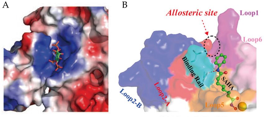
Int. J. Mol. Sci. 2020, 21, 8828                                                                      2 of 25

     Luckily, these abnormalities can be altered by HDAC inhibitors (HDACis). First discovered as
inducers of cell growth processes, HDACis show great potential in inhibiting HDACs activity and
treating many diseases [6]. Some of them have been approved by the U.S. FDA (Food and Drug
Administration), although they are pan-inhibitors with little or no specific isoform selectivity [15]. Here,
we specifically focus on the most promising drug targets, Class I HDACs. Except for HDAC8 that is fully
active in isolation, HDAC1, 2 and 3 form the catalytic subunit of multiprotein complexes to mediate
gene transcription [16]. More specifically, HDAC1 and HDAC2 form the catalytic core of multiple
corepressor complexes, including NuRD (nucleosome remodeling and deacetylase), Sin3 (switch
intensive 3) and CoREST (corepressor of RE1-silencing transcription), MiDAC (mitotic deacetylase),
while HDAC3 forms the key component of SMRT/NCoR (silencing mediator of retinoic acid and
thyroid hormone receptors/nuclear receptor corepressor) [15]. As part of these complexes, the HDACs
become maximally activated and are targeted to specific regions of chromatin.
     In this review, after briefly introducing the enzyme mechanism of Class I HDACs, we specifically
focus on the structures of Class I HDACs and their complexes and summarize the development of some
representative pan-, isoform-selective and complex-selective inhibitors and their mechanism insights.
We aim to provide an up-to-date reference for targeted design and screening of Class I HDACis.

2. HDACs

2.1. HDACs Substrates
      HDACs are capable of catalyzing the removal of the N-acetyl group from acetylated lysine
residues in histones and non-histone proteins [17,18]. The substrates of HDACs are rather complicated,
owing to the overlapping functions of different HDACs and different substrate preferences within
HDAC complexes [19]. When one HDAC is knocked down, its activity can be replaced by other
isoforms [1]. However, for HDAC8, there is no evidence for histones being its substrates in vivo [17].
Additionally, HDACs impact the functions of more than 50 non-histone substrates (e.g., p53, NF-κB,
STAT3 and Hsp90) that regulate cellular development, proliferation, differentiation and death [20].
For example, the acetylation/deacetylation of tumor suppressor protein p53 regulates its transcriptional
activity and is related to apoptosis and autophagy, which plays critical roles in eliminating tumor
cells [21]. Inhibiting HDACs allows p53-induced transcription kept in an active state, leading to tumor
cell death [2,21]. Another example is the signal transducers and activators of transcription 3 (STAT3),
which is found highly expressed in diffuse large B-cell lymphoma (DLBCL), regulates gene expression
with the aid of HDACs [22]. As HDACs involve different types of substrates, the abnormalities of them
correlate with diseases to varying degrees [1,8].

2.2. FDA Approved HDAC Inhibitors
      HDACis can antagonize the function of HDACs, increase the level of acetylated histones, and show
potential towards cancers, neurological diseases, inflammatory diseases, and so on [23–25]. In tumor
cells, HDACis induce cell apoptosis, cell cycle arrest, senescence, differentiation, autophagy and
increase tumor immunogenicity [8]. For example, inhibitors such as SAHA and sodium butyrate (NaB)
inhibit cell proliferation, arrest cell cycle at G0/G1 phase, and induce mitochondrial related apoptosis
in triple-negative breast cancer (TNBC) cells [26]. Additionally, HDACis also affect the immune system
and tumor microenvironment by regulating the differentiation, function and survival of different
immune cells, thus inhibiting tumor angiogenesis and metastasis/invasion [1,8]. In neurological
diseases, HDACis can induce neuroprotection and the expression of neurotrophins [8,27].
      Through the unremitting efforts of investigators over the years, six HDACis have been approved
by the FDA mainly for cancer treatment (Figure 1), and many others are in clinical trials [28]. The first
drug, vorinostat (suberoylanilide hydroxamic acid, SAHA, Compound 1), was approved by the FDA for
the treatment of refractory cutaneous T-cell lymphoma (CTCL) in 2006 [28,29]. A series of clinical trials
have confirmed its effects and toxicity in the treatment of CTCL [30–32]. In addition, SAHA has shown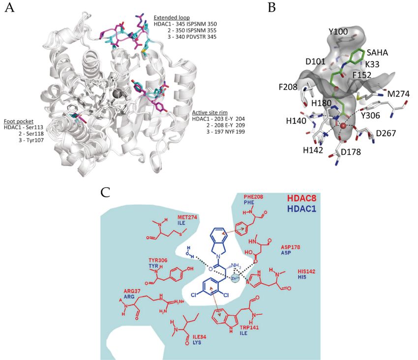
Int. J. Mol. Sci. 2020, 21, 8828 3 of 25
effectiveness in a variety of solid and hematological tumors such as head and neck cancer [33], Hodgkin
Int. J. Mol. Sci. 2020, 21, x FOR PEER REVIEW 3 of 25
lymphoma (HL) and DLBCL [34]. The second drug, romidepsin (FK228, Compound 2), was reported
in 1994 and
trialsapproved
have confirmed in 2009 [29,35].
its effects andBased
toxicityon twotreatment
in the large phaseof CTCLII studies
[30–32]. [36,37], romidepsin
In addition, SAHA has has been
shown
used in the effectiveness
treatment in a variety
of relapsed orof solid and hematological
refractory peripheral T-cell tumors such as head (PTCL)
lymphomas and neck[38,39].
cancer [33],Chidamide
Hodgkin lymphoma (HL) and DLBCL [34]. The second drug, romidepsin (FK228, Compound 2), was
(Compound 3) was approved by the Chinese National Medical Products Administration (NMPA)
reported in 1994 and approved in 2009 [29,35]. Based on two large phase II studies [36,37], romidepsin
for the treatment
has been used ofinrelapsed
the treatment or refractory
of relapsed or PTCL in 2014
refractory [40,41]
peripheral andlymphomas
T-cell breast cancer(PTCL)(combined
[38,39]. with
exemestane) in 2019 [42]. Belinostat (Compound 4) is also used
Chidamide (Compound 3) was approved by the Chinese National Medical Products Administration for treating relapsed or refractory
(NMPA)
PTCL [43,44]. Aforphasethe treatment
II studyofled relapsed
to FDA or approval
refractory PTCL in 2014 [40,41]
of belinostat [45].and breast cancer(Compound
Panobinostat (combined 5) was
approved for treating multiple myeloma based on a phase III study (PANORAMA1)refractory
with exemestane) in 2019 [42]. Belinostat (Compound 4) is also used for treating relapsed or [46], while it also
PTCL [43,44]. A phase II study led to FDA approval of belinostat [45]. Panobinostat (Compound 5)
shows anti-HIV latency effect in vivo in a clinical trial [47,48]. Pracinostat (Compound 6) is the latest
was approved for treating multiple myeloma based on a phase III study (PANORAMA1) [46], while
approved drug
it also for the
shows treatment
anti-HIV latencyof acute
effect myeloid
in vivo leukemia
in a clinical (AML)
trial [47,48]. in 2016 (Compound
Pracinostat [28,49,50]. 6) Although
is the these
inhibitors show
latest quitedrug
approved a promising efficiency
for the treatment of acuteinmyeloid
clinical treatment,
leukemia (AML)they in 2016exhibit poor
[28,49,50]. selectivity and
Although
these
significant sideinhibitors
effectsshow [51–53].quiteGiven
a promising
that, efficiency in clinical
several points treatment,
pushed they exhibit poor
the investigation ofselectivity
selective HDACs
and significant side effects [51–53]. Given that, several points
inhibitors. First, selective HDACs inhibitors may be helpful to reduce those side effects pushed the investigation of selective
[54]. Second,
HDACs inhibitors. First, selective HDACs inhibitors may be helpful to reduce those side effects [54].
some isoforms of HDACs are directly related to different types of diseases; thus, treatment may be
Second, some isoforms of HDACs are directly related to different types of diseases; thus, treatment
more specific
may beand more effective.
specific and Foreffective.
example, ForPCI-34051 specifically
example, PCI-34051 inhibits
specifically HDAC8
inhibits HDAC8 andandhashas been found
to be a specific
been found cytotoxic agentcytotoxic
to be a specific for Jurkat, agentHuT78 andHuT78
for Jurkat, Molt-4 andcell
Molt-4linescell[55].
lines Additionally,
[55]. Additionally, it has been
reporteditthat selectively
has been reportedinhibiting HDAC3
that selectively mayHDAC3
inhibiting help tomay discover
help to some
discover antiatherosclerotic
some antiatheroscleroticdrugs [28,56].
drugs
Therefore, there[28,56].
is anTherefore,
urgent need there to
is an urgent isoform-selective
develop need to develop isoform-selective
or complex-specific or complex-specific
HDAC inhibitors.
HDAC inhibitors.
Structures
Figure 1.Figure of Food
1. Structures andand
of Food Drug
DrugAdministration (FDA)-approved
Administration (FDA)-approved ClassClass I histone
I histone deacetylases
deacetylases
(HDAC)(HDAC)
inhibitors.
inhibitors.
3. Structures of Class
3. Structures I HDACs
of Class I HDACs
3.1. Structure
3.1. Structure and Catalytic
and Catalytic Mechanism
Mechanism ofofMonomeric
Monomeric HDACs
HDACs
Structural studies have been particularly useful in understanding and refining the mode of
Structural studies have been particularly useful in understanding and refining the mode of inhibitor
inhibitor binding to Class I HDACs; conversely, inhibitor studies have also promoted our
binding to Class I HDACs; conversely, inhibitor studies have also promoted our understanding of
the structure of HDACs. Since Somoza et al. reported the first crystal structure of HDACs in 2004,
to date, most crystal structural information of monomer HDACs is about HDAC8 [57–61]. The global
structures of Class I HDACs look similar (Figure 2A) because they all contain a large catalytic domain,
which consist of a central parallel β-sheet surrounded by several α-helices linked with loops. In addition,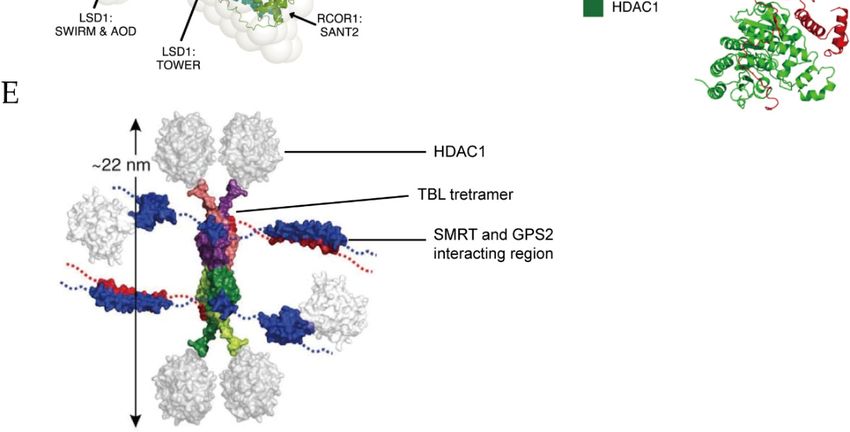
understanding of the structure of HDACs. Since Somoza et al. reported the first crystal structure of
 HDACs in 2004, to date, most crystal structural information of monomer HDACs is about HDAC8
 [57–61]. The global structures of Class I HDACs look similar (Figure 2A) because they all contain a
 large
Int.     catalytic
     J. Mol. Sci. 2020,domain,
                        21, 8828 which consist of a central parallel β-sheet surrounded by several α-helices
                                                                                                      4 of 25
 linked with loops. In addition, they share a 35–160 amino acid unstructured C-terminal tail except
 for HDAC8, which is used to recruit protein complexes and to be post-translationally modified
they share a 35–160 amino acid unstructured C-terminal tail except for HDAC8, which is used to recruit
 [15,57,62]. The active sites of Class I HDACs are almost identical, and the entrance of the active sites
protein complexes and to be post-translationally modified [15,57,62]. The active sites of Class I HDACs
 located at the surface of these enzymes [15]. In the case of HDAC8, the active site contains an
are almost identical, and the entrance of the active sites located at the surface of these enzymes [15].
 approximate 12-Å-deep narrow hydrophobic tunnel formed by hydrophobic residues Phe152,
In the case of HDAC8, the active site contains an approximate 12-Å-deep narrow hydrophobic tunnel
 Phe208, His180, Gly151, Met274 and Tyr306, where a zinc ion lies at its bottom as a member of the
formed by hydrophobic residues Phe152, Phe208, His180, Gly151, Met274 and Tyr306, where a zinc ion
 catalytic pocket (Figure 2B) [57,62]. The zinc ion is pentacoordinate and bound to Asp178 (Oδ1),
lies at its bottom as a member of the catalytic pocket (Figure 2B) [57,62]. The zinc ion is pentacoordinate
 His180 (Nδ1) and Asp267 (Oδ1), while the other two coordination sites are occupied by the acetyl
and bound to Asp178 (Oδ1), His180 (Nδ1) and Asp267 (Oδ1), while the other two coordination sites
 moiety (carbonyl oxygen) of the substrate and a water molecule [57].
are occupied by the acetyl moiety (carbonyl oxygen) of the substrate and a water molecule [57].
        However, several differences also exist between HDAC8 and HDAC1-3. For example, Loop1 of
        However, several differences also exist between HDAC8 and HDAC1-3. For example, Loop1 of
 HDAC8 is shorter than the corresponding one in HDAC1-3 [28]. Additionally, lacking the
HDAC8 is shorter than the corresponding one in HDAC1-3 [28]. Additionally, lacking the unstructured
 unstructured C-terminal tail may explain why HDAC8 can work as a monomeric protein [57]. The
C-terminal tail may explain why HDAC8 can work as a monomeric protein [57]. The surface around
 surface around the active site also plays an important role in substrate-binding [15]. A unique solvent-
the active site also plays an important role in substrate-binding [15]. A unique solvent-exposure residue
 exposure residue Try198 in the surface of HDAC3, which is near to the active site, may be related to
Try198 in the surface of HDAC3, which is near to the active site, may be related to substrate specificity
 substrate specificity (Figure 2A) [28]. Another structural difference between HDAC3 and HDAC1/2
(Figure 2A) [28]. Another structural difference between HDAC3 and HDAC1/2 is the extended loop,
 is the extended loop, which also shows a sequence distinction (Figure 2A) [15]. Moreover, a 14-Å
which also shows a sequence distinction (Figure 2A) [15]. Moreover, a 14-Å “foot pocket” was found
 “foot pocket” was found lying perpendicular to the end of the hydrophobic tunnel in Class I HDACs,
lying perpendicular to the end of the hydrophobic tunnel in Class I HDACs, which may be an exit
 which may be an exit of the acetate product [63,64]. However, the foot pocket in HDAC8 is narrower
of the acetate product [63,64]. However, the foot pocket in HDAC8 is narrower than in HDAC1-3
 than in HDAC1-3 because the large side chain of Trp141 in HDAC8 occupies this space (Figure 2C)
because the large side chain of Trp141 in HDAC8 occupies this space (Figure 2C) [15,64]. Additionally,
 [15,64]. Additionally, Ser113/Ser118 of HDAC1/2 is altered to tyrosine in HDAC3, which leads to a
Ser113/Ser118 of HDAC1/2 is altered to tyrosine in HDAC3, which leads to a steric hindrance so that
 steric hindrance so that bulky functional-groups of inhibitors are inaccessible to the foot pocket
bulky functional-groups of inhibitors are inaccessible to the foot pocket (Figure 2A) [28].
 (Figure 2A) [28].

      Figure2.2. (A)
     Figure       (A) Superposition
                      Superposition ofof the
                                          the structures
                                               structures of
                                                           ofHDAC1–3
                                                             HDAC1–3 (PDB  (PDB (Protein
                                                                                 (ProteinData
                                                                                           DataBank)
                                                                                                 Bank)codes:
                                                                                                        codes: 5ICN,
                                                                                                               5ICN,
     4LY1
      4LY1and
           and4A69).
                  4A69).Significant
                         Significantresidue
                                     residuedifferences
                                                differencesare
                                                            arehighlighted
                                                                highlightedin incyan
                                                                                 cyan(HDAC1
                                                                                       (HDAC1and and2)
                                                                                                     2)and
                                                                                                        andmagenta
                                                                                                            magenta
     (HDAC3).
      (HDAC3).Adapted
                   Adaptedwith
                            withpermission
                                  permissionfromfromMillard
                                                      Millard2017
                                                               2017[15].
                                                                    [15]. (B)
                                                                          (B) The
                                                                              Theactive
                                                                                   activesite
                                                                                         site of
                                                                                              of HDAC8
                                                                                                 HDAC8 (PDB
                                                                                                          (PDB code:
                                                                                                                code:
     4QA2). Adapted with permission from Chakrabarti 2015 [17]. (C) The foot pocket of HDAC8. Prominent
     amino-acid side-chain differences between HDAC8 and HDAC1 in the foot pocket are shown. Adapted
     with permission from Whitehead 2011 [64].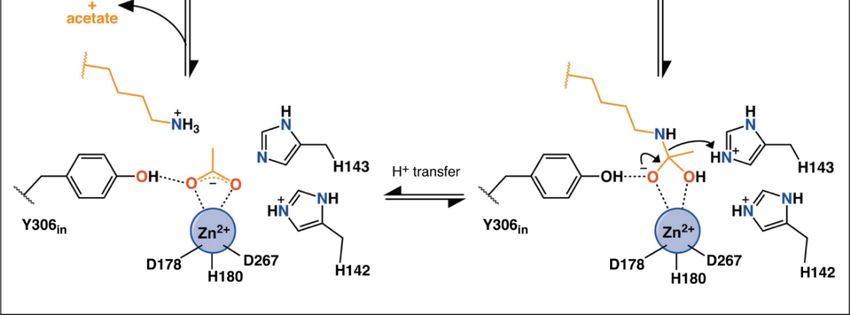
Int. J. Mol. Sci. 2020, 21, x FOR PEER REVIEW 5 of 25
Int. J. Mol. Sci. 2020, 21, 8828 5 of 25
4QA2). Adapted with permission from Chakrabarti 2015 [17]. (C) The foot pocket of HDAC8.
Prominent amino-acid side-chain differences between HDAC8 and HDAC1 in the foot pocket are
shown. Adapted with permission from Whitehead 2011 [64].
Classes I HDACs are metal-dependent enzymes that use a metal−water as the nucleophile during
catalysis. Classes
The catalytic
I HDACsmechanism of HDACs has
are metal-dependent been previously
enzymes that use a summarized
metal−water as bythePorter et al. [65,66].
nucleophile
As shown
duringincatalysis.
Figure The
3, incatalytic
a substrate-free
mechanism state, two ofhas
of HDACs thebeen
coordination
previously sites are occupied
summarized byetwater
by Porter
molecules [66]. As
al. [65,66]. When theinsubstrate
shown Figure 3, approaches the catalytic
in a substrate-free pocket,
state, two of theHis143 in HDAC8
coordination helps
sites are to activate
occupied
the water molecule,
by water moleculeswhich
[66]. then
Whennucleophilically attacks the
the substrate approaches thecatalytic
carbonyl carbon.
pocket, His143
His143 then protonates
in HDAC8 helps
to activate
the amine group theand
water molecule,
promotes thewhich thenofnucleophilically
leaving attacks theand
deacetylated substrates carbonyl carbon.
acetate. His143
During thethen
catalytic
protonates
process, Try306 the amine group
undergoes and promotes
an induced-fit the leaving
alteration of deacetylated
between substrates
“in” and “out” and acetate. During
conformations. Meanwhile,
thestays
His142 catalytic process,and
protonated Try306 undergoes
maintains an induced-fit
the electrostatic alteration between “in” and “out”
environment.
conformations. Meanwhile, His142 stays protonated and maintains the electrostatic environment.
Figure 3. Possible
Figure mechanism
3. Possible of catalysis
mechanism by by
of catalysis HDAC8. Adapted
HDAC8. Adaptedwith
withpermission
permission from
from Porter
Porter 2019 [66].
[66].
The activity of HDACs can also be regulated by monovalent metal cations or phosphorylation [7,67].
A second The activity of HDACs
metal-binding site was canfound
also be regulated by monovalent
approximately 7 Å away frommetalthecations
zincorion
phosphorylation
in Class I HDACs,
[7,67]. A second metal-binding site was found approximately
which impacts the catalytic mechanism [58,62]. The second metal can be potassium,7 Å away from the zinc ion in Class
calciumI or
HDACs, which impacts the catalytic mechanism [58,62]. The second metal can be potassium, calcium
sodium ions, depending on the salt contained during crystallization [58]. In the case of HDAC8,
or sodium ions, depending on the salt contained during crystallization [58]. In the case of HDAC8,
when a potassium ion occupies this site, it will be hexa-coordinated by six oxygen from Asp176
when a potassium ion occupies this site, it will be hexa-coordinated by six oxygen from Asp176
(main-chain carbonyl oxygen and Oδ1), Asp178 (main-chain carbonyl oxygen), His180 (main-chain
(main-chain carbonyl oxygen and Oδ1), Asp178 (main-chain carbonyl oxygen), His180 (main-chain
carbonyl oxygen),
carbonyl oxygen), Ser199
Ser199(Oγ) and
(Oγ) andLeu200
Leu200(main-chain carbonyloxygen),
(main-chain carbonyl oxygen), andand
twotwo of these
of these residues
residues
also chelate withwith
also chelate zinczinc
ionion[62].
[62].AAseries
seriesof
ofcomputational simulation
computational simulation studies
studies havehave
shownshownthat that
the the
second metal-binding
second metal-binding sitesite
also influences
also influencesthethe catalytic pocketthrough
catalytic pocket through altering
altering the the structure
structure of theof the
catalytic
catalytic site, site, facilitating
facilitating thethe stabilizationofofdeprotonation
stabilization deprotonation states
statesofofinhibitors [60,68].
inhibitors [60,68].
Additionally, a unique phosphorylation on Ser39
Additionally, a unique phosphorylation on Ser39 in HDAC8 impacts in HDAC8 impacts protein
proteinstructure and and
structure
decreases the enzyme activity [69]. Leng et al. illustrated that phosphorylation of Ser39 the
decreases the enzyme activity [69]. Leng et al. illustrated that phosphorylation of Ser39 distorts distorts
Loop1, which lines at the side of the active site, thus perturbing local structure [7]. It is worth
the Loop1, which lines at the side of the active site, thus perturbing local structure [7]. It is worth
mentioning that there is also phosphorylation at other sites of HDAC1-3 but activating the enzymes,
mentioning that there is also phosphorylation at other sites of HDAC1-3 but activating the enzymes,
which will be discussed below.
which will be discussed below.
3.2. Structure of HDAC Complexes
To date, HDAC8 is the only reported HDACs that can function as a monomer, while all other Class
I HDACs must function as a component of multiprotein complexes. The major challenge with structural
studies of HDAC complexes is that HDAC1, 2 and 3 work as subunits of large protein complexes and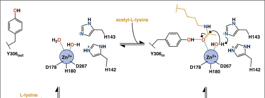
Int. J. Mol. Sci. 2020, 21, 8828 6 of 25
have distinct functions, and can exist in different complexes [15,70]. NuRD, Sin3 and CoREST are the
major Class I HDAC multiprotein complexes. Early studies have shown that HDAC1, HDAC2, RBBP4
(RbAp48) and RBBP7 (RbAp46) form the core histone deacetylase complex, which exists in both NuRD
and Sin3 macromolecular complexes [71].
The NuRD complex possesses both ATPase and histone deacetylase activities [72], participating
in transcriptional repression, chromatin assembly, cell cycle progression and genomic stability [73].
Thus far, at least seven protein families have been found as the components of NuRD: two catalytic
subunits including HDAC1/2 and CHD3/4 (chromodomain helicase DNA-binding protein 3/4,
also known as Mi-2α/Mi-2β), MTA1/2/3 (metastasis tumor-associated protein 1/2/3), MBD2/3
(methylated CpG-binding domain protein 2/3), RBBP4/7 (retinoblastoma-binding protein 4/7, also called
RbAp48/46), GATAD2A/2B (GATA zinc finger domain containing 2A/2B, i.e., p66α/p66β) and CDK2AP1
(cyclin-dependent kinase 2-associated protein 1) [74]. A single-particle negative-stain electron
microscopy (EM) method coupled with small-angle X-ray scattering (SAXS) and chemical crosslinking
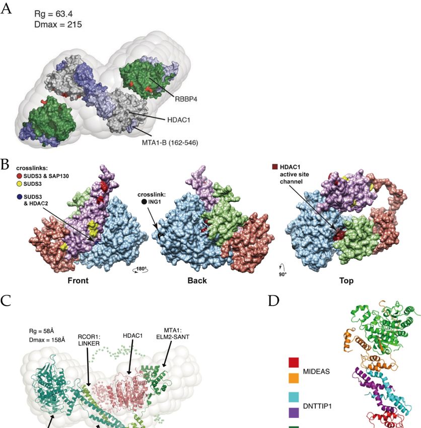
has been applied to reveal the core structure of the NuRD complex [75]. As shown in Figure 4A,
its substructure is composed of HDAC1/2, MTA1/2/3 and RBBP4/7 with a binding stoichiometry of 2:2:4.
Dimeric MTA1 functions as a backbone to recruit two HDAC1 and four RBBP4 separately and forms
an elongated zig-zag conformation through the highly conserved ELM2-SANT and R1/2 domains,
of MTA1. Additionally, GATAD2A/B bridges CHD3/4 and MBD2/3, the latter may play a key role in
linking them with the core NuRD complex [76]. The detail roles of other subunits in the formation of
NuRD complex can be found in a recent review [76].
The Sin3 complex regulates gene transcription at the promoter and transcribed regions, engaging in
cell processes such as Notch signaling and mitochondrial functions [77]. Sin3 complex contains Sin3A/B
proteins, HDAC1/2, RBBP4/7, SUDS3 (suppressor of defective silencing 3) and SAP30 (sin3-associated
protein p30). The Sin3 complex can also be divided into Sin3A or Sin3B complexes depending on which
subunit (Sin3A or Sin3B) it contains [78]. Using an affinity purification mass spectrometry (AP-MS)
based approach, Washburn’s group revealed that SUDS3 presents in both Sin3A and Sin3B complexes,
while SAP30 is only utilized in the Sin3A complex [79]. Later, by integrating chemical crosslinking
MS (XL-MS) with AP-MS, they modeled the substructure of the Sin3A complex [80]. Figure 4B shows
that Sin3A protein exists as a backbone so that the other subunits, including HDAC1/2, SAP30 and
SUDS3, can assemble. The active site of HDAC1 locates at the binding interface of HDAC1 and SAP30.
It is important to mention that, when targeting gene, Sin3 requires the aid of additional DNA-binding
proteins due to its lack of DNA-binding activity [81].
The CoREST complex has both demethylation and deacetylation activities, silencing the expression
of cancer and neurological disorders related genes [82,83]. It consists of CoREST1-3 (also called
RCOR1-3) proteins, LSD1 (lysine-specific demethylase 1) and HDAC1/2 [84]. The crystal structure of
the LSD1/CoREST revealed the interaction between LSD1 and CoREST, both of them bind to DNA,
while the latter also interacts with histones [85,86]. EM study established a bi-lobed structure with
LSD1 and HDAC1 at two opposite sides of the CoREST, where RCOR1 acts as a long string linking the
other two components (Figure 4C) [87].
The MiDAC complex contains HDAC1/2, DNTTIP1 (deoxynucleotidyl-transferase
terminal-interacting protein 1) and the mitotic deacetylase-associated SANT domain (MIDEAS)
corepressor protein [15], playing regulatory roles in gene expression of neuronal and embryonic
development [88,89]. A previous study suggested that MiDAC is a tetrameric complex that contains
four copies of HDAC1/2, DNTTIP1 and MIDEAS, respectively [90]. Cryo-EM structure showed that
in the dimeric subcomplex, two HDAC1 are on both sides of MiDAC (Figure 4D) [89]. In addition,
the ELM2 domain of MIDEAS does not directly form dimerization as MTA1 in the NuRD complex
does. It is DNTTIP1 that actually mediates the dimeric assembly.
The SMRT/NCoR complex is associated with development and homeostasis in inflammation,
neuronal and cardiovascular diseases [91]. A highly conserved N-terminal region of SMRT/NCoR
protein recruits at least three proteins: HDAC3, GPS2 (G-protein pathway suppressor 2) and TBL1Int. J. Mol. Sci. 2020, 21, x FOR PEER REVIEW 7 of 25
Int. J. Mol. Sci. 2020, 21, 8828 7 of 25
The SMRT/NCoR complex is associated with development and homeostasis in inflammation,
neuronal and cardiovascular diseases [91]. A highly conserved N-terminal region of SMRT/NCoR
(transducin
protein beta-like
recruits at1)least
[92].three
Crystal and NMR
proteins: structures
HDAC3, showed that
GPS2 (G-protein SMRT
pathway interacts2)with
suppressor andHDAC3
TBL1 at
regions near thebeta-like
(transducin active site1) [92]. the N-terminus
andCrystal of TBL1 protein
and NMR structures showed forms a tetrameric
that SMRT interaction
interacts with HDAC3 with
SMRTatand
regions
GPS2near the active
(Figure 4E)site and the N-terminus of TBL1 protein forms a tetrameric interaction with
[93,94].
SMRT and GPS2 (Figure 4E) [93,94].
Figure 4. (A) Structure of the core NuRD complex (HDAC1/RBBP4/MTA1 (162-546)). Adapted with
permission from Millard 2016 [75]. (B) Substructure of the Sin3 complex. Adapted with permission from
Banks 2020 [80]. (C) Structure of the CoREST complex. Adapted with permission from Song 2020 [87].
(D) Structure of the MiDAC complex in dimeric form. Adapted with permission from Turnbull 2020 [89].
(E) Structure of the SMRT/NCoR complex. Adapted with permission from Oberoi 2011 [93].Figure 4. (A) Structure of the core NuRD complex (HDAC1/RBBP4/MTA1 (162-546)). Adapted with
permission from Millard 2016 [75]. (B) Substructure of the Sin3 complex. Adapted with permission
from Banks 2020 [80]. (C) Structure of the CoREST complex. Adapted with permission from Song 2020
[87]. (D) Structure of the MiDAC complex in dimeric form. Adapted with permission from Turnbull
Int. J. Mol.
2020Sci. (E) 21,
2020,
[89]. 8828 of the SMRT/NCoR complex. Adapted with permission from Oberoi 2011 [93].8 of 25
Structure
3.3. Allosteric Sites and Regulations
3.3. Allosteric Sites and Regulations
Phosphorylation of HDAC1-3 stimulates the enzyme activity, which is opposite to that of
Phosphorylation of HDAC1-3 stimulates the enzyme activity, which is opposite to that of
HDAC8 [6]. In addition, the enzymatic activity of HDAC1-3 in complexes has been shown to be
HDAC8 [6]. In addition, the enzymatic activity of HDAC1-3 in complexes has been shown to be
regulated by inositol phosphates, which bind in a pocket sandwiched between the HDAC and
regulated by inositol phosphates, which bind in a pocket sandwiched between the HDAC and
corepressor proteins [16]. More specifically, in the HDAC3/SMRT crystal structure, inositol
corepressor proteins [16]. More specifically, in the HDAC3/SMRT crystal structure, inositol phosphate
phosphate binds to a few conserved key residues (His17, Gly21, Lys25, Arg265, Arg301 in HDAC3
binds to a few conserved key residues (His17, Gly21, Lys25, Arg265, Arg301 in HDAC3 and Lys449,
and Lys449, Tyr470, Tyr471, Lys474, Lys 475 in SMRT, Figure 5A) [94,95]. Allosteric communication
Tyr470, Tyr471, Lys474, Lys 475 in SMRT, Figure 5A) [94,95]. Allosteric communication between
between the inositol-binding site and the active site has been observed, which facilitates the activation
the inositol-binding site and the active site has been observed, which facilitates the activation of
of enzyme activity [16].
enzyme activity [16].
Moreover, another allosteric site on the surface of HDAC2 and near the active site of the enzyme
Moreover, another allosteric site on the surface of HDAC2 and near the active site of the enzyme
(Figure 5B) has been disclosed by computational methods [96,97]. The QM/MM study revealed the
(Figure 5B) has been disclosed by computational methods [96,97]. The QM/MM study revealed the
flexibility of Loop2 in HDAC2, the conformational changes of X-D dyad in Loop2 (also called binding
flexibility of Loop2 in HDAC2, the conformational changes of X-D dyad in Loop2 (also called binding
rail) directly induce the switch of the substrate-binding tunnel [96].
rail) directly induce the switch of the substrate-binding tunnel [96].
Recently, an NMR study revealed that Helix1-Loop1-Helix2 is an allosteric site of HDAC8 [98].
Recently, an NMR study revealed that Helix1-Loop1-Helix2 is an allosteric site of HDAC8 [98].
A bidirectional regulatory effect exists between the Helix1-Loop1-Helix2 region and the active site of
A bidirectional regulatory effect exists between the Helix1-Loop1-Helix2 region and the active site of
HDAC8; thus, Ser39 and Met40 may be key residues in this allosteric regulation. Overall, phosphate-
HDAC8; thus, Ser39 and Met40 may be key residues in this allosteric regulation. Overall, phosphate-binding
binding sites, binding rail and Helix1-Loop1-Helix2 region may be targets for the design of allosteric
sites, binding rail and Helix1-Loop1-Helix2 region may be targets for the design of allosteric inhibitors.
inhibitors.
Figure 5. (A) The binding site of inositol phosphate with HDAC3-SMRT complex. The inositol
phosphate
Figure 5. (A)is shown as a stick
The binding model.
site Adaptedwith
of inositol permission
phosphate from Watson complex.
with HDAC3-SMRT 2012 [94]. The
(B) Relative
inositol
position
phosphate is shown as a stick model. Adaptedwith permission from Watson 2012 [94]. (B)2017
of the allosteric site and binding rail on HDAC2. Adapted with permission from Zhou [96].
Relative
position of the allosteric site and binding rail on HDAC2. Adapted with permission from Zhou 2017
4. Pan-Inhibitors
[96].
Although high-resolution structures of these Class I HDACs have been determined, developing
4. Pan-Inhibitors
truly isoform-selective HDAC inhibitors has proven challenging due to the structural similarity
of the active sites of these enzymes. Up to date, all six HDAC inhibitors approved by FDA or
Although high-resolution structures of these Class I HDACs have been determined, developing
NMPA are pan-inhibitors [99]. Additionally, a number of pan-inhibitors, such as entinostat (MS-275),
truly isoform-selective HDAC inhibitors has proven challenging due to the structural similarity of
are under clinical trials or investigation [100]. A general mechanism of pan-inhibitors to act against
the active sites of these enzymes. Up to date, all six HDAC inhibitors approved by FDA or NMPA
Zn2+ -dependent HDACs is to occupy the active site, thus competitively inhibiting substrate-binding
are pan-inhibitors [99]. Additionally, a number of pan-inhibitors, such as entinostat (MS-275), are
to HDACs [101]. They mainly follow a classic pharmacophore model (Figure 1), consisting of2+a
under clinical trials or investigation [100]. A general mechanism of pan-inhibitors to act against Zn -
cap, a linker, and a zinc-binding group (ZBG), which corresponds to surface recognition region,
dependent HDACs is to occupy the active site, thus competitively inhibiting substrate-binding to
substrate-binding tunnel, zinc chelation site and foot pocket in the target protein, respectively [96].
According to their chemical structures, it can be categorized into four types, including hydroxamic
acids, benzamides, cyclic peptides or depsipeptides and aliphatic carboxylic acids [102,103]. Here,
we will briefly outline the four types of pan-inhibitors. More pan-inhibitor development can be found
in several recent reviews [5,28,99,104].Int. J. Mol. Sci. 2020, 21, 8828 9 of 25
4.1. Hydroxamic Acids
Hydroxamic acids are the most common pan-inhibitors in investigations, represented by SAHA,
belinostat, pracinostat, panobinostat, etc. As shown in Figure 2B, the carbonyl oxygen and hydroxyl
oxygen of hydroxamic bicoordinates with a zinc ion. Meanwhile, His142, His143 and Try306 (in HDAC8)
stabilize the interaction [62,68]. Due to its fair performance in vitro stability, good solubility and
easy synthesis, hydroxamic acids are often preferred in the design of novel HDACis [105]. Improved
inhibiting effects and selectivity of this category of inhibitors have been demonstrated by modifying
the cap and linker region [106]. However, its over-high metal-binding ability can lead to undesirable
coordination to other zinc-dependent enzymes such as aminopeptidases, matrix metalloproteinases
and carbonic anhydrases, thus causing low selectivity, off-target effects and severe toxicity [99,102,105].
4.2. Benzamides
Compared with hydroxamic acids, benzamides such as chidamide and entinostat show better
selectivity toward Class I HDACs [107]. In the crystal structure of HDAC2-inhibitor, the primary
amine nitrogen and amide oxygen bi-coordinate with zinc ion, but with lower affinity than that
of hydroxamic [108]. Meanwhile, the residues surrounding ZBG and foot region form a hydrogen
bond network to stabilize inhibitor-binding. Benzamide ring also provides a modifiable site target
foot pockets in Class I HDACs [108]. However, its slower binding rate constants may attribute to
compromised drug effects.
4.3. Cyclic Peptides
Romidepsin is the only approved cyclic peptide inhibitor that is selectivity to Class I HDACs [109].
The mechanism of action of this kind of inhibitors is initiated by the reduction of the disulfide bond,
thus releases a thiol group to coordinate the zinc ion in the active site [99,110]. It has been demonstrated
that modification on the cap region can increase their biological activity and selectivity [111]. Yet,
the toxicity and easy oxidation remain the major challenges in the development of cyclic peptide
inhibitors [51,110].
4.4. Aliphatic Carboxylic Acids
Low toxicity and easy synthesis are the main features of aliphatic carboxylic acids [19,106].
The specific mechanism of them is not yet clear, most probably through binding to the active
site [112]. A docking study also inferred that they might occupy the substrate-binding tunnel as
general pan-inhibitors do [113]. Inhibiting HDACs by valproic acid showed potential effects in solid or
CNS (central nervous system) tumors and neurological diseases [114,115]. Sodium phenylbutyrate is
currently under clinical trials in lymphoma or solid tumors [116]. However, weak zinc-binding ability
leads to low inhibitory effects; thus, aliphatic carboxylic acids are often used in combination with other
drugs in clinical trials [99].
5. Isoform-Selective Inhibitors
A problem with the currently available pan-HDAC inhibitors is that they have limited
specificity and target multiple deacetylases, greatly limiting their clinical uses due to significant
side effects [117]. The design of isoform-selective inhibitors has become the main focus and is
being actively undertaken [112,117,118]. Here, we introduce the current status of selective inhibitor
development according to their targeted isoforms.
5.1. HDAC8-Selective Inhibitors
HDAC8 is a unique member of Class I HDACs due to its structural distinctions from HDAC1-3.
First, as mentioned above, the absence of 35–160 amino acids in the C-terminus may explain why
it works as a monomer [17]. Second, the Loop1 of HDAC8 is highly flexible and forms a large partInt. J. Mol. Sci. 2020, 21, 8828 10 of 25
of the active site, which extends to the protein surface; thus, HDAC8 has a wider active site pocket
with a larger surface opening than HDAC1-3 [57]. Third, the phosphorylation is also distinctive in
HDAC8 than the other isoforms. Moreover, as to the development of HDAC8-selective inhibitors,
the exploration of structural features of HDAC8 goes deeper.
PCI-34051(Figure 6, Compound 7) and NCC-149 (Figure 6, Compound 8) are the most widely
investigated HDAC8-selective inhibitors. PCI-34051 has >200-fold selectivity towards HDAC8 over
other isoforms; it is found effective or cytotoxic in T-cell lymphoma, leukemia and other types of
tumor cells, such as Jurkat, HuT78 and Molt-4 cell [29]. NCC-149 shows a potent inhibiting effect
in T-cell lymphoma growth and >500 fold selectivity over HDAC1 and 2, while >30 fold selectivity
over HDAC6 in Class IIb [119]. However, the specific mechanism and binding structure of these two
inhibitors with human HDAC8 (hHDAC8) are poorly understood. Marek et al. revealed that the
binding mode of PCI-34051 and NCC-149 with Schistosoma mansoni HDAC8 (smHDAC8) through
X-ray crystallization [120]. The two inhibitors coordinate with zinc ion in the active site of HDAC8 by
hydroxamic groups, as general hydroxamic acid inhibitors do, but with an “L” shape conformation.
The cap group of PCI-34051 and NCC-149 interacts with the Y341 (Y306 in hHDACs) and inserts into
the cavity formed by Loop1 and Loop6. All together, they form an HDAC8 selective pocket. In contrast,
this pocket is blocked by protruding residues of Loop1 and Loop6 in HDAC1-3. The derivatives of
PCI-34051 and NCC-149 are under development, exhibiting great potential [121–123].
Most HDACis have a linker group composed of a long fatty chain, as shown in SAHA (Figure 1,
Compound 1). However, Krennhrubec et al. demonstrated that “linkerless” hydroxamic acids (Figure 6,
Compounds 9–14) can specifically target HDAC8 [124]. The idea is originated from the discovery
of an HDAC8 selective sub-pocket near the active site [57], confirmed by crystal structures [125].
The hydroxamic group binds to zinc ion: meanwhile, the steric hindrance effect of the bulky aryl group
of inhibitors causes the split of F152 and M274, exposing the sub-pocket [125]. It is worth mentioning
that “linkerless” does not mean that there is no linker region but a much shorter linker. “Linkerless”
inhibitors show selectivity towards HDAC8 over HDAC1 and HDAC6 and give prospect in AML,
neurodegenerative diseases as well as genetic disorders [124,126].
Additionally, Taha et al. developed a series of HDAC8 inhibitors by modifying the cap region,
yielding good selectivity towards HDAC8 over HDAC1-3 (Figure 6, Compounds 15–16) [127].
Their inhibitory effect in neuroblastoma was verified by cellular experiments [127]. By targeting
the foot region of HDAC8, Whitehead et al. designed several amino-acid derivatives with HDAC8
selectivity (Figure 6, Compounds 17–18) [64]. Crystal structures revealed that the amide group of
inhibitors coordinates with zinc ion, and the foot groups insert into the foot pocket in the active site of
protein [64]. The foot pocket in HDAC1 is narrower than in HDAC8, the differences in residues of
foot pocket between HDAC8 and HDAC1 may be responsible for the observed isoform selectivity [64].
Furthermore, other HDAC8-selective inhibitors that contain novel active or selective groups are under
development [128–131].
5.2. HDAC1/2-Selective Inhibitors
Compared to HDAC8-selective inhibitors, the development of HDAC1/2/3-selective inhibitors has
lagged behind owing to their high sequence similarities (85% homological identity between HDAC1
and HDAC2 and 64% between HDAC1 and HDAC3) [6,132]. In addition, fewer structural studies
make it even more challenging to design HDAC1/2-selective inhibitors.
The first HDAC2-selective inhibitor (Figure 7, Compound 19) was discovered by Zhou et al.,
whose selectivity depends on inhibition time [133]. The IC50 values show that after 24h inhibition,
this inhibitor displays good selectivity towards HDAC2 over HDAC1&3. QM/MM simulation inferred
that different ions in the second metal-binding site have different binding kinetics, which leads to the
time-dependence selectivity effect of β-hydroxymethyl chalcone inhibitors.Int. J. Mol. Sci. 2020, 21, 8828 11 of 25
Int. J. Mol. Sci. 2020, 21, x FOR PEER REVIEW 11 of 25
Figure 6. Selective inhibitors of HDAC8.
Figure 6. Selective inhibitors of HDAC8.
By targeting the foot pocket, Bressi et al. designed a series of N-(2-amino-5-substituted
5.2. HDAC1/2-Selective
phenyl)benzamides (Figure Inhibitors
7, Compound 20) that are effective to HDAC2, but their selectivity was
not fully explored [134]. The
Compared to HDAC8-selective crystal structure revealed
inhibitors, that the HDAC2
the development foot pocket consistsinhibitors
of HDAC1/2/3-selective of Tyr29,
Met35, Phe114 and Leu144, and the phenyl groups of inhibitors can insert into
has lagged behind owing to their high sequence similarities (85% homological identity betweenthis foot pocket [134].
HDAC1SHI-1:2
andisHDAC2
anotherand
type64%
of HDAC1/HDAC2-selective
between HDAC1 and HDAC3) inhibitors (Figure
[6,132]. In 7, Compound
addition, 21–22)
fewer [132].
structural
The docking study showed that the carbonyl and aniline groups
studies make it even more challenging to design HDAC1/2-selective inhibitors. are bound to zinc ion while the
phenyl
Theinteracts with the foot pocket.
first HDAC2-selective However,
inhibitor (FigureTry96 in HDAC319)
7, Compound (contains a larger moiety
was discovered by Zhouthan
et the
al.,
corresponding residue, Ser113 in HDAC1) causes this site inaccessible for SHI-1:2.
whose selectivity depends on inhibition time [133]. The IC50 values show that after 24h inhibition,
this inhibitor displays good selectivity towards HDAC2 over HDAC1&3. QM/MM simulation
inferred that different ions in the second metal-binding site have different binding kinetics, which
leads to the time-dependence selectivity effect of β-hydroxymethyl chalcone inhibitors.phenyl)benzamides (Figure 7, Compound 20) that are effective to HDAC2, but their selectivity was
not fully explored [134]. The crystal structure revealed that the HDAC2 foot pocket consists of Tyr29,
Met35, Phe114 and Leu144, and the phenyl groups of inhibitors can insert into this foot pocket [134].
SHI-1:2 is another type of HDAC1/HDAC2-selective inhibitors (Figure 7, Compound 21–22)
[132]. The docking study showed that the carbonyl and aniline groups are bound to zinc ion while
Int. J. Mol. Sci. 2020, 21, 8828 12 of 25
the phenyl interacts with the foot pocket. However, Try96 in HDAC3 (contains a larger moiety than
the corresponding residue, Ser113 in HDAC1) causes this site inaccessible for SHI-1:2.
Additionally,ininthe
Additionally, thesubstrate-binding
substrate-binding tunnel,
tunnel, replacing
replacing Glu98
Glu98 andand Try204
Try204 in HDAC1/2
in HDAC1/2 with
with other
other amino
amino acids may acidslead
maytolead to structural
structural distinctions,
distinctions, formingforming
a targeta for
target for selective
selective inhibitor
inhibitor design design
[135].
[135].
OH
O O
HN
H2 N
19 20
O O
N N
H H
OH NH2
N
21 22
Figure
Figure 7. Selective inhibitors
7. Selective inhibitors of
of HDAC1/2.
HDAC1/2.
5.3. HDAC3-Selective Inhibitors
5.3. HDAC3-Selective Inhibitors
A structural alignment reveals that five key residues in HDAC3 are different from HDAC1
A structural alignment reveals that five key residues in HDAC3 are different from HDAC1 and
and HDAC2, including Val13, Leu29, Asp92, Tyr107 and Phe199, which may lead to structural
HDAC2, including Val13, Leu29, Asp92, Tyr107 and Phe199, which may lead to structural divergence
divergence [136]. For example, Try107 in HDAC3 (serine in HDAC1 and HDAC2) causes steric
[136]. For example, Try107 in HDAC3 (serine in HDAC1 and HDAC2) causes steric hindrance and
hindrance and thus excludes the binding of inhibitors with large functional groups [136].
thus excludes the binding of inhibitors with large functional groups [136].
Benzamide inhibitors show great selectivity in inhibiting HDAC3, thus have earned the favors of
Benzamide inhibitors show great selectivity in inhibiting HDAC3, thus have earned the favors
investigators [28]. RGFP966 (Figure 8, Compound 23) is a famous selective inhibitor towards HDAC3
of investigators [28]. RGFP966 (Figure 8, Compound 23) is a famous selective inhibitor towards
over other Class I HDACs, with effects in hepatoma carcinoma cell [137,138]. BRD3308 (Figure 8,
HDAC3 over other Class I HDACs, with effects in hepatoma carcinoma cell [137,138]. BRD3308
Compound 24) was first discovered effective in diabetes with >10 folds selectivity over HDAC1 and
(Figure 8, Compound 24) was first discovered effective in diabetes with >10 folds selectivity over
HDAC2 [139]. A docking study indicated that the selectivity originates from the conformational
HDAC1 and HDAC2 [139]. A docking study indicated that the selectivity originates from the
differences of Try107 and Leu144 [139]. Moreover, Marson et al. discovered an inhibitor (Figure 8,
conformational differences of Try107 and Leu144 [139]. Moreover, Marson et al. discovered an
Compound 25) contained heterocyclic capping group effective for HDAC3-NCoR1 over other monomer
inhibitor (Figure 8, Compound 25) contained heterocyclic capping group effective for HDAC3-
Class I HDACs [140]. Moreover, PD-106 (Figure 8, Compound 26), RGFP109 (Figure 8, Compound 27),
NCoR1 over other monomer Class I HDACs [140]. Moreover, PD-106 (Figure 8, Compound 26),
and other benzamides also exhibit HDAC3 selective [141–145]. However, the lack of structural details
RGFP109 (Figure 8, Compound 27), and other benzamides also exhibit HDAC3 selective [141–145].
makes it difficult to gain deep insight into their selective mechanisms.
However, the lack of structural details makes it difficult to gain deep insight into their selective
In addition, McClure et al. developed a group of allosteric inhibitors that show potential in AML
mechanisms.
(Figure 8, Compounds 28–30) [97]. As suggested by Zhou’s study, these inhibitors may bind to the
In addition, McClure et al. developed a group of allosteric inhibitors that show potential in AML
allosteric site (see Section 3.3) of Class I HDACs, but lead to a close conformation of Phe144 and
(Figure 8, Compounds 28–30) [97]. As suggested by Zhou’s study, these inhibitors may bind to the
Phe200 in the substrate tunnel of HDAC3 [96,97]. The specific mechanism of allosteric regulation and
allosteric site (see Section 3.3) of Class I HDACs, but lead to a close conformation of Phe144 and
inhibition has not yet been disclosed.Int. J. Mol. Sci. 2020, 21, x FOR PEER REVIEW 13 of 25
Phe200 in the substrate tunnel of HDAC3 [96,97]. The specific mechanism of allosteric regulation and
Int. J. Mol. Sci. 2020, 21, 8828 13 of 25
inhibition has not yet been disclosed.
Figure 8. Selective inhibitors of HDAC3.
Figure 8. Selective inhibitors of HDAC3.
6. Complex-Specific Inhibitors
6. Complex-Specific Inhibitors
Although several HDAC isoform-selective inhibitors have been reported, developing truly
Although several
isoform-selective HDAC HDAC isoform-selective
inhibitors inhibitors have
has proven challenging due to been
the reported,
structuraldeveloping
similarity oftruly
the
isoform-selective
active sites of these HDAC inhibitors
enzymes. Given hastheproven challenging
fact that most of the dueClass
to the structural
I HDACs similarity
must function ofas
thea
active sites
catalytic of these
subunit enzymes. Given
of gene-regulatory the fact that
complexes, most of novel
developing the Class I HDACs
inhibitors mustspecific
targeting function as a
HDAC
catalytic
complexes subunit
offersof
angene-regulatory
alternative but complexes,
yet attractive developing novelSeveral
strategy [15]. inhibitors targeting
strategies specific
have emergedHDACfor
complexes offers an
the development of alternative
this type ofbut yet attractive
inhibitors, strategy
including [15]. Several
utilizing specificstrategies have emerged
inhibitor-binding for
kinetics,
the development
developing of this
dual action type of disrupting
inhibitors, inhibitors,protein–protein
including utilizing specificand
interactions, inhibitor-binding kinetics,
targeting other subunits
developing dual action
in HDAC complexes inhibitors, disrupting protein–protein interactions, and targeting other
[15,83,146–155].
subunits
Usingin aHDAC complexes [15,83,146–155].
chemoproteomics method combined affinity capture and quantitative mass spectrometry,
Using and
Bantscheff a chemoproteomics
coworkers demonstrated method thatcombined
benzamides affinity capture
inhibitors can haveand different
quantitative mass
affinities to
spectrometry, Bantscheff and coworkers demonstrated that benzamides inhibitors
distinct HDAC complexes [147,148]. They found that benzamide inhibitors such as CI-994 (tacedinaline, can have different
affinities to distinct HDAC
Figure 9, Compound 31) andcomplexes
BML210 (Figure [147,148]. They found
9, Compound 32)that benzamide
are able to bind inhibitors
to the NuRD,such as CI-
CoREST
994
and (tacedinaline,
MiDAC complexes Figure 9, Compound
with distinct binding31) and BML210
kinetics (Figureno9,binding
but exhibit Compound 32)toare
affinity theable
Sin3tocomplex.
bind to
the NuRD,
Fuller et al. CoREST
discoveredanda MiDAC complexes
CoREST-specific with distinct
inhibitor binding kinetics
called Rodin-A (Figure 9, but exhibit no33)
Compound binding
[149].
affinity
This to theshow
inhibitor Sin3 selectivity
complex. not Fuller
onlyettowards
al. discovered
the CoRESTa CoREST-specific
complex but also inhibitor
to HDAC1 called
andRodin-A
HDAC2
(Figure 9, Compound
at the monomeric 33) [149].
protein level. This inhibitor
In addition, show
low selectivity not
hematological sideonly towards
effects make the CoREST
Rodin-A complex
a promising
but also to HDAC1
compound and HDAC2
for neurologic disordersat the monomeric protein level. In addition, low hematological side
[149].
effects
Asmake Rodin-A
mentioned a promising
in previous compound
sections, some offortheneurologic
HDAC complexes disorders
(NuRD[149].and CoREST) possess two
different enzyme activities simultaneously. Dual-action inhibitors which containand
As mentioned in previous sections, some of the HDAC complexes (NuRD twoCoREST) possess
pharmacophores
two different
in a single enzyme
molecule activities
may target simultaneously.
both activities Dual-action
of these enzymes inhibitors inhibitor,
[15]. A dual-action which corin
contain two
(Figure 9,
Compound 34), can effectively target the CoREST complex and show potential in treating many tumorInt. J. Mol. Sci. 2020, 21, x FOR PEER REVIEW 14 of 25
pharmacophores
Int. in8828
J. Mol. Sci. 2020, 21, a single
molecule may target both activities of these enzymes [15]. A dual-action
14 of 25
inhibitor, corin (Figure 9, Compound 34), can effectively target the CoREST complex and show
potential in treating many tumor cells [83]. 4SC-202 (domatinostat, Figure 9, Compound 35) is another
cells [83]. 4SC-202 (domatinostat, Figure 9, Compound 35) is another dual-action inhibitor designed for
dual-action inhibitor designed for targeting the CoREST complex, currently under development in
targeting the CoREST complex, currently under development in the treatment of colorectal cancer and
the treatment of colorectal cancer and hematological malignancies [15,150,151].
hematological malignancies [15,150,151].
Additionally, disturbing the interface of adjacent subunits in HDAC complexes can be a good
Additionally, disturbing the interface of adjacent subunits in HDAC complexes can be a good
idea, as suggested in Schwabe’s review [15]. Waxman’s group found a decoy peptide can interfere
idea, as suggested in Schwabe’s review [15]. Waxman’s group found a decoy peptide can interfere with
with the binding interface of Sin3A/B and other partner proteins, which may explain its specific
the binding interface of Sin3A/B and other partner proteins, which may explain its specific inhibitory
inhibitory effect on the Sin3 complex [154]. Latterly, they have screened out some compounds and
effect on the Sin3 complex [154]. Latterly, they have screened out some compounds and peptides as the
peptides as the candidates for inhibiting the interaction of Sin3 protein with another partner protein,
candidates for inhibiting the interaction of Sin3 protein with another partner protein, MAD [152,155].
MAD [152,155].
Targeting subunits other than HDACs in these complexes also provide an idea. For example,
Targeting subunits other than HDACs in these complexes also provide an idea. For example,
resveratrol (Figure 9, Compound 36) can decrease the expression of MTA1 in prostate cancer cells,
resveratrol (Figure 9, Compound 36) can decrease the expression of MTA1 in prostate cancer cells,
which then reduces the amount of MTA1:HDAC1 complexes [146]. However, resveratrol is also an
which then reduces the amount of MTA1:HDAC1 complexes [146]. However, resveratrol is also an
activator of SIRT1 with treatment potential in diverse diseases such as neurodegenerative diseases,
activator of SIRT1 with treatment potential in diverse diseases such as neurodegenerative diseases,
cancers and cardiovascular diseases with distinct pharmacological mechanisms [156,157]. In this
cancers and cardiovascular diseases with distinct pharmacological mechanisms [156,157]. In this
regard, resveratrol cannot be classified as a complex-specific inhibitor, but it provides a conception for
regard, resveratrol cannot be classified as a complex-specific inhibitor, but it provides a conception
drug design.
for drug design.
Figure 9. Specific-complex inhibitors.
Figure 9. Specific-complex inhibitors.
7. Conclusions and Perspectives
7. Conclusions and Perspectives
Class I HDACs have been wildly investigated, given their important roles in epigenetic regulation.
Class I HDACs
The effectiveness have inhibitors
of HDAC been wildly
has investigated, given their
also been confirmed important
in many disease roles in epigenetic
treatments such as
regulation.
cancers, The effectiveness
neurological of HDAC
diseases inhibitors hasand
and inflammations also infections
been confirmed in many
[8]. This disease
review treatments
highlights the
such as cancers,
structural studiesneurological diseases and inflammations
in Class I HDACs their complexes and
as infections [8]. This
well as pan-, review highlights
isoform-selective and
the structural studies
complex-selective in Class
inhibitor I HDACs and
development. their
Due to complexes as well asand
the poor selectivity pan-, isoform-selective
undesirable and
side effects
that occur in pan-inhibitors, the development of selective inhibitors has attracted the attention ofYou can also read