Contemporary Distribution, Estimated Age, and Prehistoric Migrations of Old World Monkey Retroviruses - MDPI
←
→
Page content transcription
If your browser does not render page correctly, please read the page content below
Review
Contemporary Distribution, Estimated Age, and Prehistoric
Migrations of Old World Monkey Retroviruses
Antoinette C. van der Kuyl

                                          Laboratory of Experimental Virology, Department of Medical Microbiology, Amsterdam UMC,
                                          University of Amsterdam, Meibergdreef 9, 1105 AZ Amsterdam, The Netherlands;
                                          a.c.vanderkuyl@amsterdamumc.nl; Tel.: +31-205-666-778

                                          Abstract: Old World monkeys (OWM), simians inhabiting Africa and Asia, are currently affected by
                                          at least four infectious retroviruses, namely, simian foamy virus (SFV), simian immunodeficiency
                                          virus (SIV), simian T-lymphotropic virus (STLV), and simian type D retrovirus (SRV). OWM also
                                          show chromosomal evidence of having been infected in the past with four more retroviral species,
                                          baboon endogenous virus (BaEV), Papio cynocephalus endogenous virus (PcEV), simian endogenous
                                          retrovirus (SERV), and Rhesus endogenous retrovirus-K (RhERV-K/SERV-K1). For some of the
                                          viruses, transmission to other primates still occurs, resulting, for instance, in the HIV pandemic.
                                          Retroviruses are intimately connected with their host as they are normally spread by close contact.
                                          In this review, an attempt to reconstruct the distribution and history of OWM retroviruses will be
                                          made. A literature overview of the species infected by any of the eight retroviruses as well as an age
                                          estimation of the pathogens will be given. In addition, primate genomes from databases have been
                                          re-analyzed for the presence of endogenous retrovirus integrations. Results suggest that some of
                                          the oldest retroviruses, SERV and PcEV, have travelled with their hosts to Asia during the Miocene,
                                          when a higher global temperature allowed simian expansions. In contrast, younger viruses, such as

                                   SIV and SRV, probably due to the lack of a primate continuum between the continents in later times,
                                          have been restricted to Africa and Asia, respectively.
Citation: van der Kuyl, A.C.
Contemporary Distribution,
                                          Keywords: retrovirus; endogenous; exogenous; Old World monkey; BaEV; PcEV; SERV; SERV-K1;
Estimated Age, and Prehistoric
                                          RhERV-K; SFV; SIV; SRV; STLV
Migrations of Old World Monkey
Retroviruses. Epidemiologia 2021, 2,
46–67. https://doi.org/
10.3390/epidemiologia2010005
                                          1. Introduction
Received: 15 December 2020                     The primate fossil record suggests that the radiation of Old World monkeys (OWM)
Accepted: 29 January 2021                 began in the Oligocene or early Miocene (around 25–30 million years ago (mya)), with
Published: 3 February 2021                the divergence of apes, including the ancestors of humans, and OWM (reviewed in [1],
                                          see also [2–4]). Subsequently, Colobinae and Cercopithecinae diverged around 16 mya,
Publisher’s Note: MDPI stays neutral      Papionini and Cercopithecini then split around 10–12 mya, and macaques and papionins
with regard to jurisdictional claims in   separated approximately 7.6 ± 1.3 mya [1]. Over the course of evolution, retroviruses
published maps and institutional affil-   have been infecting primate species. Evidence for this can be found in primate genomes,
iations.                                  where endogenous (i.e., germ line propagated) proviruses bear witness of past infections.
                                          The origin of the retrovirus family itself dates back to the early Paleozoic, approximately
                                          460–550 mya [5]. Most likely, retroviruses originate from the long terminal repeat (LTR)-
                                          containing retrotransposons found in eukaryotic genomes, by the addition of an envelope
Copyright: © 2021 by the author.          (env) gene [6,7]. Indeed, one such LTR-retroelement, HERV-L, has similarity to foamy
Licensee MDPI, Basel, Switzerland.        viruses in its polymerase (pol) gene [8].
This article is an open access article         At present, two retrovirus species infect humans, namely the lentivirus human immun-
distributed under the terms and           odeficiency virus (HIV) and the deltaretrovirus human T-lymphotropic virus (HTLV). HIV
conditions of the Creative Commons        has been transmitted from African non-human primates quite recently, probably around
Attribution (CC BY) license (https://
                                          the beginning of the 20th century [9–11]. So far, five independent transmissions have been
creativecommons.org/licenses/by/
                                          recorded, from chimpanzee and gorilla (HIV-1 group M, N, O, and P), and from sooty
4.0/).

Epidemiologia 2021, 2, 46–67. https://doi.org/10.3390/epidemiologia2010005                        https://www.mdpi.com/journal/epidemiologia
Epidemiologia 2021, 2                                                                                            47

                        mangabey (HIV-2), respectively [12–17]. There is, however, evidence that all eight HIV-2
                        subtypes in fact each represent independent transmissions of simian immunodeficiency
                        virus from sooty mangabeys (SIVsm) [18–20], increasing the number of SIV transmission
                        events considerably. Strains of HTLV have also been spreading from monkeys and apes
                        to humans in the recent past, likely between 3000 and 378,000 years ago, depending on
                        the particular virus type [21–28]. Moreover, transmissions of the virus are ongoing [29,30].
                        A third retrovirus, simian foamy virus (SFV), is likewise known for its ongoing, zoonotic
                        transmissions from non-human primates to humans (reviewed in [31–33]), though a true
                        human variant has not emerged yet. A further retrovirus, simian retrovirus (SRV), initially
                        seen as the cause of simian AIDS in primate facilities, is largely an endemic pathogen
                        of Asian macaques [34,35]. Serological evidence for incidental SRV infection in persons
                        occupationally exposed to non-human primates has been reported [36].
                              For the existence of other simian retroviruses, namely, baboon endogenous virus
                        (BaEV), Papio cynocephalus endogenous virus (PcEV), simian endogenous retrovirus
                        (SERV) and rhesus endogenous retrovirus (RhERV-K/SERV-K1), all evidence we have
                        comes from the “fossil” record. These viruses have disappeared from circulation but have
                        left their proviral genomes in OWM chromosomes. All four appear to be OWM specific,
                        as their sequences have not been found in hominoid genomes, and neither have novel
                        infections in humans or apes been documented. Summarizing, the eight retroviral species
                        listed here spread among African primates, belong to different retroviral families, and have
                        various tendencies to target the germ-cell line. The exogenous primate retroviruses can
                        all be transmitted to humans. This review and hypothesis paper will discuss, partly on
                        the basis of new research, the epidemiology (who, where, and when?) and putative age
                        of primate retroviruses from the Oligocene till present, including both the extant and the
                        extinct members of the family, to deduce probable OWM retrovirus dispersal routes.

                        2. Materials and Methods
                        2.1. Survey Methodology
                             The PubMed database (www.ncbi.nlm.nih.gov/pubmed) and Google Scholar (scholar.
                        google.nl) were manually searched using the name of the virus ”simian immunodeficiency
                        virus”, “simian foamy virus”, “simian T-lymphotropic virus”, “Papio cynocephalus en-
                        dogenous virus”, “simian endogenous retrovirus”, “baboon endogenous virus”, or their
                        abbreviation (“SIV”, “SFV”, “STLV”, “BaEV”, “PcEV”, “RhERV”, “SERV”, respectively)
                        and a second term appropriate to the topic, such as “age”, “prevalence”, “phylogeny”,
                        “evolution”, “dating”, “presence”, “detection”, “monkey”, etc. Abstracts were inspected
                        for relevance; from the selected abstracts, only those of which a full-length publication
                        could be retrieved were read and included. In addition, references in the retrieved papers
                        were inspected and used when appropriate.

                        2.2. Identifying Endogenous Retrovirus Sequences in OWM Genomes
                             PcEV (GenBank acc. no. AF142988 [37]) and SERV 23.1 (GenBank acc. no. U85505; [38]),
                        both full-length proviruses obtained from a Papio cynocephalus (yellow baboon) chromo-
                        somal DNA library, were used to identify endogenous PcEV and SERV nucleotide se-
                        quences in OWM genomes from GenBank (www.ncbi.nlm.nih.gov/genome/) and Ensembl
                        (www.ensembl.org/index.html) databases by similarity search using the BLASTn/BLAT
                        algorithm provided. In addition, BaEV, another full-length provirus (GenBank acc. no.
                        D10032; [39]), was used to search OWM genomes for BaEV integrations. CERV1 (GenBank
                        acc. no. AY692036) and CERV2 (GenBank acc. no. AY692037) to search for CERV-like gam-
                        maretroviral sequences; SERV-K1 (GenBank acc. no. BK009405) and HERV-K5 (GenBank
                        acc. no. DQ112093) were used to query OWM genomes for ERV-K family members.
                             Default settings were used in the NCBI BLASTn search, as the aim of the research was
                        to retrieve and describe specifically PcEV, SERV and BaEV integrations, and not more dis-
                        tantly related endogenous viruses, searches were optimized for “highly similar sequences”
                        (megablast). Only when no results were retrieved with megablast, the discontiguous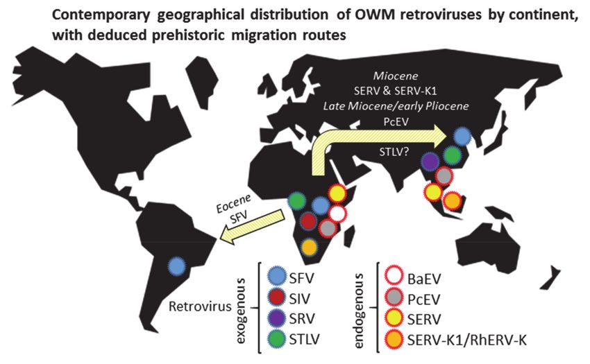
Epidemiologia 2021, 2                                                                                                 48

                        megablast option (“more dissimilar sequences”) was used. In Ensembl BLAST searches,
                        search sensitivity was the default (“normal”) and filtering low complexity regions or fil-
                        tering query sequences with RepeatMasker was disabled. When no full-length proviruses
                        were retrieved, virus presence was defined by detecting fragments with >90% homology to
                        both coding and non-coding (long terminal repeat, LTR) fragments of the specific virus.
                        Primate genomes queried were: Cercocebus atys, Cercopithecus mona, Cercopithecus neglectus,
                        Chlorocebus sabaeus (formerly known as Cercopithecus aethiops sabaeus), Colobus angolensis,
                        Erythrocebus patas, Macaca fascicularis, Macaca fuscata, Macaca mulatta, Macaca nemestrina,
                        Mandrillus leucophaeus, Mandrillus sphinx, Papio anubis, Piliocolobus tephrosceles, Rhinopithecus
                        bieti, Rhinopithecus roxellana, Theropithecus gelada, and Trachypithecus francoisi.

                        2.3. Analysis of Proviral Sequences
                              BLAST results were downloaded from the databases and aligned using ClustalW as im-
                        plemented in BioEdit (bioedit.software.informer.com). Alignments were optimized through
                        visual inspection. Sequence distances were calculated with the Kimura-2-parameter
                        method and evolutionary relationships were inferred using the Neighbor-joining method
                        with bootstrapping as implemented in MEGA6 [40,41]. Gaps/missing data treatment was
                        set to “partial deletion” with a cut-off value of 80%. No outgroup was defined. Alignments
                        are available as Supplementary Files 1 and 2.

                        3. Results
                        3.1. Exogenous Retroviruses in OWM Species
                             Four retrovirus species are currently circulating in OWM, namely SFV, SIV, SRV, and
                        STLV. SFV is also widespread in apes and New World monkeys (NWM). STLV is present in
                        both Asian and African monkey species and in apes plus humans, while SIV is solely found
                        in African primate species (including African apes), and recently, in humans (Table 1).
                             Several limitations apply when reconstructing the history of exogenous retroviruses
                        from epidemiological data:
                        •    Sampling is not systematic; some species remain untested because they may be difficult
                             to reach.
                        •    Sampling may not be optimal: it is done at the wrong age (for instance SIV is a sexually
                             transmitted infection with juveniles normally being negative for the virus), the wrong
                             type of sample is taken, or the viral load is below the detection level.
                        •    The distinction between an exogenous and endogenous retrovirus is not always
                             clear (e.g., murine leukemia virus, MuLV, feline leukemia virus, FeLV, and koala
                             retrovirus, KoRV, have both infectious and endogenous variants). Moreover, every
                             type of retrovirus has the capacity to enter the mammalian germ line, so Mendelian
                             inheritance is not a distinguishing characteristic [42–44].

                        3.1.1. Simian Foamy Virus (SFV)
                             Spumaviruses, also known as foamy viruses due to the effect they induce in cell
                        cultures, are a widely spread, distinct type of retrovirus, which share some similarities
                        with hepadnaviruses with regard to their replication cycle [45]. Infectious spumaviruses
                        are found in OWM, NWM and apes, where they induce little pathology, as replication only
                        takes place in epithelial cells of the oral cavity [45–47]. The detection of endogenous FV-like
                        sequences in a wide variety of amphibians, fish and mammals suggests a considerable
                        history for the group and indeed, phylogenetic analysis infers FVs as the oldest retrovirus
                        lineage, emerging >450 mya [48]. The origin of prosimian FV was calculated to the Mesozoic
                        (~82.5 mya). The divergence between OWM and ape SFV was dated to the Oligocene
                        (~30 mya), with the OWM node emerging ~16 to 18 mya [49]. Species-specific FV clades are
                        more recent; for instance, orangutan genus and subspecies-specific clades have been dated
                        to the late Pliocene (>4.7 mya), and the Pleistocene (>1.7 mya), respectively [50]. Cross-
                        species transmissions and recombination events, however, complicate the reconstruction of
                        spumavirus history [51,52].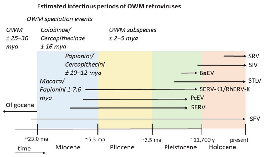
Epidemiologia 2021, 2                                                                                                                          49

                    Table 1. Overview of Old World primate species naturally infected with exogenous retroviruses.

  Retrovirus Species                                                                                       Other Primate
                                African OWM Species +                    Asian OWM Species +                                     References
        Genus                                                                                                Species +
                            Cercopithecinae        Colobinae        Cercopithecinae      Colobinae
                             Cercopithecini:
                               Chlorocebus
                              Erythrocebus
                               Papionini:                                                                      Gorilla
  Simian foamy virus
                               Cercocebus           Colobus           Papionini:         Pygathrix            Hylobates
         (SFV) 1                                                                                                                 [50,53–56]
                               Lophocebus          Procolobus          Macaca          Trachypithecus           Pan
    Simii-spumavirus
                                 Macaca                                                                        Pongo
                               Mandrillus
                                  Papio
                              Theropithecus
                             Cercopithecini:
                             Allenopithecus
      Simian                  Cercopithecus
  immuno-deficiency            Chlorocebus           Colobus                                                  Gorilla
       virus                  Miopithecus          Piliocolobus          None               None            Homo (HIV)           [54,57–62]
      (SIV) 2                  Papionini:          Procolobus                                                  Pan
     Lentivirus                Cercocebus
                               Lophocebus
                               Mandrillus
     Simian type D
      retrovirus 3                                                    Papionini:
                                  None                None                             Semnopithecus?           None               [63–67]
         (SRV)                                                         Macaca
     Betaretrovirus
                             Cercopithecini:
                             Allenopithecus
                              Cercopithecus
                               Chlorocebus
       Simian                 Erythrocebus                                                                    Gorilla
 T-lymphotropic virus         Miopithecus          Piliocolobus       Papionini:                           Homo (HTLV)
                                                                                          Presbytis                           [22,24,54,68–77]
       (STLV)                  Papionini:          Procolobus          Macaca                              Hylobates Pan
      Deltavirus               Cercocebus                                                                     Pongo
                               Lophocebus
                                 Macaca
                               Mandrillus
                                  Papio
      1SFV is also widespread in New World monkeys (NWM) species [51,55,78]. 2 Three natural infections of a yellow, an olive and a chacma
      baboon with a Chlorocebus (African green monkey, AGM) SIV strain, respectively, have been reported [79–81]. SIV isolated from an
      Erythrocebus monkey likely also results from SIVagm cross-species transmission [82]. 3 Simian type D betaretroviruses are recombinants
      with a betaretroviral gag-pol sequence and a gammaretrovirus env gene.

                                         An overview of the estimated contemporary distribution of OWM retroviruses and
                                    the deduced prehistoric virus migration routes, is given in Figure 1.

                                    3.1.2. Simian Immunodeficiency Virus (SIV)
                                         SIV infection has been detected in African, but not Asian primates or NWM, suggesting
                                    that origin and spread of the virus occurred after the separation of OWM and NWM, and
                                    also after the major migrations of African primates to Asia in the Miocene period.
                                         Two independent integrations of an endogenous lentivirus, termed pSIV, with a
                                    phylogenetic position in between the feline lentivirus feline immunodeficiency virus (FIV)
                                    cluster and modern simian SIV strains, have been detected in the genome of Malagasy
                                    lemur species, suggesting that SIV precursors infecting prosimian primates already existed
                                    ~4.3 mya [83–86]. Since Madagascar and Africa have been separated for at least 160 mya,
Epidemiologia 2021, 2 50
transitional pSIV was somehow transmitted across a substantial body of water, indicating
that lentivirus dispersal does not always need a land bridge [83].
Figure 1. Estimated present-day distribution by continent and proposed prehistoric migration routes of eight retroviruses
that are, or have been, circulating in Old World monkeys (OWM). Eocene: ~33.9 to 56 mya; Miocene: ~5.3 to 23 mya;
Pliocene: ~2.5 to 5.3 mya. The continental positions shown represent the current situation, the time periods shown are
estimates. During the Eocene, South America and Africa were much closer to each other. During the Miocene, a junction
existed between Africa and Asia due to the closure of the Tethys Seaway.
SIV is remarkably widespread, having infected over 40 African primate species and
at least 9 genera (Table 1). Prevalence in populations can be high. For instance, up to 80%
of wild Chlorocebus females test positive for SIVagm infection [87,88]. SIV strains exhibit
relatively large nucleotide diversity, suggestive of great age [87,89]. Initial calculations,
however, indicated that SIV lineages evolved in historic times, on a time scale of only tens
or hundreds of years [90,91]. Subsequent analysis of SIV isolates from primate popula-
tions on the island of Bioko, which is located 32 km off the west coast of Africa and has
been separated from mainland Africa for at least 10,000 years, pushed the emergence of
present-day SIV back to ≥32,000 years before present [92]. Confusingly, African green mon-
key (AGM, genus Chlorocebus) subspecies each harbor a distinct lineage of SIVagm. This
suggests a long-time presence of the virus, with an origin dating to somewhere between
AGM subspeciation, estimated at 1.5–3 mya, and subsequent monkey migrations during
the Plio-Pleistocene [88]. Should SIV indeed have a relatively short history in African
primates, then it is understandable that the virus did not reach Asian monkey species.
The Plio-Pleistocene is known for its lower global temperatures and, in the Pleistocene,
repeated glaciation. Such climate conditions discouraged primate settlements in Europe
and Arabia that could bridge the gap between Africa and Asia [93]. Theropithecus species
did disperse to Europe in the early Pleistocene [94]. However, modern Theropithecus is not
infected by SIV, nor are baboons, which currently still inhabit Saudi Arabia and Yemen.
Papio hamadryas likely migrated from Africa to Arabia in the late Pleistocene and remained
there ever since [95,96]. Looking at the spread of SIV, it is commonly found in arboreal
primates inhabiting tropical forests rather than terrestrial, savannah or shrubland-dwelling
monkeys such as macaques and baboons (Table 1). The exception to this observation areEpidemiologia 2021, 2 51
green monkeys. As a widespread species in sub-Saharan Africa, where they can be found
in numerous environments, AGMs may function as an intermediary spreading SIV to sa-
vannah monkeys. It is interesting to note that at three occasions, wild baboons were found
to be infected with SIVagm strains [79–81]. A Senegalese patas monkey (Erythrocebus patas),
another ground-dwelling species living among AGMs, was likewise reported to carry a
SIVagm strain [82].
Concluding, a relatively young age has been calculated for SIV based on nucleotide
sequences and from geographical separation. Such a young age is supported by its absence
in Asian primates, suggesting a post-Miocene origin. Probably, the virus originates from the
tropical forests of sub-Saharan Africa and is now spreading to ground-dwelling primates
through AGMs, which are widespread in Africa, have high SIV prevalence, and thrive
in many habitats. AGMs themselves have probably obtained SIV from cross-species
transmission to one of their subspecies with subsequent onwards transmission to the other
AGM subspecies in the past [97].
3.1.3. Simian Type D retrovirus (SRV)
SRV, a primate betaretrovirus, which can induce considerable pathology in captive
macaques, was found to have natural reservoirs in the Asian macaque species
Macaca nemestrina, Macaca fascicularis, and Macaca mulatta [63,64,66,98]. In total, eight
phylogenetically related SRV genotypes have been identified, all in macaques except SRV-6.
This variant was detected in wild Hanuman langurs, Semnopithecus entellus, from India [67],
suggesting that SRV-like viruses could have a broader species distribution. However, it
has been noted that SRV is closely related to endogenous primate type D betaretrovirus
SERV sequences. Especially the single SRV-7 sequence available, a partial fragment of pol,
shows high homology to SERV pol from baboons (unpublished observation, and see [66,99]).
Jayashree Nandi et al. therefore suggested that SRV-6 and -7 may be the result of recombi-
nation events between exogenous SRV and endogenous SERV genomes [66], which would
explain their considerable genetic distance from SRV 1–5 and SRV-8. As SRV-6 also bears
resemblance to an endogenous type D retrovirus (PO-1-Lu from Trachypithecus obscurus,
a langur species from East Asia), it could also be a reactivation of such a provirus [100].
PO-1-Lu was shown to be very similar to Mason–Pfizer monkey virus (MPMV) isolated
from a rhesus macaque, which turned out to be a variant of SRV-3 [101,102]. These diffi-
culties show that type D endogenous and exogenous retroviral sequences are intertwined
and that recombination, reactivation and/or germ line integration has probably happened
several times, which greatly confuses SRV phylogeny.
Little is known about the origin and age of SRV, except for its spread in primate centers
since at least the 1970s [99,103]. SRV 1–5 and SRV-8, which are genetically and serologically
closely related, spread only within the genus Macaca, and have the ability to induce severe
disease. Such characteristics are typical of a recently evolved virus species, making the
putative recombinant SRV-types 6 and 7 detected in Indian langurs a distinct viral species.
Increasing the search for both infectious and endogenous SRV-like retroviruses in Asian
monkeys should help gain a better understanding of the distribution and evolutionary
connections within this virus family.
3.1.4. Simian T-Lymphotropic Virus (STLV)
The deltaretrovirus STLV comprises multiple types and subtypes and has a broad
distribution in OWM and apes, but is not found in NWM [104]. Phylogenetic analysis
of LTR sequences suggests an African, rather than Asian origin for PTLV (primate T-
lymphotropic virus, the collective name for STLV and HTLV, the Homo-specific variants that
arose after cross-species transmission) [105,106]. Three STLV types have been recognized,
all of which also infect humans [107]. HTLV-1 and HTLV-2 have by now been scattered
around the world by human migration, while HTLV-3 is only found in Africa [107]. A
fourth, distantly related type, HTLV-4, has been isolated from an individual in Cameroon,
but the search for a simian counterpart has so far been unsuccessful [108].Epidemiologia 2021, 2 52
Pathogenicity in humans and non-human primates has only convincingly been doc-
umented for PTLV-1 (the cluster to which both STLV-1 and HTLV-1 belong, reviewed
in [76,109]). PTLV mostly replicates through clonal expansion, which results in a low
mutation rate and a high genetic stability [110]. However, diversity is significant in this
large virus family, making phylogenetic analysis of PTLV challenging. Often, studies are
based on relatively limited information. For instance, transmission of STLV-1 to humans
was estimated to have occurred 27,300 ± 8200 years ago based only on the analysis of LTR
and env gene sequences [21]. However, further transmissions have taken place over time
or are still ongoing (discussed in [76]), whereby geographic proximity is the basis of most
shared evolutionary relationships [23]. In fact, each HTLV-1 subtype is probably the result
of independent transmissions from either African or Asian simians [24,111]. Analysis using
a relaxed molecular clock and only first and second codon positions of the tax and env
genes, respectively, suggested that HTLV-4 diverged from HTLV-2/STLV-2 approximately
49,800–378,000 years ago [28]. In this study, the most recent common ancestor (MRCA)
of the major PTLV clades was calculated to have existed between 214,650 (tax gene) and
385,100 (env gene) years ago [28]. Using amino acid sequences from 20 PTLV strains and a
bovine deltaretrovirus as outgroup, another study suggested a much older date of origin for
the PTLV MRCA, namely, >1.3 mya [26]. A third study, based on entire genomes without
LTRs, suggested an age between 632,129 and 946,936 years for the whole PTLV cluster, and
dated the subclades of PTLV-1, PTLV-2, and PTLV-3 to 53,000–79,684, 191,621–286,730, and
63,294–94,770 years ago, respectively [112].
Overall, it is clear that PTLV comprises a clade of ancient, genetically diverse viruses,
where repeated interspecies transmissions as well as viruses migrating between Asia and
Africa pose a challenge for those trying to reconstruct the history of the virus. Despite some
variation in estimated divergence dates, the above studies all agree on a late Pleistocene
origin for PTLV, with the three main types likely arising in the last 200,000 years.
3.2. Endogenous Retrovirus Integrations Predating OWM Speciation
Endogenous retrovirus genomes and parts of such proviruses abound in vertebrates.
Most have entered the germ line long before the rise of the order of primates. The primate
germ line was thus already seeded with many of such proviral remains before the diver-
sification of extant OWM species. Examples of ancient proviruses, with their calculated
time of integration, are human endogenous retrovirus-H (HERV-H, >40 mya), HERV-W
(±63 mya), HERV-S (±43 mya), HERV-R (±33 mya), HERV-I (±33 mya), and HERV-E
(10.7–41.3 mya) [113–118]. With the exception of HERV-S, which has some similarity to
foamy viruses, all these HERVs belong to the gammaretrovirus group.
The situation for the supergroup of primate betaretroviruses, represented by HERV-K
viruses in humans, and CERV-K in chimpanzees, is more complex [119,120]. Sequences
homologous to HERV-K have been detected in all Old World primate species, with an
estimated integration time of 28 mya [121]. However, specific members of the family show
a more limited distribution and a later germ line introduction, suggestive of repeated
activation and reinfection processes over time (discussed in Section 3.3.4).
3.3. Endogenous Retroviruses Specific for OWM
Four endogenous retroviruses are exclusively present in OWM genomes, namely,
BaEV, PcEV, SERV, and SERV-K1/RhERV-K. Of these viruses, full-length proviral genomes
can be detected in at least some OWM species [37–39,122,123]. In AGM (Vero) or baboon
BEF-3 cell lines, virus particles containing SERV or BaEV genomes, respectively, can be
produced upon stimulation [124,125]. For PcEV, expression has only been confirmed at
the RNA level [126]. It is unknown whether this RNA comprises full-length genomes
nor whether it is packaged. Similarly, for SERV-K1, only env mRNA expression has been
verified [123]. Distribution of the four endogenous retroviruses among OWM species is
shown in Table 2; corresponding geography and deduced virus migrations are depicted in
Figure 1.Epidemiologia 2021, 2 53
Several limitations apply when reconstructing the history of endogenous retroviruses
from genome assemblies:
• Not all infected species may still roam the earth
• Not all infected species may contain germ line integrations
• Not all species have had their genomes sequenced
• Not all proviral sequences are likely due to bona fide viral infection and integration;
they may for instance be acquired by hybridization between species
• Proviruses may have been completely or partially lost from the germ line, making
identification challenging
• The quality of a genome assembly may be insufficient for provirus detection
• Heterozygous proviral insertions (ERV insertion polymorphism) are not included in
genome assemblies [127,128]
• The distinction between endogenous and exogenous retroviruses is not always clear
(see above, and the comment in [129])
Table 2. Overview of OWM species harboring OWM-specific endogenous retrovirus genomes.
Retrovirus Species
African OWM Species + Asian OWM Species + References
Genus
Cercopithecinae Colobinae Cercopithecinae Colobinae
Cercopithecini:
Chlorocebus
Papionini:
Baboon endogenous virus
Cercocebus [130]
(BaEV) None None None
Mandrillus Figure A1
Gammaretrovirus
Lophocebus
Papio
Theropithecus
Papio cynocephalus Papionini:
endogous virus Lophocebus Papionini: [131]
Colobus None
(PcEV) Papio Macaca Figure A1
Gammaretrovirus Theropithecus
Cercopithecini:
Simian endogenous Cercopithecus
retrovirus Chlorocebus
Papionini: [38,132,133]
(SERV) Erythrocebus None Rhinopithecus
Macaca Figure A1
Betaretrovirus Miopithecus
(Simian type D) Papionini:
Macaca
Papionini:
Simian endogenous
Cercocebus
retrovirus-K1 (SERV-K1/ Papionini:
Mandrillus Colobus Trachypithecus Figure A2
RhERV-K) Macaca
Papio
Betaretrovirus
Theropithecus
3.3.1. Baboon Endogenous Virus (BaEV)
BaEV was discovered as an integrated provirus in baboon DNA in 1978 and was
completely sequenced in 1987 [39,134]. Non-infectious virus particles containing BaEV
sequences can be induced from the Vero cell line, which is derived from Chlorocebus
kidney epithelial tissue [124]. BaEV is a recombinant virus, originating from the now also
endogenous monkey retroviruses PcEV and SERV [37]. Most BaEV proviral integrations
are defective, although complete proviruses do exist in baboons and geladas (Appendix A
Figure A1A) [122,125,134]. Although BaEV was at first thought to be widespread in
primates, a PCR analysis of 24 African monkey species suggested that BaEV integrations are
limited to few species (Table 2) [130]. In phylogenetic reconstructions, BaEV sequences didEpidemiologia 2021, 2 54
not follow the monkey phylogeny, but clustered according to habitat, implying that living
together facilitates cross-species transmissions [130]. The BaEV_forest and BaEV_savannah
strains were estimated to have diverged 24,000–400,000 years ago, using evolutionary rates
calculated for other retroviruses [130]. Analyzing LTR divergence in full-length BaEV
genomes from one baboon and two Theropithecus gelada showed a single (baboon) or no
(gelada) substitutions between the 50 and 30 LTR, respectively (unpublished observation).
As retroviral LTRs are identical at the time of integration, after which they start to diverge
with the host mutation rate, little or no LTR variation is indicative of a relatively recent
origin, probably at most ~300,000 years ago for proviruses with identical LTRs [135].
3.3.2. Papio Cynocephalus Endogenous Virus (PcEV)
The full-length endogenous gammaretrovirus PcEV was found by screening a baboon
(Papio cynocephalus) genomic library [37]. Additional species harboring PcEV proviral
sequences were identified during the analysis of OWM monkey DNA (Table 2, [131]).
Interestingly, PcEV sequences (including LTRs) were found in all examined papionin
species, but also in colobines of the genus Colobus: Colobus guereza and Colobus angolensis
(Appendix A Figure A1B, [131]). PcEV_baboon was not found in Cercopithecini, although
a variant virus is likely present there [131]. The upper limit of PcEV integration was set at
9 mya based on LTR divergence and species distribution, such as the dispersal of macaques
into Asia around 5.5–7 mya [131]. Evidence for recent activity is given by proviruses with no
or only a single nucleotide difference between their 50 and 30 LTRs, which can be identified
in gelada and baboon genomic libraries, respectively (unpublished observation). In the
rhesus macaque genome, PcEV was identified as one of the retroviruses with relatively
recent germ line activity [136] (see erratum in [126] as PcEV was mistakenly labelled BaEV
in the earlier publication). PcEV integration sites in two olive baboons (no. 15944 and no.
1X1155) differ, suggesting that the proviruses are not fixed in this subspecies (unpublished
observation). In addition, baboon PcEV integration sites are empty in the rhesus macaque
genome, which points to independent integrations after speciation and not inheritance
from a common ancestor (unpublished observation).
So, as PcEV is present in Colobus, but not in Cercopithecini, the exogenous period of
PcEV can be estimated to have started around the macaque/papionin split, 7.6 ± 1.3 mya,
until about 150,000–300,000 years ago. An ancient cross-species transmission to an ancestor
of present-day Colobus species could explain its presence there [131].
3.3.3. Simian Endogenous Retrovirus (SERV)
Similar to PcEV, SERV, a full-length integrated type D betaretrovirus was first described
from a baboon genomic library [38]. Subsequently, a widespread distribution in OWM with
a relatively large nucleotide divergence between the viral sequences suggested a relative
old age for the proviruses (Table 2) [38,132]. Heterozygous integrations abound, at least in
Chlorocebus sabaeus, which suggests that many are so recent that they have not had sufficient
time to become fixed in the population [128]. Fixation of traits is highly dependent upon
effective population size, which depends on population size and generation time. For
primate populations 1–3 million years are usually sufficient to fix most markers [137],
suggesting that polymorphic insertions are less than 1–3 my old. A number of OWM
genomes contain several full-length SERV genomes, often with open reading frames for
at least some viral proteins (unpublished observation, Appendix A Figure A1C). SERV
sequences do not cluster according to host species, implying that they integrated after
speciation and that ancient cross-species transmissions occurred [38]. SERV particles can be
expressed from Vero cells, but those are non-infectious in cell lines known to be permissive
for type D retroviruses [124].
Full-length proviruses, intact reading frames, virus expression and heterozygous
insertions do suggest a young age, but SERV has been calculated to have integrated on
average 6.16 ± 3.41 (range 0-21.62) mya in Asian colobines, which, however, carry a distinct
variant of SERV, and somewhat later, 3.42 ± 2.20 (range 0.27–14.09) mya, in cercopithecineEpidemiologia 2021, 2 55
species; that is, monkeys of the subfamily Cercopithecinae; thus, both Cercopithecini and
Papionini [132]. It was therefore suggested that SERV originated within the last 8 million
years and continued its exogenous activity until quite recently. For instance, in Asian
colobines, species specific, genus specific, but also shared integrations are seen [132].
Highly divergent SERV 50 and 30 LTRs can be found in cercopithecines. In contrast, in
Theropithecus and Chlorocebus there are also a few proviruses with only 1–2 nt substitutions
between the LTRs (unpublished observation), indeed pointing to a lengthy period of germ
line activity. Rhinopithecus roxellana SERV integrations with open reading frames were found
to be significantly younger than the ones with frame-shift mutations [132]. Furthermore,
heterozygous SERV insertions are presumed to be more recent than integrations that have
been fixed in a population.
3.3.4. Simian Endogenous Retrovirus-K1 (SERV-K1)/RhERV-K
The primate non-D type betaretrovirus superfamily, to which all ERV-K viruses belong,
has a complex history. Deep-rooted proviral integrations are present in all OWM and homi-
noids, but there is also evidence of later activity. In both humans and chimpanzees, the
possession of open reading frames combined with low LTR diversity in some proviruses as
well as the existence of polymorphic insertions, are illustrative of such recent activity [127].
In case of chimpanzee CERV-K proviruses, integration times postdate the Homo–Pan di-
vergence [127]. Another study dated the average integration time of rhesus macaque
ERV-K (RhERV-K) at 10.3 mya, although almost identical LTRs in three complete proviruses
suggested that some integratedEpidemiologia 2021, 2 56
SERV-K1 LTRs in papionins are almost identical in sequence and could thus predate
speciation. A quick inspection of Blast hits, however, suggests that the integration sites do
not match, so that the proviral integrations must largely have been independent events
(result not shown).
The uneven distribution of SERV-K1 complete proviruses suggests that either the
infectious virus did not always reach the OWM germ line, that the integrations were
lost, or that they are somehow not in the genome assemblies. Overall, due its apparently
highly infectious nature over long periods of time, and a tendency to recombine, the
epidemiological trail of the ERV-K virus family is not easy to follow. The recent spread,
on two continents, of two related SERV-K viruses in two very different monkey genera,
papionins and Colobus/Trachypithecus, is thus an interesting subject for further study.
Infectious period estimates of the eight OWM retroviruses in relation to the geological
time scale are summarized in Figure 2.
Figure 2. Estimated infectious periods of eight retroviruses that are, or have been, circulating in
OWM. Putative speciation events in OWM are indicated. The geological time line is not drawn
to scale.
4. Discussion
The order of primates probably dates to the Paleocene (56–65 mya). Primate fossils
appeared simultaneously in Europe, Asia, and Africa in the early Eocene (55–50 mya),
suggesting that by then, the order was firmly established. Since primates are relatively
intolerant of cooler climates, their history is largely determined by climatic variations,
which in their turn are linked to tectonic and cosmic events [93]. The rapid evolution and
dispersal of primate species was likely facilitated by a steep rise in global temperature
at the Paleocene/Eocene boundary, which resulted in tropical climates even at higher
latitudes [93]. Moreover, firm geographical connections between Asia, Europe, and North
America existed at that time, giving the early primates sufficient territory to expand.
Global temperatures declined during the Eocene, although warmer periods, for instance
in the Miocene, would continue to occur. The much colder conditions in the Oligocene
and especially in the more recent Pliocene and Pleistocene periods forced primates to
retreat from previously inhabited regions. Under dryer, colder conditions, terrestrial
papionins, like macaques and baboons, could from time to time establish themselves in
such climates, but the arboreal monkeys of the tropical forests were unable to adapt. For
instance, colobines migrated successfully from Africa to Eurasia in the late Miocene but
these dispersals stopped in the Plio/Pleistocene; Papio and Theropithecus were the onlyEpidemiologia 2021, 2 57
species then trekking to Eurasia through Arabia [139]. Macaques migrated from Africa
to Eurasia around 5.5–7 mya in the late Miocene or early Pleistocene [138]. Only a single,
ancestral species, Macaca sylvanus, is now left in (northern) Africa; European macaques
have gone extinct in the late Pleistocene [140]. Modern Asian macaque species show no
genomic evidence of later introductions, suggesting that subsequent migrations, if any,
have been unsuccessful [138].
With the above described primate evolution in mind, an attempt to retrace the history
of one of their successful parasites, the retrovirus family, was made. Retroviruses are
generally transmitted by close contact, for instance by the exchange of saliva or blood
(biting), through sexual encounters, or from mother-to child, implying that these pathogens
are closely associated with their host species. Infections by aerosols, the oral–fecal route or
by insect vectors are relatively rare or are unlikely; only equine infectious anemia virus,
EIAV, and possibly bovine leukemia virus, BLV, can sometimes be transmitted by flies
from viremic hosts [141,142]. So, in general, the history of a retrovirus is closely related
to that of its host. Of course, transmission of viruses can be hampered by cellular and
genetic variation between species. For SIV, immune system activity, including the actions
of restriction factors such as TRIM5alpha and APOBEC3G, as well as amino acid variation
in for instance the coreceptor protein, have all been implicated in the prevention of cross-
species transmission (for a review, see [143]). Such inhibitory mechanisms could lead to
false conclusions as to why certain species are free of infection but would hardly play a
role in conclusions drawn from infected species.
Investigating the spread and estimated age of eight OWM-infecting retroviral species
suggests that SFV, the most widely spread virus of all, with a distribution over three
continents, is also the oldest virus in the group. Ancestral primates most likely migrated,
possibly by rafting, to South America during the Paleogene when that continent was
relatively close to Africa [93]. As the nodal ancestor of OWM and NWM SFV has been
dated to ~65 mya [49], the virus could have arrived in South America together with its host.
A nodal ancestor by definition predates its progeny, and need not have been a primate
virus, which would date the emergence of genuine SFV-NWM and SFV-OWM after 65 mya.
Various approaches have estimated the node separating NWM and OWM at ~40 mya
in the Eocene, which would fit with the ancestry of the virus lineages [2]. However, as
cross-species transmission and recombination have been documented for foamy viruses,
alternative scenarios with a later introduction in NWM could also be a possibility.
The next oldest viruses, SERV and SERV-K1 (RhERV-K), are now known only in an en-
dogenous form. Their putative Miocene origin, together with a wide distribution in OWM
species in both Africa and Asia, suggests that SERV and SERV-K1 have been travelling
the land bridge(s) between the continents established after the final closure of the Tethys
Seaway ~14 mya [144]. The relatively warm climate of the Miocene facilitated the dispersal
of arboreal, tropical colobines as well as terrestrial macaques; the genera underwent an
extensive diversifying radiation in Asia [139]. A third virus with a similar distribution,
both in species and geography, is STLV. However, phylogenetic analysis indicated that the
common ancestor of STLV genotypes dates to ~1 mya, so that spread of STLV cannot readily
be explained by Miocene migrations. A later introduction with rapid onward transmissions
and adaptation could be hypothesized, though PTLV is mainly transmitted through sexual
contact, blood, or saliva exposure (biting), or vertically from mother to child, which makes
spread in and between primate groups relatively slow [145,146]. Alternatively, STLV might
be much older than estimated from sequence data, as we have possibly not yet sampled all
PTLV variation, or the evolutionary models used to analyse PTLV may be incorrect [76,147].
Another virus that likely migrated with its host from Africa to Asia is PcEV, which is
found in African papionins as well as in Asian macaques. Such a distribution suggests that
PcEV is at least 5.5–7 mya old, but not much older as the virus is not found in the germ
line of the Cercopithecini, implying that migration with its macaque host took place in the
late Miocene or early Pliocene. Recombination of PcEV with SERV, when at least one of
the two viruses was actively replicating, gave rise to BaEV, which, by definition, shouldEpidemiologia 2021, 2 58
be younger than its parents. Indeed, BaEV is not found in Asian macaques, suggesting
its infectious period postdates the divergence of macaques from the other papionins. The
putative Pleistocene origin of BaEV prohibited its spread to Asia, as by then the climate
was no longer favorable for monkey expansions. BaEV may have been present in European
baboons, having gone extinct there together with its host. Moreover, it would be interesting
to investigate whether or not African Macaca sylvanus carry PcEV and/or BaEV integrations.
Much younger OWM retroviruses appear to be SIV and SRV; SIV has an exclusively
African distribution while SRV is seemingly only found in Asian macaques. The unin-
habitable nature after the Miocene period of areas in key corridors for primates between
Africa and Asia, with subsequent isolation of monkey species on both continents, likely
impeded virus transfer between continents for these recently emerged viruses [148–152].
For a relatively young virus that owes its present global spread to a recent leap to mankind,
SIV has a remarkably broad distribution in African primates. Unexpectedly, the list of
SIV-infected species shows only anecdotal detection in terrestrial papionins such as ba-
boons and geladas, while infections are common in monkeys and apes inhabiting the
tropical forests of sub-Saharan Africa, including the forest-dwelling papionins Mandrillus,
Cercocebus, and Lophocebus. An exception here are AGMs (Chlorocebus spp.), found just
about everywhere in sub-Saharan Africa, which carry a great diversity of SIV strains and
have high SIV prevalence. They may have facilitated SIV cross-species transmissions in
the past, similar to modern AGM transmitting SIVagm to individual baboons and patas
monkeys. An example of another retrovirus shared between AGMs and papionins is BaEV.
For endogenous viruses that have gone extinct, once infected species may be missed
because no germ line introductions occurred or were lost in subsequent generations. Still,
the available data on endogenous viruses can be used to infer a plausible evolutionary
trajectory. Retroviruses that use the neutral amino acid transporter ASCT2 as a cellular
receptor, which is expressed on oocytes, spermatozoa, and pre-implantation embryos, have
a tendency to enter the germ line [153–155]. Such viruses, here represented by the type D
viruses SERV and BaEV, are thus well equipped to create a broad genomic track, and will
therefore be less likely to disappear from sight. Indeed, SERV has an extensive distribution
in OWM, while—putative—non-ASCT2 using PcEV has a relatively limited distribution
in papionin genomes only. However, although BaEV is able to the ASCT2 receptor, it
has a more limited distribution than SERV, which may either be a true representation of
the species infected in the past but could also be due to other causes. For instance, BaEV
can use the neutral amino acid transporter ASCT1 as an additional receptor, which other
type D viruses cannot [156]. Such variation in receptor use may suggest an alternative
trajectory in the host, although ASCT1 has a similar broad tissue expression as ASCT2 [157].
Alternatively, superinfection resistance (SIR), whereby for instance a homologous Env
protein occupies the viral receptor so that new infections cannot take place, could play a
role here (for a review, see [158]). The expression of endogenous SERV Env would make
such a scenario possible. In human placenta, a HERV-F Env protein has been shown to
inhibit betaretroviral Env protein mediated cell fusion through ASCT2 binding [159].
Interestingly, all four now endogenous OWM retroviruses have disappeared from
the circulation, suggesting that virus endogenization ultimately means the end of the
contagious variant. SIR could be such a possible mechanism to limit virus spread. Thus,
opting for germ line transmission seems to be an indicator for extinction of infectious
retrovirus lineages, while old age, as exemplified by the current infectivity of SFV, does not
need to be.
5. Conclusions
Current distribution of primates, prehistoric primate evolution and expansion together
with virus prevalence and sequence information have been used to infer the history of
primate retroviruses. African and Asian monkey species are, or have been, infected by at
least eight distinct retroviral species. Four of those have now become part of the germ line
while four others are currently in the infectious stage, including the most ancient retrovirus,Epidemiologia 2021, 2 59
SFV. Evidence suggests that the older retroviruses—PcEV, SERV, SERV-K1—have migrated
with their hosts to Asia ~5 to 10 mya, while the more recent ones—BaEV, SIV, and SRV—are
limited to their respective continents of origin. For STLV, the reconstruction of its history
proved more difficult, as its relatively recent age and widespread distribution does not
correspond with Miocene migration.
Supplementary Materials: The following are available online at https://www.mdpi.com/2673-3
986/2/1/5/s1, File S1: Alignment of OWM ERV sequences, File S2: Alignment of OWM SERV-K
LTR sequences.
Funding: This research received no external funding.
Institutional Review Board Statement: Not applicable.
Informed Consent Statement: Not applicable.
Data Availability Statement: The data presented in this study are available in Supplementary Files
1 and 2.
Conflicts of Interest: The author declares no conflict of interest.
Appendix A
Figure A1. Evolutionary relationships of full-length or near full-length proviruses of BaEV (A), PcEV
(B), and SERV (C) in OWM.Epidemiologia 2021, 2 60
The evolutionary history of integrated proviruses of BaEV (panel A), PcEV (panel B)
and SERV (panel C) in OWM genomes with the corresponding reference virus sequence
was inferred using the Neighbor-Joining method [160]. Bootstrap values of 500 replicates
are shown next to the branches [161]. The tree is drawn to scale, with branch lengths in the
same units as those of the evolutionary distances used to infer the phylogenetic tree. The
evolutionary distances were computed using the Kimura 2-parameter method [40] and are
given as the number of base substitutions per site. There were a total of 8492 positions in the
dataset for BaEV, 8282 for PcEV, and 8382 for SERV. Evolutionary analyses were conducted
in MEGA6 [41]. For some species, such as Macaca nemestrina, Mandrillus leucophaeus, and
Cercocebus atys, the assembled genome sequences were of relative low quality, in other
instances it was ascertained that all viral elements, gag-pol-env coding regions and LTRs,
were present with high similarity (>95%), although the proviruses themselves were likely
fragmented in those species and full-length or near full-length proviruses could not be
retrieved. The primate species name is embedded within the sequence name. The sequence
alignment file is available as Supplementary File 1.
Appendix B
Figure A2. Evolutionary relationships of SERV-K1 LTR sequences in OWM.
The evolutionary history of LTR sequences of SERV-K1 in OWM genomes and those of
the rhesus macaque reference virus were inferred using the Neighbor-Joining method [160].
For complete proviruses, both the 50 LTR and 30 LTR were included in the analysis. Boot-
strap values of 500 replicates are shown next to the branches [161]. The tree is drawn to
scale, with branch lengths in the same units as those of the evolutionary distances used to
infer the phylogenetic tree. The evolutionary distances were computed using the Kimura
2-parameter method [40]. Evolutionary analyses were conducted in MEGA6 [41]. ThereEpidemiologia 2021, 2 61
were a total of 572 positions in the dataset. The primate species name is embedded within
the sequence name. The sequence alignment file is available as Supplementary File 2.
References
1. Elton, S. Environmental correlates of the cercopithecoid radiations. Folia Primatol. 2007, 78, 344–364. [CrossRef] [PubMed]
2. Steiper, M.E.; Young, N.M. Primate molecular divergence dates. Mol. Phylogenet. Evol. 2006, 41, 384–394. [CrossRef] [PubMed]
3. Stevens, N.J.; Seiffert, E.R.; O’Connor, P.M.; Roberts, E.M.; Schmitz, M.D.; Krause, C.; Gorscak, E.; Ngasala, S.; Hieronymus, T.L.;
Temu, J. Palaeontological evidence for an Oligocene divergence between Old World monkeys and apes. Nature 2013, 497, 611–614.
[CrossRef]
4. Raaum, R.L.; Sterner, K.N.; Noviello, C.M.; Stewart, C.-B.; Disotell, T.R. Catarrhine primate divergence dates estimated from
complete mitochondrial genomes: Concordance with fossil and nuclear DNA evidence. J. Hum. Evol. 2005, 48, 237–257. [CrossRef]
[PubMed]
5. Hayward, A. Origin of the retroviruses: When, where, and how? Curr. Opin. Virol. 2017, 25, 23–27. [CrossRef] [PubMed]
6. Lerat, E.; Capy, P. Retrotransposons and retroviruses: Analysis of the envelope gene. Mol. Biol. Evol. 1999, 16, 1198–1207.
[CrossRef] [PubMed]
7. Koonin, E.V.; Dolja, V.V. Virus world as an evolutionary network of viruses and capsidless selfish elements. Microbiol. Mol. Biol.
Rev. 2014, 78, 278–303. [CrossRef] [PubMed]
8. Cordonnier, A.; Casella, J.F.; Heidmann, T. Isolation of novel human endogenous retrovirus-like elements with foamy virus-related
pol sequence. J. Virol. 1995, 69, 5890–5897. [CrossRef]
9. Zhu, T.; Korber, B.T.; Nahmias, A.J.; Hooper, E.; Sharp, P.M.; Ho, D.D. An African HIV-1 sequence from 1959 and implications for
the origin of the epidemic. Nature 1998, 391, 594–597. [CrossRef]
10. Worobey, M.; Gemmel, M.; Teuwen, D.E.; Haselkorn, T.; Kunstman, K.; Bunce, M.; Muyembe, J.-J.; Kabongo, J.-M.M.; Kalengayi,
R.M.; Van Marck, E.; et al. Direct evidence of extensive diversity of HIV-1 in Kinshasa by 1960. Nature 2008, 455, 661–664.
[CrossRef]
11. Faria, N.R.; Rambaut, A.; Suchard, M.A.; Baele, G.; Bedford, T.; Ward, M.J.; Tatem, A.J.; Sousa, J.D.; Arinaminpathy, N.; Pépin,
J.; et al. HIV epidemiology. The early spread and epidemic ignition of HIV-1 in human populations. Science 2014, 346, 56–61.
[CrossRef] [PubMed]
12. Hirsch, V.M.; Olmsted, R.A.; Murphey-Corb, M.; Purcell, R.H.; Johnson, P.R. An African primate lentivirus (SIVsmclosely related
to HIV-2. Nature 1989, 339, 389–392. [CrossRef] [PubMed]
13. Plantier, J.-C.; Leoz, M.; Dickerson, J.E.; De Oliveira, F.; Cordonnier, F.; Lemée, V.; Damond, F.; Robertson, D.L.; Simon, F. A new
human immunodeficiency virus derived from gorillas. Nat. Med. 2009, 15, 871–872. [CrossRef]
14. D’Arc, M.; Ayouba, A.; Esteban, A.; Learn, G.H.; Boué, V.; Liegeois, F.; Etienne, L.; Tagg, N.; Leendertz, F.H.; Boesch, C.; et al.
Origin of the HIV-1 group O epidemic in western lowland gorillas. Proc. Natl. Acad. Sci. USA 2015, 112, E1343–E1352. [CrossRef]
[PubMed]
15. Gao, F.; Bailes, E.; Robertson, D.L.; Chen, Y.; Rodenburg, C.M.; Michael, S.F.; Cummins, L.B.; Arthur, L.O.; Peeters, M.; Shaw,
G.M.; et al. Origin of HIV-1 in the chimpanzee Pan troglodytes troglodytes. Nature 1999, 397, 436–441. [CrossRef]
16. Huet, T.; Cheynier, R.; Meyerhans, A.; Roelants, G.; Wain-Hobson, S. Genetic organization of a chimpanzee lentivirus related to
HIV-1. Nature 1990, 345, 356–359. [CrossRef]
17. Roques, P.; Robertson, D.L.; Souquière, S.; Apetrei, C.; Nerrienet, E.; Barré-Sinoussi, F.; Müller-Trutwin, M.; Simon, F. Phylogenetic
characteristics of three new HIV-1 N strains and implications for the origin of group N. AIDS 2004, 18, 1371–1381. [CrossRef]
18. Chen, Z.; Telfier, P.; Gettie, A.; Reed, P.; Zhang, L.; Ho, D.D.; Marx, P.A. Genetic characterization of new West African simian
immunodeficiency virus SIVsm: Geographic clustering of household-derived SIV strains with human immunodeficiency virus
type 2 subtypes and genetically diverse viruses from a single feral sooty mangabey troop. J. Virol. 1996, 70, 3617–3627. [CrossRef]
19. Chen, Z.; Luckay, A.; Sodora, D.L.; Telfer, P.; Reed, P.; Gettie, A.; Kanu, J.M.; Sadek, R.F.; Yee, J.; Ho, D.D.; et al. Human
immunodeficiency virus type 2 (HIV-2) seroprevalence and characterization of a distinct HIV-2 genetic subtype from the natural
range of simian immunodeficiency virus-infected sooty mangabeys. J. Virol. 1997, 71, 3953–3960. [CrossRef]
20. Santiago, M.L.; Range, F.; Keele, B.F.; Li, Y.; Bailes, E.; Bibollet-Ruche, F.; Fruteau, C.; Noë, R.; Peeters, M.; Brookfield, J.F.Y.;
et al. Simian immunodeficiency virus infection in free-ranging sooty mangabeys (cercocebus atys atys) from the Taï Forest, Côte
d’Ivoire: Implications for the origin of epidemic human immunodeficiency virus type 2. J. Virol. 2005, 79, 12515–12527. [CrossRef]
21. Van Dooren, S.; Salemi, M.; Vandamme, A.-M. Dating the origin of the African human T-cell lymphotropic virus type-I (HTLV-I)
subtypes. Mol. Biol. Evol. 2001, 18, 661–671. [CrossRef] [PubMed]
22. Van Dooren, S.; Verschoor, E.J.; Fagrouch, Z.; Vandamme, A.-M. Phylogeny of primate T lymphotropic virus type 1 (PTLV-1)
including various new Asian and African non-human primate strains. Infect. Genet. Evol. 2007, 7, 374–381. [CrossRef] [PubMed]
23. Pecon-Slattery, J.; Franchini, G.; Gessain, A. Genomic evolution, patterns of global dissemination, and interspecies transmission
of human and simian T-cell leukemia/lymphotropic viruses. Genome Res. 1999, 9, 525–540.
24. Reid, M.J.; Switzer, W.M.; Schillaci, M.A.; Ragonnet-Cronin, M.; Joanisse, I.; Caminiti, K.; Lowenberger, C.; Galdikas, B.M.F.;
Sandstrom, P.A.; Brooks, J.I. Detailed phylogenetic analysis of primate T-lymphotropic virus type 1 (PTLV-1) sequences from
orangutans (Pongo pygmaeus) reveals new insights into the evolutionary history of PTLV-1 in Asia. Infect. Genet. Evol. 2016, 43,
434–450. [CrossRef]Epidemiologia 2021, 2 62
25. Hron, T.; Elleder, D.; Gifford, R.J. Deltaretroviruses have circulated since at least the Paleogene and infected a broad range of
mammalian species. Retrovirology 2019, 16, 33. [CrossRef]
26. Salemi, M.; Desmyter, J.; Vandamme, A.-M. Tempo and mode of human and simian T-lymphotropic virus (HTLV/STLV) evolution
revealed by analyses of full-genome sequences. Mol. Biol. Evol. 2000, 17, 374–386. [CrossRef]
27. Afonso, P.V.; Cassar, O.; Gessain, A. Molecular epidemiology, genetic variability and evolution of HTLV-1 with special emphasis
on African genotypes. Retrovirology 2019, 16, 39. [CrossRef]
28. Switzer, W.M.; Salemi, M.; Qari, S.H.; Jia, H.; Gray, R.R.; Katzourakis, A.; Marriott, S.J.; Pryor, K.N.; Wolfe, N.D.; Burke, D.S.; et al.
Ancient, independent evolution and distinct molecular features of the novel human T-lymphotropic virus type 4. Retrovirology
2009, 6, 9. [CrossRef]
29. Zheng, H.; Wolfe, N.D.; Sintasath, D.M.; Tamoufe, U.; Lebreton, M.; Djoko, C.F.; Diffo, J.L.D.; Pike, B.L.; Heneine, W.; Switzer, W.M.
Emergence of a novel and highly divergent HTLV-3 in a primate hunter in Cameroon. Virology 2010, 401, 137–145. [CrossRef]
30. Filippone, C.; Betsem, E.; Tortevoye, P.; Cassar, O.; Bassot, S.; Froment, A.; Fontanet, A.; Gessain, A. A Severe Bite from a
Nonhuman Primate Is a Major Risk Factor for HTLV-1 Infection in Hunters From Central Africa. Clin. Infect. Dis. 2015, 60,
1667–1676. [CrossRef]
31. Pinto-Santini, D.M.; Stenbak, C.R.; Linial, M.L. Foamy virus zoonotic infections. Retrovirology 2017, 14, 55. [CrossRef] [PubMed]
32. Heneine, W.; Schweizer, M.; Sandstrom, P.; Folks, T. Human infection with foamy viruses. Curr. Top Microbiol. Immunol. 2003, 277,
181–196. [CrossRef] [PubMed]
33. Mouinga-Ondémé, A.; Kazanji, M. Simian foamy virus in non-human primates and cross-species transmission to humans in
Gabon: An emerging zoonotic disease in Central Africa? Viruses 2013, 5, 1536–1552. [CrossRef]
34. Power, M.D.; Marx, P.A.; Bryant, M.L.; Gardner, M.B.; Barr, P.J.; Luciw, P.A. Nucleotide sequence of SRV-1, a type D simian
acquired immune deficiency syndrome retrovirus. Science 1986, 231, 1567–1572. [CrossRef] [PubMed]
35. Gardner, M.B. The history of simian AIDS. J. Med. Primatol. 1996, 25, 148–157. [CrossRef]
36. Lerche, N.W.; Switzer, W.M.; Yee, J.L.; Shanmugam, V.; Rosenthal, A.N.; Chapman, L.E.; Folks, T.M.; Heneine, W. Evidence of
infection with simian Type D retrovirus in persons occupationally exposed to nonhuman primates. J. Virol. 2001, 75, 1783–1789.
[CrossRef]
37. Mang, R.; Goudsmit, J.; Van Der Kuyl, A.C. Novel endogenous Type C retrovirus in baboons: Complete sequence, providing
evidence for baboon endogenous virus gag-pol ancestry. J. Virol. 1999, 73, 7021–7026. [CrossRef]
38. Van Der Kuyl, A.C.; Mang, R.; Dekker, J.T.; Goudsmit, J. Complete nucleotide sequence of simian endogenous type D retrovirus
with intact genome organization: Evidence for ancestry to simian retrovirus and baboon endogenous virus. J. Virol. 1997, 71,
3666–3676. [CrossRef]
39. Kato, S.; Matsuo, K.; Nishimura, N.; Takahashi, N.; Takano, T. The entire nucleotide sequence of baboon endogenous virus DNA:
A chimeric genome structure of murine type C and simian type D retroviruses. Jpn. J. Genet. 1987, 62, 127–137. [CrossRef]
40. Kimura, M. A simple method for estimating evolutionary rates of base substitutions through comparative studies of nucleotide
sequences. J. Mol. Evol. 1980, 16, 111–120. [CrossRef]
41. Tamura, K.; Stecher, G.; Peterson, D.; Filipski, A.; Kumar, S. MEGA6: Molecular evolutionary genetics analysis version 6.0. Mol.
Biol. Evol. 2013, 30, 2725–2729. [CrossRef] [PubMed]
42. Katzourakis, A.; Tristem, M.; Pybus, O.G.; Gifford, R.J. Discovery and analysis of the first endogenous lentivirus. Proc. Natl. Acad.
Sci. USA 2007, 104, 6261–6265. [CrossRef] [PubMed]
43. Farkasova, H.; Hron, T.; Paces, J.; Hulva, P.; Benda, P.; Gifford, R.J.; Elleder, D. Discovery of an endogenous Deltaretrovirus in the
genome of long-fingered bats (Chiroptera: Miniopteridae). Proc. Natl. Acad. Sci. USA 2017, 114, 3145–3150. [CrossRef] [PubMed]
44. Han, G.-Z.; Worobey, M. An endogenous foamy virus in the aye-aye (Daubentonia madagascariensis). J. Virol. 2012, 86, 7696–7698.
[CrossRef] [PubMed]
45. Khan, A.S.; Bodem, J.; Buseyne, F.; Gessain, A.; Johnson, W.; Kuhn, J.H.; Kuzmak, J.; Lindemann, D.; Linial, M.L.; Löchelt, M.;
et al. Spumaretroviruses: Updated taxonomy and nomenclature. Virology 2018, 516, 158–164. [CrossRef]
46. Falcone, V.; Leupold, J.; Clotten, J.; Urbanyi, E.; Herchenröder, O.; Spatz, W.; Volk, B.; Böhm, N.; Toniolo, A.; Neumann-Haefelin,
D.; et al. Sites of simian foamy virus persistence in naturally infected African green monkeys: Latent provirus is ubiquitous,
whereas viral replication is restricted to the oral mucosa. Virology 1999, 257, 7–14. [CrossRef]
47. Murray, S.; Picker, L.J.; Axthelm, M.K.; Hudkins, K.; Alpers, C.E.; Linial, M.L. Replication in a superficial epithelial cell niche
explains the lack of pathogenicity of primate foamy virus infections. J. Virol. 2008, 82, 5981–5985. [CrossRef]
48. Aiewsakun, P.; Katzourakis, A. Marine origin of retroviruses in the early Palaeozoic Era. Nat. Commun. 2017, 8, 13954. [CrossRef]
49. Shankar, A.; Sibley, S.D.; Goldberg, T.L.; Switzer, W.M. Molecular analysis of the complete genome of a simian foamy virus
infecting hylobates pileatus (pileated gibbon) reveals ancient co-evolution with lesser apes. Viruses 2019, 11, 605. [CrossRef]
50. Reid, M.J.C.; Switzer, W.M.; Schillaci, M.A.; Klegarth, A.R.; Campbell, E.; Ragonnet-Cronin, M.; Joanisse, I.; Caminiti, K.;
Lowenberger, C.; Galdikas, B.M.F.; et al. Bayesian inference reveals ancient origin of simian foamy virus in orangutans. Infect.
Genet. Evol. 2017, 51, 54–66. [CrossRef]
51. Ghersi, B.M.; Jia, H.; Aiewsakun, P.; Katzourakis, A.; Mendoza, P.; Bausch, D.G.; Kasper, M.R.; Montgomery, J.M.; Switzer, W.M.
Wide distribution and ancient evolutionary history of simian foamy viruses in New World primates. Retrovirology 2015, 12, 89.
[CrossRef] [PubMed]You can also read