Genetic Factors of Non-Obstructive Azoospermia: Consequences on Patients' and Offspring Health - MDPI
←
→
Page content transcription
If your browser does not render page correctly, please read the page content below
Journal of
              Clinical Medicine

Review
Genetic Factors of Non-Obstructive Azoospermia:
Consequences on Patients’ and Offspring Health
Csilla Krausz * and Francesca Cioppi

                                          Department of Experimental and Clinical Sciences “Mario Serio”, University of Florence, 50139 Florence, Italy;
                                          francesca.cioppi@unifi.it
                                          * Correspondence: csilla.krausz@unifi.it

                                          Abstract: Non-Obstructive Azoospermia (NOA) affects about 1% of men in the general population
                                          and is characterized by clinical heterogeneity implying the involvement of several different acquired
                                          and genetic factors. NOA men are at higher risk to be carriers of known genetic anomalies such as
                                          karyotype abnormalities and Y-chromosome microdeletions in respect to oligo-normozoospermic
                                          men. In recent years, a growing number of novel monogenic causes have been identified through
                                          Whole Exome Sequencing (WES). Genetic testing is useful for diagnostic and pre-TESE prognostic
                                          purposes as well as for its potential relevance for general health. Several epidemiological observations
                                          show a link between azoospermia and higher morbidity and mortality rate, suggesting a common
                                          etiology for NOA and some chronic diseases, including cancer. Since on average 50% of NOA
                                          patients has a positive TESE outcome, the identification of genetic factors in NOA patients has
                                          relevance also to the offspring’s health. Although still debated, the observed increased risk of certain
                                          neurodevelopmental disorders, as well as impaired cardiometabolic and reproductive health profile
                                          in children conceived with ICSI from NOA fathers may indicate the involvement of transmissible
                                genetic factors. This review provides an update on the reproductive and general health consequences

                                          of known genetic factors causing NOA, including offspring’s health.
Citation: Krausz, C.; Cioppi, F.
Genetic Factors of Non-Obstructive
                                          Keywords: azoospermia; infertility; genetics; exome; WES; Y chromosome; cancer; NOA; genes;
Azoospermia: Consequences on
                                          general health; ICSI; offspring health
Patients’ and Offspring Health. J.
Clin. Med. 2021, 10, 4009. https://
doi.org/10.3390/jcm10174009

Academic Editor: Giovanni M. Colpi        1. Introduction
                                                Azoospermia (absence of spermatozoa in the ejaculate) is a relatively frequent cause
Received: 30 July 2021                    of infertility occurring in about 1–2% of men in the general population. Its origin can be
Accepted: 31 August 2021                  congenital or acquired and can be divided into: (i) hypothalamic–pituitary axis dysfunction,
Published: 5 September 2021               (ii) primary quantitative spermatogenic disturbances, and (iii) urogenital duct obstruc-
                                          tion causing obstructive azoospermia (OA), including anatomic and genetic (e.g., CFTR
Publisher’s Note: MDPI stays neutral      mutation causes) [1]. While central hypogonadism is a rare etiology of Non-Obstructive
with regard to jurisdictional claims in   Azoospermia (NOA), accounting for approximately 5% of cases, primary testicular failure
published maps and institutional affil-   is responsible for the large majority of azoospermia (>75%) [2].
iations.
                                                NOA is a symptom which can be the consequence of different types of testicular
                                          failure such as: (i) Sertoli-Cell-Only Syndrome (SCOS), (ii) Maturation Arrest (MA) at
                                          different stages of germ cell maturation (such as Spermatogonial and Spermatocyte Arrest
                                          (SGA, SCA)), (iii) hypospermatogenesis; (iv) mixed forms. Similar to histology, follicle-
Copyright: © 2021 by the authors.         stimulating hormone (FSH) and luteinizing hormone (LH) levels, testis volume, and degree
Licensee MDPI, Basel, Switzerland.        of androgenization can vary among NOA men. This intrinsic clinical heterogeneity implies
This article is an open access article    the involvement of several different acquired and congenital genetic factors. The known
distributed under the terms and           genetic factors underlying the NOA phenotype account for almost 30% of cases and
conditions of the Creative Commons
                                          include primarily chromosomal abnormalities (such as 47, XXY Klinefelter syndrome and
Attribution (CC BY) license (https://
                                          46, XX male), followed by Y-chromosome microdeletions and monogenic defects. Three
creativecommons.org/licenses/by/
                                          comprehensive reviews on this topic were recently published providing a complete list of
4.0/).

J. Clin. Med. 2021, 10, 4009. https://doi.org/10.3390/jcm10174009                                                 https://www.mdpi.com/journal/jcm
J. Clin. Med. 2021, 10, 4009 2 of 17
NOA-related genetic factors [3–5]. NOA is receiving a growing attention, not only because
it is the most severe infertility phenotype but also because epidemiological observations
show a link between azoospermia and a higher incidence of morbidity and lower life
expectancy [6–14] (Table 1).
Table 1. List of studies reporting increased mortality and/or morbidity in azoospermic men.
Increased Mortality Rate Increased Morbidity Rate
Reference
(HR) (Yes/No)
n.a. Yes * [8]
2.29, 95% CI: 1.12–4.65 n.a. [9]
n.a. Yes ** [11]
3.66, 95% CI: 2.18–6.16 n.a. [13]
2.01, 95% CI: 1.60–2.53 n.a. [14]
HR: Hazard Ratio; n.a.: not available; * Cancer risk (HR = 2.9, 95% CI:1.4–5.4); ** The top three related-conditions
are: (i) renal disease (HR = 2.26, 95% CI:1.20–4.27), (ii) alcohol abuse (HR = 1.94, 95% CI:1.11–3.39), (iii) depression
(HR = 1.45, 95% CI:1.13–1.85).
It is worth noting that a 10-fold increased risk of hypogonadism among azoospermic
men has been reported [15], which by itself can be linked to adverse health outcomes, i.e.,
higher risks of metabolic syndrome [16], cardiovascular disease [17], rheumatic autoim-
mune diseases [18] and overall mortality [16]. In addition, a significantly increased risk of
developing testis cancer in infertile men has been well-documented [19,20]. In particular,
men with azoospermia present a 2.9 times higher risk to develop cancer in respect to the
general population [8].
Following the above observations, semen phenotype has been proposed as a biomarker
of general health [12,13,19,21]. Since on average 50% of NOA patients will have a posi-
tive Testicular Sperm Extraction (TESE) outcome, the routine testing for known genetic
anomalies has relevance not only for the carrier but also for his future child. Elucidating
the genetic causes underlying azoospermia would allow improving the management of pa-
J. Clin. Med. 2021, 10, x FOR PEER REVIEW
tients, identifying those azoospermic men who are unlikely to have testicular spermatozoa,
those who are at higher risk for general health problems and would also have an impact
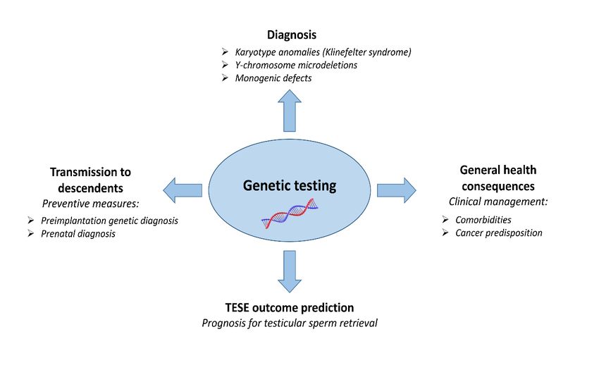
on the health of their descendants (Figure 1).
Figure 1. Clinical relevance of genetic testing in azoospermic men.
Figure 1. Clinical relevance of genetic testing in azoospermic men.
2. Consequences of Chromosomal Anomalies
2.1. Klinefelter Syndrome (47,XXY)J. Clin. Med. 2021, 10, 4009 3 of 17
This review focuses on the reproductive and general health consequences of known
genetic factors causing NOA including offspring’s health.
2. Consequences of Chromosomal Anomalies
2.1. Klinefelter Syndrome (47,XXY)
Is the most common genetic disorder causing NOA, which is characterized by the
presence of an extra X chromosome. Its prevalence is 0.1–0.2% in newborn male infants,
and it increases in relation to the age of diagnosis. Its frequency has been estimated as 3–4%
among infertile males and 10–12% in azoospermic subjects [22,23]. The severity of the
clinical phenotype of KS males may vary, and testosterone level, number of CAG repeats
in the androgen receptor and/or supernumerary X chromosome could be involved in the
clinical signs/symptoms of KS [24].
Reproductive consequences: the sex chromosome aneuploidy leads to a progressive
deterioration of the testicular tissue and both the germinal epithelium and testosterone-
producing Leydig cells are affected. There is a progressive deposition of ialine, which
is responsible for the typical hard consistency of the testes. Azoospermia is present
in about 95% of KS patients [25]. However, very rarely, non-mosaic KS patients can
have spermatozoa in their ejaculate, leading to spontaneous pregnancy. The success rate
for the recovery of spermatozoa through microsurgical TESE (m-TESE) in KS men is
34–44% [26]. As for other NOA patients, also in this case, the fertility status of the female
partner is essential for achieving pregnancy through Intracytoplasmic Sperm Injection
(ICSI). A growing number of KS patients are diagnosed during their fetal life, through
pre-natal genetic diagnosis. This novel trend raises the issue about the correct management
of these patients during their transition period from childhood to adulthood [23]. There are
still debated questions such as the right timing for testosterone replacement therapy (for its
potential interference with residual spermatogenesis) and m-TESE in young post-pubertal
KS boys [27,28].
General health consequences: besides azoospermia, a wide spectrum of clinical man-
ifestations including several comorbidities are present, i.e., metabolic syndrome, type 2
diabetes mellitus, anaemia, cardiovascular diseases (ischemic heart disease, deep vein
thrombosis, lung embolism), osteopenia/osteoporosis, breast cancer, extra-gonadal germ
cell tumours, non-Hodgkin lymphoma, haematological cancers and some autoimmune
diseases and psychiatric disorders [23,25,29,30]. Part of the above pathological conditions
are the consequence of impaired testosterone production (e.g., metabolic syndrome, os-
teopenia/osteoporosis), others may be due to X-linked gene dosage effect or epigenetic
factors [3]. Given the complexity of this disease, patients care in dedicated multidisciplinary
centres is advocated [23,31].
Consequences on offspring’s health: it is expected that spermatozoa from KS subjects
are likely to be originated from euploid spermatogonia, i.e., the testis shows a mosaic
condition where the majority of tubules contains 46,XXY spermatogonia while in a few of
them spermatogonia carry a normal chromosomal asset (46,XY) [32]. Accordingly, data in
the literature do not show an increased risk of having a KS child compared to infertile men
with normal karyotype [32]. In fact, more than 200 healthy offspring were born worldwide
from KS fathers and only a few cases of 47,XXY fetus/newborns were reported [33–35].
Despite the encouraging data that KS offspring seem not to be affected by the genetic
disease of the father, it remains still an open question whether Preimplantation Genetic
Diagnosis (PGD) or pre-natal genetic analyses should be recommended [23].
2.2. 46,XX Testicular/ovo-Testicular Disorder of Sex Development (DSD)
Also known as 46,XX male, referring to a rare, heterogeneous clinical condition with
an incidence of about 1:20,000–25,000 male newborns [36,37]. The phenotype is largely
dependent on the presence or absence of the master gene of male sex determination (SRY),
mapping to the short arm of Y chromosome.J. Clin. Med. 2021, 10, 4009 4 of 17
Reproductive consequences: due to the lack of Y chromosome linked AZF regions,
which are essential for physiological spermatogenesis, all patients with this genetic anomaly
are azoospermic. In addition, the gonadal development may be affected.
General health: apart from NOA, additional features characterize these patients. Testos-
terone levels may range from normal to low with increased FSH and LH levels leading to
the progressive development of hypogonadism [37,38]. Short stature, due to the absence of
growth-regulation genes on the Y chromosome, is also a relatively common finding.
Consequences on offspring’s health: the chance to find spermatozoa in the testes of a
46,XX male with sperm harvesting methods is zero. If the couple desires to have children,
sperm donation is the only viable option, or adoption.
3. Consequences of Y-Chromosome Microdeletions
The loss of specific chromosomal sequences on the long arm of the Y (Yq) is a the
most frequent molecular genetic cause of NOA [39]. The so called AZoospermia Factor
(AZF) regions [40,41] contain genes involved in spermatogenesis and their removal causes
different reproductive phenotypes. Many AZF genes are multicopy genes and most of
them are involved in post-transcriptional and post-translational control in germ cells [42].
The AZF regions are surrounded by highly homologous repeated sequences with the same
direction, representing an optimal substrate for Non-Allelic Homologous Recombination
(NAHR) leading to deletions. The frequency of AZF deletions in the general population
is 1:4000 but in NOA patients it can be as high as 7–10% [39,43]. The most frequently
affected region is the AZFc region accounting for >60% of deletions. Due to the peculiar
structure of this region, with many potential NAHR substrates, partial deletions with
different breakpoints may occur at a relatively high frequency [44]. Among them, the gr/gr
deletion, removing half of the AZFc gene content, is considered a proven genetic risk factor
for oligozoospermia [45].
Reproductive consequences: depending on which type of AZF regions is removed,
the semen phenotype can be azoospermia or severe oligozoospermia [39]. The complete
removal of the AZFa region (approximately 792 kb) causes SCOS, whereas the complete
removal of the AZFb deletion (with the extension marker sY1192 absent) leads to meiotic
arrest [46]. In both conditions the probability of finding testicular spermatozoa through
TESE is virtually zero. The complete removal of the AZFc is associated with a highly
variable phenotype, ranging from the complete absence of germ cells in the testis (SCOS)
to severe oligozoospermia. The TESE success rate in these patients is around 50%, but it is
highly variable in different reports.
General health: haploinsufficiency of the SHOX gene, located in the pseudoautosomal
region PAR1 of the Y chromosome, has been reported by Jorgez and colleagues in men with
AZF microdeletion and normal karyotype [47]. The authors proposed that AZF deletion
carriers are at higher risk for incurring SHOX-haploinsufficiency, which is responsible
for short stature and skeletal anomalies. This alarming finding was not confirmed in a
subsequent large, multicentre study [48]. In accordance with this latter study, Castro and
colleagues reported PAR abnormalities only in those AZF deletion carriers who presented
concomitant karyotype anomalies (isochromosome Yp and/ or Y nullisomy) [49]. In
addition to PAR abnormalities, 5/7 patients with terminal AZFbc deletion and abnormal
karyotype presented neuropsychiatric disorders. The authors hypothesize that CNVs in
the pseudoautosomal regions (PARs) and/or the removal of MSY genes (some of them are
expressed also in the brain) may play a role in the observed neuropsychiatric disorders [49].
However, the association between neuropsychiatric disorders and terminal AZFbc deletions
needs further confirmation especially in view of the lack of such neurodevelopmental
disorders in 46,XX males [37].
Consequences on offspring’s health: complete AZFc and partial AZFa or AZFb dele-
tions are compatible with the presence of spermatozoa in the ejaculate or in the testis,
therefore these patients will obligatorily transmit the deletion to their male descendants.
Recent meta-analysis reported a reduced fertilization rate, but a similar clinical pregnancyJ. Clin. Med. 2021, 10, 4009 5 of 17
rate, miscarriage rate, live birth rate and baby boy rate to those couple where the male
partner did not carry AZF deletions [50]. It is expected that the semen phenotype of the
son will be either azoospermia or oligozoospermia, however the exact semen phenotype
is not predictable, since the genetic background and exposure to environmental factors
may modulate the phenotypic expression of AZFc deletions. Some studies reported an
association between Yq microdeletions and an overall Y-chromosomal instability, which
might result in the formation of 45,X0 bearing spermatozoa [51,52]. This finding is in
accordance with the relatively high incidence of AZF deletion in patients bearing a mosaic
46,XY/45,X0 karyotype with sexual ambiguity and/or Turner stigmata [53–56]. The PGD
has been performed by two groups with conflicting data about the risk of monosomy X in
embryos [57,58]. The limited data on children born from AZF deletion carriers show that
they are apparently healthy [59].
4. Consequences of Monogenic Defects
Known monogenic anomalies with definitive clinical evidence are relatively rare in
NOA [3]. Among them two X-linked genes reached diagnostic relevance: the AR and the
TEX11 genes.
4.1. AR Gene
The androgen receptor (AR) is a DNA-binding transcription factor, which is criti-
cal for several biological functions including male sex development. Upon binding of
testosterone to the cytoplasmic AR, the complex translocates into the nucleus and binds
to the regulatory regions of specific chromosomal DNA sequences to activate androgen
dependent genes. Mutations in AR gene are responsible for the androgen insensivity syn-
drome (AIS), with an estimated prevalence of 1:20,000 to 1:64,000 live male births [60]. This
condition is associated with a high variety of phenotypes, ranging from complete androgen
insensitivity (CAIS) with a female phenotype (Morris syndrome) to milder degrees of
undervirilization (partial form or PAIS; Refenstein syndrome) or men with only infertility
(mild form or MAIS) [61]. Beside pathogenic mutations in the coding exons of the AR
causing AIS, a polymorphic CAG repeat in exon 1 has a functional effect on the receptor’s
activity. The number of the CAG repeats is inversely associated with the ligand-induced
transactivational activity of the receptor and, in physiological conditions, (CAG)n directly
correlates with serum testosterone levels [62]. This polymorphism has been associated
with various androgen-dependent conditions including impaired sperm production (for
review see [63]).
Reproductive consequences: in the PAIS/MAIS form of disease, patients may present
with quantitative spermatogenic disturbances, i.e., azoospermia or oligozoospermia. The
negative effect of longer (CAG)n on spermatogenesis is a debated issue. Although the
majority of studies report a higher than average (CAG)n in infertile patients, it is not
possible to define a cut-off value above which infertility risk is increased and to estimate
the effect size of such a risk [63].
General health: a positive correlation between CAG repeat number and depressed
mood, anxiety, and low bone mineral density with accelerated age-dependent bone loss
have been reported [64,65]. Smaller CAG repeat number is associated with benign pro-
static hypertrophy [66] and faster prostate growth during testosterone treatment [67]. The
polymorphic range in the general population is up to 39 CAG repeats, the expansion over
39 CAG is a pathological condition leading to the Kennedy disease [68]. Kennedy disease
is a rare form of X-linked spinal and bulbar muscular atrophy (SBMA), characterized
by progressive neuromuscular atrophy and ataxia [69] and a progressive set up of mild
androgen insensitivity associated to varying traits of hypogonadism, including gyneco-
mastia, testicular atrophy, disorders of spermatogenesis, elevated serum gonadotropins,
and diabetes mellitus [70].
Consequences on offspring’s health: AR mutations compatible with sperm produc-
tion will be obligatory transmitted to the female offspring with potential health conse-J. Clin. Med. 2021, 10, 4009 6 of 17
quences on her future male children. Concerning the (CAG)n repeats, it is worth noting
that repeat expansions are inherently dynamic, often changing size when transmitted to
the next generation [71]. This phenomenon, known as clinical anticipation, explains the
tendency for disease severity to increase in successive generations of a family. Patients
affected by Kennedy’s disease may conceive their own biological children and, similarly to
AR mutations, the expanded CAG repeats will be transmitted to the female child, who can
generate a male offspring affected by Kennedy disease. As far as the polymorphic range
of CAG repeats (up to 39 CAG) is concerned, the proposed relationship between longer
CAG tract and male infertility indicates a theoretical higher risk for oligozoospermic men
to conceive a female child presenting a pathological expansion of CAG repeats leading to a
future son with Kennedy disease [60,71].
4.2. TEX11 Gene
This gene belongs to the family of Testis Expressed genes, and it is crucial for chro-
mosome synapsis and formation of crossovers during meiosis. By using high-resolution
array-Comparative Genomic Hybridization (a-CGH) to screen men with NOA, a recurring
deletion of three exons of TEX11 in two patients has been identified [72]. Furthermore, by
sequencing TEX11 in larger groups of azoospermic men, more disease-causing mutations
were detected [72–75]. Overall, mutations in TEX11 were identified in more than 1% of
azoospermic men and in as many as 15% of patients with meiotic arrest.
Reproductive consequences: recessive mutations in this gene lead to NOA due to
MA [72–74]. Very recently, Krausz and colleagues demonstrated that defects in the human
gene showed a complete metaphase arrest, suggested by a residual spermatocytic develop-
ment together with the dramatic increase in the number of apoptotic metaphases [75].
General health: apart from NOA, no additional features have been reported in mu-
tated men.
Consequences on offspring’s health: the chance to find mature spermatozoa in the
testes of a man carrying loss of function TEX11 mutations is virtually zero. If the couple
desires to have children, sperm donation is the only viable option, or adoption.
4.3. Shared Genes between Spermatogenesis and Tumorigenesis
As stated in the introduction, an increased risk of various cancers has been docu-
mented in NOA patients which in part may be due to defects in biological pathways
regulating genomic integrity [8,21,76–80]. It is plausible that spermatogenesis and tu-
morigenesis may share common genetic factors, especially those involved in stem cell re-
newal/differentiation, mismatch repair mechanisms and apoptosis. Particularly, germline
alterations in DNA repair genes, which are fundamental for maintaining the genomic
integrity and stability in the early stages of the male germline, may confer hereditable
predisposition to impaired spermatogenesis and cancer.
Recent studies integrating omics and literature search revealed a significant genetic
overlap between male infertility and particular types of cancer, including urologic neo-
plasms/carcinomas and B cell lymphoma [81,82]. By using mouse model data such as
Mouse Genome Informatics (MGI) database, the integration of human orthologues to
mouse male factor infertility with a curated list of known cancer genes (COSMIC genes)
has identified 25 candidate genes that may confer risk of experiencing both conditions in
humans [21]. In particular, there is a five-fold enrichment of COSMIC genes in the MGI
male infertility list compared with genes that are not on the MGI list, suggesting that this
overlap is highly non-random [21].
Apart from the bioinformatics models and epidemiological observations, there is a
growing number of genes predisposing to cancer, which have been found mutated in men
affected by NOA.J. Clin. Med. 2021, 10, 4009 7 of 17
4.3.1. Rare Pathogenic Mutations
A recent example is related to FANCA mutations, which may cause both the classic
early onset and the rarely observed late-onset Fanconi Anaemia (FA). Both manifesta-
tions are characterized by genomic instability leading to progressive bone marrow failure,
congenital malformations and predisposition to typical cancers such as head and neck
squamous cell carcinoma and leukaemia [83]. By performing exome analysis in NOA
patients, Krausz and colleagues (2019), identified three subjects affected by SCOS with
biallelic FANCA mutations [79]. All three subjects were unaware about having Fanconi
anaemia, although two of them showed slightly abnormal blood cell count at the time of
the genetic diagnosis. This study was the first in the literature reporting the accidental
finding of Late onset FA (occult FA) in the absence of severe comorbidities of FA. In fact,
occult FA is usually diagnosed in subjects following the diagnosis of typical malignancies.
The three patients are now under surveillance by oncohematologists. This paper showed
the importance of checking blood count, especially in patients presenting idiopathic SCOS,
since the combined phenotype of SCOS with borderline low blood cell count indicates a
higher risk for occult FA. Given that the carrier frequency of FANCA defects is relatively
rare in the general population, pre-ICSI screening in the female partners of male carriers is
not recommended. However, in case of consanguinity in the couple PGD should be offered
given the severity of FA.
Fanconi anaemia and related malignancies can also be caused by recessive mutations
in the XRCC2 gene [84]. Interestingly, a homozygous XRCC2 mutation has been reported
in a consanguineous family causing isolated meiotic arrest without cancer predisposi-
tion [85]. This observation leads the authors to conclude that meiosis-specific mutations
may exist when the linker region of XRCC2, essential for protein–protein interactions, is
affected [85,86]. In support of this, knock-in mice carrying the same XRCC2 mutation
exhibited only meiotic arrest, leading to azoospermia in males and premature ovarian
failure in females [85].
Another member of the FA pathway, FANCM, involved in DNA double-strand breaks
(DSB) repair, was reported as the cause of NOA [78,80]. The FANCM gene is significantly
associated with hereditary breast and ovarian cancers [87], in line with published data on
female homozygous knock-out (KO) mice [88,89]. Recessive mutations in this gene seem to
cause a wide spectrum of seminal phenotypes, ranging from oligoasthenozoospermia to
azoospermia due to SCOS [78,80].
Biallelic mutations in two other DNA DBS repair genes, MCM8 and TEX15 were re-
ported in azoospermia and oligo/crypto/azoospermia, respectively [90–93]. Very recently,
germline mutations in the MCM8 gene following a recessive pattern of inheritance, were
detected in cancer patients [94]. One male patient affected by Lynch syndrome with fertility
problems and two patients affected by breast cancer were found to be carriers of biallelic
MCM8 mutations, suggesting a role of this gene in the germline predisposition to breast
cancer and hereditary colorectal cancer (CRC) [94]. Concerning TEX15, a rare heterozygous
mutation predicted as deleterious by four bioinformatics tools was found to be significantly
associated with prostate cancer risk [95].
Also the X-linked WNK3 gene, involved in cell signalling, survival and proliferation
has been linked both to NOA and cancer [96]. AWNK3 mutation has been found to co-
segregate with NOA due to SCOS in a family from Oman [97]. Concerning the role of
this gene in oncology, several WNK3 mutations in patient-derived xenografts of colorectal
cancer liver metastasis were predicted to be deleterious, which might contribute to the
initiation and progression of distant metastasis [98].
4.3.2. Genetic Polymorphisms
Besides rare mutations, common polymorphisms have been reported in a total of 8
mismatch repair genes, which could account for a shared aetiology between tumorigenesis
and quantitative spermatogenic failure [21].J. Clin. Med. 2021, 10, 4009 8 of 17
Homozygous or compound heterozygous mutations in the MLH1 gene have been
reported in the early-onset hereditary cancer disorder Lynch syndrome, as well as in
haematological malignancies and brain tumours [99], often associated with features of
neurofibromatosis type 1 (NF1) syndrome [100]. Besides its known carcinogenic role, an
intronic SNP in MLH1 seem to be a risk factor for the development of azoospermia or
oligozoospermia [101].
Germline MLH3 variants have been reported in hereditary Lynch syndrome-associated
brain tumours patients [102], and a common polymorphism (C2531T) in the 3’UTR of the
gene has been associated with clinical outcomes of colorectal cancer, in terms of increased
risks of relapse or metastasis in patients with heterozygous genotype [103]. Interestingly, Xu
and colleagues have observed an increased risk of azoospermia or severe oligozoospermia
associated with the above-mentioned polymorphism in 30 UTR of the MLH3 gene [104].
MSH5 has been reported as a pleiotropic susceptibility locus for lung, prostate, colorec-
tal and serous ovarian cancers [105,106], and several polymorphisms in this gene have been
associated with quantitative spermatogenic defects [101,104]. Further, one low-frequency
MSH5 variant associated with an increased risk of NOA has been reported in Han Chinese
men [107].
Biallelic germline mutations of the PMS2 gene cause the constitutional mismatch
repair deficiency, characterized by early-onset malignancies [108]. In addition, a founder
heterozygous frameshift mutation in the same gene is responsible for the Lynch syn-
drome [109]. Concerning the role of PMS2 gene in spermatogenesis, the presence of a
common polymorphism in the gene leads to a reduced interaction of MLH1 and PMS2
proteins, which may result in impaired sperm production [101].
Carriers of mutations in the ATM gene have been reported to have a higher mor-
tality rate and an earlier age at death from cancer and ischemic heart disease than non-
carriers [110]. Besides this finding, germline loss-of-function ATM mutations seem to be
enriched in men with prostate cancer and multiple primary malignancies [111]. Concerning
the role of this gene in spermatogenesis, both the homozygous and heterozygous genotypes
for a common variant in the ATM gene promoter were associated with an increased risk
for idiopathic NOA [112].
Two SNPs in the XRCC1 gene were associated with increased bladder cancer risk
among Asians [113], whilst another one, the R339Q, has been implicated in susceptibility
for both idiopathic azoospermia and different types of cancer, such as hepatocellular cancer
in Asians and breast cancer in Indians [114–118].
An identical SNP (C8092A) in 30 UTR of the ERCC1 gene has independently been
linked to both idiopathic azoospermia and various types of cancer, including breast carci-
noma, head and neck carcinoma, adult glioma [119–122].
In this context, the identification of shared genetic aetiologies between azoosper-
mia and cancer may have a significant clinical impact, for improving patient care and
genetic counselling.
5. Health Issues in ICSI Offspring from NOA Fathers
The introduction of ICSI among Assisted Reproductive Techniques (ART) has opened
an unforeseen perspective for fatherhood in NOA patients. NOA men may father their
own biological child by using non-ejaculated spermatozoa, retrieved by conventional or
micro-TESE with an average success rate of 50%. As stated above, it is well known that
NOA patients are at higher risk for genetic anomalies than he general population; therefore,
concerns were raised regarding offspring’s health.
Various parameters have been evaluated in ICSI children (from birth to young adult-
hood) born to fathers affected by spermatogenic disturbances.
Many reports describe a high frequency of chromosomal abnormalities in ICSI babies,
especially of the sex chromosomes, even when peripheral chromosome studies in the par-
ents are normal [123–125]. A possible explanation for this phenomenon could depend on
the testicular tubular alteration, which may determine abnormalities in the meiotic processJ. Clin. Med. 2021, 10, 4009 9 of 17
leading to chromosomal anomalies in the spermatozoa [126]. Therefore, other forms of
chromosome diploidy beyond sex chromosomes should be expected as well [127,128].
Overall, the risk of having chromosomal abnormalities, particularly sexual chromosome
aneuploidy, is approximately 1% in children conceived through ICSI, which is higher than
that of naturally conceived children (~0.2%) and of those conceived with conventional
in vitro fertilization (IVF) (~0.7%) (see reference in [129]). In addition, children conceived
by IVF and/or ICSI are at significantly increased risk for birth defects, although no risk
difference between children conceived with the two ARTs has been observed [130]. A sys-
tematic review and meta-analysis showed that congenital malformations in ICSI-conceived
children when compared to naturally conceived children translates into an increased risk
of 7.1% of having a malformation for individuals born after ICSI versus 4.0% for naturally
conceived children [131]. The most commonly observed congenital malformation involves
the genitourinary tract which is significantly more frequent in ICSI children compared to
both naturally conceived children and IVF children [132,133].
Besides chromosomal and birth defects, cognitive and neurodevelopmental disorders
in offspring from an ICSI father have also been evaluated [134,135]. In one study a modestly
increased risk of mental retardation and autism was reported in ICSI derived children [136],
but this finding was not replicated in independent studies [137–139]. The largest of these
studies, involving 10,718 children conceived with ICSI, 19,445 children conceived with IVF
and 2,510,166 spontaneously conceived children, observed the greatest risk of mental retar-
dation in children conceived through ICSI (RR 2.35, 95%; CI = 1.03–2.09) [136]. Importantly,
treatment factors, i.e., ICSI and embryo cryopreservation, also appear to influence this
risk [136]. In addition, an increased risk of autism in children conceived with ICSI using
surgically extracted sperm (RR 4.60, 95%; CI = 2.14–9.88) was also observed [136]. This
finding was not confirmed by Kissin and colleagues in the group of children conceived
with ICSI for male factor infertility (HR 1.23, 95%; CI = 0.92–1.64) [139]. On the other hand,
the severity of male factor does not seem to influence the cognitive development in early
childhood [140–142].
In addition to neurodevelopmental aspects, other long-term outcomes of children
conceived via ICSI due to severe male factor have been evaluated, but findings are conflict-
ing and it is difficult to evaluate the impact of NOA on these disorders [135]. Among the
large population registry studies that have examined growth and cardiometabolic factors,
there is evidence that ICSI adolescents may be at risk of increased adiposity, especially
girls [143–147]. Very recently, in male ICSI adolescents significant higher estradiol and
lower testosterone/estradiol ratio, as well as a tendency towards lower inhibin B levels,
was found [148]. Concerning reproductive outcomes in men conceived with ICSI, there is
some evidence for impaired spermatogenesis [149–151]. In fact, a Belgian study, evaluating
young men in the age interval 18–22 years, found reduced semen parameters among men
conceived with ICSI, reporting a median sperm count and total motile sperm count being
half that of their spontaneously conceived peers [151]. In addition, ICSI men showed a ten-
dency to have lower inhibin B levels and higher FSH levels compared with spontaneously
conceived peers [151].
Despite the growing number of studies, several uncertainties remain about whether
any increases in risk are due to NOA or to the ICSI procedure itself [135]. To date, the global
number of babies born as a result of ART techniques, such as ICSI, is more than 8 million
(ESHRE: https://www.eshre.eu/ 31 August 2021), therefore it should be of paramount
importance to reach to a final conclusion on safety issues. It is expected that with the
extensive use of ICSI for non-male factor, a comparison of short and long-term outcomes
between ICSI children derived from male factor versus non-male factor will elucidate the
impact of azoospermia on the descendant’s health.
6. Conclusions
Azoospermia, the most severe form of infertility, may represent a biomarker of overall
health, serving as a harbinger for higher morbidity and mortality. As reported above, certainJ. Clin. Med. 2021, 10, 4009 10 of 17
chromosomal anomalies and gene defects underlying azoospermia can be responsible for a
wide spectrum of health issues beside azoospermia, including metabolic/cardiovascular
disorders, autoimmune diseases, hypogonadism, syndromic conditions and cancers. After
the exclusion of all known acquired causes and after performing routine genetic testing, the
etiology remains unknown in a substantial proportion of patients and it could be related
to yet unidentified genetic/epigenetic factors [3]). The clinical impact of discovering such
“hidden” genetic factors is important to predict not only the fertility status but also the
general health of these men. For instance, by performing a-CGH analyses, a “CNV burden”
(especially deletions) in idiopathic infertile patients have been reported by three research
groups [152–154], suggesting a higher genomic instability potentially relevant also for
general health. CNV burden together with the above listed shared monogenic factors could
be one of the many possible explanations for the higher morbidity and lower life expectancy
observed in infertile men in respect to fertile men [6,7,19,152]. Similarly to monogenic
disorders, the inheritance of an unstable genome may also have clinical consequences on
the offspring’s health.
Thanks to the diffusion of Whole Exome Sequencing (WES) in the frame of fruitful
international collaborations, the number of genes involved in NOA is rapidly
increasing [3,5,155]. Exome analysis has proven to be very efficient in diagnosing the
cause of meiotic arrest [75], with potential implications for TESE prognosis. WES allowed
the identification of many novel genes, potentially relevant also for tumorigenesis. It can
be hypothesized that inherited genetic/epigenetic factors are responsible for the increased
risk of certain neurodevelopmental disorders, as well as impaired cardiometabolic and
reproductive health profile in children conceived with ICSI from NOA fathers. In this
context, the discovery of genetic cause underlying azoospermia would allow not only to
improve the management of NOA patients, but also to predict the clinical consequences on
the offspring inheriting the certain gene defect(s) (Figure 1).
While the list of genetic defects with potential impact on general health increases, it is
important to note that apart from a few exceptions, we are still missing a direct evidence for
a clear-cut genetic link between NOA and higher morbidity, especially in terms of cancer
predisposition. Multicentre efforts are needed in order to collect long-term follow-up data
on large groups of genetically well-characterized NOA patients. Apart from the routine
karyotype and Y chromosome deletion analysis, we hope that WES analysis will become
soon part of the genetic diagnostic work-up of NOA patients allowing diagnosis, TESE
prognosis and prevention for general health.
Author Contributions: C.K. conceived the manuscript. C.K. and F.C. designed and wrote the
manuscript. Both authors have read and agreed to the published version of the manuscript.
Funding: The publication of this review article was supported by Next Fertility Procrea, Lugano,
Switzerland.
Institutional Review Board Statement: Not applicable.
Informed Consent Statement: Not applicable.
Data Availability Statement: Not applicable.
Conflicts of Interest: The authors declare no conflict of interest.
References
1. Tournaye, H.; Krausz, C.; Oates, R.D. Novel concepts in the aetiology of male reproductive impairment. Lancet Diabetes Endocrinol.
2017, 5, 544–553. [CrossRef]
2. Krausz, C.; Riera-Escamilla, A. Genetics of male infertility. Nat. Rev. Urol. 2018, 15, 369–384. [CrossRef] [PubMed]
3. Cioppi, F.; Rosta, V.; Krausz, C. Genetics of Azoospermia. Int. J. Mol. Sci. 2021, 22, 3264. [CrossRef]
4. Kasak, L.; Laan, M. Monogenic causes of non-obstructive azoospermia: Challenges, established knowledge, limitations and
perspectives. Hum. Genet. 2021, 140, 135–154. [CrossRef]J. Clin. Med. 2021, 10, 4009 11 of 17
5. Capalbo, A.; Poli, M.; Riera-Escamilla, A.; Shukla, V.; Høffding, M.K.; Krausz, C.; Hoffmann, E.R.; Simon, C. Preconception
genome medicine: Current state and future perspectives to improve infertility diagnosis and reproductive and health outcomes
based on individual genomic data. Hum. Reprod. Update 2021, 27, 254–279. [CrossRef] [PubMed]
6. Jensen, T.K.; Jacobsen, R.; Christensen, K.; Nielsen, N.C.; Bostofte, E. Good Semen Quality and Life Expectancy: A Cohort Study
of 43,277 Men. Am. J. Epidemiol. 2009, 170, 559–565. [CrossRef] [PubMed]
7. Salonia, A.; Matloob, R.; Gallina, A.; Abdollah, F.; Saccà, A.; Briganti, A.; Suardi, N.; Colombo, R.; Rocchini, L.; Guazzoni, G.; et al.
Are Infertile Men Less Healthy than Fertile Men? Results of a Prospective Case-Control Survey. Eur. Urol. 2009, 56, 1025–1032.
[CrossRef]
8. Eisenberg, M.L.; Betts, P.; Herder, D.; Lamb, D.J.; Lipshultz, L.I. Increased risk of cancer among azoospermic men. Fertil. Steril.
2013, 100, 681–685.e1. [CrossRef]
9. Eisenberg, M.L.; Li, S.; Behr, B.; Cullen, M.R.; Galusha, D.; Lamb, D.J.; Lipshultz, L.I. Semen quality, infertility and mortality in
the USA. Hum. Reprod. 2014, 29, 1567–1574. [CrossRef]
10. Ventimiglia, E.; Capogrosso, P.; Boeri, L.; Serino, A.; Colicchia, M.; Ippolito, S.; Scano, R.; Papaleo, E.; Damiano, R.; Montorsi, F.;
et al. Infertility as a proxy of general male health: Results of a cross-sectional survey. Fertil. Steril. 2015, 104, 48–55. [CrossRef]
11. Eisenberg, M.L.; Shufeng, L.; Cullen, M.R.; Baker, L.C. Increased risk of incident chronic medical conditions in infertile men:
Analysis of United States claims data. Fertil. Steril. 2016, 105, 629–636. [CrossRef] [PubMed]
12. Choy, J.T.; Eisenberg, M.L. Male infertility as a window to health. Fertil. Steril. 2018, 110, 810–814. [CrossRef] [PubMed]
13. Glazer, C.H.; Eisenberg, M.L.; Tøttenborg, S.S.; Giwercman, A.; Flachs, E.M.; Bräuner, E.V.; Vassard, D.; Pinborg, A.; Schmidt,
L.; Bonde, J.P. Male factor infertility and risk of death: A nationwide record-linkage study. Hum. Reprod. 2019, 34, 2266–2273.
[CrossRef] [PubMed]
14. Del Giudice, F.; Kasman, A.M.; Li, S.; Belladelli, F.; Ferro, M.; de Cobelli, O.; De Bernardinis, E.; Busetto, G.M.; Eisenberg, M.L.
Increased Mortality among Men Diagnosed with Impaired Fertility: Analysis of US Claims Data. Urology 2021, 147, 143–149.
[CrossRef]
15. Bobjer, J.; Naumovska, M.; Giwercman, Y.L.; Giwercman, A. High prevalence of androgen deficiency and abnormal lipid profile
in infertile men with non-obstructive azoospermia. Int. J. Androl. 2012, 35, 688–694. [CrossRef] [PubMed]
16. Ferlin, A.; Garolla, A.; Ghezzi, M.; Selice, R.; Pelago, P.; Caretta, N.; Di Mambro, A.; Valente, U.; De Rocco Ponce, M.; Dipresa,
S.; et al. Sperm Count and Hypogonadism as Markers of General Male Health. Eur. Urol. Focus 2021, 7, 205–213. [CrossRef]
[PubMed]
17. Corona, G.; Rastrelli, G.; Vignozzi, L.; Mannucci, E.; Maggi, M. Testosterone, cardiovascular disease and the metabolic syndrome.
Best Pract. Res. Clin. Endocrinol. Metab. 2011, 25, 337–353. [CrossRef]
18. Baillargeon, J.; Al Snih, S.; Raji, M.A.; Urban, R.J.; Sharma, G.; Sheffield-Moore, M.; Lopez, D.S.; Baillargeon, G.; Kuo, Y.F.
Hypogonadism and the risk of rheumatic autoimmune disease. Clin. Rheumatol. 2016, 35, 2983–2987. [CrossRef]
19. Eisenberg, M.L.; Li, S.; Brooks, J.D.; Cullen, M.R.; Baker, L.C. Increased Risk of Cancer in Infertile Men: Analysis of U.S. Claims
Data. J. Urol. 2015, 193, 1596–1601. [CrossRef]
20. Del Giudice, F.; Kasman, A.M.; Ferro, M.; Sciarra, A.; De Bernardinis, E.; Belladelli, F.; Salonia, A.; Eisenberg, M.L. Clinical
correlation among male infertility and overall male health: A systematic review of the literature. Investig. Clin. Urol. 2020, 61,
355–371. [CrossRef]
21. Nagirnaja, L.; Aston, K.I.; Conrad, D.F. Genetic intersection of male infertility and cancer. Fertil. Steril. 2018, 109, 20–26. [CrossRef]
[PubMed]
22. Vloeberghs, V.; Verheyen, G.; Santos-Ribeiro, S.; Staessen, C.; Verpoest, W.; Gies, I.; Tournaye, H. Is genetic fatherhood within
reach for all azoospermic Klinefelter men? PLoS ONE 2018, 13, e0200300. [CrossRef]
23. Zitzmann, M.; Aksglaede, L.; Corona, G.; Isidori, A.M.; Juul, A.; T’Sjoen, G.; Kliesch, S.; D’Hauwers, K.; Toppari, J.; Słowikowska-
Hilczer, J.; et al. European academy of andrology guidelines on Klinefelter Syndrome Endorsing Organization: European Society
of Endocrinology. Andrology 2021, 9, 145–167. [CrossRef]
24. Visootsak, J.; Graham, J.M. Klinefelter syndrome and other sex chromosomal aneuploidies. Orphanet J. Rare Dis. 2006, 1, 42.
[CrossRef] [PubMed]
25. Gravholt, C.H.; Chang, S.; Wallentin, M.; Fedder, J.; Moore, P.; Skakkebæk, A. Klinefelter syndrome: Integrating genetics,
neuropsychology, and endocrinology. Endocr. Rev. 2018, 39, 389–423. [CrossRef]
26. Corona, G.; Pizzocaro, A.; Lanfranco, F.; Garolla, A.; Pelliccione, F.; Vignozzi, L.; Ferlin, A.; Foresta, C.; Jannini, E.A.; Maggi, M.;
et al. Sperm recovery and ICSI outcomes in Klinefelter syndrome: A systematic review and meta-analysis. Hum. Reprod. Update
2017, 23, 265–275. [CrossRef] [PubMed]
27. Rohayem, J.; Nieschlag, E.; Zitzmann, M.; Kliesch, S. Testicular function during puberty and young adulthood in patients with
Klinefelter’s syndrome with and without spermatozoa in seminal fluid. Andrology 2016, 4, 1178–1186. [CrossRef] [PubMed]
28. Franik, S.; Hoeijmakers, Y.; D’Hauwers, K.; Braat, D.D.; Nelen, W.L.; Smeets, D.; Claahsen-van der Grinten, H.L.; Ramos, L.;
Fleischer, K. Klinefelter syndrome and fertility: Sperm preservation should not be offered to children with Klinefelter syndrome.
Hum. Reprod. 2016, 31, 1952–1959. [CrossRef]
29. Seminog, O.O.; Seminog, A.B.; Yeates, D.; Goldacre, M.J. Associations between Klinefelter’s syndrome and autoimmune diseases:
English national record linkage studies. Autoimmunity 2015, 48, 125–128. [CrossRef]J. Clin. Med. 2021, 10, 4009 12 of 17
30. Panimolle, F.; Tiberti, C.; Granato, S.; Semeraro, A.; Gianfrilli, D.; Anzuini, A.; Lenzi, A.; Radicioni, A. Screening of endocrine
organ-specific humoral autoimmunity in 47,XXY Klinefelter’s syndrome reveals a significant increase in diabetes-specific
immunoreactivity in comparison with healthy control men. Endocrine 2016, 52, 157–164. [CrossRef]
31. Nieschlag, E.; Ferlin, A.; Gravholt, C.H.; Gromoll, J.; Köhler, B.; Lejeune, H.; Rogol, A.D.; Wistuba, J. The Klinefelter syndrome:
Current management and research challenges. Andrology 2016, 4, 545–549. [CrossRef]
32. Greco, E.; Scarselli, F.; Minasi, M.G.; Casciani, V.; Zavaglia, D.; Dente, D.; Tesarik, J.; Franco, G. Birth of 16 healthy children after
ICSI in cases of nonmosaic Klinefelter syndrome. Hum. Reprod. 2013, 28, 1155–1160. [CrossRef]
33. Denschlag, D.; Tempfer, C.; Kunze, M.; Wolff, G.; Keck, C. Assisted reproductive techniques in patients with Klinefelter syndrome:
A critical review. Fertil. Steril. 2004, 82, 775–779. [CrossRef]
34. Fullerton, G.; Hamilton, M.; Maheshwari, A. Should non-mosaic Klinefelter syndrome men be labelled as infertile in 2009? Hum.
Reprod. 2010, 25, 588–597. [CrossRef] [PubMed]
35. Brilli, S.; Forti, G. Managing infertility in patients with Klinefelter syndrome. Expert Rev. Endocrinol. Metab. 2014, 9, 239–250.
[CrossRef]
36. McElreavey, K.; Vilain, E.; Abbas, N.; Herskowitz, I.; Fellous, M. A regulatory cascade hypothesis for mammalian sex determina-
tion: SRY represses a negative regulator of male development. Proc. Natl. Acad. Sci. USA 1993, 90, 3368–3372. [CrossRef]
37. Vorona, E.; Zitzmann, M.; Gromoll, J.; Schüring, A.N.; Nieschlag, E. Clinical, endocrinological, and epigenetic features of the
46,XX male syndrome, compared with 47,XXY Klinefelter patients. J. Clin. Endocrinol. Metab. 2007, 92, 3458–3465. [CrossRef]
38. Kousta, E.; Papathanasiou, A.; Skordis, N. Sex determination and disorders of sex development according to the revised
nomenclature and classification in 46,XX individuals. Hormones 2010, 9, 218–231. [CrossRef] [PubMed]
39. Krausz, C.; Hoefsloot, L.; Simoni, M.; Tüttelmann, F.; European Academy of Andrology. European Molecular Genetics Quality
Network EAA/EMQN best practice guidelines for molecular diagnosis of Y-chromosomal microdeletions: State-of-the-art 2013.
Andrology 2014, 2, 5–19. [CrossRef]
40. Tiepolo, L.; Zuffardi, O. Localization of factors controlling spermatogenesis in the nonfluorescent portion of the human Y
chromosome long arm. Hum. Genet. 1976, 34, 119–124. [CrossRef] [PubMed]
41. Vogt, P.H.; Edelmann, A.; Kirsch, S.; Henegariu, O.; Hirschmann, P.; Kiesewetter, F.; Köhn, F.M.; Schill, W.B.; Farah, S.; Ramos,
C.; et al. Human Y chromosome azoospermia factors (AZF) mapped to different subregions in Yq11. Hum. Mol. Genet. 1996, 5,
933–943. [CrossRef]
42. Skaletsky, H.; Kuroda-Kawaguchi, T.; Minx, P.J.; Cordum, H.S.; Hillier, L.; Brown, L.G.; Repping, S.; Pyntikova, T.; Ali, J.; Bieri, T.;
et al. The male-specific region of the human Y chromosome is a mosaic of discrete sequence classes. Nature 2003, 423, 825–837.
[CrossRef]
43. Lo Giacco, D.; Chianese, C.; Sánchez-Curbelo, J.; Bassas, L.; Ruiz, P.; Rajmil, O.; Sarquella, J.; Vives, A.; Ruiz-Castañé, E.; Oliva, R.;
et al. Clinical relevance of Y-linked CNV screening in male infertility: New insights based on the 8-year experience of a diagnostic
genetic laboratory. Eur. J. Hum. Genet. 2014, 22, 754–761. [CrossRef] [PubMed]
44. Rozen, S.G.; Marszalek, J.D.; Irenze, K.; Skaletsky, H.; Brown, L.G.; Oates, R.D.; Silber, S.J.; Ardlie, K.; Page, D.C. AZFc Deletions
and Spermatogenic Failure: A Population-Based Survey of 20,000 Y Chromosomes. Am. J. Hum. Genet. 2012, 91, 890–896.
[CrossRef] [PubMed]
45. Krausz, C.; Casamonti, E. Spermatogenic failure and the Y chromosome. Hum. Genet. 2017, 136, 637–655. [CrossRef] [PubMed]
46. Stouffs, K.; Vloeberghs, V.; Gheldof, A.; Tournaye, H.; Seneca, S. Are AZFb deletions always incompatible with sperm production?
Andrology 2017, 5, 691–694. [CrossRef] [PubMed]
47. Jorgez, C.J.; Weedin, J.W.; Sahin, A.; Tannour-Louet, M.; Han, S.; Bournat, J.C.; Mielnik, A.; Cheung, S.W.; Nangia, A.K.; Schlegel,
P.N.; et al. Aberrations in pseudoautosomal regions (PARs) found in infertile men with Y-chromosome microdeletions. J. Clin.
Endocrinol. Metab. 2011, 96, E674–E679. [CrossRef] [PubMed]
48. Chianese, C.; Lo Giacco, D.; Tüttelmann, F.; Ferlin, A.; Ntostis, P.; Vinci, S.; Balercia, G.; Ars, E.; Ruiz-Castañé, E.; Giglio, S.; et al.
Y-chromosome microdeletions are not associated with SHOX haploinsufficiency. Hum. Reprod. 2013, 28, 3155–3160. [CrossRef]
49. Castro, A.; Rodríguez, F.; Flórez, M.; López, P.; Curotto, B.; Martínez, D.; Maturana, A.; Lardone, M.C.; Palma, C.; Mericq, V.; et al.
Pseudoautosomal abnormalities in terminal AZFb+c deletions are associated with isochromosomes Yp and may lead to abnormal
growth and neuropsychiatric function. Hum. Reprod. 2017, 32, 465–475. [CrossRef]
50. Li, X.; Li, X.; Sun, Y.; Han, J.; Ma, H.; Sun, Y. Effect of Y Chromosome Microdeletions on the Pregnancy Outcome of Assisted
Reproduction Technology: A Meta-analysis. Reprod. Sci. 2021, 28, 2413–2421. [CrossRef]
51. Siffroi, J.P.; Le Bourhis, C.; Krausz, C.; Barbaux, S.; Quintana-Murci, L.; Kanafani, S.; Rouba, H.; Bujan, L.; Bourrouillou, G.; Seifer,
I.; et al. Sex chromosome mosaicism in males carrying Y chromosome long arm deletions. Hum. Reprod. 2000, 15, 2559–2562.
[CrossRef]
52. Jaruzelska, J.; Korcz, A.; Wojda, A.; Jedrzejczak, P.; Bierla, J.; Surmacz, T.; Pawelczyk, L.; Page, D.C.; Kotecki, M. Mosaicism for
45,X cell line may accentuate the severity of spermatogenic defects in men with AZFc deletion. J. Med. Genet. 2001, 38, 798–802.
[CrossRef]
53. Papadimas, J.; Goulis, D.G.; Giannouli, C.; Papanicolaou, A.; Tarlatzis, B.; Bontis, J.N. Ambiguous genitalia, 45,X/46,XY mosaic
karyotype, and Y chromosome microdeletions in a 17-year-old man. Fertil. Steril. 2001, 76, 1261–1263. [CrossRef]J. Clin. Med. 2021, 10, 4009 13 of 17
54. Papanikolaou, A.D.; Goulis, D.G.; Giannouli, C.; Gounioti, C.; Bontis, J.N.; Papadimas, J. Intratubular germ cell neoplasia in
a man with ambiguous genitalia, 45,X/46,XY mosaic karyotype, and Y chromosome microdeletions. Endocr. Pathol. 2003, 14,
177–182. [CrossRef] [PubMed]
55. Patsalis, P.C.; Sismani, C.; Quintana-Murci, L.; Taleb-Bekkouche, F.; Krausz, C.; McElreavey, K. Effects of transmission of Y
chromosome AZFc deletions. Lancet 2002, 360, 1222–1224. [CrossRef]
56. Patsalis, P.C.; Skordis, N.; Sismani, C.; Kousoulidou, L.; Koumbaris, G.; Eftychi, C.; Stavrides, G.; Ioulianos, A.; Kitsiou-Tzeli,
S.; Galla-Voumvouraki, A.; et al. Identification of high frequency of Y chromosome deletions in patients with sex chromosome
mosaicism and correlation with the clinical phenotype and Y-chromosome instability. Am. J. Med. Genet. 2005, 135, 145–149.
[CrossRef] [PubMed]
57. Mateu, E.; Rodrigo, L.; Martínez, M.C.; Peinado, V.; Milán, M.; Gil-Salom, M.; Martínez-Jabaloyas, J.M.; Remohí, J.; Pellicer, A.;
Rubio, C. Aneuploidies in embryos and spermatozoa from patients with Y chromosome microdeletions. Fertil. Steril. 2010, 94,
2874–2877. [CrossRef] [PubMed]
58. Stouffs, K.; Lissens, W.; Tournaye, H.; Van Steirteghem, A.; Liebaers, I. The choice and outcome of the fertility treatment of 38
couples in whom the male partner has a Yq microdeletion. Hum. Reprod. 2005, 20, 1887–1896. [CrossRef]
59. Golin, A.P.; Yuen, W.; Flannigan, R. The effects of Y chromosome microdeletions on in vitro fertilization outcomes, health
abnormalities in offspring and recurrent pregnancy loss. Transl. Androl. Urol. 2021, 10, 1457–1466. [CrossRef] [PubMed]
60. Francomano, D.; Greco, E.A.; Lenzi, A.; Aversa, A. CAG repeat testing of androgen receptor polymorphism: Is this necessary for
the best clinical management of hypogonadism? J. Sex. Med. 2013, 10, 2373–2381. [CrossRef]
61. Krausz, C.; Cioppi, F.; Riera-Escamilla, A. Testing for genetic contributions to infertility: Potential clinical impact. Expert Rev. Mol.
Diagn. 2018, 18, 331–346. [CrossRef] [PubMed]
62. Crabbe, P.; Bogaert, V.; De Bacquer, D.; Goemaere, S.; Zmierczak, H.; Kaufman, J.M. Part of the interindividual variation in
serum testosterone levels in healthy men reflects differences in androgen sensitivity and feedback set point: Contribution of the
androgen receptor polyglutamine tract polymorphism. J. Clin. Endocrinol. Metab. 2007, 92, 3604–3610. [CrossRef] [PubMed]
63. Davis-Dao, C.A.; Tuazon, E.D.; Sokol, R.Z.; Cortessis, V.K. Male Infertility and Variation in CAG Repeat Length in the Androgen
Receptor Gene: A Meta-analysis. J. Clin. Endocrinol. Metab. 2007, 92, 4319–4326. [CrossRef]
64. Zitzmann, M.; Brune, M.; Kornmann, B.; Gromoll, J.; von Eckardstein, S.; von Eckardstein, A.; Nieschlag, E. The CAG repeat
polymorphism in the AR gene affects high density lipoprotein cholesterol and arterial vasoreactivity. J. Clin. Endocrinol. Metab.
2001, 86, 4867–4873. [CrossRef]
65. Schneider, G.; Nienhaus, K.; Gromoll, J.; Heuft, G.; Nieschlag, E.; Zitzmann, M. Sex hormone levels, genetic androgen receptor
polymorphism, and anxiety in ≥50-year-old males. J. Sex. Med. 2011, 8, 3452–3464. [CrossRef]
66. Mitsumori, K.; Terai, A.; Oka, H.; Segawa, T.; Ogura, K.; Yoshida, O.; Ogawa, O. Androgen Receptor CAG Repeat Length
Polymorphism in Benign Prostatic Hyperplasia (BPH): Correlation with Adenoma Growth. Prostate 1999, 41, 253–257. [CrossRef]
67. Zitzmann, M.; Depenbusch, M.; Gromoll, J.; Nieschlag, E. Prostate volume and growth in testosterone-substituted hypogonadal
men are dependent on the CAG repeat polymorphism of the androgen receptor gene: A longitudinal pharmacogenetic study. J.
Clin. Endocrinol. Metab. 2003, 88, 2049–2054. [CrossRef]
68. La Spada, A.R.; Wilson, E.M.; Lubahn, D.B.; Harding, A.E.; Fischbeck, K.H. Androgen receptor gene mutations in X-linked spinal
and bulbar muscular atrophy. Nature 1991, 352, 77–79. [CrossRef] [PubMed]
69. Gelmann, E.P. Molecular biology of the androgen receptor. J. Clin. Oncol. 2002, 20, 3001–3015. [CrossRef]
70. Sobue, G.; Doyu, M.; Morishima, T.; Mukai, E.; Yasuda, T.; Kachi, T.; Mitsuma, T. Aberrant androgen action and increased size of
tandem CAG repeat in androgen receptor gene in X-linked recessive bulbospinal neuronopathy. J. Neurol. Sci. 1994, 121, 167–171.
[CrossRef]
71. Paulson, H. Repeat expansion diseases. Handb. Clin. Neurol. 2018, 147, 105. [CrossRef] [PubMed]
72. Yatsenko, A.N.; Georgiadis, A.P.; Röpke, A.; Berman, A.J.; Jaffe, T.; Olszewska, M.; Westernströer, B.; Sanfilippo, J.; Kurpisz,
M.; Rajkovic, A.; et al. X-linked TEX11 mutations, meiotic arrest, and azoospermia in infertile men. N. Engl. J. Med. 2015, 372,
2097–2107. [CrossRef] [PubMed]
73. Yang, F.; Silber, S.; Leu, N.A.; Oates, R.D.; Marszalek, J.D.; Skaletsky, H.; Brown, L.G.; Rozen, S.; Page, D.C.; Wang, P.J. TEX11 is
mutated in infertile men with azoospermia and regulates genome-wide recombination rates in mouse. EMBO Mol. Med. 2015, 7,
1198–1210. [CrossRef] [PubMed]
74. Sha, Y.; Zheng, L.; Ji, Z.; Mei, L.; Ding, L.; Lin, S.; Wang, X.; Yang, X.; Li, P. A novel TEX11 mutation induces azoospermia: A case
report of infertile brothers and literature review. BMC Med. Genet. 2018, 19, 63. [CrossRef] [PubMed]
75. Krausz, C.; Riera-Escamilla, A.; Moreno-Mendoza, D.; Holleman, K.; Cioppi, F.; Algaba, F.; Pybus, M.; Friedrich, C.; Wyrwoll, M.J.;
Casamonti, E.; et al. Genetic dissection of spermatogenic arrest through exome analysis: Clinical implications for the management
of azoospermic men. Genet. Med. 2020, 22, 1956–1966. [CrossRef]
76. Chalmel, F.; Lardenois, A.; Primig, M. Toward understanding the core meiotic transcriptome in mammals and its implications for
somatic cancer. Ann. N. Y. Acad. Sci. 2007, 1120, 1–15. [CrossRef]
77. Hanson, H.A.; Anderson, R.E.; Aston, K.I.; Carrell, D.T.; Smith, K.R.; Hotaling, J.M. Subfertility increases risk of testicular cancer:
Evidence from population-based semen samples. Fertil. Steril. 2016, 105, 322–328. [CrossRef]You can also read