The Role of Micronutrients in the Infection and Subsequent Response to Hepatitis C Virus - MDPI
←
→
Page content transcription
If your browser does not render page correctly, please read the page content below
cells
Review
The Role of Micronutrients in the Infection and
Subsequent Response to Hepatitis C Virus
Sunil Gupta 1,† , Scott A. Read 1,2,† , Nicholas A. Shackel 3 , Lionel Hebbard 4 , Jacob George 2 and
Golo Ahlenstiel 1,2,5, *
 1    Blacktown Clinical School, Western Sydney University, Blacktown, NSW 2148, Australia;
      sunil.gupta@live.com.au (S.G.); S.read@westernsydney.edu.au (S.A.R.)
 2    Storr Liver Centre, The Westmead Institute for Medical Research, University of Sydney, Westmead 2145,
      Australia; Jacob.george@sydney.edu.au
 3    Department of Medicine, University of New South Wales, Kensington, NSW 2052, Australia;
      n.shackel@unsw.edu.au
 4    Department of Molecular and Cell Biology, Centre for Molecular Therapeutics, James Cook University,
      Australian Institute of Tropical Health and Medicine, Townsville, QLD 4814, Australia;
      lionel.hebbard@jcu.edu.au
 5    Department of Medicine, Blacktown Hospital, Blacktown, NSW 2148, Australia
 *    Correspondence: g.ahlenstiel@westernsydney.edu.au; Tel.: +612-9851-6073; Fax: +612-9851-6050
 †    Both authors contributed equally.

 Received: 20 May 2019; Accepted: 13 June 2019; Published: 17 June 2019

 Abstract: Micronutrient deficiencies develop for a variety of reasons, whether geographic,
 socioeconomic, nutritional, or as a result of disease pathologies such as chronic viral infection.
 As micronutrients are essential for a strong immune response, deficiencies can significantly dampen
 both the innate and the adaptive arms of antiviral immunity. The innate immune response in
 particular is crucial to protect against hepatitis C virus (HCV), a hepatotropic virus that maintains
 chronic infection in up to 80% of individuals if left untreated. While many micronutrients are required
 for HCV replication, an overlapping group of micronutrients are also necessary to enact a potent
 immune response. As the liver is responsible for the storage and metabolism of many micronutrients,
 HCV persistence can influence the micronutrients’ steady state to benefit viral persistence both
 directly and by weakening the antiviral response. This review will focus on common micronutrients
 such as zinc, iron, copper, selenium, vitamin A, vitamin B12, vitamin D and vitamin E. We will explore
 their role in the pathogenesis of HCV infection and in the response to antiviral therapy. While chronic
 hepatitis C virus infection drives deficiencies in micronutrients such as zinc, selenium, vitamin
 A and B12, it also stimulates copper and iron excess; these micronutrients influence antioxidant,
 inflammatory and immune responses to HCV.

 Keywords: hepatitis C virus; micronutrients; micronutrient deficiency; liver; innate immunity

1. Introduction
      Micronutrients are trace elements and vitamins obtained from our diet that are essential to sustain
life and optimal physiological function [1,2]. Deficiencies affect over 2 billion people and are largely
associated with malnutrition or poor diet [2,3]. Many micronutrients are necessary to elicit an effective
immune response to viral infections but are also utilized by viruses such as the hepatitis C virus
(HCV) to propagate [4]. HCV is an RNA flavivirus that infects hepatocytes and is typically transmitted
through exposure to infected body fluids, including blood transfusion and injecting drug use [5,6].
Like many viruses, HCV has the ability to impede antiviral and apoptotic responses to favor its own
persistence [7]. Consequently, approximately 80% of untreated infections progress to chronic hepatitis

Cells 2019, 8, 603; doi:10.3390/cells8060603                                        www.mdpi.com/journal/cells
Cells 2019, 8, 603 2 of 20
C (CHC), with persistent viremia and hepatic inflammation [8]. Given that the liver is responsible
for the storage and metabolism of many micronutrients such as iron and zinc [9,10], chronic HCV
replication has the potential to negatively influence this state. As such, there is a delicate balance
between the maintenance of micronutrient stores and micronutrient availability during infection.
HCV infection induces an acute inflammatory response driven by pro-inflammatory cytokines
such as IL-6 and TNF-α that can lead to mitochondrial dysfunction and hepatocyte oxidative stress [11].
Chronic hepatocyte damage stimulates persistent inflammation that can lead to the development of
liver fibrosis and, ultimately, cirrhosis and cancer [12]. Importantly, numerous micronutrients exist
as key components of the hepatic antioxidant response, which can become significantly impaired
upon micronutrient deficiency. A prime example is zinc, that is tightly bound to metallothionein (MT)
chaperone proteins in the liver [13]. MTs function as intracellular sensors of oxidative stress and heavy
metal dysregulation and act to detoxify and scavenge free radicals [14,15].
In CHC, micronutrient deficiency is common, with deficiencies in zinc, vitamin A and vitamin
D reported in up to 48.4%, 54.3% and 43% of patients, respectively [16–18]. Whilst micronutrient
deficiencies may be mediated by HCV replication and associated inflammation, they may be further
exacerbated by malnutrition due to lifestyle factors associated with chronic viral hepatitis [19]. Up to
14% of patients with CHC are malnourished, with deficiency of some micronutrients such as zinc
reported in up to 90% of patients with compensated cirrhosis and in 98% with decompensated cirrhosis,
a manifestation of caloric and protein restriction [20]. This review summarizes the metabolic and
immunological roles of the common micronutrients zinc, iron, copper, selenium, vitamin A, vitamin
B12, vitamin D and vitamin E. We will comment on the clinical importance of deficiency or excess of
these micronutrients, their role in HCV pathogenesis and the associated immune response.
2. Zinc
Zinc is an essential trace element important for growth and development. It is found in a variety
of foods including meats, cereals, grains, beans and dairy products [21,22]. Up to 10% of the human
proteome binds zinc [23]. Protein-bound zinc plays an essential catalytic role in metalloenzyme activity,
transcription factor binding and gene regulation [24]. In particular, zinc is required for optimal innate
immune defenses including phagocytosis, natural killer cell activity, generation of oxidative bursts,
cytokine production and complement activity (reviewed in [25,26]). Zinc distribution within the
body is widespread, with the greatest stores found in skeletal muscle (57%), bone (29%) and liver
(6%) [9]. Within the blood, 60% of zinc is loosely bound to albumin and 30% of it is tightly bound
to macroglobulin [27]. Signs of zinc deficiency include growth retardation, hair loss, diarrhea and
delayed development of secondary sexual characteristics [28].
2.1. Zinc Deficiency in HCV
In acute HCV infection, inflammatory cytokines such as IL-6 stimulate hepatic zinc uptake via the
Zip14 zinc transporter, resulting in transient hypozincemia [29]. Elevated cytosolic zinc up-regulates
MT expression, which exerts numerous anti-oxidant and anti-viral effects [13]. In CHC, studies have
demonstrated that up to 48.4% of patients develop persistent hypozincemia (Cells 2019, 8, 603 3 of 20
2.2. The Effect of Zinc on HCV Pathogenesis, Immune Response and Treatment
HCV infection stimulates the expression of the antiviral Th1 cytokines IL-2 and IFN-γ which
are key drivers of the cytotoxic immune response required to clear acute infection [35]. Human
studies show that zinc deficiency leads to a reduction in cytotoxic T cell populations [36], a decrease
in natural killer (NK) cell activity [37] and a down-regulation of the Th1 response [38]. In particular,
dietary zinc restriction reduces IL-2 and IFN-γ, with no effect on the Th2 cytokines IL-4 and IL-10 [38].
Consequently, low levels of circulating cytotoxic T cells and thymic atrophy due to zinc deficiency
may be explained in part, by a reduction in IL-2, a key driver of cytotoxic T cell proliferation and HCV
clearance [39]. Importantly, excess zinc supplementation can also reduce the expression of key antiviral
cytokines such as IFN-γ by reducing IRF1 expression in regulatory T cells, as observed in vitro [40].
It can lead to impairment of lymphocyte activation and granulocyte chemotaxis and phagocytosis in
humans [41]. Consequently, it is vital to measure zinc status prior to zinc supplementation, as excess
zinc can interfere with a potent antiviral response.
It has been demonstrated in vitro that zinc may play an important role as a negative regulator of
HCV replication in genome-length HCV RNA-replicating cells, albeit via an unknown mechanism [42].
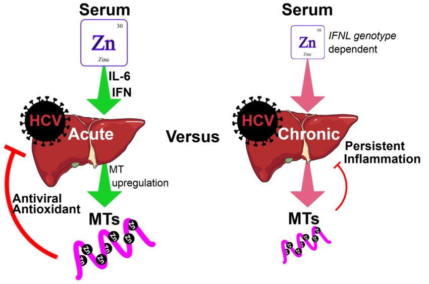
This is possibly via the induction of MTs that possess mild antiviral activity (Figure 1) [13]. Whilst acute
HCV infection drives MT induction in vitro, patients with CHC have low serum zinc and low hepatic
MT expression [43,44]. This suggests that chronic HCV infection has a markedly different effect on zinc
distribution compared to acute HCV infection. Furthermore, low hepatic MT expression is associated
with increased hepatic inflammation and fibrosis, suggesting that MTs and zinc protect against chronic
inflammation [44]. In support of this, studies have shown that elevated hepatic MT expression is
associated with improved liver function as demonstrated by reduced alanine aminotransferase (ALT)
and aspartate aminotransferase (AST) levels [45].
Hepatic zinc and MT expression also appear to affect HCV treatment response. In fact,
zinc supplementation has been shown to increase hepatic MT expression and enhance the response
to IFN-α therapies [46]. Nagamine et al. demonstrated that over a 24-week period of IFN-α therapy,
CHC treatment responders had higher pre-treatment serum zinc levels and higher post-treatment
hepatic MT expression [47]. Similarly, another study demonstrated that higher baseline zinc levels
resulted in a larger decline in serum zinc post-IFN-α treatment, and in these conditions, zinc likely
localized to the liver, stimulating MT expression and anti-viral activity [32]. A more recent in vitro
study by our group supports these findings, showing that zinc supplementation in MT knockdown
cells did not appear to inhibit HCV replication [13].
Liver transcriptomic data and in vitro studies with the Huh-7 hepatoma cell line demonstrated
that along with driving MT expression, zinc potently inhibited signaling by IFN-λ3 [43], a cytokine
with a central role in the pathogenesis and clearance of HCV infection [43,48]. CHC serum zinc
levels and hepatic MT expression exhibited strong inverse correlations with inflammatory and
interferon-stimulated genes (ISGs) in the liver [43]. Interestingly, because serum zinc levels were
elevated in CHC patients with the IFN-λ3 rs12979860 CC (responder) genotype, these data suggest
that zinc may sensitize the anti-viral response by reducing baseline ISG expression to facilitate strong
antiviral responses with minimal interferon refractoriness upon antiviral treatment [49].Cells 2019, 8, 603 4 of 20
Cells 2019, 8, x FOR PEER REVIEW 4 of 21
Figure1.1. Role
Figure Role ofof zinc
zinc and
and metallothioneins
metallothioneins (MTs)(MTs) in inacute
acuteand
andchronic
chronichepatitis
hepatitisCCvirus
virus(HCV)
(HCV)
infections.
infections.InInacute
acuteHCVHCVinfection,
infection,pro-inflammatory
pro-inflammatorycytokines
cytokinessuch
suchasasIL-6
IL-6and
andIFN-λ
IFN-λstimulate
stimulatethe
the
redistribution
redistributionofofserum
serumzinczincinto
intothe
theliver.
liver.Elevated
Elevatedhepatic
hepaticzinc
zincstimulates
stimulates thetheinduction
induction ofofMTs,
MTs,
which
whichserve as as
serve potent
potentantioxidants via their
antioxidants binding
via their and release
binding of zinc, of
and release butzinc,
also but
display
alsomild antiviral
display mild
activity.
antiviral activity. For reasons that remain poorly defined, chronic HCV infection resultshepatic
For reasons that remain poorly defined, chronic HCV infection results in low serum and in low
zinc.
serumConsequently,
and hepatic liver
zinc.MT expression isliver
Consequently, reduced, and the liver
MT expression is subjectand
is reduced, to chronic inflammation
the liver is subject to
due to persistent
chronic viral replication
inflammation due to and oxidative
persistent stress.
viral Further, chronic
replication hepatitisstress.
and oxidative C (CHC) patients
Further, with
chronic
the rs12979860 CC interferon lambda (IFNL) genotype possess increased serum
hepatitis C (CHC) patients with the rs12979860 CC interferon lambda (IFNL) genotype possess zinc and demonstrate
improved
increasedresponses
serum zinc to antiviral treatment,improved
and demonstrate supporting the immuno-stimulatory
responses role of supporting
to antiviral treatment, zinc. the
3. Ironimmuno-stimulatory role of zinc.
As the most abundant trace element in the human body, iron plays a key role in DNA and
3. Iron
protein synthesis, erythrocyte production, electron transport, cellular respiration, cell proliferation and
As the most abundant trace element in the human body, iron plays a key role in DNA and
regulation of gene expression [50]. Dietary iron is absorbed through the divalent metal transporter
protein synthesis, erythrocyte production, electron transport, cellular respiration, cell proliferation
1 (DMT1) by duodenal and jejunal enterocytes [51]. It is then exported by ferroportin (FPN) into
and regulation of gene expression [50]. Dietary iron is absorbed through the divalent metal
the bloodstream where it becomes bound to transferrin and is utilized by the muscle and erythroid
transporter 1 (DMT1) by duodenal and jejunal enterocytes [51]. It is then exported by ferroportin
compartments. In addition, iron is stored as ferritin or hemosiderin in enterocytes, macrophages and
(FPN) into the bloodstream where it becomes bound to transferrin and is utilized by the muscle and
hepatocytes [51]. The best-absorbed form of iron (“heme”) is found in meat, poultry and fish, whereas
erythroid compartments. In addition, iron is stored as ferritin or hemosiderin in enterocytes,
non-heme iron is found in leafy green vegetables, seeds of legumes, fruits and dairy products [52].
macrophages and hepatocytes [51]. The best-absorbed form of iron (“heme”) is found in meat,
Iron deficiency can lead to fatigue, anemia, infertility in females and depression [52]. Being an oxidant
poultry and fish, whereas non-heme iron is found in leafy green vegetables, seeds of legumes, fruits
with free radical activity, excess iron leads to the breakdown of cellular membranes, ultimately leading
and dairy products [52]. Iron deficiency can lead to fatigue, anemia, infertility in females and
to damage in organs such as the liver, kidneys, heart and lungs [53].
depression [52]. Being an oxidant with free radical activity, excess iron leads to the breakdown of
cellular
3.1. membranes,
Iron Excess in HCVultimately leading to damage in organs such as the liver, kidneys, heart and
lungs [53].
Iron overload is a prominent feature of CHC, with 10–42% of patients demonstrating hepatic iron
accumulation [54,55].
3.1. Iron Excess in HCV Elevated serum ferritin and transferrin saturation are similarly common in up
to 40% of patients [55], where they significantly correlate with hepatic fibrosis [56,57]. Importantly,
while Iron overload
serum ferritiniscan
a prominent feature
correlate with liverofiron,
CHC, it with 10–42%
can also of patients
be elevated demonstrating
in the hepatic
absence of hepatic
iron accumulation [54,55]. Elevated serum ferritin and transferrin saturation are similarly common
in up to 40% of patients [55], where they significantly correlate with hepatic fibrosis [56,57].
Importantly, while serum ferritin can correlate with liver iron, it can also be elevated in the absence
4Cells 2019, 8, 603 5 of 20
iron overload and be a marker of liver inflammation [58]. Consequently, because of the inflammatory
nature of CHC, liver biopsy is the gold standard for the assessment of hepatic iron overload.
HCV has been shown to influence iron absorption via oxidative stress-mediated down-regulation
of hepcidin expression [59,60]. Hepcidin is a peptide hormone produced by the liver that binds
FPN to induce its ubiquitination, internalization and degradation [61]. Consequently, HCV-mediated
down-regulation of hepcidin results in elevated enterocyte FPN levels and increased intestinal
absorption of iron [62]. Fujita et al. have shown that the hepcidin-to-ferritin ratio is significantly lower
in HCV patients compared to controls or patients with hepatitis B virus (HBV), suggesting a possible
association [63].
3.2. The Effect of Iron on HCV Pathogenesis, Immune Response and Treatment
The antiviral role of iron has generated conflicting results in vitro. Using human hepatocyte
cell lines, iron has been shown to both enhance [64] and inhibit [65] the replication of HCV. Iron can
promote HCV translation through altering the affinity of common cellular factors, the HCV internal
ribosome entry site (IRES), via increased expression of eukaryotic initiation factor 3 (EIF3), and the
intracellular La ribonucleoprotein [66]. Conversely, iron can inactivate the viral polymerase NS5B [67].
In CHC patients, iron levels have been positively associated with hepatic ALT, suggesting that
iron-mediated stimulation of viral replication in vitro may be relevant in vivo [68]. Iron deposition
in the liver of HCV patients triggers reactive oxygen species (ROS), which can induce secondary
lipid peroxidation and ultimately lead to mitochondrial dysfunction and protein and nuclei acid
damage [68,69]. Interestingly, phlebotomy has been shown to reduce liver transaminases and gamma
glutamyltransferase (GGT) without affecting HCV viral load [70–72].
The antiviral effect of therapeutic phlebotomy in patients with CHC and iron overload has been
inconclusive. Fargion et al. studied 114 patients with hepatic iron concentrations (HIC) ≥700 mcg/g
in men and ≥500 mcg/g in women [73]. Compared with IFN-α therapy alone, those who received
phlebotomy followed by IFN-α showed a trend towards a greater sustained virologic response
(SVR) at an odds ratio of 2.32 (p = 0.082) [73]. In agreement with these data, lower HIC and serum
ferritin were measured in patients who responded to IFN-α treatment [74]. Conversely, baseline HIC
had no effect on the response to IFN-α therapy in beta-thalassemia major patients with CHC and
transfusion-acquired iron overload [75]. With regards to previously treated CHC non-responders to
IFN-α therapy, phlebotomy had no effect on SVR following re-treatment; however, there was a decrease
in liver injury with reduced AST and improved necro-inflammatory changes on liver biopsy [76].
4. Selenium
In trace amounts, selenium is indispensable for the maintenance of good health [77]; it can be
obtained from cereals, grains, fish, meat and dairy products [21,22]. Selenium is a constituent of
glutathione peroxidase (GPx), an enzyme that protects against damage induced by free radicals at a
cellular level [78]. Selenium is also essential in augmenting antiviral immunity [79] and assisting in
the detoxification of liver enzymes [80]. Other major selenium-containing proteins in blood include
selenoprotein-P and albumin [81–83]. Deficiency can lead to fatal cardiomyopathies such as Keshan
disease [84] and degenerative disorders such as Kashin–Beck disease [85].
4.1. Selenium Deficiency in HCV
The presence of selenium deficiency in CHC remains uncertain. CHC infection has been shown to
result in a reduction of blood selenium levels (99.8 ± 11.0 µg/L in CHC versus 117.5 ± 15.7 µg/L in
healthy controls) that decrease even further following the development of HCV-related cirrhosis (84.7 ±
16.4 µg/L) [86]. These data are supported by studies that demonstrate a decline in serum selenium
levels in proportion to the degree of hepatic fibrosis, as determined by tissue sampling [87]. In addition,
GPx activity is reduced along with selenium levels in CHC (r = 0.374, p = 0.148), providing a possible
mechanism by which CHC stimulates oxidative stress due to selenium deficiency [87]. While othersCells 2019, 8, 603 6 of 20
have found no significant reduction in serum selenium in CHC patients [88], alcohol intake was a
major confounding variable as it affects selenium levels [89].
With regard to the mechanism by which selenium becomes deficient in CHC patients,
Thuluvath et al. have demonstrated that urinary excretion of selenium was not significantly different
in cirrhotic patients compared to controls, although this study was not specific to viral hepatitis [90].
Nonetheless, these data suggest that reduced selenium levels may be secondary to malabsorption in
the small intestine [87]. Further studies are required in CHC patients to determine the underlying
cause for selenium deficiency.
4.2. The Effect of Selenium on HCV Pathogenesis, Immune Response and Treatment
RNA viruses, including HCV, encode selenium-dependent GPx genes [91,92]. In view of this
finding, it is possible that the sequestration of selenium to facilitate viral replication could generate a
shortage of circulating selenium [93]. In vitro, HCV can inhibit the expression of gastrointestinal-GPx,
a GPx that is also expressed in the liver, resulting in an increase in viral replication [94]. These data are
supported by clinical data demonstrating a negative correlation between HCV viral load and selenium
(r = −0.730) and GPx activity (r = −0.675) [95]. In that study, plasma GPx activity, plasma selenium
and erythrocyte selenium levels were significantly lower in CHC patients than in controls; however,
hepatic selenium was not measured [95].
In CHC patients, low serum selenium levels were found to positively associate with low GPx
activity (r = 0.374, p = 0.0148) but not with HCV genotype or HCV-RNA load [87]. While these data do
not indicate that HCV directly decreases selenium, they raise the possibility of selenium replacement
as a therapeutic supplement to boost anti-oxidant and antiviral defenses. This supports animal models
of HCV infection where reduced selenium levels resulted in the accumulation of lipid peroxides [96],
which led to increased expression of VEGF and IL-8, accelerating the development of HCC [96],
whilst also stimulating hepatic fibrosis via hepatic stellate cell activation [97].
The effect of selenium has not been examined in the context of HCV treatment but has been
assessed for its anti-inflammatory properties. Berkson et al. analyzed the effect of selenium alpha-lipoic
acid and silymarin supplementation in three CHC patients and demonstrated an improvement in
ALT [93]. A more recent randomized, placebo-controlled, double-blinded trial assessed the effect of
ascorbic acid (500 mg), vitamin E (945 IU) and selenium (200 µg) versus placebo for six months. During
supplementation, the group administered the antioxidants had higher erythrocyte GPx; however,
no differences were observed in serum ALT or HCV viral load [98]. While limited, these data suggest
that selenium supplementation alone may not be adequate to suppress inflammation in CHC.
5. Copper
Copper is an essential trace element that is present in mineral-rich foods like meats, oysters, nuts,
seeds, dark chocolate and whole grains [52]. The main protein carrier of copper in the human body is
ceruloplasmin (95%), whilst a small amount of copper is bound to albumin [99,100]. Once absorbed from
the digestive tract, copper is transported to the liver and is essential for erythrocyte production [101].
Up to 50% of copper content is stored in muscles and bone, 8% in the brain and 8–15% in the
liver [100]. Copper has the ability to accept electrons, which makes it important in redox processes [102].
As such, it is a functional component of several essential enzymes including cytochrome oxidase and
superoxide dismutase, which serve to catalyse the reduction of oxygen molecules to water and of free
radicals to hydrogen peroxide [102]. Copper deficiency can cause anemia, defective keratinization and
pigmentation [103], whilst copper excess can cause acne, alopecia, strokes, pancreatic dysfunction and
osteoporosis [104].
5.1. Copper Excess in HCV
Acute HCV infection stimulates an increase in serum copper [105], which is further exacerbated
in CHC and fibrotic liver disease [105–107]. Hepatic copper is increased in CHC [107], where it isCells 2019, 8, 603 7 of 20
primarily bound to MTs (Cu–MTs), thus contributing to hepatic copper overload [108]. The mechanism
by which HCV stimulates copper accumulation in the liver remains unknown, however, Cu–MTs have
been shown to stimulate hydroxyl radical generation in rats, thus driving liver damage and, perhaps,
fibrosis [109,110]. In a recent human study, hepatic copper content increased significantly as hepatic
fibrosis advanced and correlated positively with bilirubin (r = 0.466, p = 0.0023) and type IV collagen
(r = 0.402, p = 0.0086) [109]. As copper is excreted solely in the bile, HCV-mediated inhibition of bile
acid secretion may indeed result in the retention of biliary copper [111,112]. Interestingly, copper
and zinc metabolism are strongly linked in the liver. Zinc over-supplementation can lead to copper
deficiency via MT-mediated inhibition of copper absorption in the gut [101,113].
5.2. The Effect of Copper on HCV Pathogenesis, Immune Response and Treatment
While there are no direct links between copper excess and the pathogenesis in CHC, unbound
copper efficiently catalyzes the formation of ROS and is a potential driver of oxidative liver damage [114].
Oxidative stress is further aggravated by a decline in glutathione in CHC patients, which serves as a
key hepatic antioxidant by binding free copper [115].
No clinical studies have examined the antiviral role of copper supplementation or depletion in
humans; however, copper in various forms can exhibit antiviral properties [116,117]. In particular,
cuprous oxide nanoparticles (CO-NPs), a p-type semiconductor that possesses unique physical and
chemical properties [118,119], have been shown to inhibit the infectivity of HCV cell cultures at a
non-cytotoxic concentration. In addition, CO-NPs can inhibit the attachment and entry of genotype 1a,
1b and 2a HCV pseudoparticles (HCVpp), with no effect on HCV replication [120].
6. Vitamin A
Vitamin A represents a group of fat-soluble retinoids including retinol and retinoic acid.
The majority of liver vitamin A is stored in hepatic stellate cells (HSCs) and hepatocytes [10]. Retinoic
acid (RA) is thought to play a role in potentiating the innate immune response by binding to cellular
retinoic acid-binding protein (CRABP) 1 and 2 [121]. Vitamin A can be obtained from vegetables,
dairy products, eggs and fruits [21,22].
6.1. Vitamin A Deficiency in HCV
In the setting of CHC, vitamin A deficiency is common. A study of 199 treatment-naive CHC
patients found that serum vitamin A was significantly lower compared to healthy controls (256 ng/mL
versus 742 ng/mL; P < 0.0001) [122]. Peres et al. showed that in patients with CHC, vitamin A deficiency
was present in 54.3% of patients, with a serum retinol decline in those with cirrhosis and hepatocellular
carcinoma (HCC) [16]. In support, a 2002 study by Yadav et al. found diminished liver retinol levels in
CHC patients with moderate to severe fibrosis, compared with those with mild fibrosis [123]. Notably,
because only free retinol was assessed, any inference regarding total liver vitamin A stores cannot
be made. Nonetheless, these data suggest that vitamin A deficiency is associated primarily with
the development of liver fibrosis. HSCs lose their retinoid content once they become activated as
a consequence of CHC, resulting in the formation of a collagenous extracellular matrix that drives
liver fibrosis [10]. Consequently, they are a likely candidate for the drastic reduction of hepatic retinol
observed in fibrotic CHC patients.
6.2. The Effect of Vitamin A on HCV Pathogenesis, Immune Response and Treatment
Vitamin A has been shown to both promote and inhibit HCV replication in vitro. Retinoic acid
binding to CRABP1 can activate lipid metabolism gene expression in hepatocytes, thus providing a
platform for HCV replication complexes and subsequent viral propagation [124,125]. A separate study
has demonstrated that administration of all-trans-retinoic acid (ATRA), an active metabolite of vitamin
A, results in the down-regulation of the HCV replicon, at least in part via the induction of GPx [94].Cells 2019, 8, 603 8 of 20
In addition, ATRA has been shown to increase the expression of IFN-α receptor IFNAR1, enhancing
the antiviral effect of IFN-α.
Because of its antiviral effect in vitro, a recent study administered ATRA in combination with
IFN-α to a cohort of CHC patients that had previously failed therapy [126]. While none of the patients
achieved SVR, ATRA demonstrated a direct antiviral and synergistic effect with pegylated-IFN-α,
resulting in a reduction in HCV viral load 12 weeks after the completion of treatment [127]. A possible
mechanism by which ATRA stimulates the antiviral response is through the up-regulation of the
dsRNA helicase enzyme retinoic acid-inducible gene-I (RIG-I) [128]. RIG-I is responsible for HCV
dsRNA recognition and subsequent IFN-α and IFN-λ production as part of the innate antiviral response
in the liver [126,129]. In agreement, combined vitamin A and D deficiency was found to be a strong
independent predictor of nonresponse to antiviral therapy [122]. It should be noted that vitamin A
supplementation in clinical practice is limited by its possible hepatotoxicity [130].
7. Vitamin B12
Vitamin B12 (cobalamin) is a water-soluble vitamin obtained through the consumption of meat,
liver, fish, dairy products and fortified cereals [131,132]. It is co-absorbed with intrinsic factor, a product
of gastric parietal cells, in the terminal ileum. Cobalamin is vital for normal neurologic function,
erythrocyte production and DNA synthesis [131,132]. Deficiency can result in hyperpigmentation,
vitiligo, glossitis, anemia, cognitive impairment, gait abnormalities and areflexia [133]. Importantly,
vitamin B12 is stored in high concentration in the human liver [134]. A review of the literature did not
reveal any studies with data to suggest the presence of vitamin B12 deficiency in CHC.
The Effect of Vitamin B12 on HCV Pathogenesis, Immune Response and Treatment
HCV utilizes a cap-independent initiation of translation through an IRES that is present in the viral
5’ untranslated region (UTR) [135]. As vitamin B12 is highly concentrated in the liver [134] and has a
strong affinity for RNA pseudoknots [136], the effect of cobalamin has been examined with respect
to IRES function. Vitamin B12 was shown to inhibit HCV IRES-dependent initiation of translation
in vitro and was suggested to represent an evolutionary mechanism serving to limit HCV replication
and promote persistence [137]. However, it also suggests that vitamin B12 may offer a therapeutic
option for CHC treatment [137].
Human studies have demonstrated conflicting results regarding baseline serum vitamin B12 and
response to IFN-based therapy. While Rosenberg et al. found significantly lower B12 in non-responders
(NR) when compared to responders (R) (262 pM NR versus 331 pM R) [138], Mechie et al. showed the
opposite effect (616 ng/L NR versus 333 ng/L R) [139]. Elevated B12 measured in cirrhotic patients
may, however, reflect their poor hepatic vitamin B12 clearance [140]. Consequently, cirrhosis may
impair responses to IFN-based therapies and increase serum vitamin B12, independently. Supporting
the beneficial role of baseline vitamin B12, serum homocysteine levels are reduced (Cells 2019, 8, 603 9 of 20
and immunomodulatory properties. More specifically, it inhibits T cell proliferation, expression of
interleukin-2, expression of IFN-γ and CD8 T-lymphocyte-mediated cytotoxicity [146]. These extra-
skeletal effects are relevant in the pathogenesis and treatment of many causes of chronic liver
disease [147,149].
8.1. Vitamin D Deficiency in HCV
Vitamin D deficiency (Cells 2019, 8, 603 10 of 20
The Effect of Vitamin E on HCV Pathogenesis, Immune Response and Treatment
The anti-oxidant effect of vitamin E on HCV treatment has been trialed in various studies.
High-dose vitamin E treatment (twice the daily dose of 400 IU) in patients with CHC refractory to
IFN-α therapy showed significant reductions in AST and ALT by eight weeks. This was temporary,
as transaminases increased after treatment cessation [165]. In 2014, a study administering 400 IU of
vitamin E twice daily to a group of patients with genotype 3 CHC similarly showed a reduction in
serum ALT after 12 weeks, from 122.6 ± 80.1 IU/L to 68.4 ± 25.3 IU/L, p = 0.016 [168]. These data are
supported by viral hepatitis studies demonstrating a significant reduction in vitamin E in patients with
elevated hepatic inflammation (ALT) [166].
Falasca et al. demonstrated that a silybin–phospholipid and vitamin E complex (SPV complex)
in CHC resulted in a persistent reduction in serum ALT (p = 0.02) and AST (p = 0.01). There were
also a significant increase in IL-2 (p = 0.03) and a decrease in IL-6 (p = 0.02), suggesting that the
complex exerted hepato-protective and anti-inflammatory effects [169]. In 2003, a group analyzed
the role of vitamin E in oxidative stress in CHC patients who had partially responded to previous
treatment. The level of thioredoxin (TRX), a stress inducible multifunctional protein that is secreted
during oxidative stress, was compared pre- and post-treatment with vitamin E 500 mg for three months.
The results revealed a marked improvement, with reduction in TRX and ALT [170].
10. Conclusions
Micronutrients play a significant role in HCV infection and replication, associated inflammation
and fibrosis, and response to therapy (Table 1). Micronutrient deficiencies are common in CHC, with
deficiencies in zinc, vitamin A and vitamin D reaching rates close to 50% [16–18]. In patients with
CHC-related liver disease, deficiency of some micronutrients such as zinc can reach 90% in compensated
cirrhosis [20]. This remains relevant in the epidemiology of hepatitis C infections, with developing
countries accounting for over 95% of the worlds’ malnourished population [171], whilst having some
of the highest rates of CHC [172]. Furthermore, as demonstrated in our review, the virus itself may
exacerbate some of these deficiencies.
Zinc supplementation is perhaps most beneficial, as zinc has been shown to negatively regulate
HCV replication and reduce copper excess via activation of MTs in the gut; it also shows promise in the
setting of DAA therapy. Selenium and vitamin E supplementation have not yielded promising results,
with minimal and often temporary improvements in liver transaminases and no effect on HCV viral
load. On the other hand, vitamin B12 replacement appears promising, showing a strong correlation
with SVR, although it is yet to be studied in the setting of DAA therapy. Deficiencies in vitamin A
and D are independent predictors of nonresponse to antiviral therapy; however, further research is
required regarding their use as supplements.
Phlebotomy for iron overload in the setting of CHC does not appear to have therapeutic benefit in
achieving SVR, whilst there are no studies assessing chelation therapy for copper overload in CHC.
Copper nanotechnology is promising for the production of anti-viral agents; however, no human
studies to date have been conducted.
With micronutrient deficiencies common in the setting of CHC, supplementation with zinc or
vitamin B12 alongside conventional therapies should be considered, particularly in developing nations
with high rates of malnutrition and limited access to resources [172]. Despite the high cure rate with
direct-acting antivirals for HCV, the high incidence of micronutrient deficiencies supports the notion of
screening and supplementation, as the impact of these micronutrients reaches far beyond HCV alone.Cells 2019, 8, 603 11 of 20
Table 1. Summary of the role of micronutrients in HCV infection and the immune response.
Micronutrient Mechanism of Deficiency/Excess Role in HCV Life Cycle Role in Tissue Damage / Fibrosis Role in Treatment Response
Zinc inhibits proliferation and collagen
Zinc supplementation reduces HCV replication
Acute HCV stimulates IL-6 induction, Zinc is a negative regulator of HCV synthesis in HSCs by increasing matrix
Zinc in vitro, improves response to IFN-α [42,45,46]
stimulating hepatocyte uptake of zinc via replication in genome-length metalloproteinase 13 [171]. Promotes
(Deficiency) and results in higher rate of HCV clearance
the Zip14 zinc transporter [29]. RNA-replicating cells [13,42]. apoptosis of HSCs and reduces type-IV
[173,174].
collagen [172]. Inhibits IFN-λ3 [43].
Iron deposition in the liver of HCV patients
Through HCV protein-mediated Iron promotes HCV via initiation factor 3,
triggers reactive oxygen species, inducing lipid Phlebotomy modestly increases SVR [73]. In IFN
Iron oxidative stress, hepcidin is lowered, La proteins, and binding of cellular
peroxidation and mitochondrial dysfunction therapy non-responders, reduces AST and
(Excess) increasing FPN-mediated iron absorption factors to HCV IRES [66]. Inhibits NS5B
[68,69]. Iron is associated with a higher necro-inflammation [76].
[175]. polymerase activity [67].
prevalence of HCC in HCV patients [69].
Selenium levels decline in proportion with
HCV inhibits expression of Selenium with alpha-lipoic acid and silymarin
hepatic fibrosis [87] and result in accumulation
Selenium Possible malabsorption [87,90] or viral selenium-dependent GPx, promoting improves ALT [93]. When given with vitamin E
of lipid peroxides. This leads to expression of
(Deficiency) sequestration [93]. intracellular HCV propagation and and ascorbic acid, it does not affect ALT or HCV
VEGF and IL-8, accelerating the growth of
higher viral loads [94,95]. RNA viral load [98].
HCC [96].
Hepatic copper increases with hepatic fibrosis
Hepatic copper-MT accumulation
Cuprous oxide nanoparticles (CO-NPs) and correlates positively with type IV collagen
Copper contribute to copper overload [108].
inhibit the infectivity of HCV in vitro [108]. MT-bound copper stimulates hydroxyl No data available.
(Excess) Reduction in biliary copper secretion
[120]. radicals in rats, driving liver damage and
[112]
fibrosis [109].
HCV replication is up-regulated in cells
Diminished liver retinol levels are found in ATRA mediates retinoic acid-inducible gene-I
Vitamin A expressing CRABP1 via lipid droplet
No clear mechanism identified. CHC patients with moderate to severe fibrosis [128], leading to transcription of type-1 IFNs,
(Deficiency) formation [125]. ATRA reduces viral
compared to those with mild fibrosis [123] enhancing the effect of IFN-α on HCV [126,129].
replication [94].
HCV may use a virally encoded protein Vitamin B12 supplementation with
Vitamin B12
No clear mechanism identified. or cellular factor, such as B12, that targets No clear association identified. pegylated-IFN-α and ribavirin therapy improves
(Deficiency)
HCV IRES to regulate translation [137]. SVR [143,144].
Vitamin D predicts response in HCV-2/3 patients
Vitamin D3 inhibits HCV replication via Low vitamin D levels in CHC are linked to
Vitamin D from Europe [153] and Asia [158], and in HCV-4
No clear mechanism identified. expression of IFN-β [155]. No direct severe fibrosis [153] and higher levels of nitric
(Deficiency) patients from Africa [159]. Studies in HCV-1
antiviral effect [157]. oxide metabolites [154].
found no association [149,161,162].
High-dose vitamin E results in reductions in ALT
and AST [165,168]. In combination with
Vitamin E
No clear mechanism identified. No clear association identified. No clear association identified. silybin–phospholipid, vitamin E reduced IFN-γ,
(Deficiency)
TNF-α and IL-6, suggesting an
anti-inflammatory effect [169].
ALT, alanine aminotransferase; AST, aspartate aminotransferase; ATRA, all-trans-retinoic acid; CRABP1, cellular retinoic acid-binding protein; FPN, ferroportin; GPx, glutathione
peroxidase; HSC, hepatic stellate cell; HCC, hepatocellular carcinoma; IFN, interferon; IRES, internal ribosome entry site; MAPK, mitogen-activated protein kinase; SVR, sustained
virologic response.Cells 2019, 8, 603 12 of 20
Funding: This work was supported by the Ainsworth bequest to Western Sydney University and the Robert W.
Storr Bequest to the Sydney Medical Foundation, University of Sydney.
Conflicts of Interest: The authors declare no conflict of interest.
References
1. McMillan, D.C.; Maguire, D.; Talwar, D. Relationship between nutritional status and the systemic
inflammatory response: Micronutrients. Proc. Nutr. Soc. 2019, 78, 56–67. [CrossRef] [PubMed]
2. Bailey, R.L.; West, K.P., Jr.; Black, R.E. The epidemiology of global micronutrient deficiencies. Ann. Nutr.
Metabol. 2015, 66, 22–33. [CrossRef] [PubMed]
3. Howson, C.P.; Kennedy, E.T.; Horwitz, A. Prevention of Micronutrient Deficiencies: Tools for Policymakers and
Public Health Workers; Committee on Micronutrient Deficiencies, Board on International Health, Food and
Nutrition Board: Washington, DC, USA, 1998.
4. Rashed, M.N. The role of trace elements on hepatitis virus infections: A review. J. Trace Element. Med. Biol.
2011, 25, 181–187. [CrossRef] [PubMed]
5. Robertson, B.; Myers, G.; Howard, C.; Brettin, T.; Bukh, J.; Gaschen, B.; Gojobori, T.; Maertens, G.;
Mizokami, M.; Nainan, O. Classification, nomenclature, and database development for hepatitis C virus
(HCV) and related viruses: Proposals for standardization. Arch. Virol. 1998, 143, 2493–2503. [CrossRef]
[PubMed]
6. Alter, M.J. HCV routes of transmission: What goes around comes around. Semin. Liver Dis. 2011, 31, 340–346.
[CrossRef]
7. Fischer, R.; Baumert, T.; Blum, H.E. Hepatitis C virus infection and apoptosis. World J. Gastroenterol. 2007, 13,
4865. [CrossRef] [PubMed]
8. Sy, T.; Jamal, M.M. Epidemiology of hepatitis C virus (HCV) infection. Int. J. Med. Sci. 2006, 3, 41. [CrossRef]
9. Mills, C.F. Zinc in Human Biology; Springer: Berlin, Germany, 2013; pp. 1–4.
10. Lee, Y.S.; Jeong, W.I. Retinoic acids and hepatic stellate cells in liver disease. J. Gastroenterol. Hepatol.y 2012,
27, 75–79. [CrossRef]
11. Falasca, K.; Ucciferri, C.; Dalessandro, M.; Zingariello, P.; Mancino, P.; Petrarca, C.; Pizzigallo, E.; Conti, P.;
Vecchiet, J. Cytokine patterns correlate with liver damage in patients with chronic hepatitis B and C. Ann. Clin.
Lab. Sci. 2006, 36, 144–150.
12. Chen, S.L.; Morgan, T.R. The natural history of hepatitis C virus (HCV) infection. Int. J. Med. Sci. 2006, 3, 47.
[CrossRef]
13. Read, S.A.; Parnell, G.; Booth, D.; Douglas, M.W.; George, J.; Ahlenstiel, G. The antiviral role of zinc and
metallothioneins in hepatitis C infection. J. Viral Hepat. 2018, 25, 491–501. [CrossRef] [PubMed]
14. Maret, W. Metallothionein redox biology in the cytoprotective and cytotoxic functions of zinc. Exp. Gerontol.
2008, 43, 363–369. [CrossRef] [PubMed]
15. Sciavolino, P.J.; Vilček, J. Regulation of metallothionein gene expression by TNF-α and IFN-β in human
fibroblasts. Cytokine 1995, 7, 242–250. [CrossRef] [PubMed]
16. Peres, W.; Chaves, G.; Gonçalves, J.; Ramalho, A.; Coelho, H. Vitamin A deficiency in patients with hepatitis
C virus-related chronic liver disease. Br. J. Nutr. 2011, 106, 1724–1731. [CrossRef] [PubMed]
17. Backstedt, D.; Pedersen, M.; Choi, M.; Seetharam, A. 25-Vitamin D levels in chronic hepatitis C infection:
Association with cirrhosis and sustained virologic response. Ann. Gastroenterol. 2017, 30, 344. [CrossRef]
[PubMed]
18. Ko, Y.-L.; Morihara, D.; Shibata, K.; Yamauchi, R.; Fukuda, H.; Kunimoto, H.; Takata, K.; Tanaka, T.;
Inomata, S.; Yokoyama, K. Factors attenuating zinc deficiency improvement in direct-acting antiviral
agent-treated chronic hepatitis C virus infection. Nutrients 2018, 10, 1620. [CrossRef]
19. Hayashi, F.; Momoki, C.; Yuikawa, M.; Simotani, Y.; Kawamura, E.; Hagihara, A.; Fujii, H.; Kobayashi, S.;
Iwai, S.; Morikawa, H. Nutritional status in relation to lifestyle in patients with compensated viral cirrhosis.
World J. Gastroenterol. 2012, 18, 5759. [CrossRef]
20. Ismail, F.W.; Khan, R.A.; Kamani, L.; Wadalawala, A.A.; Shah, H.A.; Hamid, S.; Jafri, W. Nutritional status in
patients with hepatitis C. J. College Phys. Surg. Pakistan 2012, 22, 139.
21. Pennington, J.; Young, B. Iron, zinc, copper, manganese, selenium, and iodine in foods from the United States
total diet study. J.Food Compos. Anal. 1990, 3, 166–184. [CrossRef]Cells 2019, 8, 603 13 of 20
22. Olza, J.; Aranceta-Bartrina, J.; González-Gross, M.; Ortega, R.; Serra-Majem, L.; Varela-Moreiras, G.; Gil, Á.
Reported dietary intake and food sources of zinc, selenium, and vitamins A, E and C in the Spanish
population: Findings from the ANIBES study. Nutrients 2017, 9, 697. [CrossRef]
23. Andreini, C.; Banci, L.; Bertini, I.; Rosato, A. Counting the zinc-proteins encoded in the human genome.
J. Proteome Res. 2006, 5, 196–201. [CrossRef] [PubMed]
24. Tuerk, M.J.; Fazel, N. Zinc deficiency. Curr. Opin. Gastroenterol. 2009, 25, 136–143. [CrossRef] [PubMed]
25. Fraker, P.J.; King, L.E. Reprogramming of the immune system during zinc deficiency. Ann. Rev. Nutr. 2004,
24, 277–298. [CrossRef] [PubMed]
26. Overbeck, S.; Rink, L.; Haase, H. Modulating the immune response by oral zinc supplementation: A single
approach for multiple diseases. Arch. Immunol. Et Ther. Exp. 2008, 56, 15–30. [CrossRef] [PubMed]
27. Prasad, A.S.; Oberleas, D. Binding of zinc to amino acids and serum proteins in vitro. Translat. Res. 1970, 76,
416–425.
28. Prasad, A.S. Clinical, endocrinological and biochemical effects of zinc deficiency. Clin. Endocrinol. Metabol.
1985, 14, 567–589. [CrossRef]
29. Lichten, L.A.; Cousins, R.J. Mammalian zinc transporters: Nutritional and physiologic regulation. Annu.
Rev. Nutr. 2009, 29, 153–176. [CrossRef] [PubMed]
30. Grüngreiff, K.; Reinhold, D. Zinc: A complementary factor in the treatment of chronic hepatitis C? Mol. Med.
Rep. 2010, 3, 371–375. [CrossRef]
31. Gupta, S.H.H.; Read, S.; Wijaya, R.; George, J.; Ahlenstiel, G. The effect of fibrosis and direct-acting antiviral
therapy on serum zinc levels in chronic hepatitis C infection. J. Gastroenterol. Hepatol. 2018, 33, 34–81.
32. Grüngreiff, K.; Reinhold, D.; Ansorge, S. Serum concentrations of sIL-2R, IL-6, TGF-β1, neopterin, and zinc
in chronic hepatitis C patients treated with interferon-alpha. Cytokine 1999, 11, 1076–1080. [CrossRef]
33. Keeling, P.; Ruse, W.; Bull, J.; Hannigan, B.; Thompson, R. Direct measurement of the hepatointestinal
extraction of zinc in cirrhosis and hepatitis. Clin. Sci. 1981, 61, 441–444. [CrossRef] [PubMed]
34. Capocaccia, L.; Merli, M.; Piat, C.; Servi, R.; Zullo, A.; Riggio, O. Zinc and other trace elements in liver
cirrhosis. Italian J. Gastroenterol. 1991, 23, 386–391.
35. Cacciarelli, T.V.; Martinez, O.M.; Gish, R.G.; Villanueva, J.C.; Krams, S.M. Immunoregulatory cytokines in
chronic hepatitis C virus infection: Pre-and posttreatment with interferon alfa. Hepatology 1996, 24, 6–9.
[CrossRef]
36. Beck, F.W.; Kaplan, J.; Fine, N.; Handschu, W.; Prasad, A.S. Decreased expression of CD73 (ecto-50 -
nucleotidase) in the CD8+ subset is associated with zinc deficiency in human patients. J. Lab. Clin. Med.
1997, 130, 147–156. [CrossRef]
37. Tapazoglou, E.; Prasad, A.S.; Hill, G.; Brewer, G.J.; Kaplan, J. Decreased natural killer cell activity in patients
with zinc deficiency with sickle cell disease. J. Lab. Clin. Med. 1985, 105, 19–22. [PubMed]
38. Beck, F.; Prasad, A.; Kaplan, J.; Fitzgerald, J.; Brewer, G. Changes in cytokine production and T cell
subpopulations in experimentally induced zinc-deficient humans. Am. J. Physiol. Endocrinol. Metabol. 1997,
272, E1002–E1007. [CrossRef]
39. Kaltenberg, J.; Plum, L.M.; Ober-Blöbaum, J.L.; Hönscheid, A.; Rink, L.; Haase, H. Zinc signals promote
IL-2-dependent proliferation of T cells. Eur. J. Immunol. 2010, 40, 1496–1503. [CrossRef]
40. Maywald, M.; Rink, L. Zinc supplementation induces CD4+ CD25+ Foxp3+ antigen-specific regulatory T
cells and suppresses IFN-γ production by upregulation of Foxp3 and KLF-10 and downregulation of IRF-1.
Eur. J. Nutr. 2017, 56, 1859–1869. [CrossRef]
41. Chandra, R.K. Excessive intake of zinc impairs immune responses. JAMA 1984, 252, 1443–1446. [CrossRef]
42. Yuasa, K.; Naganuma, A.; Sato, K.; Ikeda, M.; Kato, N.; Takagi, H.; Mori, M. Zinc is a negative regulator of
hepatitis C virus RNA replication. Liver Int. 2006, 26, 1111–1118. [CrossRef]
43. Read, S.A.; O’Connor, K.S.; Suppiah, V.; Ahlenstiel, C.L.; Obeid, S.; Cook, K.M.; Cunningham, A.;
Douglas, M.W.; Hogg, P.J.; Booth, D. Zinc is a potent and specific inhibitor of IFN-λ3 signalling. Nat. Commun.
2017, 8, 15245. [CrossRef] [PubMed]
44. Carrera, G.; Paternain, J.L.; Carrere, N.; Folch, J.; Courtade-Saïdi, M.; Orfila, C.; Vinel, J.P.; Alric, L.; Pipy, B.
Hepatic metallothionein in patients with chronic hepatitis C: Relationship with severity of liver disease and
response to treatment. Am. J. Gastroenterol. 2003, 98, 1142. [PubMed]Cells 2019, 8, 603 14 of 20
45. Matsuoka, S.; Matsumura, H.; Nakamura, H.; Oshiro, S.; Arakawa, Y.; Hayashi, J.; Sekine, N.; Nirei, K.;
Yamagami, H.; Ogawa, M. Zinc supplementation improves the outcome of chronic hepatitis C and liver
cirrhosis. J. Clin. Biochem. Nutr. 2009, 45, 292–303. [CrossRef] [PubMed]
46. Takagi, H.; Nagamine, T.; Abe, T.; Takayama, H.; Sato, K.; Otsuka, T.; Kakizaki, S.; Hashimoto, Y.;
Matsumoto, T.; Kojima, A. Zinc supplementation enhances the response to interferon therapy in patients
with chronic hepatitis C. J. Viral Hepat. 2001, 8, 367–371. [CrossRef] [PubMed]
47. Nagamine, T.; Takagi, H.; Hashimoto, Y.; Takayama, H.; Shimoda, R.; Nomura, N.; Suzuki, K.; Mori, M.;
Nakajima, K. The possible role of zinc and metallothionein in the liver on the therapeutic effect of IFN-α to
hepatitis C patients. Biol. Trace Elem. Res. 1997, 58, 65. [CrossRef] [PubMed]
48. Read, S.A.; Tay, E.S.; Shahidi, M.; O’Connor, K.S.; Booth, D.R.; George, J.; Douglas, M.W. Hepatitis C virus
driven AXL expression suppresses the hepatic type I interferon response. PLoS ONE 2015, 10, e0136227.
[CrossRef] [PubMed]
49. Sarasin-Filipowicz, M.; Oakeley, E.J.; Duong, F.H.; Christen, V.; Terracciano, L.; Filipowicz, W.; Heim, M.H.
Interferon signaling and treatment outcome in chronic hepatitis C. Proc. Nat. Acad. Sci. USA 2008, 105,
7034–7039. [CrossRef]
50. Lieu, P.T.; Heiskala, M.; Peterson, P.A.; Yang, Y. The roles of iron in health and disease. Mol. Asp. Med. 2001,
22, 1–87. [CrossRef]
51. Winter, W.E.; Bazydlo, L.A.; Harris, N.S. The molecular biology of human iron metabolism. Lab. Med. 2014,
45, 92–102. [CrossRef]
52. Fraga, C.G. Relevance, essentiality and toxicity of trace elements in human health. Mol. Asp. Med. 2005, 26,
235–244. [CrossRef]
53. Gordeuk, V.R.; Bacon, B.R.; Brittenham, G.M. Iron overload: Causes and consequences. Annu. Rev. Nutr.
1987, 7, 485–508. [CrossRef] [PubMed]
54. Hezode, C.; Cazeneuve, C.; Coue, O.; Roudot-Thoraval, F.; Lonjon, I.; Bastie, A.; Duvoux, C.; Pawlotsky, J.M.;
Zafrani, E.S.; Amselem, S.; et al. Liver iron accumulation in patients with chronic active hepatitis C:
Prevalence and role of hemochromatosis gene mutations and relationship with hepatic histological lesions.
J. Hepatol. 1999, 31, 979–984. [CrossRef]
55. Riggio, O.; Montagnese, F.; Fiore, P.; Folino, S.; Giambartolomei, S.; Gandin, C.; Merlis, M.; Quinti, I.;
Violante, N.; Caroli, S. Iron overload in patients with chronic viral hepatitis: How common is it? Am. J.
Gastroenterol. 1997, 92, 1298–1301. [PubMed]
56. Fabris, C.; Toniutto, P.; Scott, C.A.; Falleti, E.; Avellini, C.; Del Forno, M.; Mattiuzzo, M.; Branca, B.; Pirisi, M.
Serum iron indices as a measure of iron deposits in chronic hepatitis C. Clin. Chim. Acta 2001, 304, 49–55.
[CrossRef]
57. Metwally, M.A.; Zein, C.O.; Zein, N.N. Clinical significance of hepatic iron deposition and serum iron values
in patients with chronic hepatitis C infection. Am. J. Gastroenterol. 2004, 99, 286. [CrossRef]
58. Arber, N.; Konikoff, F.M.; Moshkowitz, M.; Baratz, M.; Hallak, A.; Santo, M.; Halpern, Z.; Weiss, H.; Gilat, T.
Increased Serum iron and iron saturation without liver iron accumulation distinguish chronic hepatitis-C
from other chronic liver-diseases. Dig. Dis. Sci. 1994, 39, 2656–2659. [CrossRef] [PubMed]
59. Nishina, S.; Hino, K.; Korenaga, M.; Vecchi, C.; Pietrangelo, A.; Mizukami, Y.; Furutani, T.; Sakai, A.;
Okuda, M.; Hidaka, I.; et al. Hepatitis C virus-induced reactive oxygen species raise hepatic iron level in
mice by reducing hepcidin transcription. Gastroenterology 2008, 134, 226–238. [CrossRef]
60. Miura, K.; Taura, K.; Kodama, Y.; Schnabl, B.; Brenner, D.A. Hepatitis C virus-induced oxidative stress
suppresses hepcidin expression through increased histone deacetylase activity. Hepatology 2008, 48, 1420–1429.
[CrossRef]
61. Abboud, S.; Haile, D.J. A novel mammalian iron-regulated protein involved in intracellular iron metabolism.
J. Biol. Chem. 2000, 275, 19906–19912. [CrossRef]
62. Nemeth, E.; Tuttle, M.S.; Powelson, J.; Vaughn, M.B.; Donovan, A.; Ward, D.M.; Ganz, T.; Kaplan, J. Hepcidin
regulates cellular iron efflux by binding to ferroportin and inducing its internalization. Science 2004, 306,
2090–2093. [CrossRef]
63. Fujita, N.; Sugimoto, R.; Takeo, M.; Urawa, N.; Mifuji, R.; Tanaka, H.; Kobayashi, Y.; Iwasa, M.; Watanabe, S.;
Adachi, Y. Hepcidin expression in the liver: Relatively low level in patients with chronic hepatitis C. Mol. Med.
2007, 13, 97. [CrossRef] [PubMed]Cells 2019, 8, 603 15 of 20
64. Kakizaki, S.; Takagi, H.; Horiguchi, N.; Toyoda, M.; Takayama, H.; Nagamine, T.; Mori, M.; Kato, N.
Iron enhances hepatitis C virus replication in cultured human hepatocytes. Liver 2000, 20, 125–128.
[CrossRef] [PubMed]
65. Fillebeen, C.; Pantopoulos, K. Iron inhibits replication of infectious hepatitis C virus in permissive Huh7.5.1
cells. J. Hepatol. 2010, 53, 995–999. [CrossRef] [PubMed]
66. Wang, Q.; Liu, Y.; An, D.; Diao, H.; Xu, W.; He, X.; Sun, R.; Wei, L.; Li, L. Regulation of hepatitis C virus
translation initiation by iron: Role of eIF3 and La protein. Virus Res. 2012, 167, 302–309. [CrossRef] [PubMed]
67. Fillebeen, C.; Rivas-Estilla, A.M.; Bisaillon, M.; Ponka, P.; Muckenthaler, M.; Hentze, M.W.; Koromilas, A.E.;
Pantopoulos, K. Iron inactivates the RNA polymerase NS5B and suppresses subgenomic replication of
hepatitis C virus. J. Biol. Chem. 2005, 280, 9049–9057. [CrossRef] [PubMed]
68. Bassett, S.E.; Di Bisceglie, A.M.; Bacon, B.R.; Sharp, R.M.; Govindarajan, S.; Hubbard, G.B.; Brasky, K.M.;
Lanford, R.E. Effects of iron loading on pathogenicity in hepatitis C virus–infected chimpanzees. Hepatology
1999, 29, 1884–1892. [CrossRef] [PubMed]
69. Meneghini, R. Iron homeostasis, oxidative stress, and DNA damage. Free Radic. Biol. Med. 1997, 23, 783–792.
[CrossRef]
70. Fong, T.-L.; Han, S.H.; Tsai, N.C.; Morgan, T.R.; Mizokami, M.; QianP, D.; Phan, C.; Goad, K.; Redeker, A.G.
A pilot randomized, controlled trial of the effect of iron depletion on long-term response to α-interferon in
patients with chronic hepatitis C. J. Hepatol. 1998, 28, 369–374. [CrossRef]
71. Sartori, M.; Andorno, S.; Rigamonti, C.; Boldorini, R. Chronic hepatitis C treated with phlebotomy alone:
Biochemical and histological outcome. Digest. Liver Dis. 2001, 33, 157–162. [CrossRef]
72. Yano, M.; Hayashi, H.; Wakusawa, S.; Sanae, F.; Takikawa, T.; Shiono, Y.; Arao, M.; Ukai, K.; Ito, H.;
Watanabe, K. Long term effects of phlebotomy on biochemical and histological parameters of chronic
hepatitis C. Am. J. Gastroenterol. 2002, 97, 133. [CrossRef]
73. Fargion, S.; Fracanzani, A.L.; Rossini, A.; Borzio, M.; Riggio, O.; Belloni, G.; Bissoli, F.; Ceriani, R.; Ballarè, M.;
Massari, M. Iron reduction and sustained response to interferon-α therapy in patients with chronic hepatitis
C: Results of an Italian multicenter randomized study. Am. J. Gastroenterol. 2002, 97, 1204–1210. [PubMed]
74. Barton, A.L.; Banner, B.F.; Cable, E.E.; Bonkovsky, H.L. Distribution of iron in the liver predicts the response
of chronic hepatitis C infection to interferon therapy. Am. J. Clin. Pathol. 1995, 103, 419–424. [CrossRef]
[PubMed]
75. Sievert, W.; Pianko, S.; Warner, S.; Bowden, S.; Simpson, I.; Bowden, D.; Locarnini, S. Hepatic iron overload
does not prevent a sustained virological response to interferon-α therapy: A long term follow-up study in
hepatitis C-infected patients with β thalassemia major. Am. J. Gastroenterol. 2002, 97, 982. [CrossRef]
76. Di Bisceglie, A.M.; Bonkovsky, H.L.; Chopra, S.; Flamm, S.; Reddy, R.K.; Grace, N.; Killenberg, P.; Hunt, C.;
Tamburro, C.; Tavill, A.S. Iron reduction as an adjuvant to interferon therapy in patients with chronic hepatitis
C who have previously not responded to interferon: A multicenter, prospective, randomized, controlled trial.
Hepatology 2000, 32, 135–138. [CrossRef] [PubMed]
77. Goldhaber, S.B. Trace element risk assessment: Essentiality vs. toxicity. Regulat. Toxicol. Pharmacol. 2003, 38,
232–242. [CrossRef]
78. Rotruck, J.T.; Pope, A.L.; Ganther, H.E.; Swanson, A.; Hafeman, D.G.; Hoekstra, W. Selenium: Biochemical
role as a component of glutathione peroxidase. Science 1973, 179, 588–590. [CrossRef] [PubMed]
79. Hoffmann, P.R.; Berry, M.J. The influence of selenium on immune responses. Mol. Nutr. Food Res. 2008, 52,
1273–1280. [CrossRef]
80. Khan, M.S.; Dilawar, S.; Ali, I.; Rauf, N. The possible role of selenium concentration in hepatitis B and C
patients. Saudi J. Gastroenterol. 2012, 18, 106. [CrossRef]
81. Harrison, I.; Littlejohn, D.; Fell, G.S. Distribution of selenium in human blood plasma and serum. Analyst
1996, 121, 189–194. [CrossRef]
82. Takahashi, K.; Cohen, H.J. Selenium-dependent glutathione peroxidase protein and activity: Immunological
investigations on cellular and plasma enzymes. Blood 1986, 68, 640–645.
83. Åkesson, B.; Bellew, T.; Burk, R.F. Purification of selenoprotein P from human plasma. Biochim. Biophys. Acta
Protein Struct. Mol. Enzymol. 1994, 1204, 243–249. [CrossRef]
84. Li, G.; Wang, F.; Kang, D.; Li, C. Keshan disease: An endemic cardiomyopathy in China. Human Pathol. 1985,
16, 602–609. [CrossRef]You can also read