Emerging Immunotherapy for Acute Myeloid Leukemia - MDPI
←
→
Page content transcription
If your browser does not render page correctly, please read the page content below
International Journal of
               Molecular Sciences

Review
Emerging Immunotherapy for Acute Myeloid Leukemia
Rikako Tabata 1,2,† , SungGi Chi 1,† , Junichiro Yuda 1                      and Yosuke Minami 1, *

                                           1   Department of Hematology, National Cancer Center Hospital East, Kashiwa 277-8577, Japan;
                                               tabata.rikako@kameda.jp (R.T.); schi@east.ncc.go.jp (S.C.); jyuda@east.ncc.go.jp (J.Y.)
                                           2   Department of Hematology, Kameda Medical Center, Kamogawa 296-8602, Japan
                                           *   Correspondence: yominami@east.ncc.go.jp; Tel.: +81-4-7133-1111; Fax: +81-7133-6502
                                           †   These authors contributed equally.

                                           Abstract: Several immune checkpoint molecules and immune targets in leukemic cells have been
                                           investigated. Recent studies have suggested the potential clinical benefits of immuno-oncology
                                           (IO) therapy against acute myeloid leukemia (AML), especially targeting CD33, CD123, and CLL-
                                           1, as well as immune checkpoint inhibitors (e.g., anti-PD (programmed cell death)-1 and anti-
                                           CTLA4 (cytotoxic T-lymphocyte-associated protein 4) antibodies) with or without conventional
                                           chemotherapy. Early-phase clinical trials of chimeric antigen receptor (CAR)-T or natural killer (NK)
                                           cells for relapsed/refractory AML showed complete remission (CR) or marked reduction of marrow
                                           blasts in a few enrolled patients. Bi-/tri-specific antibodies (e.g., bispecific T-cell engager (BiTE) and
                                           dual-affinity retargeting (DART)) exhibited 11–67% CR rates with 13–78% risk of cytokine-releasing
                                           syndrome (CRS). Conventional chemotherapy in combination with anti-PD-1/anti-CTLA4 antibody
                                           for relapsed/refractory AML showed 10–36% CR rates with 7–24 month-long median survival.
                                           The current advantages of IO therapy in the field of AML are summarized herein. However, although
                                           cancer vaccination should be included in the concept of IO therapy, it is not mentioned in this review
                                           because of the paucity of relevant evidence.

                                    Keywords: acute myeloid leukemia (AML); immune check-point inhibitor (ICI); bispecific T-cell
Citation: Tabata, R.; Chi, S.; Yuda, J.;
                                           engager (BiTE); dual-affinity retargeting (DART); trispecific killer cell engager (TriKE); chimeric
Minami, Y. Emerging Immunotherapy          antigen receptor (CAR)
for Acute Myeloid Leukemia. Int. J.
Mol. Sci. 2021, 22, 1944. https://
doi.org/10.3390/ijms22041944
                                           1. Introduction
Academic Editor: Lih Kuo                         Since the Food and Drug Administration (FDA) approved ipilimumab, the first-in-
Received: 23 January 2021
                                           class anti-CTLA-4 (cytotoxic T-lymphocyte-associated protein 4) antibody, for melanoma in
Accepted: 12 February 2021
                                           March 2011 [1], immune checkpoint inhibitors (ICIs) such as nivolumab, pembrolizumab,
Published: 16 February 2021
                                           atezolizumab, and durvalumab have been eagerly developed and are now practically avail-
                                           able for a variety of malignant tumors. For the majority of solid tumors, immuno-oncology
Publisher’s Note: MDPI stays neutral
                                           (IO) therapy has been involved in mainstream cancer treatment along with molecular
with regard to jurisdictional claims in
                                           targeted therapy, conventional chemotherapy, and radiation therapy. In the field of hema-
published maps and institutional affil-
                                           tologic malignancies, the first bispecific T-cell engager (BiTE) blinatumomab, a CD19-
iations.
                                           and CD3-targeted bispecific antibody, achieved complete remission with 6.7 month-long
                                           duration in 32% of the patients with B-cell lymphoblastic leukemia (B-ALL) in a clinical
                                           study [2] and has shown efficacy in non-Hodgkin lymphomas (NHLs) [3,4]. Currently,
                                           chimeric antigen receptor (CAR)-T therapy also accounts for an essential part of IO therapy
Copyright: © 2021 by the authors.
                                           against hematologic malignancies. The first-in-class CD19-targeted CAR-T tisagenlecleucel
Licensee MDPI, Basel, Switzerland.
                                           (also known as tisa-cel) was approved by the FDA for relapsed/refractory B-ALL in August
This article is an open access article
                                           2017 and diffuse large B cell lymphoma (DLBCL) in May 2018, with overall remission rates
distributed under the terms and
                                           of 83% and 52%, respectively [5,6]. A multicenter phase 1/2 study ZUMA-1 also showed
conditions of the Creative Commons
                                           somwhat better response rates (82%) of another CD19-targeted CAR-T axicabtagene ciloleu-
Attribution (CC BY) license (https://
creativecommons.org/licenses/by/
                                           cel (also known as axi-cel) for refractory DLBCL [7]. Traditional monoclonal antibodies
4.0/).
                                           (e.g., rituximab, alemtuzumab, obinutuzumab, and mogamulizumab) and antibody-drug

Int. J. Mol. Sci. 2021, 22, 1944. https://doi.org/10.3390/ijms22041944                                         https://www.mdpi.com/journal/ijms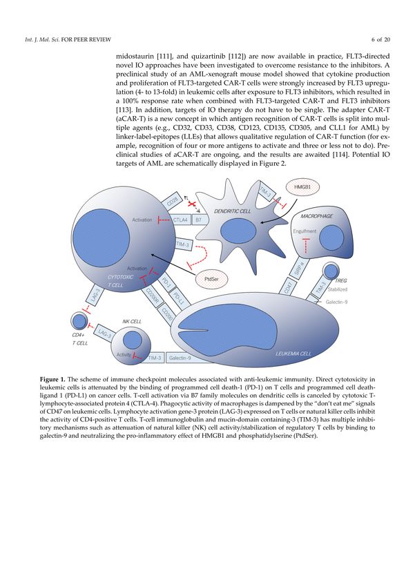
Int. J. Mol. Sci. 2021, 22, 1944                                                                                          2 of 20

                                   conjugates (ADCs) (e.g., gemtuzumab ozogamicin (GO) and brentuximab vedotin) are also
                                   used in IO therapy. Novel agents of CAR-Ts, ADCs, and bispecific antibodies are currently
                                   under development.
                                         Intensive chemotherapy (e.g., anthracyclines and cytarabine) with or without hematopoi-
                                   etic stem cell transplantation (SCT) has been the mainstay of the curative treatment of
                                   acute myeloid leukemia (AML), presenting a dismal prognosis for relapsed/refractory or
                                   intolerable cases which mostly require palliative care. For CD33-positive AML, GO showed
                                   an overall response rate of 63% in relapsed/refractory cases [8], along with survival benefit
                                   in newly diagnosed cases when combined with conventional chemotherapy [9]. Due to
                                   the paucity of favorable immune targets in leukemic cells, IO approaches for AML have
                                   been limited. However, as immune inhibitory molecules and cancer-related antigens on
                                   leukemic cells have been discovered, novel IO drugs have been recently developed in the
                                   field of AML and are expected to become another treatment option. Recent representative
                                   advantages in terms of IO therapy are summarized in this review.

                                   2. Molecules Involved in IO Therapy
                                   2.1. Immune Checkpoint Molecules
                                   2.1.1. Programmed Cell Death 1 (PD-1), Programmed Cell Death-Ligand 1 (PD-L1),
                                   and Cytotoxic T-Lymphocyte-Associated Protein 4 (CTLA-4)
                                         The inhibitory surface receptor programmed cell death 1 (PD-1) (UniProtKB-Q15116)
                                   on activated T cells is encoded by the PDCD1 gene on chromosome 2q37.3. Along with
                                   its ligands PD-L1 and PD-L2, also known as CD274 and CD273, PD-1 plays an impor-
                                   tant role in maintaining self-tolerance [10] and is often involved in immune escape in
                                   cancer by inhibiting the direct cytotoxic activity of effector CD8-positive T cells on tumor
                                   cells [11]. CTLA-4 on activated T cells, which is encoded by the CTLA4 gene on chromo-
                                   some 2q33.2, also has a crucial role in attenuating T cell activation in peripheral lymph
                                   nodes by preventing CD28 on T cells to bind its co-stimulatory counterparts B7 family
                                   ligands (CD80 and CD86) on antigen-presenting cells [12,13]. An in vivo study of murine
                                   myelogenous leukemia suggested that blockade of B7-1 (CD80) and not B7-2 (CD86) by
                                   CTLA-4 contributed to the attenuation of anti-leukemic immunity [14].
                                         An observational study at the MD Anderson Cancer Center analyzed bone marrow
                                   and peripheral blood specimens from 124 patients with myelodysplastic syndrome (MDS),
                                   chronic myelo-monocytic leukemia (CMML), and AML who received hypomethylating
                                   agents (HMAs) and reported that PD-1 and PD-L1 expression on CD34-positive cells were
                                   found in 7% and 20% of the patients, respectively [15]. In 57% of previously untreated
                                   patients, PD-L1 and PD-L2 expression on peripheral blood mononuclear cells (PBMNCs)
                                   increased more than twice during the first cycle of HMA. These patients had a shorter
                                   median survival than those who did not (4.7–6.6 vs. 11.7–12.5 months), suggesting the
                                   negative impact of PD-L1 and PD-L2 on the anti-tumor effect of HMAs. Upregulation of
                                   CTLA-4 on PBMNCs was also observed in 8% of the patients. Another study suggested
                                   that PD-L1 expression was higher in relapsed cases and associated with poor prognosis [16].
                                   Epigenetic analysis of 197 AML specimens revealed that the less methylated promoters
                                   of PD-L1 and PD-L2 gene in leukemic cells were an independent negative prognostic
                                   factor [17]. Analysis of bone marrow samples from nine refractory/relapsed AML patients
                                   showed a higher proportion (22%) of CD8-positive T cells co-expressing PD-1 and larger
                                   T-cell clonal expansion measured by T-cell receptor rearrangement compared with healthy
                                   donor samples [18]. PD-1 and OX40 on bone marrow T cells were more frequently found
                                   in relapsed AML samples than in newly diagnosed ones [19]. A report from China showed
                                   that PD-1 expression was seen in 33.8% of the peripheral CD3-positive lymphocytes in
                                   patients with previously untreated de novo AML and was correlated with the increased
                                   expression of exhaustion markers such as CD244 and CD57 [20]. However, other experi-
                                   ments suggested that PD-1 expression does not result in functional impairment of T cells,
                                   but rather correlates with a shift to memory cells [21]. Twenty-three samples from patients
                                   with AML were compared with those of 30 healthy controls. Although relatively high
Int. J. Mol. Sci. 2021, 22, 1944 3 of 20
(>30%) PD-1 expression on CD8-positive T cells was observed in 3 of 23 (13%) AML sam-
ples, the median percentages did not differ significantly compared with healthy controls
(median 15.6%). Other immune inhibitory markers, CD244, CD160, and TIM-3, were also
not significantly expressed. Instead, PD-1 was upregulated in peripheral blood specimens
of patients with AML who relapsed after either intensive chemotherapy or allogeneic
stem cell transplantation (allo-SCT) compared with those of the same patients at the time
of diagnosis.
2.1.2. T-Cell Immunoglobulin and Mucin-Domain Containing-3 (TIM-3)
The cell surface receptor T-cell immunoglobulin and mucin-domain containing-3
(TIM-3), also known as hepatitis A virus cellular receptor 2 (HAVcr-2), is encoded by
the HAVCR2 gene on chromosome 5q33.3. TIM-3 is normally expressed on T-helper
type 1 (Th1) lymphocytes, regulatory T cells (Treg), and natural killer (NK) cells. TIM-3
regulates macrophage activation [22], promotes immunological tolerance by inhibiting
Th1-mediated responses [23], attenuates T-cell receptor (TCR)-induced signaling in CD8-
positive T cells [24], and inhibits Th17 responses when expressed on Tregs [25]. The first
identified ligand for TIM-3 is galectin-9 [26], which is also a ligand for P4HB and CD44 [27].
It contributes to the stabilization/empowerment of induced Tregs [27], helps mesenchymal
stromal cells in suppressing T cells [28], and inhibits NK cell activity [29]. TIM-3 is also
able to bind phosphatidylserine (PtdSer) [30], HMGB1 [31], and CEACAM1 [32] to prevent
the activation of the immune response.
TIM-3 was found in approximately 6% of the bone marrow T cells in newly diagnosed
AML patients, and the proportion was larger (11.5–18.5%) in FLT3-ITD mutated cases [33].
Simultaneous expression of PD-1 and TIM-3 on peripheral T cells was associated with AML
relapse after allo-SCT [34]. An in vitro experiment using murine AML cells demonstrated
that co-expression of TIM-3 and PD-1 on CD8-positive T cells was enhanced during disease
progression and inhibition of either molecule alone did not attenuate tumor activity [35].
TIM-3 is also expressed on NK cells [36], while galectin-9 is found in AML blasts [37].
TIM-3/galectin-9 interaction leads to the production of IFN-gamma by NK cells, resulting
in indoleamine 2,3-dioxygenase 1 (IDO1) expression in AML cells [38]. IDO1-positive
AML cells gained the ability to negatively regulate NK cell degranulation, which can be
responsible for the immune escape of leukemic cells. Another in vitro study suggested
that AML cells protect themselves by producing soluble TIM-3 to form a TIM-3-galectin-9
complex that attenuates NK cell-mediated cytotoxicity [39].
TIM-3 inhibitors are currently under early phase evaluation as monotherapies or in
combination with PD-1/PD-L1 inhibitors for patients with advanced tumors. Preclinically,
CAR-T therapy bispecific for CD13 and TIM-3 has been efficiently screened by a nanobody-
based technology and shows eradication of leukemic cells in mouse models [40].
2.1.3. Lymphocyte Activation Gene-3 Protein (LAG-3)
The inhibitory receptor lymphocyte activation gene-3 protein (LAG-3), also known
as CD223, is encoded by the LAG-3 gene on chromosome 12p13.31. LAG-3 is normally
expressed on activated T cells, NK cells, and plasmacytoid dendritic cells (DCs) [41,42].
Notably, CD4-positive/CD25-high/Foxp3-positive/LAG-3-positive T cells were preferen-
tially expanded in patients with cancer [43]. LAG-3 structurally resembles CD4 and binds
to major histocompatibility complex (MHC) class II on CD4-positive T cells, resulting in
the downregulation of their antigen-mediated activity [44–46]. LAG-3 and PD-1 synergis-
tically prevent autoimmunity and promote immune escape in cancer [47,48]. An in vitro
experiment reported that T cells co-expressing LAG-3 and PD-1 were frequently seen in
bone marrow samples from patients with relapsed AML [49]. An in vivo study of murine
chronic lymphocytic leukemia showed that dual inhibition of PD-1 and LAG-3 successfully
decreased tumor load [50]. Anti-PD-1 antibodies with or without anti-LAG-3 antibody are
now being evaluated in early phase trials for solid and hematologic malignancies [51,52],
although these results have not been reported yet.Int. J. Mol. Sci. 2021, 22, 1944 4 of 20
2.1.4. Leukocyte Surface Antigen CD47
CD47, encoded by the CD47 gene on chromosome 3q13.12, is a transmembrane
glycoprotein and a ligand for signal-regulatory proteins (SIRPs). SIRPα is expressed on
macrophages, DCs, myeloid cells, neurons, and astrocytes. In terms of macrophages and
DCs, SIRPα-CD47 binding inhibits their phagocytic function through the cytoplasmic
domain of SIRPα called immunoreceptor tyrosine-based inhibition motifs (ITIMs) by
recruiting SH2 domain-containing protein tyrosine phosphatase (SHP)-1 and SHP-2 [53,54].
In other words, CD47-expressing cells are prevented from being engulfed, which have
been recognized as the “don’t eat me” signal. Another member of the family, SIRPβ2 is
expressed on T cells and NK cells. Unlike SIRPα, binding of SIRPβ2 on T cells to CD47 on
antigen-presenting cells results in antigen-specific T cell proliferation and T cell activation,
although its affinity for CD47 is weaker than that of SIRPα [55]. Although CD47 is broadly
expressed in a variety of normal tissues, it is upregulated in human leukemic cells as well as
circulating hematopoietic stem cells (HSCs) [56], and are also related to poor prognoses [57].
A preclinical study in which human tumor cells were co-cultured with SIRPαFc (TTI-621)
that binds to CD47 demonstrated the anti-tumor effect of CD47-blockade on various solid
and hematologic malignancies [58]. A phase Ib study of anti-CD47 monoclonal (Hu5F9-G4)
antibody in combination with rituximab for relapsed or refractory B-cell non-Hodgkin
lymphomas showed good tolerability and CR rates of 36% [59], although drug-related
anemia occurred as an adverse event [60].
2.1.5. Other Checkpoint Molecules
Similar to the CD47 receptor SIRPα, the B- and T-lymphocyte attenuator (BTLA;
also known as CD272) and CD200R (also called OX-2 receptor) have cytoplasmic ITIMs
which induce inhibitory signaling in immune effector cells [61–63]. CD200 overexpression
in AML/MDS cells was associated with higher relapse rates and poor prognoses [64].
Blockade of the BTLA and CD200R/CD200 axis has demonstrated enhanced anti-tumor
immunity in preclinical settings [65,66]. Nevertheless, no clinical trials have been conducted
to date. B7-homolog 3 (B7-H3; also known as CD276) is one of the B7 family molecules
normally expressed on activated APCs and negatively regulates T-cell activation [67].
B7-H3 has been discovered in a variety of solid and hematologic malignancies, and is
expected to be a pan-cancer target for IO therapy [68]. A preclinical experiment in which
B7-H3-targeted CAR-T cells were co-cultured with 10 samples from AML patients in vitro
and administered to human AML-transplanted mice showed significant cytotoxicity in
four (40%) patient-derived samples and prolonged survival of the xenograft mice [69].
Representative immune checkpoint molecules are summarized in Figure 1.Int. J. Mol. Sci. 2021, 22, 1944 5 of 20
Figure 1. The scheme of immune checkpoint molecules associated with anti-leukemic immunity. Direct cytotoxicity in
leukemic cells is attenuated by the binding of programmed cell death-1 (PD-1) on T cells and programmed cell death-ligand
1 (PD-L1) on cancer cells. T-cell activation via B7 family molecules on dendritic cells is canceled by cytotoxic T-lymphocyte-
associated protein 4 (CTLA-4). Phagocytic activity of macrophages is dampened by the “don’t eat me” signals of CD47
on leukemic cells. Lymphocyte activation gene-3 protein (LAG-3) expressed on T cells or natural killer cells inhibit the
activity of CD4-positive T cells. T-cell immunoglobulin and mucin-domain containing-3 (TIM-3) has multiple inhibitory
mechanisms such as attenuation of natural killer (NK) cell activity/stabilization of regulatory T cells by binding to galectin-9
and neutralizing the pro-inflammatory effect of HMGB1 and phosphatidylserine (PtdSer).
2.2. Potential Immune Targets on Leukemic Cells
2.2.1. Interleukin-3 Receptor Subunit Alpha (IL-3RA) or CD123
Interleukin-3 receptor subunit alpha (IL-3RA), also known as CD123, contains the
alpha-subunit of the receptor for IL-3 and is encoded by the IL3RA gene on the X chromo-
some. Studies have indicated that IL-3RA is overexpressed in AML cells and other hemato-
logic malignancies but is scant in normal hematopoietic stem cells [70–72]. Ehninger et al.
reported that 77.9% (232/298) of AML patients were positive for CD123, whereas almost
none of the healthy donors were [72]. The presence of CD123 on leukemic stem cells is
known to be related to the risk of treatment failure [73]. A study analyzing patient speci-
mens showed that co-expression of CD123, CD25, and CD99 in CD34-positive leukemic
cells was frequently observed in AML with FLT3-ITD mutations [74]. Yan and his col-
leagues demonstrated that the expression of CD123 and CD47 in leukemic stem cells
increased in chemo-resistant cell lines compared with chemo-naive ones [75]. Interestingly,
romidepsin, an HDAC inhibitor, re-sensitized these resistant cells in vitro. A dual-affinity
retargeting (DART) molecule targeting both CD123 and CD3, called MGD006, induced
dose-dependent killing of AML cell lines in vitro and in vivo [76]. A preclinical study using
AML-transplanted mice showed selective anti-tumor effects of CD123-directed CAR-T
on leukemic cells [77]. Similarly, engineered T cells that secrete bispecific CD123/CD3
antibody exhibited anti-leukemic effects in a xenograft mouse model [78]. Monoclonal anti-
bodies and ADCs directing CD123 have also shown efficacy in preclinical studies [79–81].Int. J. Mol. Sci. 2021, 22, 1944 6 of 20
2.2.2. Myeloid Cell Surface Antigen CD33
CD33 is a sialic-acid-binding immunoglobulin-like lectin expressed in monocytic/myeloid
lineage cells and is encoded by the CD33 gene on chromosome 19q13.41. CD33 is expressed
on myeloid cell lines from the progenitor to well-differentiated cells, including neutrophils,
monocytes, and tissue-resident macrophages [82]. CD33 shows positive immunostaining
in at least 80–90% of patients with AML, thereby indicating its presence. While CD33
expression levels largely differ between patients, its high expression can be seen in AML
with NPM1 mutation [83]. GO, a CD33-targeted ADC, has shown clinical efficacy and has
been administered to AML patients in practice (mentioned in later sections). Some stud-
ies have suggested that the expression levels of CD33 may be positively related to the
anti-leukemic effect of GO treatment [84–86]. The first-in-ever CD33/CD3-targeted BiTE,
called AMG330, prolonged the survival of human AML-transplanted immunodeficient
mice [87] and has been tested in early phase trials (mentioned in a later chapter). A novel
bifunctional checkpoint inhibitory T-cell engager (CiTE), a bispecific protein for CD3 and
CD33 conjugated with the extracellular domain of PD-1, has been recently developed and
was found to improve AML in a murine xenograft model [88].
2.2.3. C-Type Lectin-Like Molecule-1 (CLL-1)
C-type lectin domain family 12 member A (CLEC12A; UniProtKB-Q5QGZ9), also
known as c-type lectin-like molecule-1 (CLL-1), is an ITIM-containing inhibitory trans-
membrane glycoprotein expressed on more than 80% of AML blasts as well as leukemic
stem cells. The protein is encoded by the CLEC12A gene on chromosome 12p13.31. Al-
though the function of CLL-1 is not fully understood, its involvement in homeostasis in
certain inflammatory situations, such as monosodium urate-induced reaction and collagen
antibody-induced arthritis has been suggested [89,90]. A previous study suggested that
CLL-1 is selectively present on LSCs but absent on normal HSCs, indicating this as an ideal
candidate for immune targets [91]. Hutten et al. showed that CLL-1 is also expressed on
myeloid and plasmacytoid DCs, enhancing delivery of tumor antigens into DCs, resulting
in efficient antigen presentation on CD8-positive T cells, which was not attenuated by CLL-
1-targeted antibodies [92]. Two xenograft models in which patient-derived AML cells were
transplanted to cynomolgus monkeys and mice, respectively, showed that CLL-1-targeted
ADCs exhibited almost complete depletion of leukemic cells and tumor growth inhibition,
respectively [93,94]. Other preclinical data have shown significant anti-leukemic potentials
of monoclonal/bispecific antibodies and CAR-Ts targeting CLL-1 [95–104]. A trispecific
killer cell engager (TriKE) targeting CLL-1 on leukemic cells and CD16/IL15 on NK cells
increased NK cell proliferation and degranulation in leukemic cells, resulting in death in ap-
proximately 15% of the AML cells in vitro, which was comparable to that of CD33-targeted
TriKE [105].
2.2.4. Other Candidates of Immune Targets
A proto-oncogene protein c-KIT, also known as CD117, is a type III receptor tyrosine
kinase expressed in 80–90% of AML blasts and is related to adverse clinical outcome [106].
A second-generation CAR-T targeting c-Kit demonstrated elimination of more than 90%
of CD117-positive AML cells in vitro and almost complete depletion (>98%) of CD117-
positive marrow cells in xenograft mice [107]. Like c-Kit, FMS-like tyrosine kinase 3 (FLT3)
is a type III receptor tyrosine kinase. FLT3 plays an important role in maintaining the
survival of normal HSCs [108] and is also expressed in the vast majority of AML cells
along with its recurrent mutations (e.g., internal tandem duplication (ITD) and tyrosine
kinase domain mutation (TKD)) [109]. While potent FLT3 inhibitors (e.g., gilteritinib [110],
midostaurin [111], and quizartinib [112]) are now available in practice, FLT3-directed novel
IO approaches have been investigated to overcome resistance to the inhibitors. A pre-
clinical study of an AML-xenograft mouse model showed that cytokine production and
proliferation of FLT3-targeted CAR-T cells were strongly increased by FLT3 upregulation
(4- to 13-fold) in leukemic cells after exposure to FLT3 inhibitors, which resulted in a 100%Int. J. Mol. Sci. 2021, 22, 1944 7 of 20
response rate when combined with FLT3-targeted CAR-T and FLT3 inhibitors [113]. In ad-
dition, targets of IO therapy do not have to be single. The adapter CAR-T (aCAR-T) is a new
concept in which antigen recognition of CAR-T cells is split into multiple agents (e.g., CD32,
CD33, CD38, CD123, CD135, CD305, and CLL1 for AML) by linker-label-epitopes (LLEs)
that allows qualitative regulation of CAR-T function (for example, recognition of four or
more antigens to activate and three or less not to do). Preclinical studies of aCAR-T are
ongoing, and the results are awaited [114]. Potential IO targets of AML are schematically
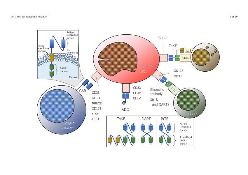
displayed in Figure 2.
Figure 2. Potential immune targets being investigated in preclinical and early-phase trials. CAR consists of an antigen-
recognizing extracellular domain and an intracellular signal domain(s). CAR-T or natural killer-targeting CD33, CLL-1,
and pan-cancer antigen NKG2D have been evaluated in phase I trials. While bispecific T-cell engager (BiTE) and trispecific
killer cell engager (TriKE) are composed of a single-chain variable fragment (scFv), dual-affinity retargeting (DART) is
composed of two cross-linked variable fragments. Bispecific antibodies (BiTEs and DARTs) targeting CD33 and CD123 have
been tested in early-phase trials. Gemtuzumab ozogamicin (GO), a CD33-directed antibody-drug conjugate, has already
been used in clinical practice.
3. Novel IO Therapy in Clinical Trials
3.1. Immune Checkpoint Inhibitors
3.1.1. Anti-PD-1/CTLA-4 Antibody
Published trials of ICIs are listed in Table 1. In a phase Ib/II study that recruited 51
patients with AML that failed prior therapy, the combination of nivolumab, an anti-PD-1
antibody, and azacitidine demonstrated 18% CR and 15% hematologic improvement [115].
The median overall survival was 9.3 months, which was favorable to historical data in
which patients were treated with azacitidine alone as a salvage therapy. The author also
reported a phase II study in which a combination of azacitidine, nivolumab, and ipili-
mumab (an anti-CTLA-4 antibody) brought about CR and CR with incomplete hematologicInt. J. Mol. Sci. 2021, 22, 1944 8 of 20
recovery (CRi) in 6 of 20 (36%) patients and 58% of 1-year survival rate with severe immune-
related adverse effects (irAEs) in 26% of the patients [116]. Farhad et al. reported that the
combination of nivolumab and conventional induction chemotherapy (e.g., idarubicin plus
cytarabine) was feasible for patients with newly diagnosed AML [117]. CR and CRi were
observed in 34 (78%) of 44 patients, including 18 cases in which their minimum residual
disease (MRD) was undetectable after completion of the induction therapy. The median
overall survival of all patients and that of patients who underwent allogeneic hematopoi-
etic stem cell transplantation (allo-HSCT) were 18.5 and 24 months, respectively. Another
anti-PD-1 inhibitor, pembrolizumab, was also evaluated in a small pilot study [118]. Ten pa-
tients with relapsed or refractory AML received pembrolizumab along with decitabine
every other cycle. CR was observed in one patient (10%) and stable disease (SD) in four
patients (40%). The median survival time was 7 months. Ipilimumab showed a response
in patients with hematologic malignancies that relapsed after allo-HSCT. In a multicenter
phase I study, 28 patients with relapsed hematologic malignancies, including 12 patients
with AML and 1 patient with MDS, were enrolled [119]. Among 5 patients (23%) who
achieved CR, four had AML and one had MDS. Another open-label phase I study that
enrolled 29 patients with solid and hematological malignancies, including two patients
with AML, who relapsed after allo-HSCT reported that tumor regression was seen in three
patients (10%) but none of them had AML [120]. The efficacy of ipilimumab was also
evaluated in MDS resistant to HMAs. In a multi-center phase I study, 29 patients received
ipilimumab after HMA failure [121]. One patient (3.4%) achieved marrow CR lasting
3 months, and 7 patients (24%) remained in SD for more than 46 weeks.Int. J. Mol. Sci. 2021, 22, 1944 9 of 20
Table 1. A summary of clinical trials of immune checkpoint inhibitors (anti-PD-1, CTLA4, and TIM-3 antibodies) for AML and/or MDS. HL: Hodgkin lymphoma, NHL: non-Hodgkin
lymphoma, MM: multiple myeloma, MPN: myeloproliferative neoplasm, ALL: acute lymphoid leukemia, CML: chronic myeloid leukemia, CLL: chronic lymphocytic leukemia, RCC: renal
cell carcinoma, allo-HSCT: allogeneic hematopoietic stem cell transplantation, HMA: hypomethylating agent, CR: complete remission, CRi: CR with incomplete hematologic recovery, HI:
hematologic improvement, PR: partial remission, SD: stable disease, OS; overall survival.
Author Object(s) Disease State Agent(s) Dosing Phase Response Rate Median Survival
3 mg/kg on Day 1, 14 (every
Daver, et al., Relapsed after Nivolumab CR/CRi 18% (6/51) 9.3 mo.
AML 4–5 weeks) Ib/II
2016 prior therapy HI 15% (5/51) [1.8–14.3]
75 mg/m2 on Day 1–7 (every
+ Azacitidine
4–5 weeks)
CR/CRi 21% (15/70)
Nivolumab
[Cohort 1] Not Reported II HI 10% (7/70) 16.1 mo.
+ Azacitidin
Daver, et al., Relapsed or Prolonged SD 9% (6/70)
AML refractory
2018 Nivolumab+
CR/CRi 36% (6/20) Not Reached (1-yr.
Azacitidin+ [Cohort 2] Not Reported II
Prolonged SD 10% (2/20) OS 58%)
Ipilimimab
12 mg/m2 on Day 1–3
Idarubicin
Ravandi, et al., AML and 1.5 g/m2 on Day 1–4 CR/CRi 78% (34/44) 18.5 mo.
Newly diagnosed + Cytarabine II
2019 high-risk MDS 3 mg/kg (every 2 weeks) *started Negative MRD 41% (18/34) [10.8–28.8]
+ Nivolumab
on Day 24
3 or 10 mg/kg (every 3 weeks for
Hematologic
4 doses
malignancies
Davids, et al., Relapsed after then every 12 weeks for upto CR 23% (5/28)
(AML, HL, NHL, Ipilimumab I/Ib Not Reported
2016 allo-HSCT 6 doses) PR 9% (2/22)
MDS, MM, MPN,
*All reseposive cases recieved
ALL)
10 mg/kg.
Malignancies
(AML, HL, NHL,
Bashey, et al., Relapsed after 0.1 to 3.0 mg/kg (every 60 days) CR 6.9% (2/29)
MM, CML, CLL, Ipilimumab I 24.7 mo.
2009 allo-HSCT *Dose-escalating model. PR 3.4% (1/29)
Breast cancer,
RCC)Int. J. Mol. Sci. 2021, 22, 1944 10 of 20
Table 1. Cont.
Author Object(s) Disease State Agent(s) Dosing Phase Response Rate Median Survival
3 or 10 mg/kg (every 3 weeks for
Zeidan, et al., Refractory to 4 doses Marrow CR 3.4% (1/29)
MDS Ipilimumab I Not Reported
2018 HMAs then every 12 weeks for upto Prolonged SD 24% (7/29)
8 doses)
Lindblad, 200 mg/body (every 3 weeks)
Relapsed or Pembrolizumab CR 10% (1/10) 7 mo.
et al., AML 20 mg/m2 on Day 8–12, 15–19 I/II
refractory + Decitabine SD 40% (4/10) [2–14]
2018 (every 6 weeks)
Escalating dose from 240 to
MBG453 800 mg/body CR/CRi 23% (7/31) Exposure
Borate, et al., AML and Ineligible to
(anti-TIM-3) (every 2 weeks or 4 weeks) Ib PR 6% (2/31)2 durations
2019 high-risk MDS standard therapy
+ Decitabine 20 mg/m2 on Day 1–5 (every Blasts halved 6% (8/31) 2.1–17.9 months
4 weeks)Int. J. Mol. Sci. 2021, 22, 1944 11 of 20
3.1.2. Anti-TIM-3 Antibody
TIM-3 blockade can be another strategy for disarming the immune-escaping mech-
anisms of tumor cells. A phase Ib study testing anti-TIM-3 antibody (MBG453) in com-
bination with decitabine for patients with high-risk MDS and AML reported that 9 of 31
(29%) patients with AML achieved partial response or better response and eight (25.8%)
additional patients showed more than a 50% reduction in marrow blasts [122]. Severe
(Grade 3–4) irAE, liver toxicity, and arthritis occurred in 7% of the patients; this result was
comparable to those of other ICI monotherapy.
3.2. CAR-T Therapy and Its Relatives
CAR-T cells are engineered peripheral T cells, often of autologous origin, which have
an extracellular antigen-recognition domain, commonly single-chain variable fragments
derived from monoclonal antibodies, conjugated with intracellular signal domains (as
shown in Figure 2). The first-generation CAR has only one signal domain (CD3 zeta chain)
and the other descendants have additional co-stimulatory structures (CD28 or 4-1BB for
2nd generation and CD28 plus 4-1BB or OX40 for 3rd generation). A variety of CAR-Ts are
now under development. While CD19-directed CAR-T cell therapy has been successful
for B-ALL, the application of the concept for AML has been delayed due to the lack of
suitable targetable surface antigens until recently. Ritchie et al. reported a phase I study
of the first-in-human and proof-of-concept CAR-T therapy targeting Lewis-Y antigen in
patients with relapsed/refractory AML (RR-AML) [123]. Although the clinical outcome
was modest, if transient, its feasible transduction efficiency (14–38%) and persistence of
CAT-T cells within the body (up to 10 months) as well as its acceptable safety profile
were displayed. Among a number of surface antigens, CD33, CD123, and CLL-1 have
been eagerly investigated for CAR-T therapy. Wang et al. conducted a clinical trial in
which a patient with refractory AML received CD33-directed autologous CAR-T therapy
followed by marked reduction of marrow blasts lasting 9 weeks [124]. An early-phase
result of CD33-targeted CAR-NK therapy (autologous NK cells with CAR) in patients
with RR-AML was reported by Tang and his colleagues [125]. One of three patients who
received CAR-NK therapy showed increased levels of serum interleukin (IL)-6 and IL-10
on day 6, followed by a decrease in the MRD level and WT1 copy-numbers, although the
clinical benefit was insufficient. Co-administration of engineered DCs with CD33-directed
CAR-T cells may enhance anti-leukemic activity through the production of DC-derived
IL-12 [126]. A novel compound CAR-T targeting both CD33 and CLL-1 was tested in
a phase I study in which a 6 year-old female with Fanconi’s anemia-associated juvenile
myelomonocytic leukemia carrying FLT3-ITD mutation that had been heavily treated
with multiple therapies, including a FLT3 inhibitor [127]. After CAR-T infusion following
lymphodepletion therapy, she achieved CR with negative MRD on day 19, which allowed
her to undergo allo-SCT. Sallman et al. reported a phase I study of CAR-T therapy targeting
NKG2D, which is expressed on a variety of solid and hematologic tumors, in which three
of seven (42%) patients with RR-AML achieved CR [128]. Although only a small number
of patients with AML have been involved in clinical trials of CAR-T therapy, the results
are promising and further investigations are expected. A summary of early phase trials is
shown in Table 2.Int. J. Mol. Sci. 2021, 22, 1944 12 of 20
Table 2. A summary of early phase trials of CAR-T and CAR-NK therapy. JMML: juvenile myelomonocytic leukemia,
Rel./ref.: relapsed or refractory, CR: complete remission, CRh: CR with partial hematologic recovery, CRi: CR with
incomplete hematologic recovery, MRD: minimal residual disease.
Authors Objects Disease State Agents Target(s) Phase Clinical Outcome
Transient decrease of
Rithchie, et al., Lewis-Y blasts in 1 of 4 patients
AML Rel./ref. CAR-T (2nd gen.) I
2013 antigen 14–38% of
Transduction efficiency
Marked reduction of
Wang, et al.,
AML Rel./ref. CAR-T (2nd gen.) CD33 I marrow blasts
2015
for 9 weeks in 1 patient
Solid tumors
Sallman, et al., and 1 CRh and 2 CRi of 7
Rel./ref. CAR-T NKG2D I
2018 hematologic patients with AML
malignancies
Liu, et al., CLL-1 and CR with negative MRD
JMML Rel./ref. Compound CAR-T I
2018 CD33 in 1 patient
Tang, et al., Decrease of MRD and
AML Rel./ref. CAR-NK CD33 I
2018 WT-1 in 1 of 3 patients
3.3. Bispecific and Trispecific Antibodies: BiTE and DART
Bispecific antibodies are artificially synthetized small molecules consisting of two
different antigen-recognition domains derived from variable regions of monoclonal an-
tibody. Both BiTEs and DARTs recognize CD3 and engage effector T cells with tumor
cells. CD33 and CD123 are popular as leukemia-specific targets in recent investigations.
BiTEs are composed of a single-chain variable fragment (Fv), and DARTs are made by
cross-linking two Fvs [129]. A CD33-targeted BiTE AMG330 was tested in a phase I study
of patients with RR-AML [130]. In total, four patients out of 35 (11.4%) participants, includ-
ing those who received a low dose of AMG330, achieved CR/CRi, and treatment-related
severe adverse effects including up to Grade 2 cytokine-releasing syndrome (CRS) were
seen in 15 (42.9%) patients. Another CD33-targeted BiTE AMV564 showed reduction of
marrow blasts in 12 of 18 (66.7%) patients with RR-AML and up to Grade 2 CRS in one
(5.6%) patient [131]. Currently, two agents of CD123-targeted antibodies, XmAb14045
(also known as SQZ622) and flotetuzumab, have been evaluated in early phase studies
of patients with RR-AML. A CD33-directed BiTE XmAb14045 achieved CR/CRi in three
of 13 (23.1%) participants who received sufficient doses of the agent and up to Grade 3
CRS in 49 of 63 (77.8%) patients who received any dose in the initial stage of the phase I
study [132]. Flotetuzumab is a CD123-/CD3-bispecific DART that has undergone a phase
I/II study [133]. Of twenty-seven evaluable patients who received its recommended dose,
five (18.5%) patients achieved CR/CRi and four (13.3%) patients suffered from Grade 3
or more severe CRS. The agent is also being evaluated in combination with an anti-PD-1
antibody, MGA012, in expectation of more potent clinical benefits [134]. A summary of
early phase trials is shown in Table 3.Int. J. Mol. Sci. 2021, 22, 1944 13 of 20
Table 3. A summary of clinical trials of bispecific antibodies for relapsed/refractory AML (RR-AML). BiTE: bispecific T-cell
engager, DART: dual-affinity retargeting molecule, ICI: immune checkpoint inhibitor, CR: complete remission, CRi: CR with
incomplete hematologic recovery, CRS: cytokine releasing syndrome.
Author Object Agent(s) Class Targets Phase Clinical Outcome
Ravandi, et al., CD33 and CR/CRi 11.4% (4 of 35)
RR-AML AMG330 BiTE I
2018 CD3 CRS 42.1% (15 of 35)
Eissenberg, et al., CD33 and CR/CRi 66.7% (12 of 18)
RR-AML AMV564 BiTE I
2018 CD3 CRS 5.7% (1 of 18)
Ravandi, et al., CD123 and CR/CRi 23.1% (3 of 13)
RR-AML XmAb14045 BiTE I
2018 CD3 CRS 77.9% (49 of 63)
Uy, et al., CD123 and CR/CRi 18.5% (5 of 27)
RR-AML Flotetuzumab DART I/II
2018 CD3 Severe CRS 13.4% (4 of 30)
CD123 and
Wei, et al., Flotetuzumab
RR-AML DART+ ICI CD3 I Not reported
2019 +MGA012
plus PD-1
3.4. Antibody-Drug Conjugate
CD33 has been recognized for decades as the most popular immune target of AML.
An anti-CD33 ADC, GO, showed CR rates of 63% with 2-year overall survival of 41%
in patients with relapsed/refractory CD33-positive AML [8]. A meta-analysis of five
randomized trials in which GO was combined with induction chemotherapy in patients
with newly diagnosed AML concluded that this combination significantly reduced re-
lapse within 5 years (hazard ratio (HR) 0.81 (0.73–0.90)) and slightly prolonged overall
survival (HR 0.90 (0.82–0.98)) without improving response rates (HR 0.91 (0.77–1.07)) [9].
GO monotherapy also showed moderate survival benefit (median survival 4.9 months
vs. 3.6 months) without increasing the severe adverse effects compared with best sup-
portive care in the elderly with newly diagnosed AML who were unsuitable for intensive
chemotherapy [135]. As mentioned in the earlier sections, novel ADCs targeting CD123
and CLL-1 (e.g., IMGN632 [81] and SL-101 [80] for CD123 and CLT030 [94] for CLL-1)
have been shown to have anti-leukemic activity. In the future, they should be evaluated in
clinical trials.
4. Conclusions
As knowledge of immune inhibitory molecules and leukemic antigens has accumu-
lated, clinical use of IO therapy for AML has come closer to reality. While conventional
chemotherapy, with or without SCT, is still pivotal in curative treatment and/or disease
control for AML because of its high proliferation intensity, molecular targeted approaches
(e.g., GO and FLT3 inhibitors) remain a practical alternative, especially for patients with
relapsed/refractory or intolerable AML to intensive treatment. Clinical experience of ICIs
in solid tumors has shown that IO therapy brings long-term disease control in at least
10–20% of the patients with metastatic cancers (more favorable in malignant melanoma,
renal cell cancer, and non-small cell lung cancer with high PD-L1 expression, and MSI-high
tumors) even after discontinuation of chemotherapy. Furthermore, concomitant use of
chemotherapy and/or radiation could enhance the efficacy of IO therapy [136,137], al-
though it has not yet been proven in hematologic malignancies. Long-term follow up of
CD19-CAR-T therapy (tisa-cel) for relapsed/refractory B-ALL showed more than 5 years
of estimated progression-free survival in approximately 40% of patients with low disease
burden [6]. Selective eradication of cancer stem cells and/or sustained establishment of
anti-tumor immunity might be, at least theoretically, able to bring about a true cure for
patients with AML who are relapsed/refractory or ineligible for curative treatment. Further
advances and clinical applications of IO therapy for AML are awaited.Int. J. Mol. Sci. 2021, 22, 1944 14 of 20
Author Contributions: R.T. and S.C. wrote the first draft and all the authors revised the manuscript.
All authors have read and agreed to the published version of the manuscript.
Funding: This paper was supported by the National Cancer Research and Development expenses
grant.
Institutional Review Board Statement: Not applicable.
Informed Consent Statement: Not applicabl.
Data Availability Statement: Not applicable.
Conflicts of Interest: Y.M. received research funding from Ono and CMIC, and honoraria from
Bristol-Myers Squibb, Novartis, Astellas, and Daiichi-Sankyo. The other authors declare no conflict
of interest.
References
1. Specenier, P. Ipilimumab in melanoma. Expert Rev. Anticancer Ther. 2016, 16, 811–826. [CrossRef]
2. Kantarjian, H.M.; Stein, A.; Gökbuget, N.; Fielding, A.K.; Schuh, A.C.; Ribera, J.-M.; Wei, A.; Dombret, H.; Foà, R.; Bassan, R.; et al.
Blinatumomab versus Chemotherapy for Advanced Acute Lymphoblastic Leukemia. N. Engl. J. Med. 2017, 376, 836–847.
[CrossRef] [PubMed]
3. Goebeler, M.-E.; Knop, S.; Viardot, A.; Kufer, P.; Topp, M.S.; Einsele, H.; Noppeney, R.; Hess, G.; Kallert, S.; Mackensen, A.; et al.
Bispecific T-Cell Engager (BiTE) Antibody Construct Blinatumomab for the Treatment of Patients With Relapsed/Refractory
Non-Hodgkin Lymphoma: Final Results From a Phase I Study. J. Clin. Oncol. 2016, 34, 1104–1111. [CrossRef]
4. Viardot, A.; Goebeler, M.-E.; Hess, G.; Neumann, S.; Pfreundschuh, M.; Adrian, N.; Zettl, F.; Libicher, M.; Sayehli, C.;
Stieglmaier, J.; et al. Phase 2 study of the bispecific T-cell engager (BiTE) antibody blinatumomab in relapsed/refractory diffuse
large B-cell lymphoma. Blood 2016, 127, 1410–1416. [CrossRef] [PubMed]
5. Schuster, S.J.; Bishop, M.R.; Tam, C.S.; Waller, E.K.; Borchmann, P.; McGuirk, J.P.; Jäger, U.; Jaglowski, S.; Andreadis, C.;
Westin, J.R.; et al. Tisagenlecleucel in Adult Relapsed or Refractory Diffuse Large B-Cell Lymphoma. N. Engl. J. Med. 2019,
380, 45–56. [CrossRef] [PubMed]
6. Park, J.H.; Rivière, I.; Gonen, M.; Wang, X.; Sénéchal, B.; Curran, K.J.; Sauter, C.; Wang, Y.; Santomasso, B.; Mead, E.; et al.
Long-Term Follow-up of CD19 CAR Therapy in Acute Lymphoblastic Leukemia. N. Engl. J. Med. 2018, 378, 449–459. [CrossRef]
[PubMed]
7. Neelapu, S.S.; Locke, F.L.; Bartlett, N.L.; Lekakis, L.J.; Miklos, D.B.; Jacobson, C.A.; Braunschweig, I.; Oluwole, O.O.; Siddiqi, T.;
Lin, Y.; et al. Axicabtagene Ciloleucel CAR T-Cell Therapy in Refractory Large B-Cell Lymphoma. N. Engl. J. Med. 2017,
377, 2531–2544. [CrossRef] [PubMed]
8. Chevallier, P.; Delaunay, J.; Turlure, P.; Pigneux, A.; Hunault, M.; Garand, R.; Guillaume, T.; Avet-Loiseau, H.; Dmytruk, N.;
Girault, S.; et al. Long-Term Disease-Free Survival After Gemtuzumab, Intermediate-Dose Cytarabine, and Mitoxantrone in
Patients With CD33+Primary Resistant or Relapsed Acute Myeloid Leukemia. J. Clin. Oncol. 2008, 26, 5192–5197. [CrossRef]
9. Hills, R.K.; Castaigne, S.; Appelbaum, F.R.; Delaunay, J.; Petersdorf, S.; Othus, M.; Estey, E.H.; Dombret, H.; Chevret, S.;
Ifrah, N.; et al. Addition of gemtuzumab ozogamicin to induction chemotherapy in adult patients with acute myeloid leukaemia:
A meta-analysis of individual patient data from randomised controlled trials. Lancet Oncol. 2014, 15, 986–996. [CrossRef]
10. Fife, B.T.; Pauken, K.E. The role of the PD-1 pathway in autoimmunity and peripheral tolerance. Ann. N. Y. Acad. Sci. 2011,
1217, 45–59. [CrossRef]
11. Berger, K.N.; Pu, J.J. PD-1 pathway and its clinical application: A 20 year journey after discovery of the complete human PD-1
gene. Gene 2018, 638, 20–25. [CrossRef]
12. Linsley, P.S.; Brady, W.; Urnes, M.; Grosmaire, L.S.; Damle, N.K.; A Ledbetter, J. CTLA-4 is a second receptor for the B cell
activation antigen B7. J. Exp. Med. 1991, 174, 561–569. [CrossRef]
13. Teft, W.A.; Kirchhof, M.G.; Madrenas, J. A MOLECULAR PERSPECTIVE OF CTLA-4 FUNCTION. Annu. Rev. Immunol. 2006,
24, 65–97. [CrossRef]
14. LaBelle, J.L.; Hanke, C.A.; Blazar, B.R.; Truitt, R.L. Negative effect of CTLA-4 on induction of T-cell immunity in vivo to B7-1+,
but not B7-2+, murine myelogenous leukemia. Blood J. Am. Soc. Hematol. 2002, 99, 2146–2153. [CrossRef] [PubMed]
15. Yang, H.; Bueso-Ramos, C.; Dinardo, C.D.; Estecio, M.R.; Davanlou, M.; Geng, Q.-R.; Fang, Z.; Nguyen, M.; Pierce, S.; Wei, Y.; et al.
Expression of PD-L1, PD-L2, PD-1 and CTLA4 in myelodysplastic syndromes is enhanced by treatment with hypomethylating
agents. Leukemia 2014, 28, 1280–1288. [CrossRef] [PubMed]
16. Chen, X.; Liu, S.; Wang, L.; Zhang, W.-G.; Ji, Y.; Ma, X. Clinical significance of B7-H1 (PD-L1) expression in human acute leukemia.
Cancer Biol. Ther. 2008, 7, 622–627. [CrossRef] [PubMed]
17. Goltz, D.; Gevensleben, H.; Grünen, S.; Dietrich, J.; Kristiansen, G.; Landsberg, J. PD-L1 (CD274) promoter methylation predicts
survival in patients with acute myeloid leukemia. Leukemia 2017, 31, 738–743. [CrossRef] [PubMed]Int. J. Mol. Sci. 2021, 22, 1944 15 of 20
18. Goswami, M.; Oetjen, K.; Mulé, B.M.P.; Sheela, M.S.; Wong, H.Y.; Liu, Q.; Calvo, K.R.; Lai, C.E.; Hourigan, C.S. Increased
Frequencies of PD-1+ CD8+ Marrow-Infiltrating Lymphocytes Associated with Highly Clonal T-Lymphocyte Expansions in
Relapsed and Refractory AML Patients but Not Healthy Adults. Blood 2016, 128, 1644. [CrossRef]
19. Daver, N.; Basu, S.; Garcia-Manero, G.; Cortes, J.E.; Ravandi, F.; Ning, J.; Xiao, L.; Juliana, L.; Kornblau, S.M.; Konopleva, M.; et al.
Defining the Immune Checkpoint Landscape in Patients (pts) with Acute Myeloid Leukemia (AML). Blood 2016, 128, 2900.
[CrossRef]
20. Tan, J.; Chen, S.; Lu, Y.; Yao, D.; Xu, L.; Zhang, Y.; Yang, L.; Chen, J.; Lai, J.; Yu, Z.; et al. Higher PD-1 expression concurrent with
exhausted CD8+ T cells in patients with de novo acute myeloid leukemia. Chin. J. Cancer Res. 2017, 29, 463–470. [CrossRef]
21. Schnorfeil, F.M.; Lichtenegger, F.S.; Emmerig, K.; Schlueter, M.; Neitz, J.S.; Draenert, R.; Hiddemann, W.; Subklewe, M. T cells are
functionally not impaired in AML: Increased PD-1 expression is only seen at time of relapse and correlates with a shift towards
the memory T cell compartment. J. Hematol. Oncol. 2015, 8, 93. [CrossRef] [PubMed]
22. Monney, L.; Sabatos, C.A.; Gaglia, J.L.; Ryu, A.; Waldner, H.; Chernova, T.; Manning, S.; Greenfield, E.A.; Coyle, A.J.;
Sobel, R.A.; et al. Th1-specific cell surface protein Tim-3 regulates macrophage activation and severity of an autoimmune
disease. Nat. Cell Biol. 2002, 415, 536–541. [CrossRef]
23. Sánchez-Fueyo, A.; Tian, J.; Picarella, D.; Domenig, C.; Zheng, X.X.; Sabatos, C.A.; Manlongat, N.; Bender, O.; Kamradt, T.;
Kuchroo, V.K.; et al. Tim-3 inhibits T helper type 1-mediated auto- and alloimmune responses and promotes immunological
tolerance. Nat Immunol. 2003, 4, 1093–1101. [CrossRef] [PubMed]
24. Tomkowicz, B.; Walsh, E.R.; Cotty, A.; Verona, R.; Sabins, N.; Kaplan, F.; Santulli-Marotto, S.; Chin, C.-N.; Mooney, J.L.; Lingham,
R.B.; et al. TIM-3 Suppresses Anti-CD3/CD28-Induced TCR Activation and IL-2 Expression through the NFAT Signaling Pathway.
PLoS ONE 2015, 10, e0140694. [CrossRef]
25. Gautron, A.-S.; Dominguez-Villar, M.; De Marcken, M.; Hafler, D.A. Enhanced suppressor function of TIM-3+FoxP3+regulatory T
cells. Eur. J. Immunol. 2014, 44, 2703–2711. [CrossRef]
26. Wang, F.; He, W.; Zhou, H.; Yuan, J.; Wu, K.; Xu, L.; Chen, Z.K. The Tim-3 ligand galectin-9 negatively regulates CD8+ alloreactive
T cell and prolongs survival of skin graft. Cell. Immunol. 2007, 250, 68–74. [CrossRef]
27. Bi, S.; Hong, P.W.; Lee, B.; Baum, L.G. Galectin-9 binding to cell surface protein disulfide isomerase regulates the redox
environment to enhance T-cell migration and HIV entry. Proc. Natl. Acad. Sci. USA 2011, 108, 10650–10655. [CrossRef] [PubMed]
28. Gieseke, F.; Kruchen, A.; Tzaribachev, N.; Bentzien, F.; Dominici, M.; Müller, I. Proinflammatory stimuli induce galectin-9 in
human mesenchymal stromal cells to suppress T-cell proliferation. Eur. J. Immunol. 2013, 43, 2741–2749. [CrossRef]
29. Golden-Mason, L.; Mcmahan, R.H.; Strong, M.; Reisdorph, R.; Mahaffey, S.; Palmer, B.E.; Cheng, L.; Kulesza, C.; Hirashima, M.;
Niki, T.; et al. Galectin-9 Functionally Impairs Natural Killer Cells in Humans and Mice. J. Virol. 2013, 87, 4835–4845. [CrossRef]
30. DeKruyff, R.H.; Bu, X.; Ballesteros, A.; Santiago, C.; Chim, Y.-L.E.; Lee, H.-H.; Karisola, P.; Pichavant, M.; Kaplan, G.G.; Umetsu,
D.T.; et al. T Cell/Transmembrane, Ig, and Mucin-3 Allelic Variants Differentially Recognize Phosphatidylserine and Mediate
Phagocytosis of Apoptotic Cells. J. Immunol. 2010, 184, 1918–1930. [CrossRef]
31. Chiba, S.; Baghdadi, M.; Akiba, H.; Yoshiyama, H.; Kinoshita, I.; Dosaka-Akita, H.; Fujioka, Y.; Ohba, Y.; Gorman, J.V.;
Colgan, J.D.; et al. Tumor-infiltrating DCs suppress nucleic acid–mediated innate immune responses through interactions
between the receptor TIM-3 and the alarmin HMGB1. Nat. Immunol. 2012, 13, 832–842. [CrossRef]
32. Huang, Y.-H.; Zhu, C.; Kondo, Y.; Anderson, A.C.; Gandhi, A.; Russell, A.F.; Dougan, S.K.; Petersen, B.-S.; Melum, E.;
Pertel, T.; et al. CEACAM1 regulates TIM-3-mediated tolerance and exhaustion. Nat. Cell Biol. 2015, 517, 386–390. [Cross-
Ref] [PubMed]
33. Li, C.; Chen, X.; Yu, X.; Zhu, Y.; Ma, C.; Xia, R.; Ma, J.; Gu, C.; Ye, L.; Wu, D. Tim-3 is highly expressed in T cells in acute myeloid
leukemia and associated with clinicopathological prognostic stratification. Int. J. Clin. Exp. Pathol. 2014, 7, 6880–6888.
34. Kong, Y.; Zhang, J.; Claxton, D.F.; Ehmann, W.C.; Rybka, W.B.; Zhu, L.; Zeng, H.; Schell, T.D.; Zheng, H. PD-1(hi)TIM-3(+) T
cells associate with and predict leukemia relapse in AML patients post allogeneic stem cell transplantation. Blood Cancer J. 2015,
5, e330. [CrossRef]
35. Zhou, Q.; Munger, M.E.; Veenstra, R.G.; Weigel, B.J.; Hirashima, M.; Munn, D.H.; Murphy, W.J.; Azuma, M.; Anderson, A.C.;
Kuchroo, V.K.; et al. Coexpression of Tim-3 and PD-1 identifies a CD8+ T-cell exhaustion phenotype in mice with disseminated
acute myelogenous leukemia. Blood J. Am. Soc. Hematol. 2011, 117, 10.
36. Han, G.; Chen, G.; Shen, B.; Li, Y. Tim-3: An Activation Marker and Activation Limiter of Innate Immune Cells. Front. Immunol.
2013, 4. [CrossRef] [PubMed]
37. Gleason, M.K.; Lenvik, T.R.; McCullar, V.; Felices, M.; O’Brien, M.S.; Cooley, S.A.; Verneris, M.R.; Cichocki, F.; Holman, C.J.;
Panoskaltsis-Mortari, A.; et al. Tim-3 is an inducible human natural killer cell receptor that enhances interferon gamma production
in response to galectin-9. Blood 2012, 119, 3064–3072. [CrossRef]
38. Folgiero, V.; Cifaldi, L.; Pira, G.L.; Goffredo, B.M.; Vinti, L.; Locatelli, F. TIM-3/Gal-9 interaction induces IFNγ-dependent IDO1
expression in acute myeloid leukemia blast cells. J. Hematol. Oncol. 2015, 8, 1–5. [CrossRef] [PubMed]
39. Silva, I.G.; Yasinska, I.M.; Sakhnevych, S.S.; Fiedler, W.; Wellbrock, J.; Bardelli, M.; Varani, L.; Hussain, R.; Siligardi, G.;
Ceccone, G.; et al. The Tim-3-galectin-9 Secretory Pathway is Involved in the Immune Escape of Human Acute Myeloid Leukemia
Cells. EBioMedicine 2017, 22, 44–57. [CrossRef]
40. He, X.; Feng, Z.; Ma, J.; Ling, S.; Cao, Y.; Gurung, B.; Wu, Y.; Katona, B.W.; O’Dwyer, K.P.; Siegel, D.L.; et al. Bispecific and split
CAR T cells targeting CD13 and TIM3 eradicate acute myeloid leukemia. Blood 2020, 135, 713–723. [CrossRef]Int. J. Mol. Sci. 2021, 22, 1944 16 of 20
41. Baixeras, E.; Huard, B.; Miossec, C.; Jitsukawa, S.; Martin, M.; Hercend, T.; Auffray, C.; Triebel, F.; Piatier-Tonneau, D. Charac-
terization of the lymphocyte activation gene 3-encoded protein. A new ligand for human leukocyte antigen class II antigens.
J. Exp. Med. 1992, 176, 327–337. [CrossRef] [PubMed]
42. Workman, C.J.; Wang, Y.; El Kasmi, K.C.; Pardoll, D.M.; Murray, P.J.; Drake, C.G.; Vignali, D.A.A. LAG-3 Regulates Plasmacytoid
Dendritic Cell Homeostasis. J. Immunol. 2009, 182, 1885–1891. [CrossRef] [PubMed]
43. Camisaschi, C.; Casati, C.; Rini, F.; Perego, M.; De Filippo, A.; Triebel, F.; Parmiani, G.; Belli, F.; Rivoltini, L.; Castelli, C. LAG-3
Expression Defines a Subset of CD4+CD25highFoxp3+ Regulatory T Cells That Are Expanded at Tumor Sites. J. Immunol. 2010,
184, 6545–6551. [CrossRef] [PubMed]
44. Triebel, F.; Jitsukawa, S.; Baixeras, E.; Roman-Roman, S.; Genevee, C.; Viegas-Pequignot, E.; Hercend, T. LAG-3, a novel
lymphocyte activation gene closely related to CD4. J. Exp. Med. 1990, 171, 1393–1405. [CrossRef] [PubMed]
45. Huard, B.; Tournier, M.; Hercend, T.; Triebel, F.; Faure, F. Lymphocyte-activation gene 3/major histocompatibility complex class II
interaction modulates the antigenic response of CD4+ T lymphocytes. Eur. J. Immunol. 1994, 24, 3216–3221. [CrossRef] [PubMed]
46. Huard, B.; Prigent, P.; Pagès, F.; Bruniquel, D.; Triebel, F. T cell major histocompatibility complex class II molecules down-regulate
CD4+ T cell clone responses following LAG-3 binding. Eur. J. Immunol. 1996, 26, 1180–1186. [CrossRef]
47. Woo, S.-R.; Turnis, M.E.; Goldberg, M.V.; Bankoti, J.; Selby, M.; Nirschl, C.J.; Bettini, M.L.; Gravano, D.M.; Vogel, P.; Liu, C.L.; et al.
Immune Inhibitory Molecules LAG-3 and PD-1 Synergistically Regulate T-cell Function to Promote Tumoral Immune Escape.
Cancer Res. 2012, 72, 917–927. [CrossRef]
48. Okazaki, T.; Okazaki, I.-M.; Wang, J.; Sugiura, D.; Nakaki, F.; Yoshida, T.; Kato, Y.; Fagarasan, S.; Muramatsu, M.; Eto, T.; et al.
PD-1 and LAG-3 inhibitory co-receptors act synergistically to prevent autoimmunity in mice. J. Exp. Med. 2011, 208, 395–407.
[CrossRef]
49. Williams, P.; Basu, S.; Garcia-Manero, G.; Hourigan, C.S.; Oetjen, K.A.; Cortes, J.E.; Ravandi, F.; Jabbour, E.J.; Al-Hamal, Z.;
Konopleva, M.; et al. The distribution of T-cell subsets and the expression of immune checkpoint receptors and ligands in patients
with newly diagnosed and relapsed acute myeloid leukemia. Cancer 2019, 125, 1470–1481. [CrossRef]
50. Wierz, M.; Pierson, S.; Guyonnet, L.; Viry, E.; Lequeux, A.; Oudin, A.; Niclou, S.P.; Ollert, M.; Berchem, G.; Janji, B.; et al.
Dual PD1/LAG3 immune checkpoint blockade limits tumor development in a murine model of chronic lymphocytic leukemia.
Blood 2018, 131, 1617–1621. [CrossRef]
51. Bristol-Myers Squibb. A Phase 1/2a Dose Escalation and Cohort Expansion Study of the Safety, Tolerability, and Efficacy of
Anti-LAG-3 (BMS-986016) in Monoclonal Antibody (BMS-986016) Administered Alone and in Combination With Anti-PD-1
Monoclonal Antibody (Nivolumab, BMS-936558) in Relapsed or Refractory B-Cell Malignancies. clinicaltrials.gov. 2019. Available
online: https://clinicaltrials.gov/ct2/show/NCT02061761 (accessed on 21 June 2020).
52. Bristol-Myers Squibb. A Phase I/2a Dose Escalation and Cohort Expansion Study of the Safety, Tolerability, and Efficacy of
Anti-LAG-3 Monoclonal Antibody (BMS-986016) Administered Alone and in Combination With Anti-PD-1 Monoclonal Antibody
(Nivolumab, BMS-936558) in Advanced Solid Tumors. clinicaltrials.gov. 2020. Available online: https://clinicaltrials.gov/ct2
/show/NCT01968109 (accessed on 21 June 2020).
53. Latour, S.; Tanaka, H.; Demeure, C.; Mateo, V.; Rubio, M.; Brown, E.J.; Maliszewski, C.; Lindberg, F.P.; Oldenborg, A.;
Ullrich, A.; et al. Bidirectional Negative Regulation of Human T and Dendritic Cells by CD47 and Its Cognate Receptor Signal-
Regulator Protein-α: Down-Regulation of IL-12 Responsiveness and Inhibition of Dendritic Cell Activation. J. Immunol. 2001,
167, 2547–2554. [CrossRef]
54. Oldenborg, P.-A.; Gresham, H.D.; Lindberg, F.P. Cd47-Signal Regulatory Protein α (Sirpα) Regulates Fcγ and Complement
Receptor–Mediated Phagocytosis. J. Exp. Med. 2001, 193, 855–862. [CrossRef] [PubMed]
55. Piccio, L.; Vermi, W.; Boles, K.S.; Fuchs, A.; Strader, C.A.; Facchetti, F.; Cella, M.; Colonna, M. Adhesion of human T cells to
antigen-presenting cells through SIRPbeta2-CD47 interaction costimulates T-cell proliferation. Blood 2005, 105, 7. [CrossRef]
[PubMed]
56. Jaiswal, S.; Jamieson, C.H.; Pang, W.W.; Park, C.Y.; Chao, M.P.; Majeti, R.; Traver, D.; Van Rooijen, N.; Weissman, I.L. CD47
Is Upregulated on Circulating Hematopoietic Stem Cells and Leukemia Cells to Avoid Phagocytosis. Cell 2009, 138, 271–285.
[CrossRef]
57. Majeti, R.; Chao, M.P.; Alizadeh, A.A.; Pang, W.W.; Jaiswal, S.; Gibbs, K.D.; Van Rooijen, N.; Weissman, I.L. CD47 Is an Adverse
Prognostic Factor and Therapeutic Antibody Target on Human Acute Myeloid Leukemia Stem Cells. Cell 2009, 138, 286–299.
[CrossRef]
58. Petrova, P.S.; Viller, N.N.; Wong, M.; Pang, X.; Lin, G.H.Y.; Dodge, K.; Chai, V.; Chen, H.; Lee, V.; House, V.; et al. TTI-621
(SIRPαFc): A CD47-Blocking Innate Immune Checkpoint Inhibitor with Broad Antitumor Activity and Minimal Erythrocyte
Binding. Clin. Cancer Res. 2017, 23, 1068–1079. [CrossRef] [PubMed]
59. Advani, R.; Flinn, I.; Popplewell, L.; Forero, A.; Bartlett, N.L.; Ghosh, N.; Kline, J.; Roschewski, M.; LaCasce, A.; Collins, G.P.; et al.
CD47 Blockade by Hu5F9-G4 and Rituximab in Non-Hodgkin’s Lymphoma. N. Engl. J. Med. 2018, 379, 1711–1721. [CrossRef]
60. Brierley, C.; Staves, J.; Roberts, C.; Johnson, H.; Vyas, P.; Goodnough, L.; Murphy, M. The effects of monoclonal anti-CD47 on
RBCs, compatibility testing, and transfusion requirements in refractory acute myeloid leukemia. Transfusion 2019, 59, 2248–2254.
[CrossRef]
61. Hobo, W.; Hutten, T.J.A.; Schaap, N.P.M.; Dolstra, H. Immune checkpoint molecules in acute myeloid leukaemia: Managing the
double-edged sword. Br. J. Haematol. 2018, 181, 38–53. [CrossRef]You can also read