Predicting single-cell gene expression profiles of imaging flow cytometry data with machine learning
←
→
Page content transcription
If your browser does not render page correctly, please read the page content below
Published online 29 October 2020                                              Nucleic Acids Research, 2020, Vol. 48, No. 20 11335–11346
                                                                                                                 doi: 10.1093/nar/gkaa926

Predicting single-cell gene expression profiles of
imaging flow cytometry data with machine learning
                                          1,2
Nikolaos-Kosmas Chlis                           , Lisa Rausch3 , Thomas Brocker3 , Jan Kranich3,* and
Fabian J. Theis1,4,*
1
 Institute of Computational Biology, Helmholtz Zentrum München, Neuherberg 85764, Germany, 2 Roche Pharma
Research and Early Development, Large Molecule Research, Roche Innovation Center Munich, Penzberg 82377,
Germany, 3 Institute for Immunology, Medical Faculty, Ludwig Maximilian University of Munich, 82152

                                                                                                                                                              Downloaded from https://academic.oup.com/nar/article/48/20/11335/5943188 by guest on 12 December 2020
Planegg-Martinsried, Germany and 4 Department of Mathematics, Technical University of Munich, Garching 85748,
Germany

Received March 12, 2020; Revised August 24, 2020; Editorial Decision October 03, 2020; Accepted October 28, 2020

ABSTRACT                                                                        INTRODUCTION
High-content imaging and single-cell genomics are                               Extracting actionable knowledge from vast volumes of data
two of the most prominent high-throughput technolo-                             acquired with modern high-throughput single-cell profiling
gies for studying cellular properties and functions at                          methods is an intriguing challenge in the field of compu-
scale. Recent studies have demonstrated that infor-                             tational biology, more so if multiple such methods are to
mation in large imaging datasets can be used to es-                             be integrated for one particular biological question. One
                                                                                of the most prominent single-cell profiling methods is flu-
timate gene mutations and to predict the cell-cycle                             orescence microscopy (1), which allows for the acquisi-
state and the cellular decision making directly from                            tion of information-rich imaging data. Imaging flow cy-
cellular morphology. Thus, high-throughput imag-                                tometry (IFC) (2) is a key extension of fluorescence mi-
ing methodologies, such as imaging flow cytometry                               croscopy that combines the high-throughput capabilities
can potentially aim beyond simple sorting of cell-                              of flow-cytometry (3) with imaging at the single-cell level.
populations. We introduce IFC-seq, a machine learn-                             IFC datasets have three main characteristics that make
ing methodology for predicting the expression pro-                              them well-suited for quantitative analysis. First, fluorescent
file of every cell in an imaging flow cytometry experi-                         markers can be used to label distinct cellular characteris-
ment. Since it is to-date unfeasible to observe single-                         tics and functions, rendering the generated datasets rich
cell gene expression and morphology in flow, we in-                             in information. Second, each cell is imaged separately. As
tegrate uncoupled imaging data with an independent                              such, there is no need for a segmentation method in down-
                                                                                stream analysis steps at the cost of losing information re-
transcriptomics dataset by leveraging common sur-
                                                                                garding the original morphology of the tissue. Third, the
face markers. We demonstrate that IFC-seq success-                              high-throughput nature of imaging flow cytometry allows
fully models gene expression of a moderate number                               for the imaging of a very large number of cells (tens of thou-
of key gene-markers for two independent imaging                                 sands or more) per experiment in a standardized fashion.
flow cytometry datasets: (i) human blood mononu-                                High-throughput image acquisition naturally leads to large
clear cells and (ii) mouse myeloid progenitor cells. In                         datasets, which calls for contemporary analysis methods in
the case of mouse myeloid progenitor cells IFC-seq                              particular machine learning for analysis and interpretation.
can predict gene expression directly from brightfield                              As an extension of flow cytometry, IFC has the potential
images in a label-free manner, using a convolutional                            to tackle diagnostic applications in a clinical setting. Flow
neural network. The proposed method promises to                                 cytometry is a key technology used to diagnose and evalu-
add gene expression information to existing and new                             ate hematopoietic neoplasia (4). While historically, diagno-
                                                                                sis of such malignancies relied strongly on morphological
imaging flow cytometry datasets, at no additional
                                                                                changes of malignant cells, modern diagnostics combines
cost.                                                                           morphological assessment with immunophenotyping and
                                                                                genetic analysis (5). The large heterogeneity of lymphomas
                                                                                and leukemias require a precise characterization of neoplas-
                                                                                tic cells, hence a large panel of specific antibodies is required

* To
   whom correspondence should be addressed. Tel: +49 89 3187 4030; Email: fabian.theis@helmholtz-muenchen.de
Correspondence may also be addressed to Jan Kranich. Email: Jan.Kranich@med.uni-muenchen.de


C The Author(s) 2020. Published by Oxford University Press on behalf of Nucleic Acids Research.
This is an Open Access article distributed under the terms of the Creative Commons Attribution License (http://creativecommons.org/licenses/by/4.0/), which
permits unrestricted reuse, distribution, and reproduction in any medium, provided the original work is properly cited.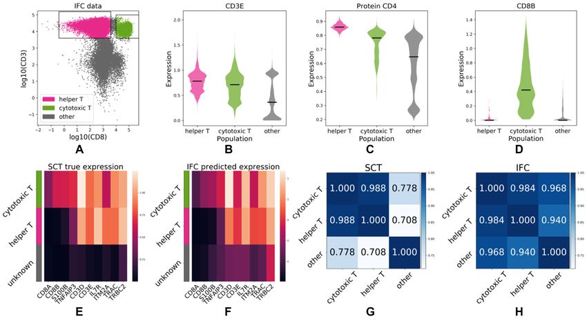
11336 Nucleic Acids Research, 2020, Vol. 48, No. 20

for reliable diagnosis (6). Recently, deep learning analysis       ing the expression profile of each individual cell of an IFC
of histology imaging data has gained attention from clini-         experiment, based on integrating a corresponding SCT ex-
cians and pathologists in the diagnosis of cancers. Convo-         periment that includes the same cell types of interest. We
lutional neural networks have achieved a success rate in the       demonstrate that our method correctly predicts and local-
classification of certain tumors that match the success rate       izes the expression of key marker genes for each cell type. We
of pathologists (7,8). Data obtained by IFC is ideally suited      also demonstrate that in some cases, the estimation of gene
for deep learning-assisted image analysis and hence can be         expression can be performed in a label-free manner from the
a valuable tool in the diagnosis of lymphomas and other            brightfield images only, based on the morphology of each
diseases affecting blood cells, such as immunodeficiencies.        cell (32). To the best of our knowledge, this is the first study
   IFC allows for imaging of cells and studying cellular           that aims to computationally augment IFC datasets with an
properties through corresponding surface markers. As the           additional SCT information modality.
measurement of surface markers occurs via fluorescently la-           The closest method to our approach is (7) where the imag-
beled antibodies, this measurement is naturally limited by         ing modality is used to predict the mutation status of select

                                                                                                                                      Downloaded from https://academic.oup.com/nar/article/48/20/11335/5943188 by guest on 12 December 2020
the number of available fluorescent channels. In turn, this        genes. Nonetheless, IFC-seq differs from (7) in the follow-
limits the cellular diversity that can be studied using a stan-    ing points: First, unlike the case of (7) where ground truth
dard IFC approach. Additionally, the view of the dataset           mutational status is available for each sample, in the case of
is inherently biased since the surface markers are selected        IFC-seq no ground truth expression information is available
prior to performing the experiment. In contrast, direct ob-        for the IFC experiments. Second, IFC-seq predicts contin-
servation of each cell’s molecular properties would allow for      uous expression instead of a binary outcome. Third, IFC-
an unbiased view of each cell’s inner workings. A natural          seq predicts the expression of hundreds of genes that corre-
example of such a high-throughput unbiased view of cel-            spond to population markers, instead of predicting a small
lular properties is single-cell omics (9). Specifically, single-   number of predetermined genes. The workflow of IFC-seq
cell transcriptomics (SCT) (10,11) corresponds to an addi-         is demonstrated in Figure 1.
tional modality of information-rich and high-throughput
datasets at the single-cell level. The novelty of SCT methods      MATERIALS AND METHODS
lies in their ability to measure the full gene expression pro-
file of each individual cell. As a result, the advent of single-   Preprocessing the SCT datasets
cell transcriptomics has led to new advancements in several        Each SCT dataset was pre-processed before being co-
areas of biology, such as hematopoiesis (12,13), embryo-           registered to its corresponding IFC dataset. First, the sur-
genesis (14,15), the airway epithelium (16,17) and the im-         face markers values of the SCT datasets were normalized to
mune system (18–20). With increasing complexity and size           [0,1]. Then, genes that were expressed in fewer than 20 cells
of these data sets (10), these biological advancements have        of the SCT experiment were excluded from further anal-
gone hand-in-hand with the development of novel statisti-          ysis. Next, the expression of all genes was logarithmized
cal and machine learning concepts for analyzing SCT data           using the natural logarithm. Subsequently, gating was per-
(21–24).                                                           formed by an expert on the surface marker values of the
   Machine learning approaches have also been developed            SCT dataset in order to identify the cellular subpopulations
for the analysis of IFC measurements, mainly focussing             of interest. It should be noted that gating information is not
on the identification and automated sorting of different           used in the predictive model, but is only used to validate the
cell types (25–28). Nonetheless, recent developments in ma-        model’s results. Last, a set of top 100 differentially expressed
chine learning methods have shown that analysis of imag-           marker genes was computed for each cellular subpopula-
ing data can be extended far beyond simple sorting of cell         tion, using the ‘rank genes groups’ function of Scanpy (24).
types. For example, it was recently demonstrated that gene         As such, differentially expressed genes were identified in a
mutational status can be predicted from imaging data (7).          purely unbiased data-driven manner and no manual identi-
Moreover imaging data can be used in order to estimate the         fication of marker genes was performed by an expert.
cell-cycle stage (27) and to predict cellular decisions such
as differentiation (29) based on morphology information.
                                                                   SCT––human cord blood mononuclear cells
Additionally, technologies that offer both IFC and SCT ca-
pabilities are expected in the near future (30,31). Datasets       The first SCT dataset corresponds to Cord Blood Mononu-
including both imaging and transcriptomic views of each            clear Cells (CBMCs) (33). The original dataset in-
individual cell will offer unprecedented quality and quan-         cludes human and mouse cells and the count ma-
tity of information. As such, they can aid our understanding       trix and surface markers are available as supplementary
of biological systems. Having SCT information available in         files GSE100866 CBMC 8K 13AB 10X-RNA umi.csv.gz
IFC experiments would not only alleviate the bias of prese-        and GSE100866 CBMC 8K 13AB 10X-ADT umi.csv.gz,
lecting surface markers prior to performing the IFC exper-         respectively. Only human cells were kept by selecting cells
iment, but would additionally allow studying cellular prop-        that express more human than mouse genes. To be pre-
erties and functions in unprecedented resolution by provid-        cise, human genes in the count matrix are characterized by
ing expression information for individual cells.                   a ‘HUMAN ’, while mouse genes are characterized by a
   However, at the moment the IFC and SCT modalities               ‘MOUSE ’ prefix in the gene name. We identified as hu-
are still acquired separately, in different experiments and        man cells, the cells that express more human than mouse
for different populations of cells. In this paper we intro-        genes. We considered a gene to be expressed in a cell if it cor-
duce IFC-seq: a machine learning methodology for predict-          responds to non-zero counts. Additionally, cells expressing
Nucleic Acids Research, 2020, Vol. 48, No. 20 11337

                                                                                                                                                              Downloaded from https://academic.oup.com/nar/article/48/20/11335/5943188 by guest on 12 December 2020
Figure 1. IFC-seq predicts gene expression of individual cells in IFC datasets. Given the values of the surface markers of the IFC experiment, IFC-seq
proceeds to predict the gene expression profile of each cell. Expression prediction is achieved via a Random Forest (RF) for regression. In standard mode,
the measured values of the markers are utilized, while in label-free mode the markers are predicted from the cells’ morphology using the brightfield images
and a convolutional neural network (CNN). Learning the correspondence between marker values and expression becomes possible by co-registering an
independent single-cell transcriptomics experiment where the same surface markers are also measured for each cell.

fewer than 200 genes were discarded from further analysis                          Data acquisition was performed as follows: Blood from
and highly variable genes were kept using the filter genes dis-                 healthy donors was diluted in Phosphate-Buffered Saline
persion method of Scanpy (24) with parameters min mean                          (PBS) carefully layered onto a Ficoll cushion (Biocoll:
= 0.0125, max mean = 3, min disp = −0.15. All subsequent                        Density 1.077 g/ml). After centrifugation the layer con-
preprocessing steps were performed as described in the pre-                     taining PMBCs was collected and washed. 5 × 10⁶ cells
vious section. After preprocessing, the dataset includes 8017                   were stained with CD3 PE-Cy7 (clone UCHT1, Biolegend),
human cells and 2768 genes, as well as CD3 and CD8 sur-                         CD8a-AF647 (clone RPA-Ta, Biolegend) and live dead fix-
face marker values measured for each cell. Subsequently,                        able violet dye (ThermoFischer). After fixation (4% PFA,
the cells were sorted into helper T-cell (CD3+CD8-) and cy-                     10 min) cells were analyzed by imaging flow cytometry. Af-
totoxic T-cell (CD3+CD8+) sub-populations based on the                          ter acquisition, TIF-images (32 × 32 pixels, 16-bit, raw) of
CD3 and CD8 marker values.                                                      live dead-CD3+CD8a−, live dead-CD3+CD8a+ and live
                                                                                dead-CD3−CD8a− were exported and used for analysis
SCT––mouse myeloid progenitor cells                                             and the CD3 and CD8 surface markers were normalized
                                                                                in [0,1].
The second SCT dataset corresponds to a publicly avail-
able dataset of mouse myeloid progenitor cells (13). Af-
ter preprocessing it includes 2730 cells with 3371 genes, as                    IFC––mouse myeloid progenitor cells
well as FcgR and CD34 surface marker values for each
cell. The preprocessing steps were performed as described                       Two separate IFC datasets were acquired for this study.
above. Gating was subsequently performed to sort the cells                      The training dataset was used to train a CNN for label-
into three sub-populations: Common Myeloid Progenitor                           free marker prediction consists of 65 008 cells. The test set
(CMP) cells, Granulocyte/Macrophage Progenitor (GMP)                            was used to evaluate the results of IFC-seq and consists of
cells and Megakaryocyte/Erythrocyte Progenitor (MEP),                           3137 cells. Both IFC datasets include brightfield, FcgR and
using the same gates as in (13).                                                CD34 images of cells, along with the measured CD34 and
                                                                                FcgR surface marker intensity values. Subsequently, CMP,
                                                                                GMP and MEP cells were identified by gating on the CD34
IFC––mice                                                                       and FcgR markers.
Sex and aged matched (8 weeks) C57BL/6 mice were pur-                              The data acquisition process was the following: BM cells
chased from Envigo. The permissions for animal experi-                          were flushed from femur and tibia with PBS + 2% fetal
ments were granted by the animal ethics committee of the                        calf serum (FCS) using syringes. Erythrocytes were lysed
Regierung von Oberbayern, Munich, Germany.                                      using an ammonium chloride potassium buffer. Number
                                                                                of live cells was determined using a CASY cell counter
                                                                                (OMNI Life Science). 5 × 10⁶ cells were stained with CD117
IFC––human peripheral blood mononuclear cells
                                                                                APC (clone 2B8, eBioscience), CD34 FITC (clone RAM34,
IFC was used to acquire data of human Peripheral Blood                          eBioscience), Sca-1 PE-Cy5 (clone D7, eBioscience), FcgR
Mononuclear Cells (PBMCs). The resulting IFC dataset                            PE-Cy7 (clone 93, Invitrogen) and Lin-1 BV421 (Biole-
corresponds to 82 109 human PBMCs with CD3 and                                  gend) and analyzed on an ImageStreamX MKII imag-
CD8 surface marker measurements for each cell. Gating                           ing flow cytometer (Luminex). TIF-images (32 × 32 pix-
on the CD3 and CD8 markers was employed to sort the                             els, 16-bit, raw) of Lin-1-CD117+Sca-1+FcgR-CD34−
cells into helper T-cell (CD3+CD8-) and cytotoxic T-cell                        MEP, Lin-1-CD117+Sca-1+FcgRintCD34int CMP, Lin-1-
(CD3+CD8+) sub-populations.                                                     CD117+Sca-1+FcgR+CD34+ GMP cells were exported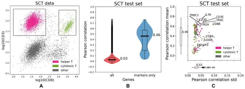
11338 Nucleic Acids Research, 2020, Vol. 48, No. 20

and used for analysis and the CD34 and FcgR surface                the helper and cytotoxic T-cells of the human dataset. The
markers were normalized in [0,1].                                  CNN was trained using Adam (37) for 50 epochs using a
                                                                   batch size of 64 on the IFC training dataset of 65 008 mouse
                                                                   myeloid progenitor cells, while 10% of the IFC dataset was
Predicting gene expression
                                                                   randomly left out of training and was used for validation.
The goal of the proposed IFC-seq method is to augment              The best model according to the validation loss was saved.
IFC datasets with expression information at the single-cell        Additionally, early stopping with a patience parameter of
level. Ideally, that would require data where the imaging          five epochs was employed during training. Moreover, data
modality and gene expression are available for the exact           augmentation was employed on the training set. Such aug-
same cell. However, while such promising techniques have           mentation corresponds to flipping the images along the ver-
been proposed (30,31) they are not yet well established and        tical, horizontal, or both axes. The network consists of 17
broadly available. Thus, IFC-seq overcomes the lack of such        convolutional layers and ∼700 000 parameters. Each acti-
datasets by co-registering an IFC experiment to a corre-           vation layer, except the last, is preceded by a batch normal-

                                                                                                                                    Downloaded from https://academic.oup.com/nar/article/48/20/11335/5943188 by guest on 12 December 2020
sponding SCT experiment that includes a common subset              ization layer (38). The neural network was implemented in
of cell-types. The co-registration step is made possible by        Keras. An overview of the CNN architecture is presented
aligning the datasets using surface markers that are present       in Figure 2 and the trained model is available online at
in both the IFC and SCT modalities. That is, we assume that        https://github.com/theislab/ifcseq.
if a cell in the IFC experiment is close in the space of surface
markers to cells in the SCT dataset, then its expression can       RESULTS
be estimated from the expression of its corresponding cells
in the SCT dataset. To ensure that surface marker values are       Overview
comparable across modalities, as part of co-registration we        Next, we will proceed to demonstrate the results of IFC-seq
independently normalize each marker within each modality           on the human and mouse test cases. For each of these two
so that its range of values extends from 0 to 1.                   test cases, the process is the following: First, we will eval-
   Consequently, we treat expression prediction as a regres-       uate the predicted expression on the left out test set of the
sion problem and predict thousands of genes per cell, given        SCT dataset. This is helpful since we have ground truth ex-
the values of the corresponding surface markers. Specif-           pression that we can compare to. Thus, this will allow for
ically, the scikit-learn (34) implementation of a Random           the quantification of the model’s predictive capability and
Forest for regression (35) was employed. The Random For-           provide an upper bound for its expected performance when
est was configured to minimize the mean absolute error,            applied to the IFC data. In both cases, we will demonstrate
‘max features’ was set to ‘sqrt’ and the ensemble consisted        that while IFC-seq can be used to predict all genes that are
of 50 trees. The Random Forest was trained separately for          included in the SCT experiment, prediction performance is
the human and mouse test cases. In the case of mouse               not uniform across all genes. Specifically, gene expression is
data the CD34 and FcgR markers are used as input while             only predicted successfully for marker genes of the cellular
CD3 and CD8 were used for the human data. Addition-                subpopulations of interest.
ally, surface marker CD4 is directly predicted for the hu-            Furthermore, we will apply IFC-seq and predict expres-
man data along with gene expression, since surface CD4 is          sion for the corresponding IFC seq experiment. Since no
a known helper T-cell marker but the correlation between           ground truth expression is available for each cell in the
the measured protein and transcript CD4 levels is low (33).        IFC data, we need to employ a different validation ap-
Each SCT experiment is split into a 70% training set and a         proach. That is, we will assess the predicted expression at
30% test set. No validation set was used when training the         the population level and quantify to what extent the pre-
Random Forest, since no hyperparameter tuning was per-             dicted expression of population-specific marker genes fol-
formed. The trained Random Forest model is trained on the          lows the same pattern as observed in the SCT experiment.
SCT dataset, it is then employed to predict the expression         That is, if IFC-seq is successful, then the expression pat-
of the corresponding (human or mouse) IFC dataset.                 terns of population-specific marker genes should be consis-
                                                                   tent across the IFC and SCT modalities. As mentioned in
Predicting surface markers in a label-free manner                  the previous section, we will also demonstrate the label-free
                                                                   capability of IFC-seq and predict gene expression directly
In the label-free mode of IFC-seq, a Convolutional Neural          from the brightfield images in the case of mouse cells. On
Network (CNN) (36) was employed to predict the surface             the other hand, IFC-seq label-free mode is not supported
marker values based only on the 32 × 32 brightfield image          in the case of human blood cells, since the T-cell subpopu-
of each cell in the IFC experiment. Since network architec-        lations of interest cannot be distinguished by morphological
tures that perform well on natural images have been shown          features alone.
to perform well on IFC data (27), we based our approach
on the popular residual CNN architecture which achieves
                                                                   Human blood mononuclear cells
state of the art results on natural images (36). It should be
noted that label-free prediction is only expected to work if       IFC-seq was employed to predict gene expression of human
there is sufficient morphological information in the bright-       blood mononuclear cells, based on the measured CD3 and
field images of the cells. As such, we will demonstrate the        CD8 markers for each of the SCT and IFC modalities. It
label-free mode IFC-seq in the case of the mouse dataset,          should be noted that the SCT dataset consists of CBMCs
since there is no sufficient morphological difference between      while the IFC dataset consists of PBMCs. However, they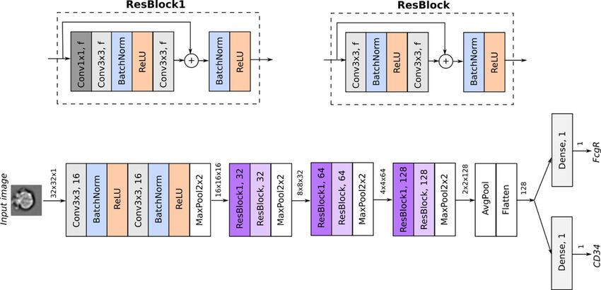
Nucleic Acids Research, 2020, Vol. 48, No. 20 11339

                                                                                                                                                              Downloaded from https://academic.oup.com/nar/article/48/20/11335/5943188 by guest on 12 December 2020
Figure 2. Overview of the architecture of the CNN for regression used to predict the surface markers in the mouse data. Given the brightfield image of
each cell, the network predicts the value of the CD34 and FcgR surface markers, whose values are continuous and lie in [0,1]. After the initial input stem,
the network employs residual blocks whose architecture is visualized on the top part of the figure. Every convolutional layer employs zero padding. Thus,
the spatial dimensions of the intermediate tensors are only reduced via pooling operations. Tensors are visualized as arrows, along with their dimensions
(height × width × channels).

both include helper and cytotoxic T-cell subpopulations.                        imentally for the IFC experiment, so this successful pre-
Additionally, the bulk of ‘other’ cells is considered as a sub-                 diction of IFC-seq is exclusively data-driven. Nonetheless,
population where T-cell specific markers are not expected to                    while CD4 is predicted to be less expressed in the cyto-
be expressed. As such, the results of IFC-Seq are assessed                      toxic and other cells, it should ideally be predicted to be
with respect to these three cellular subpopulations.                            closer to zero for these subpopulations. Moreover, CD8A
   As seen in Figure 3, IFC-seq successfully predicts gene                      and CD8B are two known cytotoxic T-cell markers (40)
expression in the case of marker genes for the subpopula-                       that IFC-Seq predicts to be highly expressed almost exclu-
tions of helper and cytotoxic T-cell. These marker genes                        sively in this subpopulation. The above results are visualized
correspond to the differentially expressed genes identified                     in Figure 4(A-D).
during preprocessing (see Materials and Methods). Over-                            Additionally, IFC-Seq correctly predicts the expression
all, the median Pearson correlation between the predicted                       patterns of several other genes that are known to be asso-
and true expression is 0.46 when considering only the top                       ciated with the subpopulations of interest. These marker
100 genes per subpopulation, while it drops to 0.03 if all                      genes include: S100B which is associated with T-cells and
genes in the SCT dataset are taken into account. Moreover,                      natural killer cells (41), TNFAIP3 which is related to im-
the top marker genes are predicted with low uncertainty,                        mune response (42), CD27 which associated with T-cell im-
as quantified by the standard deviation of the Pearson cor-                     munity (43), ITM2A which is involved in T-cell activation
relation achieved by individual trees in the Random For-                        (44), IL7R which is known to be expressed in naive T-cells
est ensemble, which equals to 0.03. Table 1 summarizes the                      (45), TRBC2 and TRAC which are related to the T-cell re-
median Pearson and Spearman correlations, as well as the                        ceptor (46), CD69 and SELL, associated with both helper
mean squared error achieved by the Random Forest, as well                       and cytotoxic T cells (47) and finally SOX4 which is associ-
as a linear regression baseline model. Last, the correlation                    ated with helper T-cells (48).
between the CD3 and CD8 surface markers and their corre-                           Figure 4E and F visualizes the expression profiles of the
sponding coding genes is visualized in Supplementary Fig-                       aforementioned markers across the SCT and IFC modal-
ure S1 in the supplement.                                                       ities. While gene expression is predicted at the single-cell
   When applied to the IFC dataset, IFC-seq correctly pre-                      level, the figure visualizes the average expression per popu-
dicts that the CD3D and CD3E are highly expressed in the                        lation in order to highlight patterns at the population level.
helper and cytotoxic T-cells. This is to be expected since                      By observing these patterns it is straightforward to distin-
both CD3D and CD3E correspond to proteins necessary                             guish the helper and cytotoxic T-cells from each other, as
for T-Cell receptor signalling (39). Moreover, surface CD4,                     well as from the bulk of other cells. To be precise, CD8A,
a known helper T-cell marker, is predicted to be highly ex-                     CD8B, S100B and TNFAIP3 only mark cytotoxic T-cells,
pressed in the helper T-cells. It should be noted that CD4                      while the remaining markers separate helper T-Cells from
was not included in the set of markers measured exper-                          the bulk of other cells. Additionally, the transcriptional sim-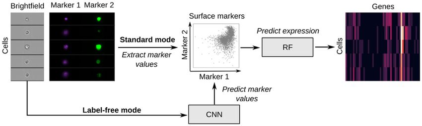
11340 Nucleic Acids Research, 2020, Vol. 48, No. 20

                                                                                                                                                                Downloaded from https://academic.oup.com/nar/article/48/20/11335/5943188 by guest on 12 December 2020
Figure 3. IFC-seq results on SCT data of human blood mononuclear cells. (A) The SCT dataset and corresponding helper and cytotoxic T-cell gates plotted
on top of the CD3 and CD8 markers. The population of ‘other’ cells corresponds to all unknown cell-types that are not included in the population-specific
gates. (B) The predicted gene expression is more accurate for the differentially expressed population-specific marker genes than for all genes present in the
SCT dataset, as quantified by the Pearson correlation between the true and predicted expression values for each cell in the SCT test set. (C) Looking at
the population-specific marker genes shows that they are predicted with low uncertainty, as quantified by the standard deviation of the per-gene Pearson
correlation computed over the trees in the Random Forest ensemble. Individual points correspond to distinct population-specific marker genes. Genes are
colored according to the respective population they are markers for. Select population markers are overlaid on top of the scatter plot.

Table 1. Predictive performance of Random Forest regression and Lin-             modalities. In the case of IFC, we present the results when
ear Regression on the SCT test of human blood mononuclear cells. The             the measured marker values are employed, as well as the
median value of each statistic across all cells is reported                      case where IFC-seq is performed in label-free mode and the
                       Pearson          Spearman           Root mean             markers are predicted from directly from the brightfield im-
Method                correlation       correlation       squared error          ages.
Random Forest             0.46             0.46                0.52
                                                                                    Similar to the case of human cells, the predicted gene ex-
Linear regression         0.38             0.35                0.52              pression is more closely correlated to true expression when
                                                                                 focusing only on population-specific marker genes, instead
                                                                                 of all genes in the SCT dataset. That is, when looking only
ilarity of populations is quantified as the Pearson correla-                     at markers the median Pearson correlation between true
tion of the population-average expression of all top marker                      and predicted expression is 0.32, as opposed to 0.08 when
genes across the populations. That is, the helper and cyto-                      looking at all genes. Additionally, the population-specific
toxic T-cells are expected to be transcriptionally more sim-                     marker genes are predicted with low uncertainty, as the me-
ilar to each other, than to the bulk of other cells. That is                     dian standard deviation of the per-gene Pearson correla-
indeed the case when the similarity is predicted with true                       tion is only 0.03. The aforementioned results are presented
expression in the SCT experiment and with IFC-seq pre-                           in Figure 5, while the relationship between the CD34 and
dicted expression for the IFC data, as shown in Figure 4G                        FcgR markers and their respective coding genes is presented
and H. However, in the case of predicted expression for the                      in Supplementary Figure S2 of the supplement. Interest-
IFC data the differences in population similarities are not                      ingly, the prediction quality of the model appears to be
as pronounced. Nonetheless, by calculating the 95% con-                          population-specific, contrary to what was observed for the
fidence intervals for the Pearson correlations via Fisher’s                      human data. However, unlike the human data where helper
transformation (49), we see that the similarity of cytotoxic                     and cytotoxic T-cells correspond to distinct clusters in the
and helper T-cells is at least 0.979 (low confidence inter-                      space of the surface markers, the subpopulations of the
val). On the other hand, the similarity of cytotoxic T-cells                     mouse data correspond to a continuous differentiation pro-
to other cells is at most 0.975 and the similarity of helper                     cess. Gene expression is best predicted for the MEP marker
T-cells to other cells is at most 0.953 (high confidence inter-                  genes, which agrees with the observation that MEP cells
vals). As such, cytotoxic and helper T-cells are significantly                   yield more distinct expression profiles (Figure 6D). On the
more similar to each other than to the bulk of other cells,                      other hand, gene expression is not predicted as well for the
even when looking at the predicted expression profiles of the                    CMP marker genes. This could be explained by the fact that
IFC data.                                                                        the CMP cells lie in a smaller range of the surface markers
                                                                                 than the GMP and MEP cells, which could result in reduced
                                                                                 sensitivity of the model in that area of the feature space.
Mouse myeloid progenitor cells
                                                                                 Last, the median Pearson correlation, Spearman correla-
Next, we present the results of IFC-seq on mouse myeloid                         tion and the mean squared error achieved by the Random
progenitor cells where gene expression was predicted based                       Forest, as well as a linear regression baseline model are pre-
on the CD3 and CD8 markers for each of the SCT and IFC                           sented in Table 2.
Nucleic Acids Research, 2020, Vol. 48, No. 20 11341
Downloaded from https://academic.oup.com/nar/article/48/20/11335/5943188 by guest on 12 December 2020
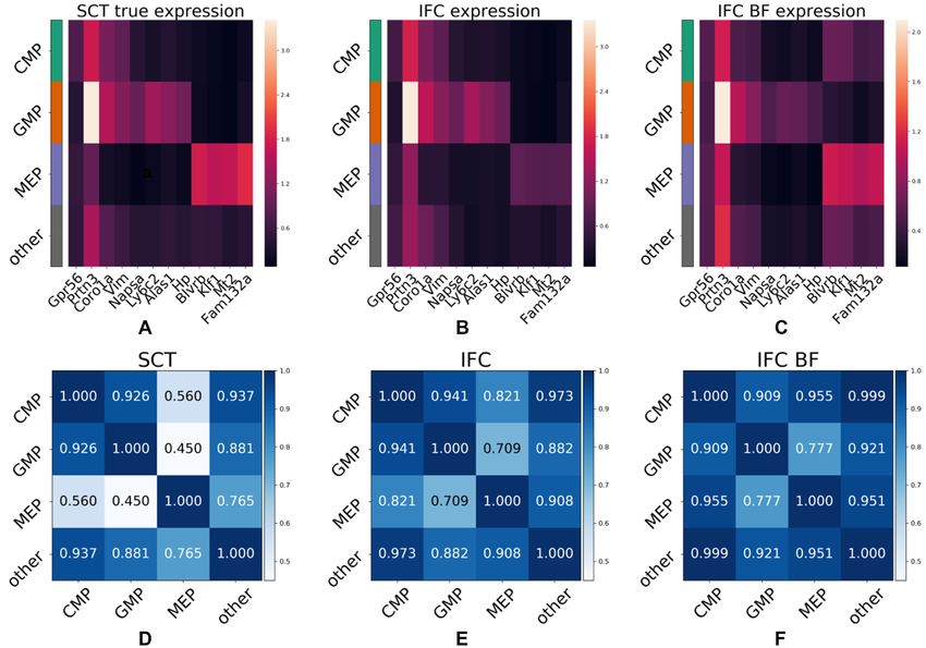
Figure 4. IFC-seq predicts gene signatures of helper and cytotoxic T-cell subpopulations in human blood mononuclear cells. (A) IFC dataset and corre-
sponding gates for helper and cytotoxic T-cells on the CD3 and CD8 markers. The population of ‘other’ cells corresponds to all unknown cell-types that
are not included in the population-specific gates. (B) CD3E, a known T-cell marker is predicted to be highly expressed in both helper and cytotoxic T-cells.
(C) Surface CD4, a helper T-cell marker, is predicted to be predominantly expressed in helper T-cells. (D) CD8B, a cytotoxic T-cell marker, is predicted
to be almost exclusively expressed on cytotoxic T-cells. (E, F) The expression profiles between marker genes agree across the SCT and IFC experiments.
Each row of the heatmap corresponds to a population while each column corresponds to the expression of a gene averaged across all cells in a population.
(G, H) The transcriptional similarity of populations is similar across the SCT and IFC modalities. That is, cytotoxic and helper T-cells are more similar to
each other than the bulk of other cells.
We subsequently apply IFC-seq on the corresponding the population specific gene expression patterns across the
IFC dataset two times, in standard and in label-free mode modalities of true SCT expression and predicted expres-
and compare the results. In label-free mode, the CD34 and sion for the IFC experiment (both in standard and label-
FcgR markers are predicted with a CNN (see Materials free modes of IFC-seq). That is, it is straightforward to
and Methods). The performance of the CNN on the IFC separate the CMP progenitor cells from their descendant
dataset, corresponding to the Pearson correlation between populations of GMP and MEP cells, as well as GMP and
the true and predicted marker values, is 0.38 ± 0.16 for MEP cells from each other, based on the population-specific
CD34 and 0.5 ± 0.016 for FcgR. The standard deviation markers mentioned above. As expected, there is loss of in-
was calculated using 10 000 bootstrap iterations (50). formation when IFC-seq is performed in label-free mode.
Examining the results on the mouse data shows that IFC- Nonetheless, it is still possible to easily distinguish GMP
seq successfully predicts the expression of key marker genes and MEP cells based on expression predicted from morpho-
for the subpopulations of interest in the IFC dataset, purely logical information alone. It is also possible to distinguish
in a data-driven manner. Specifically, IFC-seq is successful CMP cells from their two descendant populations. How-
at predicting the expression of known CMP markers, such ever, in label-free mode the predicted expression profile of
as Serpina3f (51,52) and Gpr56 (53). Next, IFC-seq predicts CMP cells is close to the profile of the background popula-
the expression of GMP markers, such as Napsa, Ly6c2, tion of other cells. Nonetheless, it should be noted that the
Alas1, Hp, as well as known GMP markers Coro1a (54), CMP cells and background population have similar expres-
Ly6c2 (55), Vim (56) and Prtn3 (55). Additionally, some sion profiles even in the case of true expression in the SCT
GMP markers like Coro1a and Vim are also expressed in experiment, as seen in Figure 6D.
the progenitor populations of CMP cells. Next, MEP mark-
ers predicted by IFC-seq include Mt2, Fam132a and known
MEP markers Blvrb (54) and Klf1 (57). DISCUSSION
Figure 6A–C visualizes gene expression averaged per In this paper, we introduced IFC-seq: a machine learning
population, while the gating strategy for the IFC data is methodology which can augment IFC datasets by predict-
shown in Supplementary Figure S3. Visual inspection of ing an additional modality of single cell transcriptomics
the gene expression heatmaps highlights agreement between for each cell at no additional cost. Predicting the expres-11342 Nucleic Acids Research, 2020, Vol. 48, No. 20
Downloaded from https://academic.oup.com/nar/article/48/20/11335/5943188 by guest on 12 December 2020
Figure 5. IFC-seq results on SCT data of mouse myeloid progenitor cells. (A) SCT data plotted on top of the CD34 and FcgR markers, along with gates
for the CMP, GMP and MEP populations of interest. The population of ‘other’ cells corresponds to all unknown cell-types that are not included in the
population-specific gates. (B) The gene expression is predicted more accurately for the population-specific marker genes, compared to the bulk of all genes
in the SCT experiment. Specifically, the median Pearson correlation between the true and predicted expression iis 0.32 for the marker while it drops to 0.08
when all genes are considered. (C) The population-specific marker genes are predicted with low uncertainty of 0.04, as quantified by the standard deviation
of the Pearson correlation achieved by individual trees in the Random Forest ensemble. Individual points correspond to distinct population-specific marker
genes. Genes are colored according to the respective population they are markers for. Select population markers are overlaid on top of the scatter plot.
sion profile of key marker genes for each single cell in the model accuracy. Such a bottleneck is potentially posed by
IFC experiment is made possible by coupling it to a cor- the fact that gene expression was predicted only from two
responding independently acquired SCT experiment using available markers in each dataset. Since the performance of
common surface protein markers. Additionally, we showed IFC-seq depends on the selection of surface markers, it is
that for the SCT datasets, where ground truth expression only applicable in cases where markers for the cellular pop-
is available, IFC-seq is successful at predicting the expres- ulations of interest are known and available during model
sion of population specific marker genes with low uncer- training. Additionally, IFC-seq is sensitive to batch effects
tainty. Naturally, since the model’s predictions are based related to intra-modality variability across independent ex-
on surface markers, it performs better for genes associated periment replicates (58), as well as inter-modality variability
with populations characterized by these markers. Last, we of the surface markers across the SCT and IFC modalities.
also showed that in some cases, such as the mouse bone In this study, we alleviated the inter-modality variability by
marrow cells where morphology is informative, it is possi- marker normalization. Moreover, we expect that upcom-
ble to directly predict the gene expression of population- ing batch effect correction methods (59,60) will further al-
specific marker genes for each cell in a label-free manner. leviate challenges related to both intra- and inter-modality
This label-free mode of IFC-seq is made possible by using variabilities. While performance of IFC-seq is bound by the
the brightfield images of the IFC experiment and leverag- co-registration step, we expect that if more relevant surface
ing a convolutional neural network as an additional step. markers become available, the predictive capability of IFC-
The main goal of this study is to provide a proof of con- seq will improve. Moreover, augmenting IFC datasets with
cept that demonstrates the feasibility of predicting gene ex- information of gene expression at the single-cell level, can
pression of key marker genes in IFC data by aligning an in- substantially increase the depth of available information,
dependent SCT experiment with overlapping cellular sub- supplementing the measured surface protein markers. In
populations. To this extent, we provide the code for IFC- fact, cell states are often determined by biological processes
seq and all data used in this publication online at https: that might not be identified by surface markers alone, yet
//github.com/theislab/ifcseq. show distinct transcriptional signatures. It is worth noting
In both test cases of human blood, as well as mouse bone that augmenting IFC datasets with the proposed method
marrow cells the proposed IFC-seq methodology success- comes at zero additional cost, assuming that the markers
fully predicted key gene markers of the populations of in- coupling the IFC to the corresponding SCT experiment are
terest. These results are promising considering the underly- available or that morphological information is sufficient in
ing limitations, such as the low resolution of the IFC images order to apply IFC-seq in label-free mode.
(32 × 32 pixels) and the complexity introduced by the co- Predicting gene expression reduces the need for surface
registration step used to couple the independent SCT and markers in certain use cases and that is useful for two main
IFC experiments via a limited subset of common surface reasons. First, the number of available fluorescence chan-
markers. The similar predictive performance of the Ran- nels is always limited. By being able to predict genes (or
dom Forest and linear regression within the SCT modal- additional markers) directly from a few known markers, or
ity suggests that non-linear effects are not a bottleneck in from brightfield images in the label-free case, some of theNucleic Acids Research, 2020, Vol. 48, No. 20 11343
Downloaded from https://academic.oup.com/nar/article/48/20/11335/5943188 by guest on 12 December 2020
Figure 6. IFC-seq predicts gene signatures of mouse myeloid progenitor cells. (A–C) Heatmaps of average gene expression for population-specific marker
genes. The true and predicted expression profiles for (A) SCT and (B) IFC, respectively, are in agreement. (C) In the case where IFC-seq is performed in
label-free mode, it is still possible to distinguish CMP, GMP and MEP cells from each-other, but it is harder to separate CMP cells from the background
of all other cells in the experiment. (D–F) The transcriptional similarity of populations shows the same picture as the previous heatmaps, in a quantified
manner. (D) CMP cells are transcriptionally similar to the background population of other cells even when considering the true expression of the SCT
experiment. Additionally, CMP cells are more transcriptionally similar to GMP than to MEP cells. A similar pattern is visible in the case of predicted
expression for the IFC data using the true marker values (E) and is still noticeable although noisier in the case of label-free mode (F).
Table 2. Predictive performance of Random Forest regression and linear speed up and significantly lower the costs of routine di-
regression on the SCT test of mouse myeloid progenitor cells. The median agnostics (63) Last but not least, it should be noted that
value of each statistic across all cells is reported the mouse myeloid progenitor dataset used to showcase the
Pearson Spearman Root mean label-free mode of IFC-seq is a particularly challenging use-
Method correlation correlation squared error case, since it has been previously shown that most CMP cells
Random Forest 0.32 0.35 0.40
are nearly indistinguishable from their offspring GMP and
Linear regression 0.32 0.35 0.39 MEP populations based only on morphological informa-
tion, with the exception of CMP cells that are close to be-
ing differentiated (29). Supplementary Figure S4 shows ex-
emplary brightfield images of CMP, GMP and MEP cells,
fluorescence channels become redundant. Thus, they are along with guided saliency maps (64) visualizing the pix-
freed and can be used with different stains in order to study els of each input image influencing the CNN’s predictions.
other cellular properties and functions. This was the case The saliency maps were computed with keras-vis (https:
in the human data, where for example CD4 was not mea- //raghakot.github.io/keras-vis) and suggest that all parts of
sured in the experiment but CD4 positive cells were identi- the input image contribute equally to both CD34 and FcgR
fied by IFC-seq. Second, there are cases where avoiding cer- predictions and that the network mainly bases its predic-
tain fluorescent stains may be a goal in itself due to potential tions on regions near the cellular boundary and in some
side effects of the staining process. The above advantages cases on regions deeper inside the cell.
become especially pronounced in the label-free case, where To quantitatively validate the performance of the pro-
analysis methods rely on cellular morphology (25,27), sub- posed method we need to be able to experimentally assess
cellular structures (61) or other label-free modalities (62). how accurately the predictions generated by IFC-seq re-
Moreover, label-free cell phenotyping has the potential to flect the ground truth gene expression at the single-cell level.11344 Nucleic Acids Research, 2020, Vol. 48, No. 20
To this extent, we would need an experimental procedure, REFERENCES
which performs imaging and sequencing on the exact same 1. Pepperkok,R. and Ellenberg,J. (2006) High-throughput fluorescence
cell in a high-throughput manner and results in a dataset in microscopy for systems biology. Nat. Rev. Mol. Cell Biol., 7, 690–696.
which both the IFC and SCT modalities are simultaneously 2. Basiji,D.A., Ortyn,W.E., Liang,L., Venkatachalam,V. and
measured for each cell. To the best of our knowledge, no Morrissey,P. (2007) Cellular image analysis and imaging by flow
cytometry. Clin. Lab. Med., 27, 653–670.
such dataset exists at the moment but recent developments 3. Brown,M. and Wittwer,C. (2000) Flow cytometry: principles and
in next generation imaging and sorting techniques such as clinical applications in hematology. Clin. Chem., 46, 1221–1229.
(30,31) suggest that this is only a matter of time. We expect 4. Pillai,V. and Dorfman,D.M. (2016) Flow cytometry of
that these new datasets where IFC and SCT modalities are nonhematopoietic neoplasms. ACY, 60, 336–343.
5. Tute,R.M. de (2011) Flow cytometry and its use in the diagnosis and
simultaneously present will not only allow us to properly management of mature lymphoid malignancies. Histopathology, 58,
validate, but also improve the performance of the proposed 90–105.
methodology. Additionally, lower-throughput experimental 6. Wood,B.L., Arroz,M., Barnett,D., DiGiuseppe,J., Greig,B.,
methods capable of imaging and sequencing individual cells Kussick,S.J., Oldaker,T., Shenkin,M., Stone,E. and Wallace,P. (2007)
Downloaded from https://academic.oup.com/nar/article/48/20/11335/5943188 by guest on 12 December 2020
are currently available (65). Such methods are not practical 2006 Bethesda International Consensus recommendations on the
immunophenotypic analysis of hematolymphoid neoplasia by flow
in the label-free case where large datasets are required to cytometry: optimal reagents and reporting for the flow cytometric
train a CNN, but could be used to train and validate the diagnosis of hematopoietic neoplasia. Cytometry Part B: Clin.
performance of IFC-seq using the measured marker values. Cytometry, 72B, S14–S22.
Having access to the expression values of key marker genes 7. Coudray,N., Ocampo,P.S., Sakellaropoulos,T., Narula,N.,
Snuderl,M., Fenyö,D., Moreira,A.L., Razavian,N. and Tsirigos,A.
would be crucial for the validation of IFC-seq, especially in (2018) Classification and mutation prediction from non–small cell
label free mode where the expression of key marker genes lung cancer histopathology images using deep learning. Nat. Med.,
could be used as a control. Nonetheless, this requires some 24, 1559.
familiarity with the cellular populations at hand. Last, IFC- 8. Djuric,U., Zadeh,G., Aldape,K. and Diamandis,P. (2017) Precision
seq can also be extended to be useful in additional imaging histology: how deep learning is poised to revitalize histomorphology
for personalized cancer care. npj Precis. Oncol., 1, 22.
modalities, other than IFC. That is, we expect IFC-seq will 9. Junker,J.P. and van Oudenaarden,A. (2014) Every cell is special:
benefit from the advent of spatial transcriptomic methods genome-wide studies add a new dimension to single-cell biology. Cell,
(66,67) for spatially resolved transcriptional information in 157, 8–11.
tissues. Using these next generation datasets it will be pos- 10. Angerer,P., Simon,L., Tritschler,S., Wolf,F.A., Fischer,D. and
Theis,F.J. (2017) Single cells make big data: new challenges and
sible to predict gene expression directly from the imaging opportunities in transcriptomics. Curr. Opin. Syst. Biol., 4, 85–91.
modality, without the need of an additional step of coupling 11. Stubbington,M.J.T., Rozenblatt-Rosen,O., Regev,A. and
different datasets using common surface markers. Teichmann,S.A. (2017) Single cell transcriptomics to explore the
immune system in health and disease. Science, 358, 58–63.
12. Moignard,V., Woodhouse,S., Haghverdi,L., Lilly,A.J., Tanaka,Y.,
SUPPLEMENTARY DATA Wilkinson,A.C., Buettner,F., Macaulay,I.C., Jawaid,W., Diamanti,E.
et al. (2015) Decoding the regulatory network of early blood
Supplementary Data are available at NAR Online. development from single-cell gene expression measurements. Nat.
Biotechnol., 33, 269–276.
13. Paul,F., Arkin,Y., Giladi,A., Jaitin,D.A., Kenigsberg,E.,
ACKNOWLEDGEMENTS Keren-Shaul,H., Winter,D., Lara-Astiaso,D., Gury,M., Weiner,A.
et al. (2015) Transcriptional heterogeneity and lineage commitment in
N.K.C. would like to thank Dr Lukas Simon for suggest- myeloid progenitors. Cell, 163, 1663–1677.
ing the human PBMC dataset, Dr Malte D. Luecken for 14. Briggs,J.A., Weinreb,C., Wagner,D.E., Megason,S., Peshkin,L.,
suggesting relevant literature, as well as Dr Carsten Marr, Kirschner,M.W. and Klein,A.M. (2018) The dynamics of gene
Valerio Lupperger and Giovanni Palla for their comments expression in vertebrate embryogenesis at single-cell resolution.
Science, 360, eaar5780.
on the manuscript. We acknowledge the Core Facility Flow 15. Wagner,D.E., Weinreb,C., Collins,Z.M., Briggs,J.A., Megason,S.G.
Cytometry at the Biomedical Center, Ludwig-Maxmilians- and Klein,A.M. (2018) Single-cell mapping of gene expression
Universität München, for providing equipment and exper- landscapes and lineage in the zebrafish embryo. Science, 360,
tise. 981–987.
16. Montoro,D.T., Haber,A.L., Biton,M., Vinarsky,V., Lin,B.,
Birket,S.E., Yuan,F., Chen,S., Leung,H.M., Villoria,J. et al. (2018) A
FUNDING revised airway epithelial hierarchy includes CFTR-expressing
ionocytes. Nature, 560, 319.
N.K.C. was supported by a DFG Fellowship through 17. Plasschaert,L.W., Žilionis,R., Choo-Wing,R., Savova,V., Knehr,J.,
the Graduate School of Quantitative Biosciences Mu- Roma,G., Klein,A.M. and Jaffe,A.B. (2018) A single-cell atlas of the
airway epithelium reveals the CFTR-rich pulmonary ionocyte.
nich (QBM); F.J.T. acknowledges support by the BMBF Nature, 560, 377.
[01IS18036A, 01IS18053A]; Helmholtz Association (In- 18. Jaitin,D.A., Kenigsberg,E., Keren-Shaul,H., Elefant,N., Paul,F.,
cubator grant sparse2big) [ZT-I-0007]; Chan Zuckerberg Zaretsky,I., Mildner,A., Cohen,N., Jung,S., Tanay,A. et al. (2014)
Initiative DAF (advised fund of Silicon Valley Commu- Massively parallel single cell RNA-Seq for marker-free
nity Foundation) [182835]; Deutsche Forschungsgemein- decomposition of tissues into cell types. Science, 343, 776–779.
19. Mahata,B., Zhang,X., Kolodziejczyk,A.A., Proserpio,V.,
schaft [SFB1054 B03 to T.B.]. Haim-Vilmovsky,L., Taylor,A.E., Hebenstreit,D., Dingler,F.A.,
Conflict of interest statement. F.J.T. reports receiving con- Moignard,V., Göttgens,B. et al. (2014) Single-Cell RNA sequencing
sulting fees from Roche Diagnostics GmbH and Cellarity reveals T helper cells synthesizing steroids de novo to contribute to
Inc., and ownership interest in Cellarity, Inc. and Dermag- immune homeostasis. Cell Rep., 7, 1130–1142.
20. Proserpio,V., Piccolo,A., Haim-Vilmovsky,L., Kar,G., Lönnberg,T.,
nostix. All other authors have no conflicts of interest to de- Svensson,V., Pramanik,J., Natarajan,K.N., Zhai,W., Zhang,X. et al.
clare.Nucleic Acids Research, 2020, Vol. 48, No. 20 11345
(2016) Single-cell analysis of CD4+ T-cell differentiation reveals three alpha-induced protein 3 (TNFAIP3, A20) imposes a brake on
major cell states and progressive acceleration of proliferation. antitumor activity of CD8 T cells. Proc. Natl. Acad. Sci. U.S.A., 111,
Genome Biol., 17, 103. 11115–11120.
21. Lopez,R., Regier,J., Cole,M.B., Jordan,M.I. and Yosef,N. (2018) 43. Chen,D., Gerasimčik,N., Camponeschi,A., Tan,Y., Wu,Q.,
Deep generative modeling for single-cell transcriptomics. Nat. Brynjolfsson,S., Zheng,J., Abrahamsson,J., Nordlund,J.,
Methods, 15, 1053. Lönnerholm,G. et al. (2017) CD27 expression and its association with
22. Satija,R., Farrell,J.A., Gennert,D., Schier,A.F. and Regev,A. (2015) clinical outcome in children and adults with pro-B acute
Spatial reconstruction of single-cell gene expression data. Nat. lymphoblastic leukemia. Blood Cancer J., 7, e575.
Biotechnol., 33, 495–502. 44. Kirchner,J. and Bevan,M.J. (1999) Itm2a is induced during thymocyte
23. Trapnell,C., Cacchiarelli,D., Grimsby,J., Pokharel,P., Li,S., selection and T cell activation and causes downregulation of Cd8
Morse,M., Lennon,N.J., Livak,K.J., Mikkelsen,T.S. and Rinn,J.L. when overexpressed in Cd4+Cd8+ double positive thymocytes. J.
(2014) The dynamics and regulators of cell fate decisions are revealed Exp. Med., 190, 217–228.
by pseudotemporal ordering of single cells. Nat. Biotechnol., 32, 45. Peng,Y. (2017) Forced expression of IL-7R promotes CD8 T cell
381–386. cytotoxicity to self antigen. PLoS One, 12, e0188112.
24. Wolf,F.A., Angerer,P. and Theis,F.J. (2018) SCANPY: large-scale 46. Morgan,N.V., Goddard,S., Cardno,T.S., McDonald,D., Rahman,F.,
Downloaded from https://academic.oup.com/nar/article/48/20/11335/5943188 by guest on 12 December 2020
single-cell gene expression data analysis. Genome Biol., 19, 15. Barge,D., Ciupek,A., Straatman-Iwanowska,A., Pasha,S.,
25. Blasi,T., Hennig,H., Summers,H.D., Theis,F.J., Cerveira,J., Guckian,M. et al. (2011) Mutation in the TCRa subunit constant
Patterson,J.O., Davies,D., Filby,A., Carpenter,A.E. and Rees,P. gene (TRAC) leads to a human immunodeficiency disorder
(2016) Label-free cell cycle analysis for high-throughput imaging flow characterized by a lack of TCRab+ T cells. J. Clin. Invest., 121,
cytometry. Nat. Commun., 7, 10256. 695–702.
26. Eliceiri,K.W., Berthold,M.R., Goldberg,I.G., Ibáñez,L., 47. Kumar,B.V., Ma,W., Miron,M., Granot,T., Guyer,R.S.,
Manjunath,B.S., Martone,M.E., Murphy,R.F., Peng,H., Plant,A.L., Carpenter,D.J., Senda,T., Sun,X., Ho,S.-H., Lerner,H. et al. (2017)
Roysam,B. et al. (2012) Biological imaging software tools. Nat. Human tissue-resident memory T cells are defined by core
Methods, 9, 697–710. transcriptional and functional signatures in lymphoid and mucosal
27. Eulenberg,P., Köhler,N., Blasi,T., Filby,A., Carpenter,A.E., Rees,P., sites. Cell Rep., 20, 2921–2934.
Theis,F.J. and Wolf,F.A. (2017) Reconstructing cell cycle and disease 48. Yoshitomi,H., Kobayashi,S., Miyagawa-Hayashino,A., Okahata,A.,
progression using deep learning. Nat. Commun., 8, 463. Doi,K., Nishitani,K., Murata,K., Ito,H., Tsuruyama,T., Haga,H.
28. Hennig,H., Rees,P., Blasi,T., Kamentsky,L., Hung,J., Dao,D., et al. (2018) Human Sox4 facilitates the development of
Carpenter,A.E. and Filby,A. (2017) An open-source solution for CXCL13-producing helper T cells in inflammatory environments.
advanced imaging flow cytometry data analysis using machine Nat. Commun., 9, 3762.
learning. Methods, 112, 201–210. 49. Fisher,R.A. (1921) On the ‘probable error’ of a coefficient of
29. Buggenthin,F., Buettner,F., Hoppe,P.S., Endele,M., Kroiss,M., correlation deduced from a small sample. Metron, 1, 3–32.
Strasser,M., Schwarzfischer,M., Loeffler,D., Kokkaliaris,K.D., 50. Moore,D.S., McCabe,G.P. and Craig,B.A. (2009) In: Introduction to
Hilsenbeck,O. et al. (2017) Prospective identification of hematopoietic the Practice of Statistics. W.H. Freeman.
lineage choice by deep learning. Nat. Methods, 14, 403–406. 51. Riddell,J., Gazit,R., Garrison,B.S., Guo,G., Saadatpour,A.,
30. Deplancke,B., Bues,J., Dainese,R. and Biocanin,M. (2018) Device for Mandal,P.K., Ebina,W., Volchkov,P., Yuan,G.-C., Orkin,S.H. et al.
high throughput single-cell studies. Patent PCT/IB2017/055524. (2014) Reprogramming committed murine blood cells to induced
31. Nitta,N., Sugimura,T., Isozaki,A., Mikami,H., Hiraki,K., hematopoietic stem cells with defined factors. Cell, 157, 549–564.
Sakuma,S., Iino,T., Arai,F., Endo,T., Fujiwaki,Y. et al. (2018) 52. Winkler,I.G., Hendy,J., Coughlin,P., Horvath,A. and Lévesque,J.-P.
Intelligent image-activated cell sorting. Cell, 175, 266–276. (2005) Serine protease inhibitors serpina1 and serpina3 are
32. Esfahani,Haftbaradaran and Knöll,R. (2020) Cell shape: effects on down-regulated in bone marrow during hematopoietic progenitor
gene expression and signaling. Biophys. Rev., 12, 895–901. mobilization. J. Exp. Med., 201, 1077–1088.
33. Stoeckius,M., Hafemeister,C., Stephenson,W., Houck-Loomis,B., 53. Daria,D. (2015) Defining the role of G-coupled protein receptor
Chattopadhyay,P.K., Swerdlow,H., Satija,R. and Smibert,P. (2017) Gpr56 in acute myeloid leukemia. Open Access Repositorium der
Simultaneous epitope and transcriptome measurement in single cells. Universität Ulm. Dissertation.
Nat. Methods, 14, 865–868. http://dx.doi.org/10.18725/OPARU-4533.
34. Pedregosa,F., Varoquaux,G., Gramfort,A., Michel,V., Thirion,B., 54. Chen,H., Albergante,L., Hsu,J.Y., Lareau,C.A., Bosco,G.L., Guan,J.,
Grisel,O., Blondel,M., Prettenhofer,P., Weiss,R., Dubourg,V. et al. Zhou,S., Gorban,A.N., Bauer,D.E., Aryee,M.J. et al. (2019)
(2011) Scikit-learn: machine learning in Python. J. Mach. Learn. Res., Single-cell trajectories reconstruction, exploration and mapping of
12, 2825–2830. omics data with STREAM. Nat. Commun., 10, 1903.
35. Breiman,L. (2001) Random forests. Mach. Learn., 45, 5–32. 55. Yanez,A., Ng,M.Y., Hassanzadeh-Kiabi,N. and Goodridge,H.S.
36. He,K., Zhang,X., Ren,S. and Sun,J. (2016) Deep residual learning for (2015) IRF8 acts in lineage-committed rather than oligopotent
image recognition. In: 2016 IEEE Conference on Computer Vision and progenitors to control neutrophil vs monocyte production. Blood,
Pattern Recognition (CVPR). pp. 770–778. 125, 1452–1459.
37. Kingma,D.P. and Ba,J. (2015) Adam: a method for stochastic 56. Terskikh,A.V., Miyamoto,T., Chang,C., Diatchenko,L. and
optimization. In 2015 International Conference on Learning Weissman,I.L. (2003) Gene expression analysis of purified
Representations (ICLR). Poster. hematopoietic stem cells and committed progenitors. Blood, 102,
38. Ioffe,S. and Szegedy,C. (2015) Batch normalization: accelerating deep 94–101.
network training by reducing internal covariate shift. In: International 57. Tallack,M.R., Magor,G.W., Dartigues,B., Sun,L., Huang,S.,
Conference on Machine Learning. pp. 448–456. Fittock,J.M., Fry,S.V., Glazov,E.A., Bailey,T.L. and Perkins,A.C.
39. Pacheco-Castro,A., Alvarez-Zapata,D., Serrano-Torres,P. and (2012) Novel roles for KLF1 in erythropoiesis revealed by
Regueiro,J.R. (1998) Signaling through a CD3gamma-Deficient mRNA-seq. Genome Res., 22, 2385–2398.
TCR/CD3 complex in immortalized mature CD4+ and CD8+ T 58. Tran,H.T.N., Ang,K.S., Chevrier,M., Zhang,X., Lee,N.Y.S., Goh,M.
lymphocytes. J. Immunol., 161, 3152–3160. and Chen,J. (2020) A benchmark of batch-effect correction methods
40. Janeway,C.A., Travers,P., Walport,M. and Shlomchik,M. (2001) In: for single-cell RNA sequencing data. Genome Biol., 21, 12.
Immunobiology: The Immune System in Health and Disease, 5th edn. 59. Li,X., Wang,K., Lyu,Y., Pan,H., Zhang,J., Stambolian,D.,
Taylor & Francis, Inc. Susztak,K., Reilly,M.P., Hu,G. and Li,M. (2020) Deep learning
41. Steiner,J., Marquardt,N., Pauls,I., Schiltz,K., Rahmoune,H., enables accurate clustering with batch effect removal in single-cell
Bahn,S., Bogerts,B., Schmidt,R.E. and Jacobs,R. (2011) Human RNA-seq analysis. Nat. Commun., 11, 2338.
CD8+ T cells and NK cells express and secrete S100B upon 60. Wang,T., Johnson,T.S., Shao,W., Lu,Z., Helm,B.R., Zhang,J. and
stimulation. Brain Behav. Immun., 25, 1233–1241. Huang,K. (2019) BERMUDA: a novel deep transfer learning method
42. Giordano,M., Roncagalli,R., Bourdely,P., Chasson,L., Buferne,M., for single-cell RNA sequencing batch correction reveals hidden
Yamasaki,S., Beyaert,R., Loo,G. van, Auphan-Anezin,N., high-resolution cellular subtypes. Genome Biol., 20, 165.
Schmitt-Verhulst,A.-M. et al. (2014) The tumor necrosis factorYou can also read