The Rho-GEF PIX-1 directs assembly or stability of lateral attachment structures between muscle cells - Nature
←
→
Page content transcription
If your browser does not render page correctly, please read the page content below
ARTICLE
                  https://doi.org/10.1038/s41467-020-18852-4                OPEN

                  The Rho-GEF PIX-1 directs assembly or stability of
                  lateral attachment structures between muscle cells
                  Jasmine C. Moody 1, Hiroshi Qadota1, April R. Reedy1, C. Denise Okafor2, Niveda Shanmugan1,
                  Yohei Matsunaga1, Courtney J. Christian1, Eric A. Ortlund 2 & Guy M. Benian 1 ✉
1234567890():,;

                  PIX proteins are guanine nucleotide exchange factors (GEFs) that activate Rac and Cdc42,
                  and are known to have numerous functions in various cell types. Here, we show that a PIX
                  protein has an important function in muscle. From a genetic screen in C. elegans, we found
                  that pix-1 is required for the assembly of integrin adhesion complexes (IACs) at borders
                  between muscle cells, and is required for locomotion of the animal. A pix-1 null mutant has a
                  reduced level of activated Rac in muscle. PIX-1 localizes to IACs at muscle cell boundaries, M-
                  lines and dense bodies. Mutations in genes encoding proteins at known steps of the PIX
                  signaling pathway show defects at muscle cell boundaries. A missense mutation in a highly
                  conserved residue in the RacGEF domain results in normal levels of PIX-1 protein, but a
                  reduced level of activated Rac in muscle, and abnormal IACs at muscle cell boundaries.

                  1 Department of Pathology, Emory University, Atlanta, GA 30322, USA. 2 Department of Biochemistry, Emory University, Atlanta, GA 30322, USA.
                  ✉email: pathgb@emory.edu

                  NATURE COMMUNICATIONS | (2020)11:5010 | https://doi.org/10.1038/s41467-020-18852-4 | www.nature.com/naturecommunications                       1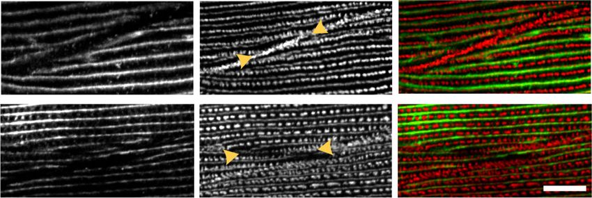
ARTICLE                                                                 NATURE COMMUNICATIONS | https://doi.org/10.1038/s41467-020-18852-4

I
    n striated muscle, sarcomeres are attached end to end to create          three types of muscle IAC-muscle cell boundaries, M-lines and
    myofibrils that extend the length of the elongated muscle cell.           dense bodies. A biochemical pathway for PIX-1 can be inferred
    The myofibrils are tightly packed and connected to each other             from studies of PIX proteins in mammals and pix-1 in several
via intermediate filaments. Myofibrils in both skeletal and cardiac            non-muscle tissues in C. elegans. Analysis of loss of function
muscle cells are connected at the periphery of the muscle cell to            mutants for genes encoding proteins at all known steps of this
the cell membrane and extracellular matrix (ECM) via costa-                  pathway show defects of muscle cell boundaries similar to that of
meres, muscle-specific integrin adhesion complexes (IACs).                    pix-1 mutants. As compared to wild type, a pix-1 null mutant and
Although detectable beneath the entire sarcolemma of skeletal                a pix-1 missense mutant show reduced levels of activated (GTP
muscle, the dystrophin-glycoprotein complex (DGC) is enriched                bound) Rac in muscle. Both deficiency and overexpression of
at costameres1. Genetic deficiencies of dystrophin and a number               wild-type PIX-1 protein result in disrupted muscle cell bound-
of DGC proteins result in muscular dystrophies. Deficiency of                 aries and decreased whole nematode locomotion, suggesting that
costameric component integrin α7, results in a congenital myo-               the level of PIX-1 protein needs to be tightly regulated. Our
pathy2. Heterozygous mutations in several other costameric                   results demonstrate that the PIX signaling pathway has an
proteins result in dilated and hypertrophic cardiomyopathy                   important function in muscle.
including vinculin, α-actinin-2, four and a half LIM protein-2
(FHL2), and ILK3.
   C. elegans is an excellent genetic model organism in which to             Results
learn new principles of sarcomere assembly, maintenance, and                 pix-1 mutants lack IACs at muscle cell boundaries. We screened
regulation4. The major striated muscle of C. elegans is found in             the Million Mutation Project (MMP)12 mutant strains for defects
the body wall and is required for locomotion. Similar to striated            in integrin adhesion complex organization. After screening
muscle in other animals, the thin filaments are attached to Z-                574 strains, we identified one strain, VC20386, that by immu-
disks (called dense bodies), and the thick filaments are attached to          nostaining showed lack of localization of one IAC component,
M-lines. However, the myofibrils are restricted to a narrow                   PAT-6 (α-parvin), at muscle cell boundaries, but normal locali-
~1.5 µm zone adjacent to the cell membrane along the outer side              zation of PAT-6 at M-lines and dense bodies (Fig. 1a). Using a
of the muscle cell, and all the dense bodies and M-lines are                 combination of outcrossing to wild-type and SNP mapping, we
anchored to the muscle cell membrane and ECM. This archi-                    determined that this phenotype is due to a nonsense mutation in
tecture, together with what is known about the molecular com-                a single gene, pix-1 (see “Methods”), with allele designation
position of these structures, indicates that nematode dense bodies           gk299374. We then obtained six additional strains from the
and M-lines also act as costameres. C. elegans body wall muscle              Caenorhabditis Genetics Center (CGC) that contained mutations
also has IACs at the muscle cell boundaries5, where they form                in pix-1. Two are intragenic deletions, one is a nonsense muta-
attachment plaques that anchor the muscle cell to a thin layer of            tion, and three are missense mutations. Except for the deletions,
ECM that lies between adjacent muscle cells.                                 these additional pix-1 alleles come from the MMP. WormBase
   IACs (aka focal adhesions or adhesomes) consist of the                    indicates that there are 17 MMP alleles of pix-1, but the ones we
transmembrane protein integrin and hundreds of different pro-                chose to study either have nonsense mutations (the original one
teins in a complex both in the ECM and especially intracellu-                from our immuno-screening, and one more), or have missense
larly6–9. IACs are important for many cell types. The adhesion of            mutations residing in conserved protein domains (see below for
cells to a matrix is crucial for both tissue formation and for cell          PIX-1 domain structure). By immunostaining, we found the
migration. In stationary cells like muscle, these complexes are              absence of PAT-6 at the muscle cell boundaries in four out of six
rather stable, but in motile cells they are dynamic, with new                of these additional strains (Fig. 1b; Supplementary Fig. 1). The
complexes assembled at the leading edge and older complexes                  two intragenic deletion alleles, gk416 and ok982, and the addi-
disassembled at the trailing edge6. Studies of platelets, leukocytes,        tional nonsense allele, gk299384, show the same boundary defect
and tissue culture cells indicate that normally when integrins are           phenotype as the original allele, gk299374, thus confirming that
expressed on the cell surface they are in a compact or bent or               loss of function of pix-1 is responsible for the phenotype. One of
inactive state, unable to bind to their extracellular targets, but can       the three missense alleles, gk893650, shows a similar phenotype,
become “activated” to bind via several triggers (chemokine to                but two of the missense alleles, gk406361 and gk713465, display a
chemokine receptor interaction, local increase in PIP2 or calpain)           wild-type boundary phenotype. Perhaps these two missense
that lead to binding of the cytoplasmic tail of β-integrin to talin.         mutants, gk406361 and gk713465, showing no phenotype are
Talin binding drives integrin to a more open conformation,                   affecting residues in PIX-1 that are not essential for function.
competent to bind extracellular targets10. Kindlin, which is                 Indeed, gk713465 is an R212Q substitution in a residue that is not
similar in domain composition to talin, is also involved in                  conserved in the RhoGEF domain (see below).
integrin activation by clustering of talin-activated integrins, at              Additional evidence that pix-1 is responsible for the muscle
least in mammalian platelets11. Although we understand the steps             boundary phenotype, is that we were able to rescue the phenotype
involved in the formation of IACs7–9, we do not know how the                 by expressing a wild-type pix-1 cDNA under the control of a
composition of an IAC is determined, and we do not know what                 muscle-specific promoter (Fig. 1c). By immunostaining, we found
determines where an IAC forms. This question is especially                   that in the nonsense mutant, pix-1(gk299374), other IAC
important for skeletal muscle cells, in which one type of IAC (the           components are missing from the muscle cell boundaries,
costamere) forms at regular intervals and anchors the peripherally           including UNC-52 (perlecan) in the ECM, PAT-3 (β-integrin)
located myofibrils to the sarcolemma and ECM at the level of                  at the muscle cell membrane, UNC-112 (kindlin), and UNC-95
Z-disks.                                                                     (Fig. 1d). Thus, we conclude that PIX-1 is required for the
   Here, we show that through a genetic screen in C. elegans,                assembly or stability of IACs at the muscle cell boundary.
identification of a signaling molecule, PIX-1 (orthologous to β-                 The standard view of integrin adhesion sites is that they act as a
PIX in mammals), that is required for assembly or stability of               physical link between the ECM and cell membrane to cortical
IACs only at muscle cell boundaries. PIX proteins consist of SH3             actin filaments. Like other cells, muscle cells are known to have
and RhoGEF domains, and a coiled-coil region, and are known to               cortical actin, however, this has not been previously reported for
act as guanine exchange factors (GEFs) for activation of the small           C. elegans muscle cells. Our attempts to visualize actin filaments
GTPases, Rac1 and/or Cdc42. Antibodies to PIX-1 localize to all              near the muscle cell boundary using phalloidin staining were not

2                NATURE COMMUNICATIONS | (2020)11:5010 | https://doi.org/10.1038/s41467-020-18852-4 | www.nature.com/naturecommunications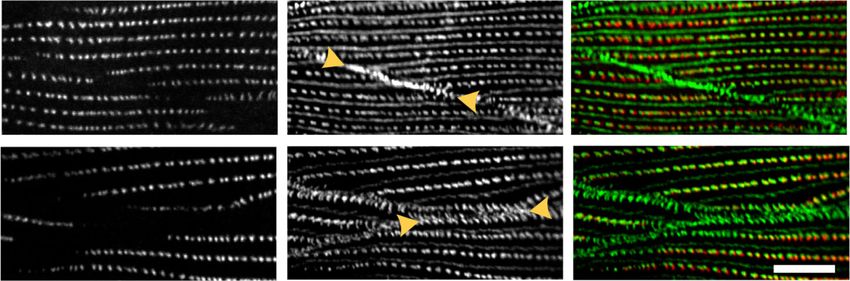
NATURE COMMUNICATIONS | https://doi.org/10.1038/s41467-020-18852-4 ARTICLE
a b SH3 RhoGEF Coiled coil
PiX-1a
Wild type
gk299384
gk406361 gk299374 * gk713465 (Q578stop)
(W44R) (Q153stop) (R212Q)
gk893650
(P190S)
VC20386
gk416 ok982
(deletion) (deletion)
: absence of PAT-6 at the muscle cell boundaries
c d Wild type pix-1 (gk299374)
UNC-52
anti-HA
anti-GFP
PAT-3
B
anti-PAT-6
UNC-95
Merge
UNC-112
Fig. 1 Identification of pix-1 as a gene required for the assembly of IACs at muscle cell boundaries. a Confocal images of several body wall muscle cells
immunostained with antibodies to PAT-6 (α-parvin) from wild type and the strain VC20366 identified by screening 574 MMP strains. Arrowheads point to
the boundaries between muscle cells. b Schematic representation of domains in C. elegans PIX-1a, and the location and nature of 7 pix-1 mutants and
evaluation of their phenotypes. The asterisk denotes the pix-1 mutant allele found in the original strain VC20366. c Muscle-specific expression of a wild-
type cDNA for PIX-1 tagged with HA rescues the phenotype of pix-1(gk299374). The transgene is sfEx61[myo-3p::HA-PIX-1; sur-5::nls::GFP], in which sur-5::
nls::GFP is the transformation marker showing GFP in nuclei. Note that PAT-6 has been restored to the muscle cell boundaries (indicated by arrowheads),
and that HA-tagged PIX-1 localizes to muscle cell boundaries, dense bodies, and M-lines. d Comparison of wild type vs. pix-1(gk299374) immunostained
with antibodies to the indicated IAC proteins and imaged by confocal microscopy. Arrowheads denote muscle cell boundaries. Note that all four proteins
are present in wild type but missing from muscle cell boundaries in the pix-1 mutant. Each image is a representative image obtained from at least 2 fixation
and immunostaining experiments, and imaging at least three different animals. Scale bars in (a), (c) and (d), 10 μm.
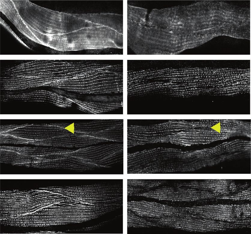
successful, for unknown reasons. As an alternative, we imaged F- cell boundary (bottom two rows in Fig. 2 and Supplementary
actin using strain KAG547 that expresses in body wall muscle Fig. 2). Therefore, although pix-1 is required for assembly or
both GFP-myosin (MHC A) and LifeAct-mCherry. LifeAct is a 17 maintenance of IACs at the muscle cell boundary, it is not
amino acid peptide that binds to F-actin and does not interfere required for the assembly of cortical F-actin to which it likely
with actin dynamics13. In wild-type animals, as shown in Fig. 2 interacts. The fact that two clearly separated F-actin lines can be
(top row), there is clearly a thin band of F-actin that lies near the observed in the pix-1 nonsense mutant indicates that the cells are
muscle cell boundary (indicated by yellow arrow), which can be separated from each other, likely due to less adhesion to the ECM
identified by three criteria: (1) its location between two adjacent lying between adjacent cells.
spindle-shaped body wall muscle cells (Supplementary Fig. 2), (2)
being thinner than a typical I-band which alternates with myosin
A-bands, and (3) not projecting throughout the depth of the Nematode PIX-1 is most similar to human β-PIX. A BLAST
myofilament lattice5 like a typical I-band, which can be discerned search reveals that C. elegans PIX-1 is most similar to mammalian
by observing less intense signal when the optical slice is taken β-PIX and α-PIX. Mammals have 2 PIX proteins, α-PIX and β-
deeper into the lattice (the label of deeper part in Fig. 2 and PIX, encoded by separate genes, whereas C. elegans has a single
Supplementary Fig. 2). If there is cortical actin underneath the gene encoding a single PIX protein14,15. Our sequence and
cell membranes of each adjacent muscle cell, why do we not domain analysis of PIX-1 and human α-PIX and β-PIX proteins
observe two closely spaced lines? The likely reason is that the cells indicates that PIX-1 is quite similar to both α-PIX and β-PIX, but
are very close to each other and these lines are not resolvable by more similar to β-PIX because PIX-1, like β-PIX, is missing the
the light microscope. We crossed the KAG547 strain into pix-1 CH domain found in α-PIX, and the SH3, RhoGEF and coiled-
(gk299374). In this pix-1 nonsense mutant, the muscle cell coil regions of PIX-1 are slightly more identical to those in β-PIX
boundaries clearly have two separated F-actin lines at the muscle (Fig. 3a).
NATURE COMMUNICATIONS | (2020)11:5010 | https://doi.org/10.1038/s41467-020-18852-4 | www.nature.com/naturecommunications 3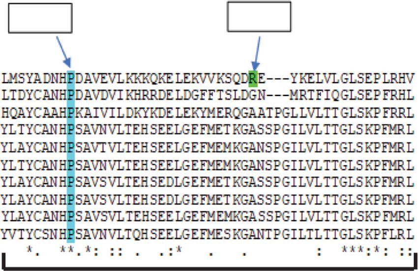
ARTICLE NATURE COMMUNICATIONS | https://doi.org/10.1038/s41467-020-18852-4
Wild type GFP-MYO-3 a Pfam predicted domain strutures
GFP-MYO-3 LifeAct LifeAct
CH SH3 RhoGEF PH Coiled Coil
Human
membrane
α-PIX
Close to
776 aa
SH3 RhoGEF PH Coiled coil
Human
β-PIX
646 aa
SH3 RhoGEF Coiled coil
C.elegans
PIX-1
Deeper
646 aa
part
Percentage identity for conserved domains of PIX-1, α-PIX, and β-PIX
PIX-1 PIX-1 PIX-1
60% 63% 31% 33% 38% 42%
pix-1(gk299374) GFP-MYO-3 SH3 RhoGEF Coiled
Coil
GFP-MYO-3 LifeAct LifeAct α-PIX 88% β-PIX α-PIX 68% β-PIX α-PIX 69% β-PIX
membrane
Close to
b GST-PAK-PBD
pull down
+GTPγS
+GTPγS
+GDP
+GDP
KDa
Deeper
part
250
150
100
75
50
Fig. 2 Live imaging of cortical F-actin at muscle cell boundaries. SIM
images of portions of two adjacent body wall muscle cells from a nematode 37
strain in which a muscle myosin MHC A was tagged with GFP by CRISPR
and LifeAct-mCherry was expressed in muscle cells from a transgene. GFP-
25 *
MYO-3 (MHC A) labels the middle of sarcomeric A-bands, and LifeAct- 20
mCherry labels I-bands, except for F-actin at the boundary between two 15
adjacent muscle cells (indicated by yellow arrows). Note how the signal 10
from the F-actin at the boundary diminishes as the focal plane changes
from close to the outer muscle cell membrane to deeper into the Ponceau S anti-HA
myofilament lattice whereas the F-actin signal from I-bands does not
c GST-PAK-PBD
GST-PAK-PBD
change. Also note that in the pix-1 nonsense mutant, gk299374, there are
two bands of cortical F-actin at the boundary. Each image is a
pull down
pull down
representative image obtained from imaging at three different animals of
Lysate
Lysate
each strain. Scale bar, 5 μm.
PIX-1 acts as a GEF for the Rac, CED-10, in muscle. PIX
pix-1 [null]
pix-1 [null]
pix-1 [null]
pix-1 [null]
wild type
wild type
wild type
wild type
proteins are known to act as guanine nucleotide exchange factors
(GEFs) for Rac1 and/or Cdc42. C. elegans has three Rac proteins
encoded by separate genes, CED-10, MIG-2, and RAC-2. As KDa
shown later, analysis of mutants in these proteins indicate that 250
only CED-10 is required for the assembly of IACs at muscle cell 150
100
boundaries. Therefore, we asked whether the activation of CED- 75
10 might be defective in a pix-1 loss of function mutant. To
50
address this question, we generated a transgenic line in which
HA-tagged CED-10 was expressed in body wall muscle using the
muscle-specific myo-3 promoter. We adapted the use of a com-
mercially available kit for Rac1 activation to measure the status of
37
25
*
CED-10 activation, that is based on the ability of GST-PAK-PBD
20
to pull down activated or GTP-bound CED-10, but not inactive
or GDP-bound CED-10. To validate our assay, worm lysates were 15
incubated with an excess of GDP or the non-hydrolysable GTPγS, 10
and the amount of activated CED-10 was compared by western Ponceau S anti-HA
blot using anti-HA. As shown in Fig. 3b, much more activated
CED-10 was pulled down with GTPγS than with GDP. Our HA-
CED-10 muscle-expressed reporter strain was then crossed into
pix-1(gk299374). As shown in Fig. 3c, less activated CED-10 was western blot and is thus likely to be a null mutant. That the
pulled out from pix-1(gk299374) than from wild type. After amount of activated CED-10 is not zero in this mutant likely
repeating this experiment three times, the mean level of activated reflects the expression of other Rac GEFs in C. elegans muscle,
CED-10 from pix-1(gk299374) was 54.0 +/− 7.6% (mean and one of these known to be UNC-73 (TRIO)16,17. Nevertheless,
standard deviation) of the level from wild type. As shown in these results indicate that PIX-1 is a GEF for the Rac, CED-10, in
Fig. 6b, pix-1(gk299374), shows no detectable PIX-1 protein by body wall muscle.
4 NATURE COMMUNICATIONS | (2020)11:5010 | https://doi.org/10.1038/s41467-020-18852-4 | www.nature.com/naturecommunications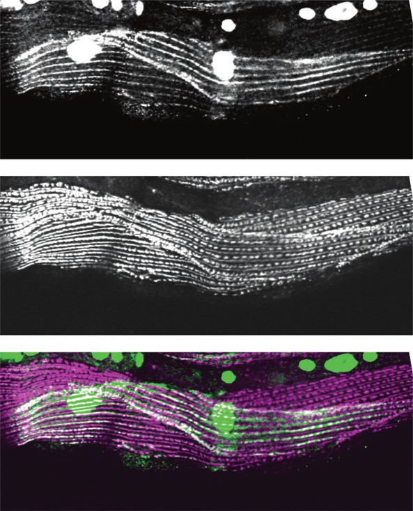
NATURE COMMUNICATIONS | https://doi.org/10.1038/s41467-020-18852-4                                                                   ARTICLE

Fig. 3 PIX-1 is most similar to human β-PIX and acts as a GEF for CED-10        pix-1(syb2137gk299374) displays normal swimming and crawling
(Rac) in muscle. a Schematic representation of predicted domains in C.          motility, and as shown in Fig. 5d, normal muscle cell boundaries.
elegans PIX-1a, human α-PIX and human β-PIX. The bottom triangles show a        We conclude that either loss of function or overexpression of pix-
comparison of percentage identities for SH3, RhoGEF, and coiled-coil            1 results in reduced locomotion and a muscle cell boundary
regions. b Validation of activation assay. Lysates were prepared from           defect.
nematodes expressing HA-CED-10 in body wall muscle using a muscle-
specific promoter, an excess of GDP or GTPγS, was added, and then beads          pix-1 mutants have normal organization of muscle sarcomeres.
coupled to GST-PAK-PBD were used to pull down activated CED-10 (i.e.            The sarcomeres of pix-1 mutants are normally organized: thin
bound to GTP or GTP + GTPγS). Samples were separated on a gel, blotted          filaments (phalloidin), thick filaments (anti-MHC A), dense
and incubated with antibodies to HA. The asterisk indicates the position of     bodies (anti-α-actinin), and M-lines (anti-UNC-89 (obscurin))
GST-PAK-PBD on the blot. Arrow indicates the position of HA-CED-                show the same localization in pix-1(gk299374) as they do in wild-
10•GTP/GTPγS on the western blot. c Deficiency of PIX-1 results in a             type muscle (Supplementary Fig. 3). Therefore, the defects in
reduced level of activated CED-10. Lysates were prepared from two strains,      locomotion in pix-1 mutants might be attributed to a defect in
each expressing HA-CED-10 in body wall muscle: wild type, and pix-1             force transmission through a lack of IACs at the lateral muscle
(gk299374), a nonsense mutant that results in no detectable PIX-1. These        cell boundaries, thus demonstrating both the functional impor-
lysates were incubated with beads coupled to GST-PAK-PBD, and used to           tance of these structures, and the importance of PIX-1 in estab-
pull down activated CED-10. Both the Ponceau S stained blot, and the result     lishing or maintaining these structures.
of the western using anti-HA are shown. The western shows, from left to
right: total HA-CED-10 in pix-1 mutant, total HA-CED-10 in wild type, HA-
CED-10•GTP in pix-1 mutant, and HA-CED-10•GTP in wild type. Asterisk            PIX-1 localizes to M-line, dense body, and adhesion plaque.
indicates the position of GST-PAK-PBD on the blot. Arrow indicates the          We developed two sets of polyclonal antibodies to PIX-1.
position of HA-CED-10 from the lysate, or HA-CED-10•GTP from the                WormBase predicts that pix-1 encodes two protein isoforms, PIX-
pulldown.                                                                       1a (646 residues) and PIX-1b (450 residues) (Fig. 6a). The first
                                                                                immunogen chosen (#1) was expected to generate antibodies that
                                                                                could detect both of these isoforms. The resulting rabbit poly-
                                                                                clonal antibodies were of low titer and allowed western blot
Similar phenotype from deficiency or overexpression of pix-1.                    detection of PIX-1a but not PIX-1b. Also, these antibodies to
In C. elegans muscle, the force of muscle contraction that bends                immunogen #1 failed to localize in muscle by immunostaining
the worm and thus propels locomotion (swimming, crawling,                       experiments. We then generated antibodies to a second immu-
burrowing), is transmitted through all three integrin attachment                nogen (#2). As shown in Fig. 6b, this higher-titer antibody
sites, the M-lines, the dense bodies and the adhesion plaques at                detected a protein of ~80 kDa, close to the expected size for PIX-
the muscle cell boundaries. Thus, mutants that are defective in                 1a (73.2 kDa) from wild type, but not from the nonsense or two
these structures, in many cases, show reduced whole-animal                      intragenic deletion alleles of pix-1. Interestingly, these antibodies
locomotion18. As shown in Fig. 4a and b, the pix-1 nonsense                     detect a PIX-1 protein of normal size and abundance from pix-1
mutant, gk299374, the pix-1 intragenic deletion, gk416 (each                    (gk893650), which expresses PIX-1 with the missense mutation
outcrossed 5× to wild type) and the pix-1 intragenic deletion,                  P190S in the RhoGEF domain. The expression of normal levels of
ok982 (outcrossed 3X to wild type), show reduced locomotion in                  intact PIX-1 protein likely explains why the muscle cell boundary
swimming in buffered water and in crawling along an agar sur-                   defect is more subtle in pix-1(gk893650). (Supplementary Figs. 1;
face. However, pix-1(gk893650) (outcrossed 5× to wild type)                     Fig. 6c; Fig. 9b, c).
which has the missense mutation P190S, and has a more subtle                       We next used these anti-PIX-1 antibodies (to immunogen #2)
boundary defect (see below), displays normal swimming and                       to immunostain body wall muscle. As shown in Fig. 6c, anti-PIX-
crawling motility (Fig. 4a, b). We next wondered whether the pix-               1 localizes to muscle cell boundaries, to M-lines and dense bodies,
1 rescued strain would show normal or near normal locomotion.                   and this staining is not detectable in the pix-1 nonsense,
To our surprise, in both swimming and crawling, the integrated                  gk299374, and intragenic deletion, gk416, mutants. However,
transgene expressing wild-type pix-1 cDNA from the muscle-                      consistent with the immunoblot results anti-PIX-1 staining is
specific promoter for myo-3, [pix-1(gk299374); sfIs20], was slower               detectable at muscle cell boundaries in pix-1(gk893650) P190S.
than wild type (Fig. 4c, d). One possibility is that the integration
occurred in a gene essential for normal locomotion, for example,                Determination of the PIX-1 signaling pathway in muscle.
a muscle or neuronal Unc gene. However, this does not appear to                 Based on studies of β-PIX in mammals and some studies of pix-1
be the explanation for slow movement: Slow movement was                         in C. elegans (but not in muscle), we hypothesized that PIX-1
observed even in a strain in which the extrachromosomal array                   functions in the biochemical pathway shown in Fig. 7a: that it
was expressed in a wild-type background, [wild type; sfEx61]                    activates a Rac/Cdc42 family member, perhaps via the scaffold
(Fig. 4c, d). Thus, the most likely explanation for reduced motility            protein GIT-1 (or additional or another scaffold protein), and this
shown by the strains carrying the transgene, is overexpression of               Rac/Cdc42 family member acts through a PAK protein kinase. C.
PIX-1, which is typical for extrachromosomal or integrated                      elegans has 3 Rac proteins, CED-10, MIG-2, and RAC-2; one
arrays. Indeed, quantitative western blotting using an antibody to              Cdc42, CDC-42; and 3 PAK protein kinases, PAK-1, PAK-2, and
PIX-1 (described below) shows that the integrated array expresses               MAX-2. There are loss or reduction of function mutants available
six times the amount of PIX-1 as found in wild type (Fig. 5a, b).               for all of the proteins indicated in Fig. 7a, except for CDC-42. As
In addition to a motility defect, overexpression of PIX-1 results in            shown in Fig. 7b (and summarized in Fig. 7a), loss of function
a defective muscle cell boundary (Fig. 5c). Finally, although we                mutants in GIT-1, CED-10, PAK-1, and PAK-2, but not MIG-2,
found a motility defect in three independently generated loss of                RAC-2 or MAX-2, result in the absence or reduced accumulation
function mutants, we were concerned that the motility defect                    of PAT-6 at muscle cell boundaries. The defect in pak-2(ok332) is
might result from mutation in a gene closely linked to pix-1. In                more subtle than the other mutants (Fig. 7b, bottom row);
order to eliminate this possibility, we used CRISPR/Cas9 to                     although there is not complete absence of PAT-6, PAT-6 appears
correct the TAA stop codon in pix-1(gk299374), to the wild-type                 less concentrated or more discontinuous at the cell boundaries
sequence of a CAA Gln codon. As shown in Fig. 4c, d, this strain,               than it does in wild type. Also, as shown in Supplementary Fig. 4,

NATURE COMMUNICATIONS | (2020)11:5010 | https://doi.org/10.1038/s41467-020-18852-4 | www.nature.com/naturecommunications                           5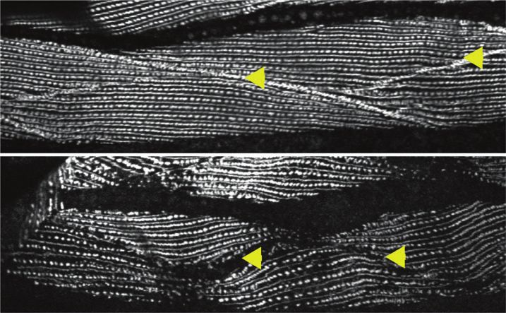
ARTICLE NATURE COMMUNICATIONS | https://doi.org/10.1038/s41467-020-18852-4
a Swimming assay b Crawling assay
2.5 0.4
n.s.
2.0
0.3 n.s.
BBPS in aqueous buffer
BBPS on agar surface
1.5
0.2
1.0
0.1
0.5
0.0 0.0
pe
6)
pe
2)
4)
)
4)
6)
2)
)
50
50
14
98
37
ty
37
41
98
ty
36
36
ild
ild
gk
ok
99
99
gk
ok
89
89
w
w
1(
1(
k2
1(
1(
k2
gk
gk
x-
x-
x-
x-
(g
(g
pi
1(
pi
1(
pi
pi
1
1
x-
x-
x-
x-
pi
pi
pi
pi
c Swimming assay d Crawling assay
2.5 0.5
n.s.
2.0 0.4
BBPS in aqueous buffer
BBPS on agar surface
n.s.
1.5 0.3
1.0 0.2
0.5 0.1
0.0 0.0
pe
pe
)
)
)
4)
20
1
1
20
74
74
74
x6
x6
ty
ty
37
ls
ls
93
93
93
fE
fE
ild
ild
sf
sf
99
29
29
29
;s
;s
w
w
);
);
k2
74
74
pe
gk
pe
gk
gk
(g
93
93
ty
ty
1(
7
7
-1
ild
13
ild
13
x-
29
29
x
pi
pi
w
w
b2
b2
gk
gk
sy
sy
1(
1(
1(
1(
x-
x-
pi
x-
pi
x-
pi
pi
Fig. 4 Both loss of function and overexpression of pix-1 results in reduced locomotion. a Swimming and b crawling assays show that loss of function
mutations in pix-1 result in reduced locomotion compared to wild type. c Swimming and d crawling assays show that both the integrated rescued strain, pix-
1(gk299374); sfIs20, and wild type expressing the rescuing transgene from an extrachromosomal array, sfEx61, have reduced locomotion. In contrast,
CRISPR/Cas9 repair of the nonsense mutation in pix-1(gk299374), called pix-1(syb2137 gk299374), results in normalization of locomotion. In the graphs,
each open circle represents the result from an independently selected animal. The exact n values vary, and these data can be found in the Source Data
Files. Student’s two-sided t test was used to test for significance. Error bars: standard deviations; ****p ≤ 0.0001; n.s.: no significant difference.
we obtained similar results for an additional allele of rac-2, gk281, there are reduced levels of PIX-1 as compared to wild type. These
and an additional allele of ced-10, n1993. However, the disruption data suggest that GIT-1 and PAK-1 are required for PIX-1
of PAT-6 organization at muscle cell boundaries is less severe for stabilization.
ced-10(n1993) than it is for ced-10(n3246). This is consistent with
the nature of the mutations: ced-10(n3246) is a G60R mutation at
a highly conserved residue in the middle of the protein, whereas A boundary defect from mutation P190S in the RhoGEF
ced-10(n1993) is a V128G mutation at the penultimate residue domain. PFAM alignment of RhoGEF domains from PIX pro-
that is only moderately conserved. teins across 10 different species shows that P190 is absolutely
We next asked whether the level of PIX-1 protein might be conserved (Fig. 9a). This conservation suggests that P190 is
affected by the deficiency of other pathway proteins. As shown in required for RhoGEF activity, and that the P190S missense
Fig. 8, using anti-PIX-1 in a quantitative western, we found that mutation results in reduced RhoGEF activity. Our alignment of
in either git-1(ok1848) or pak-1 (ok448), but not ced-10(n3246), these RhoGEF domains also indicates that PIX-1 R212 is not
6 NATURE COMMUNICATIONS | (2020)11:5010 | https://doi.org/10.1038/s41467-020-18852-4 | www.nature.com/naturecommunications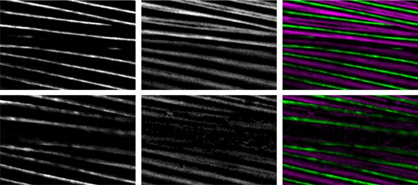
NATURE COMMUNICATIONS | https://doi.org/10.1038/s41467-020-18852-4 ARTICLE
a b 8 a SH3 RhoGEF
7
PIX-1 protein level
6 PIX-1b
pix-1(gk299374);
kDa WT sfIs20 5
150 4 SH3 RhoGEF Coiled coil
3
100 PIX-1a
anti-PIX-1 2
75 1
Immunogen Immunogen
0
#1 #2
20
20
T
anti-Histone H3
W
f ls
15
); s
e)
74
ns
93
se
29
is
gk
0S )
19 se
(m
1(
) P sen
x-
pi
89 4) n)
pi gk2 (de on)
n
o
36 (no
1( 993 leti
1( 82) leti
c
pi ok9 (de
50
7
anti-PAT-6
1( 16)
pi gk4
gk
wildtype;sfEx61
x- e
p
1(
ty
x-
x-
x-
ild
b
pi
kDa
w
250 -
150 -
100 -
75 -
d 50 - anti-PIX-1
30 -
pix-1(syb2137 gk299374)
1X
25 -
20 -
100 - anti-Paramyosin
75 -
2.5X
c PAT-6 PIX-1
wildtype
Fig. 5 Overexpression of pix-1 results in disruption and CRISPR repair
of pix-1(gk299374) results in normalization of muscle cell boundaries.
a, b Western blot analysis showing overexpression of PIX-1 in the strain
(gk299374)
[pix-1(gk299374); sfIs20] in which the muscle cell boundary defect observed
pix-1
in the nonsense mutant pix-1(gk299374) has been rescued by an integrated
array of wild-type pix-1 expressed from a muscle-specific promoter (see
Fig. 1c). a A representative western blot is shown reacted with antibodies to
PIX-1, and to the loading control histone H3. b Graphical summary of four
pix-1(gk416)
independent western blot reactions for wild type vs. pix-1(gk299374); sfIs20.
Means and standard errors of the means are shown. The two strains show
statistically different levels of PIX-1 using a two-sided students t-test with
p < 0.0001 (indicated by ****). Images of the entire western blots are
provided in the Source Data Files. c This same array when expressed in a
(gk893650)
wild-type background [wild-type; sfEx61] disrupts the muscle cell boundary.
pix-1
d pix-1(syb2137gk299374) is pix-1(gk299374) after CRISPR/Cas9 was used
to repair the nonsense mutation. Confocal images of anti-PAT-6 are shown
and reveal that the muscle cell boundary is normal. This is further evidence
that the muscle cell boundary defect is due specifically to mutation in the
pix-1 gene. Arrowheads point to muscle cell boundaries visualized by
reported that by structured illumination microscopy (SIM), in
immunostaining with anti-PAT-6. Each image is a representative image
wild-type muscle, the boundaries appear like a zipper, in which
obtained from at least 2 fixation and immunostaining experiments, and
the two sides of the zipper are closely apposed to each other (as if
imaging at least three different animals. Scale bars, 10 μm.
the zipper were closed). Zoomed-in confocal views of muscle
(Fig. 9b) using antibodies to two different IAC components,
conserved, and this might explain why pix-1(gk713465) R212Q UNC-95 and PAT-6 (α-parvin), show a closed zipper in wild
has no obvious phenotype (Fig. 1b, Supplementary Fig. 1). Closer type, whereas in pix-1(gk893650) the zipper appears open. This
examination of confocal images of pix-1(gk893650) shows that the result is also revealed by SIM imaging of PAT-6 staining (Sup-
boundary defect is more subtle in this mutant than in the non- plementary Fig. 5). A similar “open zipper” appears with anti-
sense and intragenic deletion pix-1 mutants. In Qadota et al.5, we UNC-112 (kindlin) staining (Fig. 9c), although anti-UNC-52
NATURE COMMUNICATIONS | (2020)11:5010 | https://doi.org/10.1038/s41467-020-18852-4 | www.nature.com/naturecommunications 7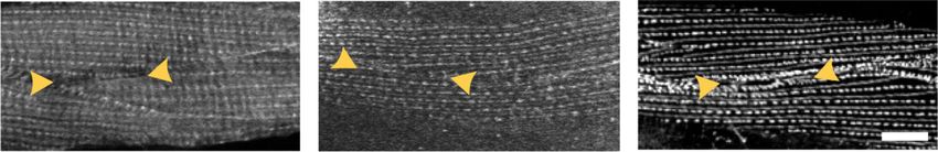
ARTICLE                                                                   NATURE COMMUNICATIONS | https://doi.org/10.1038/s41467-020-18852-4

Fig. 6 Antibodies to PIX-1 detect PIX-1a on a western blot and localize to      (perlecan) and anti-PAT-3 (β-integrin) staining show only less of
muscle cell boundaries, M-lines, and dense bodies. a Schematic                  these proteins at the boundaries of gk893650 (Fig. 9c). In sum-
representation of domains in the predicted isoforms PIX-1a and PIX-1b           mary, the immunoblot and immunolocalization results using
and regions used as immunogens to generate antibodies. b Western blot           anti-PIX-1 show that PIX-1 P190S is a stable protein that localizes
detection of a protein of expected size for PIX-1a from wild type and from      to the general vicinity of the muscle cell boundaries, but exam-
pix-1(gk893650) [P190S] but not from deletion or nonsense pix-1                 ination of other IAC components show that although each half of
mutants. Anti-paramyosin was used as a gel loading control. An image of         the zipper is formed, these halves are abnormally separated from
the entire blot reacted with anti-paramyosin is available in the Source         each other. Taken together with the conservation of P190 in the
Data Files. c Antibodies to PIX-1 localize to muscle cell boundaries and        RhoGEF domain, this suggests that RhoGEF activity is required
with less intensity to M-lines and dense bodies. Each strain was co-            for proper muscle cell boundary organization.
stained with antibodies to PAT-6 and PIX-1, and imaged by confocal.
Note the lack of PIX-1 immunostaining in the nonsense and deletion pix-1
mutants, but strong and disorganized staining in pix-1(gk893650)                P190S may alter RhoGEF structure and binding with Rac•GTP.
[P190S]. Arrowheads point to a muscle cell boundary. Each image is a            We predicted the structure of the RhoGEF domain of PIX-1 by
representative image obtained from at least 2 fixation and                       homology modeling using the NMR structure of human β-PIX19
immunostaining experiments, and imaging at least three different                as a template. We also predicted the structure of a complex
animals of each strain. Scale bar, 10 μm.                                       between PIX-1 and both GDP- and GTP-bound forms of Rac1
                                                                                GTPase, using GEF-Rac1 complexes as templates. We analyzed
                                                                                the helical conformation in PIX-1 molecular dynamic (MD)

                          a
                                                                 GIT-1 (scaffold protein,Arf GAP)
                                                        PIX-1 (βPIX)

                                           Rac/                              Rac/                       PAK-1
                                          Cdc42                             Cdc42                       (PAK)
                                                              ?
                                           GDP              (GDP)             GDP
                                                                                                        PAK-2
                                                                                                        (PAK)

                                          Rac in C.elegans: CED-10 MIG-2               RAC-2
                                          Cdc42 in C.elegans: CDC-42                                    MAX-2
                                             : Disorganization of PAT-6 at muscle cell boundary         (PAK)
                                             : Normal PAT-6 at muscle cell boundary

                          b                                                anti-PAT-6
                                                                                 (gm103)
                           (ok448)

                                                                                  mig-2
                            pak-1
                           (ok1848)

                                                                                 (ok326)
                                                                                  rac-2
                             git-1

                                                                                 (ok1904)
                                                                                  max-2
                           (n3246)
                           ced-10

                                                                              *2.5x zoom
                           (ok332)

                                                                                (ok332)
                            pak-2

                                                *
                                                                                 pak-2

Fig. 7 Mutations in genes encoding known proteins of a PIX-1 pathway result in muscle cell boundary disruption. a Putative PIX-1 biochemical pathway
based on what is known of PIX proteins in mammals and other cell types in C. elegans. Also indicated is a summary of the results shown in b. b Confocal
images of body wall muscle from the indicated mutants immunostained with anti-PAT-6. Note reduced or disorganized PAT-6 localization at muscle cell
boundaries (indicated by arrowheads) in pak-1, git-1, ced-10, and pak-2 mutants, but normal PAT-6 localization in mig-2, rac-2, and max-2 mutants. A higher
magnification view (bottom row, right) of pak-2(ok332) is shown because it has a more subtle defect than the other mutants. Results on second alleles for
rac-2 and ced-10 are shown in Supplementary Fig 3. Each image is a representative image obtained from at least 2 fixation and immunostaining
experiments, and imaging at least three different animals of each strain. Scale bar, 10 μm.

8                  NATURE COMMUNICATIONS | (2020)11:5010 | https://doi.org/10.1038/s41467-020-18852-4 | www.nature.com/naturecommunications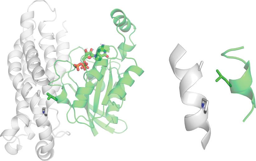
NATURE COMMUNICATIONS | https://doi.org/10.1038/s41467-020-18852-4                                                                   ARTICLE

                   2.5                                                           complex, but present 24.4% of the simulation in wild type.
                                                                                 Combined, MD analyses suggest that the P190S mutation
                                              n.s.                               enhances GTP-bound Rac1 interaction with PIX-1.
                   2.0

                                                                                 P190S reduces the level of activated CED-10 in muscle. To
  PIX-1 protein

                   1.5                                                           understand how the P190S mutation results in a normal level of
                                                                                 PIX-1 protein but disrupted muscle cell boundary structures, pix-
                                                                                 1(gk893650) [P190S] was crossed into the transgenic line in which
                   1.0                                                           HA-tagged CED-10 is expressed in body wall muscle cells, made
                                                                                 whole worm lysates and used GST-PAK-PBD to pull down GTP-
                   0.5                                                           bound CED-10. As shown in Fig. 10d, less activated CED-10 was
                                                                                 pulled out from pix-1(gk893650) [P190S] than from wild type.
                                                                                 Repeating this experiment three times, the mean level of activated
                   0.0                                                           CED-10 from P190S was 52.9 +/− 5.7% (mean and standard
                         Wildtype     git-1  ced-10 pak-1                        deviation) of the level from wild type. Therefore, the P190S
     kDa                            (ok1848) (n3246) (ok448)                     mutation in the RhoGEF domain of PIX-1 reduces its GEF
                                                                                 activity.
       100
                                                               anti-PIX-1
                  75                                                             Discussion
                                                                                 By screening a collection of adult-viable C. elegans mutants by
                  20                                           anti-Histone-H3   immunostaining, we identified a strain in which IAC components
                                                                                 are missing from the muscle cell boundaries but present and
Fig. 8 PIX-1 levels are reduced in git-1 and pak-1 mutants. Equal quantities     normally localized at M-lines and dense bodies. These boundaries
of total SDS-soluble proteins from wild type, git-1(ok1848), ced-10(n3246),      consist of cell to ECM to cell attachments. The defect in the strain
and pak-1(ok448), were resolved on a gel, blotted to the membrane and            was mapped to a single mutant gene, pix-1, which encodes a PIX
reacted with anti-PIX-1, and as a loading control with anti-histone H3. After    protein, known from previous studies to be a RhoGEF for Rac/
normalization to the amount of histone H3, the levels of PIX-1 in the            Cdc42. As compared to wild type, a pix-1 null mutant shows an
mutants were compared to the level of PIX-1 in wild type. Representative         ~50% reduction in the level of activated (GTP bound) Rac in
immunoblot results are shown below the graph. N = 4 independent western          muscle. Despite having normally organized sarcomeres, multiple
blot reactions from each strain; means and standard error of the means are       pix-1 mutants display reduced whole-animal locomotion. We
shown; git-1 and pak-1 mutants show statistically different levels from wild-    hypothesize that this reduced motility results from decreased
type based on a two-sided student’s t-test at p ≤ 0.0001 (indicated              transmission of lateral forces between muscle cells. Interestingly,
by ****). Images of the entire western blots are provided in the Source          in addition to deficiency of PIX-1, muscle-specific overexpression
Data Files.                                                                      of PIX-1 protein also results in decreased locomotion and dis-
                                                                                 rupted muscle cell boundaries. Perhaps these results reflect the
                                                                                 requirement for PIX-1 signaling to be set at just the optimal level
simulations to predict structural effects of the P190S mutation. In              for proper assembly or maintenance of IACs at the muscle cell
PIX-1 simulations, we observed that the serine eliminates the                    boundary.
helical kink at the P190 position (Fig. 10a, left pair). This same                  Antibodies to PIX-1 localize to all 3 IACs–muscle cell
effect is not observed in Rac1 complexes (Fig. 10a, right pair). The             boundaries, M-lines and dense bodies—and yet PIX-1 is only
presence of the GTPase appears to maintain the kinked con-                       required at muscle cell boundaries. One possibility is genetic
formation at the 190 position, even with the serine substitution.                redundancy, that is, there is a second PIX protein that is localized
    However, further investigation of the complexes in MD                        to M-lines and dense bodies that compensates for loss of PIX-1 at
simulations reveals that the mutation modulates the interaction                  these sites. This does not seem to be the case however, since no
of PIX-1 with Rac1 in complexes. We evaluated the (i) contact                    PIX-1 paralogs can be found by querying the C. elegans proteome.
surface area, (ii) van der Waals interactions at the interface, and              Another possibility is that there are additional RhoGEF-
(iii) root mean square fluctuations (RMSF) of protein residues.                   containing proteins with RacGEF activity like PIX-1 that are
Our analyses reveal that the introduction of the P190S mutation                  present at M-lines and dense bodies, but not at muscle cell
stabilizes a putative complex between PIX-1 and GTP-bound                        boundaries. We find that there are a total of 17 proteins in C.
Rac1. We observe an increase in the contact surface area between                 elegans that contain RhoGEF (DH) domains and are expressed in
the two proteins in the mutant compared to the wild-type PIX-1                   muscle, based on the query of SAGE data17(Supplementary
complex (Fig. 10b). We examined a predicted van der Waals                        Table 1). These 17 proteins include PIX-1, and UIG-1 and UNC-
interaction on the interface between A186 (PIX-1) and L70                        89 that have been studied previously20,21. UNC-89 is specifically
(located in the Switch II region of Rac1). This interaction, which               localized to M-lines22,23, and the DH domain of UNC-89 speci-
lies in the vicinity of P190, exists in the mutant P190S complex                 fically activates RHO-1(RhoA)24, and is thus not relevant here.
but not in the wild-type complex. In addition, RMSF analysis                     The properties of UIG-1 somewhat support this hypothesis: UIG-
reveals that the interface is more stabilized in the P190S complex               1 is localized to dense bodies and is a GEF for Cdc42. TIAM-1 is
(Supplementary Fig. 6). RMSF values are lower around the                         reported on WormBase to activate Rac. UNC-73 has two RhoGEF
interface of the mutant complex, indicative of reduced fluctua-                   domains, one that activates Rac and one that activates RhoA16.
tions and a more stable interaction.                                             The muscle intracellular locations of TIAM-1 and UNC-73 are
    All the trends that are observed here are reversed in the PIX-1              unknown.
complexes with GDP-bound Rac1. Contact surface area between                         Another possible reason that PIX-1 is required at muscle cell
PIX-1 and Rac1 is decreased in the mutant complex relative to                    boundaries but not at M-lines dense bodies can be envisioned.
wild type (Fig. 10b). Consistent with this observation, the van der              We observe the loss of IACs at muscle cell boundary when either
Waals interaction shown in Fig. 10c is lost in the mutant                        the PIX-1 pathway is reduced or increased in activity; there are

NATURE COMMUNICATIONS | (2020)11:5010 | https://doi.org/10.1038/s41467-020-18852-4 | www.nature.com/naturecommunications                           9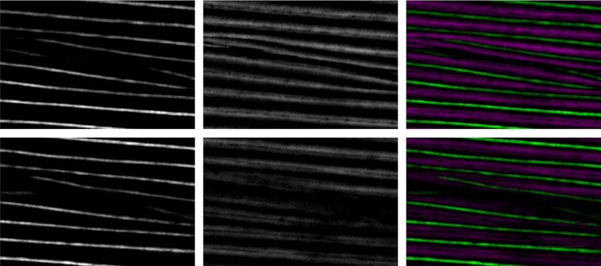
ARTICLE                                                                  NATURE COMMUNICATIONS | https://doi.org/10.1038/s41467-020-18852-4

                       a                          pix-1 (gk893650)            pix-1 (gk713465)

                                                         P1905                      R212Q

                                     C. elegans
                                     P. pecificus
                                     D.melanogaster
                                     H. sapiens
                                     P. vitticeps
                                     L. domestica
                                     R. rattus
                                     M. musculus
                                     D. leucas
                                     D. rerio

                                                                           RhoGEF

                      b                         ATN-1                      UNC-95                              Merge
                      Wildtype
                      pix-1(P190S)

                                                UNC-89                      PAT-6                              Merge
                        Wildtype
                        pix-1(P190S)

                      c                         UNC-52                      PAT-3                            UNC-112
                        pix-1(p190S)

Fig. 9 P190 is conserved in PIX RhoGEF domains and required for PIX-1 function at muscle cell boundaries. a PFAM alignment of RhoGEF domain
sequences of PIX proteins from 10 species showing that P190 is absolutely conserved. In contrast, R212 is not conserved, which might explain why pix-1
(gk713465) [R212Q] has no obvious phenotype. b Confocal microscopy of wild type and pix-1(gk893650) [P190S] mutant co-stained with antibodies to
ATN-1 (α-actinin) and UNC-95 (top two rows), and co-stained with UNC-89 (obscurin) and PAT-6 (α-parvin) (bottom two rows). Note that in pix-1
(gk893650), at the muscle cell boundary, the two halves of the zipper are separated, whereas in wild type they are together. c Confocal imaging of pix-1
(gk893650) [P190S] stained with antibodies to UNC-52 (perlecan), PAT-3(β-integrin) or UNC-112 (kindlin). Note that these IAC proteins also show
reduced or disrupted localization at muscle cell boundaries. Arrowheads bracket a muscle cell boundary. Each image is a representative image obtained
from at least 2 fixation and immunostaining experiments, and imaging at least three different animals of each strain. Scale bar, 10 μm.

boundary defects in loss of function for pix-1, git-1, ced-10, and            dis-assembling at faster rates would be more sensitive to PIX-1
pak-1, and from overexpression of pix-1. These results could be               cycling. Therefore, we have considered the hypothesis that PIX-1
interpreted as indicating a requirement for cycling of the                    is required at MCBs because muscle cell boundaries are
RacGTPase rather than its absolute activity. If IAC assembly                  more dynamic that M-lines and dense bodies. However, our
required the PIX-1 pathway, then IACs that are assembling and                 preliminary FRAP experiments with GFP tagged PAT-6 and

10                NATURE COMMUNICATIONS | (2020)11:5010 | https://doi.org/10.1038/s41467-020-18852-4 | www.nature.com/naturecommunications
NATURE COMMUNICATIONS | https://doi.org/10.1038/s41467-020-18852-4 ARTICLE
a PIX-1 PIX-1-Rac1(GTP)
b
Contact area
(Å2)
Start WT-Rac1 (GTP) 1298.3
End P190S-Rac1 (GTP) 1742.0
P190 WT-Rac1 (GDP) 2047.2
S190
P190S-Rac1 (GDP) 1597.4
(WT) (P190S) (WT) (P190S)
c
N
Distance Interaction
(Å) %
A186 WT-Rac1 (GTP) 9.8 1.2
P190S-Rac1 (GTP) 4.4 65.5
WT-Rac1 (GDP) 5.1 24.4
L70
P190S-Rac1 (GDP) 9.1 0.4
C
PIX-1 Rac1
d
pix-1 [P190S] GST-PAK-PBD
pix-1 [P190S] GST-PAK-PBD
pull down
pull down
Lysate
Lysate
pix-1 [P190S]
pix-1 [P190S]
wild type
wild type
wild type
wild type
KDa
250
150
100
75
50
37
25
*
20
Ponceau S anti-HA
UNC-97, show no differences in turnover rates at muscle cell C. elegans has a single PIX protein, in mammals, there are two
boundaries vs. dense bodies, so this idea does not seem viable. PIX proteins, α-PIX and β-PIX, and these have been shown to be
It should be emphasized that prior to our study, there were no important for development and function of nervous and immune
known genes that when mutated resulted in loss or disorganiza- systems25,26. In C. elegans, PIX-1 has been shown to be required
tion of IACs specifically at muscle cell boundaries. This easily for several events during development: control distal tip cell shape
scorable phenotype allowed us to determine that each component and migration (important for germ cell formation)15, migration
of the known PIX-1 signaling pathway is required for the of Q neuroblasts that give rise to sensory and inter-neurons27,
assembly or stability of IACs at muscle cell boundaries. Although tension-dependent morphogenesis of epidermal cells28, and for
NATURE COMMUNICATIONS | (2020)11:5010 | https://doi.org/10.1038/s41467-020-18852-4 | www.nature.com/naturecommunications 11ARTICLE NATURE COMMUNICATIONS | https://doi.org/10.1038/s41467-020-18852-4 Fig. 10 P190S may alter RhoGEF structure and interaction with Rac, and in muscle there is a reduction in activated Rac. a Comparison of helical conformations at the start (gray) and end (cyan) of PIX-1 and PIX-1-Rac1 complex MD simulations. Wild type PIX-1 maintains the predicted kinked helical conformation near P190. (This proline is on helix F in the β-PIX structure.) Mutant PIX-1 reveals a loss of the kink in the helix by the end of the simulation. In simulations of PIX-1 complexed with GTP-bound Rac1, the kinked conformation is maintained in the mutant complex. The same result is observed with GDP-bound Rac1 (not shown). b Contact surface areas were calculated for wild type and mutant PIX-1-Rac1 complexes. In GTP-bound Rac1 complexes, the mutation leads to an increase in contact surface area. In GDP-bound Rac1 complexes, the mutation leads to a decrease in the contact surface area. c The predicted PIX-1-Rac1 complex reveals an interaction on the interface near the P190 position. This interaction between A186 of PIX-1 with L70 of Rac1 (Switch II region) is quantified as an average distance between the residues and percentage of time the residue distance is
NATURE COMMUNICATIONS | https://doi.org/10.1038/s41467-020-18852-4                                                                                       ARTICLE

810 μl of fixative (50% Bouin’s fixative, 50% methanol, 1.2% β-mercaptoethanol) at       temperature for 5–10 min to solidify, images were taken using a Nikon N-SIM
room temperature for 30 min, then frozen in liquid nitrogen for 5 min, thawed and      microscope system, as described above.
continuing incubation at room temperature for an additional 30 mins. The worms
were pelleted and washed 3× with 1.4 ml of BTB solution (20 mM sodium borate
pH 9.5, 0.5% Triton X-100, 2% β-mercaptoethanol), and then resuspended in 1 ml         Protein sequence analysis. Nematode PIX-1a protein sequence was obtained
of BTB and continuing incubation with mixing for 1 h. The worms were pelleted          from Wormbase. A BLAST homology search identified human orthologs of the
again and resuspended in 1 ml of BTB and incubated for 3 hrs with mixing at room       nematode protein using the NCBI PubMed database. The domain organization for
temperature. The worms were pelleted and washed with 1 ml of BT (20 mM                 PIX-1 and its orthologs were analyzed via PFAM. Human α-PIX and β-PIX amino
sodium borate pH 9.5, 0.5% Triton X-100), pelleted, and washed 2× with 1 ml of         acid sequences were aligned with PIX-1 using pBLAST to determine percent
AbA buffer (PBS, 1% bovine serum albumin, 0.5% Triton X-100, 1 mM sodium               identities for each domain (Fig. 3a). PIX-1 RhoGEF domains from 10 organisms
azide, 1 mM EDTA), pelleted, resuspended in 1 ml of AbA and incubated with             were compared also using pBLAST (Fig. 9a).
mixing for 30 mins, pelleted and resuspended in 100 μl of AbA. Immunostaining
was conducted using 5 μl of a suspension of these fixed worms together with 20 μl
of primary antibody in AbA and incubating overnight with mixing. The animals
were pelleted and washed 4X with PBS + 0.5% Triton X-100, pelleted, removing as        Measurement of CED-10 activation state in body wall muscle. An HA-tagged
much supernatant as possible and incubating with 20 μl of secondary antibody in        cDNA encoding full-length CED-10 was amplified by PCR using the RB2 cDNA
AbA for 2 h, followed by washing 4× with PBS + 0.5% Triton X-100, and finally           library as template and was cloned into vector pKS-HA8(Nhex2), the DNA
removing as much supernatant as possible and mounting 5 μl of resuspended              sequenc- verified, and then the NheI fragment was excised and inserted into
worms with 5 μl of DAPCO solution (20 mM Tris-HCl pH 8.0, 0.2 M 1,4-diaza-             pPD95.86 designed to express HA-CED-10 in muscle from the myo-3 muscle-
bicyclo-2,2,2-octane (DABCO), 90% glycerol) on a glass slide with a coverslip and      specific promoter. This plasmid, pPD95.86-HA-CED-10, at 10 ng/μl together with
sealed with nail polish. The following primary antibodies were used at 1:200           plasmid pTG96 which expresses SUR-5-NLS-GFP as a transformation marker at
dilution except as noted: anti-PAT-6 (rat polyclonal)36, anti-UNC-52 (mouse            90 ng/μl was microinjected into wild-type animals to generate a transgenic strain
monoclonal MH2)37, anti-PAT-3 (1:100 dilution; mouse monoclonal MH25)38,39,            GB314, sfEx63[myo-3p::HA::CED-10; sur-5::NLS::GFP]. This extrachromosomal
anti-UNC-95 (rabbit polyclonal Benian-13)40, anti-UNC-112 (1:100 dilution)20,          array was integrated into the genome by ultraviolet irradiation44 with modifications
anti-MHC A (mouse monoclonal 5–6)41, anti-UNC-89(rabbit polyclonal EU30)22,            (Peter Barrett, personal communication). The resulting nematode strain is called
anti-ATN-1 (mouse monoclonal MH35)42, anti-HA (mouse monoclonal; H3663;                GB315, sfIs22 [myo-3p::HA::CED-10; sur-5::NLS::GFP]. This strain was crossed
Sigma-Aldrich), and anti-GFP (rabbit polyclonal; Thermo Fisher, A11122). Sec-          into pix-1(gk299374) 5X OC, to generate strain GB316, pix-1(gk299374); sfIs22, and
ondary antibodies, used at 1:200 dilution, included anti-rabbit Alexa 488, anti-rat    crossed into pix-1(gk893650) [P190S] 5X OC, to generate strain GB318, pix-1
Alexa 594, and anti-mouse Alexa 594, all purchased from Invitrogen. Fixation and       (gk893650); sfIs22. After growing several grams of worms from GB315, GB316, and
phalloidin-rhodamine staining staining was conducted as described43. Images were       GB318, worm powders were prepared by grinding in a mortar and pestle under
captured at room temperature with a Zeiss confocal system (LSM510) equipped            liquid nitrogen on a bed of dry ice. We modified the Rac1 Activation Assay
with an Axiovert 100 M microscope and an Apochromat x63/1.4 numerical                  Biochem Kit (cat. #BK035, Cytoskeleton, Inc.) as follows: One small spatula-full of
aperture oil immersion objective, in 1× and 2.5× zoom mode. For the images             worm powder was added to 3 ml of ice-cold Cell Lysis Buffer containing protease
presented in Fig. 2, and Supplemental Figs. 2 and 5, super-resolution microscopy       inhibitor cocktail, vortexing for 1 min, and centrifuging at maximum speed in a
was performed with a Nikon N-SIM system in 3D structured illumination mode on          microcentrifuge for 10 min at 4°. A small portion supernatant was used for protein
an Eclipse Ti-E microscope equipped with a 100×/1.49 NA oil immersion objective,       concentration determination using the BCA Assay (cat. #23225, ThermoScientific),
488- and 561-nm solid-state lasers, and an EM-CCD camera (DU-897, Andor                and multiple 200 μl aliquots of the remainder were snap frozen in liquid nitrogen
Technology). Super-resolution images were reconstructed using the N-SIM module         and stored at −70°. The protein concentrations of the lysates varied from 1.25 to
in NIS-Elements software. For all the images, confocal, and SIM, the color balances    3.4 mg/ml. Positive and negative controls were created by adding to 250 μg of total
were adjusted by using Adobe Photoshop (Adobe, San Jose, CA).                          protein of wild-type worm lysate, GTPγS to a final concentration of 0.20 mM, or
                                                                                       GDP to a final concentration of 1 mM, respectively. 250 μg of total protein from
                                                                                       each lysate were added to 10 μg of GST-PAK-PBD Beads, and incubated with
Mapping the phenotype to pix-1. The original MMP strain VC2038612 has out-             mixing for 1 h, 15 min, at 4°. Then, the beads were pelleted at 4° by spinning at
crossed to wild type N2 Bristol three times, each time selecting for the PAT-6         4000 × g for 1 min, supernatant carefully removed, and the beads were washed 1×
muscle boundary defect. Comparison of genomic sequences of VC20386 and N2              with Wash Buffer, and the beads pelleted. After removing as much supernatant as
allowed the selection of SNPs on each chromosome arm to distinguish the two            possible, 20 μl of 2× Laemmli were added, vortexed for 5 s, heated at 95° for 3 min,
strains. PCR and Sanger sequencing of these 12 segments from the third outcrossed      centrifuged at top speed for 3 min, and then 20 μl of each supernatant were run on
strain revealed that only the left arm of chromosome III and the right arm of X        a 12% SDS-PAGE and transferred to nitrocellulose. HA-CED-10 was visualized by
were derived from VC20386. For III and X, we identified genes known to be               incubating with rabbit monoclonal antibodies to HA at 1:1000 dilution (cat.
expressed in muscle17, and of these identified four genes on III and three genes on     #C29F4, Cell Signaling Technology), and reacting with ECL reagents and exposed
X, that had nonsense or non-conservative missense mutations in VC20386. RNAi           to film. We used a flat-bed scanner to image the Ponceau S staining of the blot
for three of them failed to reveal the PAT-6 boundary defect. One of the 7 genes,      using the “reflective” mode in 24 bit color, and to image the ECL reactions recorded
pix-1 on X, had a nonsense mutation. Six additional mutant alleles of pix-1 were       on film using the “film scan” mode in gray scale. The images were opened in
obtained from CGC, and four of these six mutants also showed the PAT-6                 AdobePhotoshop, and after inverting the ECL images, both the ECL bands (HA-
boundary defect (detailed in Results).                                                 CED-10) and the Ponceau S bands (GST-PAK-PBD) “mean” and “pixel” values
                                                                                       were recorded. The absolute intensity of each band was a product of these two
                                                                                       values. The HA-CED-10 band products were normalized by dividing by the GST-
Transgenic rescue. An HA-tagged cDNA encoding full-length PIX-1a was                   PAK-PBD bands for each lane.
amplified by PCR using the RB2 cDNA library (provided by Robert Barstead) as
template and was cloned into vector pPD95.86 (provided by Andrew Fire) designed
to express HA-PIX-1a in muscle by the myo-3 muscle-specific promoter. This              Swimming and crawling assays. For swimming assays, day 2 adults from two, 6
plasmid, pPD95.86-HA-PIX-1a at 10 ng/μl together with plasmid pTG96 which              cm NGM OP50 seeded plates were washed off the plates, washed free from bacteria
expresses SUR-5-NLS-GFP as a transformation marker at 90 ng/μl was micro-              and collected into M9 buffer such that the ratio of worms to buffer was 1:1. 2 ml of
injected into pix-1(gk299374) 5X o.c. to generate the strain GB286, pix-1(gk299374);   M9 buffer was added to an unseeded 6 cm NGM plate where upon 5 μl of worm
sfEx61[myo-3p::HA::PIX-1a; sur-5::NLS::GFP]. This extrachromosomal array was           suspension was added to the center of the plate. Worms were allowed 5 min to
integrated into the genome by ultraviolet irradiation44 with modifications (Peter       adapt before a video recording of their swimming motions was made using a
Barrett, personal communication). The resulting nematode strain is called GB288,       dissecting stereoscopic microscope fitted with a CMOS camera (Thorlabs). Ten,
pix-1(gk299374); sfIs20 [myo-3p::HA::PIX-1a; sur-5::NLS::GFP]. To remove back-         10 s videos were recorded for each nematode strain from different sections of the
ground mutations induced by UV, GB288 was backcrossed to pix-1(gk299374) 1X,           plate, each video tracking the motion of ~10 worms. The video data were analyzed
recovering strain GB290, and this strain was tested in locomotion assays. The          by Image J FIJI WrmTracker software46 to obtain body bends per second (BBPS).
extrachromosomal array sfEx61 [myo-3p::HA::PIX-1a; sur-5::NLS::GFP], was also          After removing outliers and animals that had moved out of frame, ~30 animals
crossed into N2 to generate the strain GB295.                                          were analyzed for each strain. The resulting BBPS values for each mutant strain
                                                                                       were compared to wild type and differences were tested for statistical significance
                                                                                       using a Student T-test.
Imaging of F-actin at the muscle cell boundary. We used strain KAG547 kindly               For crawling assays, day 2 adults were harvested as above, except that all
provided by Kathrin Gieseler (Universite Claude Bernard Lyon): GFP::MYO-3; Ex          washing steps used M9 buffer containing 0.2 g/L gelatin. Five microliters of worm
[myo-3p::LifeAct::mCherry]. Animals were washed off of plates and washed free          suspension was added to the center of a 6 cm unseeded NGM plate, and the excess
from bacteria using M9 buffer and then immobilized by incubation in 10 μM              liquid was removed. After 5 min for adaptation, worm crawling was recorded using
levamisole in M9 for 10 min. Approximately 50–100 animals in 3 μl were added to        the above-mentioned strategy for extraction of BBPS for individual worms in each
7 μl of ice-cold 25% Pluronic F127 in M945 lying on a cold glass slide, to which was   video. The resulting values for each strain were compared to wild type for statistical
added a cover slip and it was sealed with nail polish. After incubation at room        analysis using a Student T-test for significance.

NATURE COMMUNICATIONS | (2020)11:5010 | https://doi.org/10.1038/s41467-020-18852-4 | www.nature.com/naturecommunications                                                  13
You can also read