SARS-COV-2 INFECTION IN THE MOUSE OLFACTORY SYSTEM - NATURE
←
→
Page content transcription
If your browser does not render page correctly, please read the page content below
Ye et al. Cell Discovery (2021)7:49
                                    https://doi.org/10.1038/s41421-021-00290-1
                                                                                                                                                                                                        Cell Discovery
                                                                                                                                                                                                     www.nature.com/celldisc

                                     ARTICLE                                                                                                                                                    Open Access

                                    SARS-CoV-2 infection in the mouse olfactory
                                    system
                                    Qing Ye1, Jia Zhou1, Qi He2, Rui-Ting Li1, Guan Yang2, Yao Zhang2, Shu-Jia Wu2, Qi Chen1, Jia-Hui Shi2,
                                    Rong-Rong Zhang1, Hui-Ming Zhu2, Hong-Ying Qiu1, Tao Zhang2, Yong-Qiang Deng 1, Xiao-Feng Li1, Jian-Feng Liu3,
                                    Ping Xu2, Xiao Yang2 and Cheng-Feng Qin 1,4

                                      Abstract
                                      SARS-CoV-2 infection causes a wide spectrum of clinical manifestations in humans, and olfactory dysfunction is one of
                                      the most predictive and common symptoms in COVID-19 patients. However, the underlying mechanism by which
                                      SARS-CoV-2 infection leads to olfactory disorders remains elusive. Herein, we demonstrate that intranasal inoculation
                                      with SARS-CoV-2 induces robust viral replication in the olfactory epithelium (OE), not the olfactory bulb (OB), resulting
                                      in transient olfactory dysfunction in humanized ACE2 (hACE2) mice. The sustentacular cells and Bowman’s gland cells
                                      in the OE were identified as the major target cells of SARS-CoV-2 before invasion into olfactory sensory neurons (OSNs).
                                      Remarkably, SARS-CoV-2 infection triggers massive cell death and immune cell infiltration and directly impairs the
                                      uniformity of the OE structure. Combined transcriptomic and quantitative proteomic analyses revealed the induction
                                      of antiviral and inflammatory responses, as well as the downregulation of olfactory receptor (OR) genes in the OE from
                                      the infected animals. Overall, our mouse model recapitulates olfactory dysfunction in COVID-19 patients and provides
                                      critical clues for understanding the physiological basis for extrapulmonary manifestations of COVID-19.
                  1234567890():,;
1234567890():,;
                  1234567890():,;
1234567890():,;

                                    Introduction                                                                                        alteration of smell as one of the most predictive symptoms
                                      The emergence of coronavirus disease 2019 (COVID-                                                 for COVID-19 screening5,6.
                                    19), caused by the newly identified severe acute respira-                                               The perception of smell begins with odorant binding to
                                    tory syndrome coronavirus 2 (SARS-CoV-2), has led to a                                              the olfactory receptors (ORs) of olfactory sensory neurons
                                    global crisis. The clinical manifestations of SARS-CoV-2                                            (OSNs) along the upper surface of the olfactory epithe-
                                    infection predominantly involve the respiratory system,                                             lium (OE). Each OSN projects an axon into the glomer-
                                    including cough, sore throat, pneumonia, and acute                                                  ulus of the olfactory bulb (OB) and then synapses with
                                    respiratory distress syndrome (ARDS)1,2. As the disease                                             second-order neurons to convey odour information to the
                                    continues to spread widely, a significant portion of                                                 olfactory cortex. Previously, upper respiratory tract
                                    COVID-19 patients are developing anosmia, hyposmia, or                                              infections were considered a common cause of olfactory
                                    other olfactory dysfunctions according to clinical                                                  disorders. Mouse models have been used to reproduce
                                    reports3–5. Accumulated evidence has established the                                                olfactory system infection and subsequent olfactory dys-
                                                                                                                                        function7,8. For example, post viral olfactory disorders
                                                                                                                                        were observed in Sendai virus-infected mice by the buried
                                    Correspondence: Ping Xu (xuping_bprc@126.com) or Xiao Yang (yangx@bmi.                              food pellet test (BFPT), as well as the impairment of OE
                                    ac.cn) or Cheng-Feng Qin (qincf@bmi.ac.cn)
                                    1                                                                                                   and OB tissues9. However, an animal model that can
                                     State Key Laboratory of Pathogen and Biosecurity, Beijing Institute of
                                    Microbiology and Epidemiology, Beijing, China                                                       recapitulate the olfactory dysfunctions seen in COVID-19
                                    2
                                     State Key Laboratory of Proteomics, National Center for Protein Science                            patients remains lacking.
                                    (Beijing), Beijing Institute of Lifeomics, Beijing, China
                                                                                                                                           Human nasal respiratory epithelium (RE) cells exhibit
                                    Full list of author information is available at the end of the article
                                    These authors contributed equally: Qing Ye, Jia Zhou, Qi He, Rui-Ting Li, Guan                      high expression of angiotensin-converting enzyme 2
                                    Yang, Yao Zhang

                                    © The Author(s) 2021
                                                        Open Access This article is licensed under a Creative Commons Attribution 4.0 International License, which permits use, sharing, adaptation, distribution and reproduction
                                                        in any medium or format, as long as you give appropriate credit to the original author(s) and the source, provide a link to the Creative Commons license, and indicate if
                                    changes were made. The images or other third party material in this article are included in the article’s Creative Commons license, unless indicated otherwise in a credit line to the material. If
                                    material is not included in the article’s Creative Commons license and your intended use is not permitted by statutory regulation or exceeds the permitted use, you will need to obtain
                                    permission directly from the copyright holder. To view a copy of this license, visit http://creativecommons.org/licenses/by/4.0/.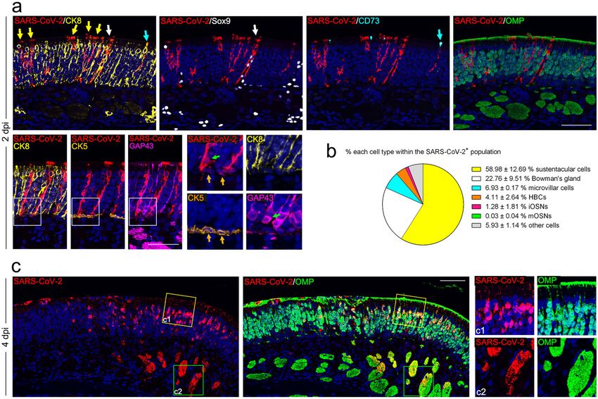
Ye et al. Cell Discovery (2021)7:49 Page 2 of 13
(ACE2)10,11, the functional receptor of SARS-CoV-212–14. (9.89 × 10⁶ RNA copies/mouse); however, the sgRNA
Single-cell RNA sequencing analyses have characterized levels in the OB and brain were below the detection
the expression profile of ACE2 in the OE of mice and threshold (Fig. 1b). As expected, in situ hybridization
humans, mainly in non-neuroepithelial cells11,15, and in a (ISH) by RNAscope demonstrated that SARS-CoV-2 RNA
recent study based on a hamster model, many SARS-CoV- was predominantly detected in OE (Supplementary
2-infected cells were observed in the OE section16,17. In Fig. S2a) but not in the OB (Supplementary Fig. S2b).
addition, vascular pericytes in the OB exhibited a high Furthermore, an immunofluorescence staining assay
level of ACE2 expression in a mouse model15; these cells detected high levels of SARS-CoV-2 N protein expression
play a key role in the maintenance of the blood–brain in the OE along the OM at 2 dpi (Fig. 1c), and only marginal
barrier, as well as the regulation of blood pressure and the expression of viral protein was observed at 8 dpi (Supple-
host immune response18. Interestingly, some respiratory mentary Fig. S3). However, we did not detect SARS-CoV-2
viruses, such as influenza virus and respiratory syncytial N protein-positive cells in the OB or other parts of the brain
virus, are able to invade the OB and other parts of the at 2 dpi and 4 dpi (Supplementary Fig. S2c).
brain to establish infection19,20. Thus, how SARS-CoV-2 To examine whether SARS-CoV-2 infection directly
invades the olfactory system and contributes to the impairs the olfactory function of infected mice, a standard
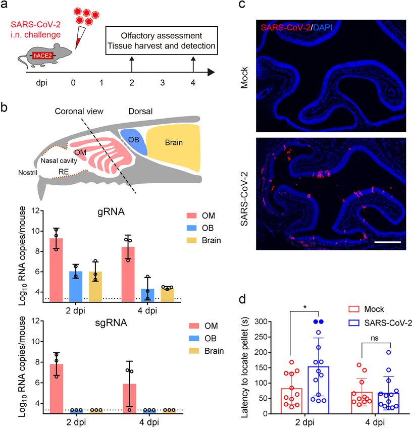
observed central nervous system (CNS) diseases remains BFPT was conducted at 2 and 4 dpi. Remarkably, a sig-
to be determined. In the present study, we demonstrate nificantly increased latency (152.8 s vs 81.8 s; P = 0.022) to
that SARS-CoV-2 infection directly causes transient locate food pellets was observed in SARS-CoV-2-infected
olfactory dysfunction in an established mouse model and mice compared with the control animals at 2 dpi (Fig. 1d).
characterize the major target cells and pathological effects Of particular note, 2 out of the 13 infected mice devel-
that contribute to olfactory dysfunction. oped severe symptoms of anosmia, as they failed to locate
the food pellet within the observation period. Interest-
Results ingly, recovery of infected mice from olfactory dysfunc-
SARS-CoV-2 targets the OE and causes transient olfactory tion was observed at 4 dpi, as the latency to locate food
dysfunction in hACE2 mice pellets was not different from that of the control animals
We previously established a humanized ACE2 (hACE2) (67.1 s vs 70.2 s; P = 0.992). Thus, these results demon-
mouse model susceptible to SARS-CoV-2 infection21. strate that SARS-CoV-2 primarily infects the OE and
Herein, to determine the impact of SARS-CoV-2 infection leads to transient olfactory dysfunction in mice.
on the olfactory system, groups of 6–8-week-old hACE2
mice were intranasally infected with 5.4 × 10⁵ plaque- SARS-CoV-2 primarily targets non-neuroepithelial cells in
forming units (PFU) of SARS-CoV-2. Mice inoculated the OE of hACE2 mice
with the same volume of culture medium were used as The OM consists of the OE and the underlying lamina
mock infection controls. At 2 and 4 days post infection propria (LP). The OE is composed of olfactory stem/pro-
(dpi), tissues from the respiratory tract and olfactory genitor cells, including the horizontal basal cells (HBCs)
system were collected from the necropsied mice and and globose basal cells (GBCs) residing in the basal region,
subjected to virological and immunological assays mature and immature OSNs, and a variety of non-
(Fig. 1a). As expected, high levels of SARS-CoV-2 RNA neuroepithelial lineages, including the sustentacular cells,
was detected in the nasal respiratory RE, trachea, and lung microvillar cells, and Bowman’s gland cells. The OSNs
at 2 and 4 dpi, and the peak viral RNA level (2.36 × 10¹¹ lining under the supporting cells project numerous den-
RNA copies/mouse) was detected in the lung at 2 dpi dritic cilia with ORs into the nasal cavity and intermingle
(Supplementary Fig. S1a), while robust viral nucleocapsid with the microvilli of sustentacular cells and microvillar
(N) protein expression was detected in the lung from cells (Supplementary Fig. S4a). Due to the asymmetrical
SARS-CoV-2-infected hACE2 mice at 2 dpi and 4 dpi but expression pattern of ACE2 on the cell membrane as well as
not in the control animals (Supplementary Fig. S1b). the unique organization of the OE, it is not easy to deter-
Strikingly, high levels of viral genomic RNA (gRNA) were mine which cell compartments express ACE2. To overcome
also detected in the olfactory mucosa (OM) at 2 dpi this, we took advantage of the tdTomato cassette down-
(5.85 × 10⁹ RNA copies/mouse) and maintained until 4 stream of the hACE2 transgene with an internal ribosome
dpi (8.93 × 10⁸ RNA copies/mouse) (Fig. 1b), while the entry site (IRES), which allows the detection of hACE2
viral RNA levels were much lower in the OB and other expression by cytoplasmic fluorescence of tdTomato (Sup-
parts of the brain at 2 dpi and decreased to marginal levels plementary Fig. S4b). Abundant expression of hACE2 along
at 4 dpi. Additionally, high levels of viral subgenomic the apical surface of the OE and within the underlying LP
RNA (sgRNA) (2.24 × 10⁸ RNA copies/mouse), indicating was detected with a human ACE2-specific monoclonal
the presence of actively replicating virus, were detected in antibody, exhibiting a similar expression pattern as tdTo-
the OM at 2 dpi, and subsequently decreased at 4 dpi mato (Supplementary Fig. S4c). A detailed characterization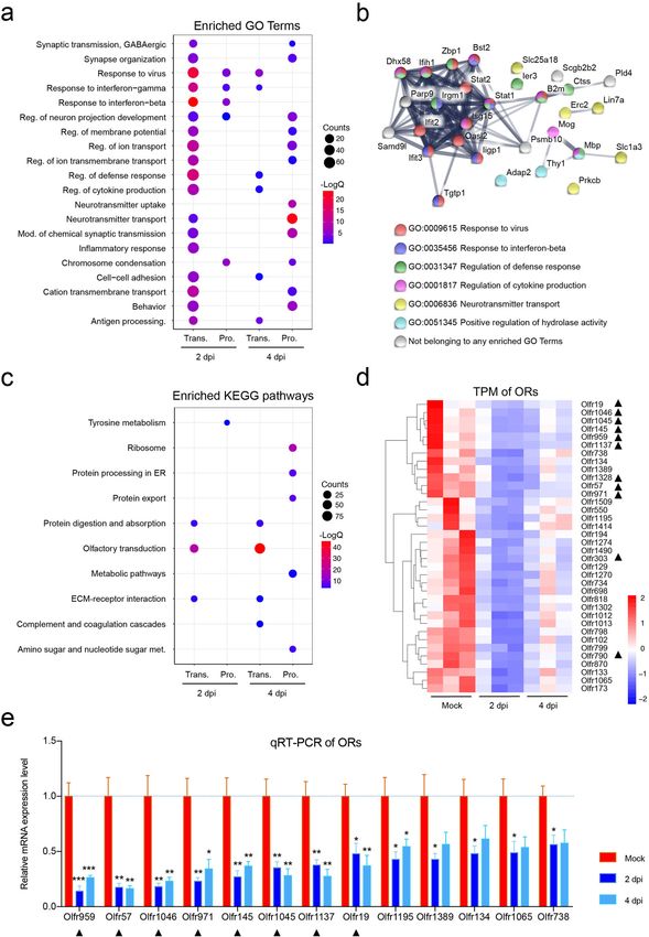
Ye et al. Cell Discovery (2021)7:49                                                                                                    Page 3 of 13

 Fig. 1 SARS-CoV-2 primarily infects the OE and causes olfactory dysfunction in hACE2 mice. a Schematic diagram of the experimental design.
 Briefly, groups of 6–8-week-old hACE2 mice were infected with 5.4 × 10⁵ PFU of SARS-CoV-2 intranasally. The olfactory function of infected mice was
 measured by the buried food pellet test at the indicated times post inoculation. Mice were sacrificed at 2 dpi and 4 dpi for viral detection and
 histopathological analysis. b Schematic view of the OM in the nasal cavity of mice in a sagittal plane. The dotted line indicates a coronal section
 (upper). Viral genomic RNA (gRNA, middle) and subgenomic RNA (sgRNA, lower) copies were determined by quantitative real-time reverse
 transcription PCR (qRT-PCR) and are shown as means ± SD from three independent replicates. c Immunostaining of the OM from SARS-CoV-2-
 infected mice for SARS-CoV-2 N protein (red) and DAPI (blue) at 2 dpi. Scale bar, 400 μm. d Buried food pellet test. The latency to locate the food
 pellets for mice infected with SARS-CoV-2 (n = 13) or DMEM (n = 11) was measured at 2 dpi and 4 dpi.

of hACE2/tdTomato-expressing cells in the OM revealed                       (Sox2 positive in the basal region), immature olfactory
that non-neuroepithelial cells, including sustentacular cells               sensory neurons (iOSNs) (GAP43 positive), and mature
(CK8 positive, Supplementary Fig. S4d, d1), the duct and                    olfactory sensory neurons (mOSNs) (OMP positive) (Sup-
acinus of Bowman’s gland cells (Sox9/CK8 positive, Sup-                     plementary Fig. S4d, d1–d4).
plementary Fig. S4d, d2, d4) in the OE and LP, respectively,                   To further characterize the primary targets of SARS-CoV-
and microvillar cells (CD73/CK8 positive, Supplementary                     2 in the OE, multiplex immunostaining assays were per-
Fig. S4e), are the primary cell types that exhibit human                    formed with antibodies against SARS-CoV-2 and specific
ACE2 expression (Supplementary Fig. S4d, f), whereas little                 cell markers. Remarkably, robust expression of SARS-CoV-
hACE2/tdTomato expression was detected in the neuroe-                       2 viral N protein was detected in the non-neuroepithelial
pithelial lineages, including HBCs (CK5 positive), GBCs                     lineage lining the outer surface of the OE at 2 and 4 dpi
Ye et al. Cell Discovery (2021)7:49 Page 4 of 13
Fig. 2 SARS-CoV-2 primarily targets non-neuroepithelial cells in the OE. a Representative multiplex immunofluorescent staining assay showing
that SARS-CoV-2 (SARS-CoV-2 N protein positive) infects sustentacular cells (CK8 positive, yellow arrows), Bowman’s gland cells (Sox9/CK8 positive,
white arrows), microvillar cells (CD73/CK8 positive, cyan arrows), HBCs (CK5 positive, gold arrows), and iOSNs (GAP43 positive, green arrows) at 2 dpi.
Little SARS-CoV-2 N protein was detected within OMP-positive mOSNs. b Statistical analysis of the percentage of each cell compartment within SARS-
CoV-2-positive cells. The data are presented as means ± SD (n = 3). c Multiplex immunofluorescent staining results showing an OM sample at 4 dpi
with SARS-CoV-2 detected in the OMP-positive mOSNs and the underlying nerve bundles. The framed areas labeled c1 and c2 are shown adjacently
at larger magnifications. Scale bar, 50 μm.
(Fig. 2a, c). Sustentacular cells (58.98%) and Bowman’s uniformity of the OE, as characterized by clusters of
gland cells (22.76%) were the major target cell types at 2 dpi, remnants on the surface of the OE (Fig. 3a), as well as
while some microvillar cells (6.93%) and HBCs (4.11%) were disorganized arrangement of supporting cells (Fig. 3b) and
also infected (Fig. 2a, b). A small population of iOSNs olfactory neurons (Fig. 3c). The integrity of the cilia layer
(1.28%) was also infected by SARS-CoV-2, while no mOSNs of mOSNs and the microvilli of supporting cells were
were infected at 2 dpi (Fig. 2a, b). Interestingly, SARS-CoV- severely damaged (Fig. 3b, c). More importantly, com-
2-positive HBCs and iOSNs were found adjacent to the pared with the mock-treated groups, profound cell
infected sustentacular cells (Fig. 2a). Additionally, a sub- apoptosis (cleaved-caspase3 positive) was observed in
stantial amount of viral protein was detected within the both the OE and LP sections of the OM from SARS-CoV-
cilia, cellular bodies, and underlying nerve bundles of 2-infected mice (Fig. 3d). Immunofluorescence co-
mOSNs at 4 dpi (Fig. 2c, c1, c2). These results indicated that staining indicated the occurrence of apoptosis in susten-
SARS-CoV-2 primarily targets the non-neuroepithelial cells tacular cells, HBCs as well as the cellular bodies and
lining the outer surface of the OE and subsequently invades underlying nerve bundles of iOSNs and mOSNs (Fig. 3d).
the neuroepithelial lineage in hACE2 mice. Additionally, infiltration of immune cells, including
macrophages (CD68 positive), dendritic cells (CD103
SARS-CoV-2 infection triggers apoptosis and immune cell positive) and neutrophils (Ly-6G positive), was evident in
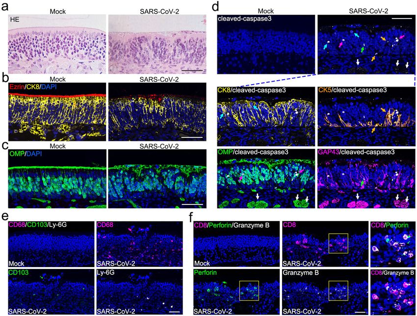
infiltration in the OE the infected OE (Fig. 3e). The profound invasion of CD8 T
We then characterized the histopathological changes in lymphocytes with high expression of the cytotoxic
the OE in response to SARS-CoV-2 infection. Strikingly, enzymes perforin and granzyme B further deteriorated the
SARS-CoV-2 infection directly impaired the structural cellularity of olfactory epithelial cells (Fig. 3f). ThisYe et al. Cell Discovery (2021)7:49 Page 5 of 13
Fig. 3 SARS-CoV-2 infection induces apoptosis and immune cell infiltration in the OE. a Representative hematoxylin and eosin (H&E) staining
results showing histopathological changes of the OE. b Representative results of multiplex immunofluorescent detection of sustentacular cells (CK8
positive) and microvilli (Ezrin positive) of the OE. c Representative results of immunofluorescent detection of mOSNs (OMP positive) of the OE. d
Apoptosis of olfactory epithelial cells (cleaved-caspase3 positive, white) after SARS-CoV-2 infection. The panels below show apoptosis of sustentacular
cells (CK8 positive, yellow; indicated by cyan arrows), HBCs (CK5 positive, gold; indicated by gold arrows), mOSNs (OMP positive, green; indicated by
magenta arrows), iOSN (GAP43 positive, magenta; indicated by green arrows), and olfactory nerve bundles (OMP/GAP43 positive; indicated by white
arrows). e Representative multiplex immunofluorescent staining results showing infiltration of macrophages (CD68 positive, magenta), dendritic cells
(CD103 positive, green), and neutrophils (Ly-6G positive, white) in the OE after infection. f Representative multiplex immunofluorescent staining
results showing infiltration of CD8 cytotoxic T lymphocytes (magenta) with expression of perforin (green) and granzyme B (white) in the olfactory
mucosa after infection. The framed areas are shown adjacently at larger magnifications. Scale bar, 50 μm.
observed physiological damage that occurs upon SARS- OE was observed in infected animals, which also co-
CoV-2 infection probably contributes to the functional expressed the markers of their lineage offspring, such as
loss of olfaction. iOSNs (Supplementary Fig. S5b, b1), sustentacular cells
(Supplementary Fig. S5b, b2), and microvillar cells (Sup-
SARS-CoV-2 infection induces regeneration of the OE plementary Fig. S5b, b3). These results suggest that the
Without infection, HBCs in the basal region of the OE impaired OE is regenerated through olfactory stem cell-
remained quiescent, as indicated by the low expression based proliferation and differentiation into olfactory
level of the proliferation marker Ki67 within CK5-positive neurons and supporting lineages, thereby restoring the
cells (Supplementary Fig. S5a, a1). SARS-CoV-2 infection normal function of the OE.
significantly increased the number of CK5/Sox2/Ki67
triple-positive cells, strongly suggesting a transition from SARS-CoV-2 infection induces antiviral and inflammatory
HBCs to actively cycling GBCs (Supplementary Fig. S5a, responses in the OE
a2). Of particular note, prominent upward growth of To decipher the mechanism underlying the observed
HBCs from the basal layer into the upper section of the olfactory dysfunction in SARS-CoV-2-infected mice at theYe et al. Cell Discovery (2021)7:49 Page 6 of 13 molecular level, combined transcriptomic and quantita- downregulation of Bsg/CD147, Tmprss2, Furin, and tive proteomic analyses of the OE and OB samples from Tfrc/transferrin receptor was also detected by RNA-seq SARS-CoV-2-infected mice were performed, and the analyses (Supplementary Fig. S10b–e). results were compared with those of the control animals. In the OE samples, a total of 929 genes and 507 proteins Discussion were regulated upon SARS-CoV-2 infection, and 40 of In the present study, we used an established mouse them were synchronously regulated at both the mRNA model to demonstrate that SARS-CoV-2 infection can and protein levels (Supplementary Fig. S6a, b). In the OB cause olfactory dysfunction and anosmia, and this samples, 286 genes and 251 proteins were up/down- experimental evidence supports the hypothesis that regulated, and only four of them were consistently regu- SARS-CoV-2 infection is the cause of olfactory dysfunc- lated at the mRNA and protein levels (Supplementary Fig. tion and anosmia in COVID-19 patients5,6,22,23. SARS- S6a, c). Gene enrichment analyses showed that SARS- CoV-2-infected mice exhibited a damaged OE, immune CoV-2 infection induced a strong antiviral IFN response cell infiltration, downregulated OR expression, and in the OE at 2 dpi, and the response decayed at 4 dpi, impaired olfactory function, largely mimicking the olfac- while no obvious changes were observed in the OB tory abnormalities of COVID-19 patients. Robust viral (Supplementary Fig. S7a). Further validation by qRT-PCR replication and direct antiviral responses were detected in confirmed the results from RNA-seq analysis (Supple- the OE of the infected mice but not in the OB and other mentary Fig. S7b, c). Notably, a strong inflammatory parts of the brain, indicating that SARS-CoV-2 mainly response in the OE was detected at both the mRNA and infected the OM in the hACE2 mouse model. A recent protein levels at 2 dpi, and this response faded at 4 dpi study also showed that the SARS-CoV-2 protein could be (Fig. 4a and Supplementary Fig. S8a). Moreover, genes detected in the OE, but not in the OB, in a hamster related to “positive regulation of cell death” and “regula- model17. One possible explanation for the absence of tion of neuron projection development” were also upre- SARS-CoV-2 in the CNS is that virus infection and gulated upon SARS-CoV-2 infection (Fig. 4a and replication in OE can effectively activate IFN-dependent Supplementary Fig. S8b, c), which was consistent with the antiviral responses (Supplementary Fig. S7a, b), which is immunostaining results (Fig. 3d–f and Supplementary Fig. an effective barrier that prevents viral invasion into the S5a, b). Further integrated omic analysis of the OE sam- CNS. In addition, the apoptosis of infected OSNs may ples showed that a total of 30 genes were upregulated at contribute to the prevention of viral spread into the CNS both the mRNA and protein levels. Of these, antiviral after the rapid infection and destruction of the OE24. genes/proteins, including Isg15, Stat1, Stat2, Oasl2, Ifit2, Additionally, SARS-CoV-2 invaded brain tissues in K18- and Ifit3, were found to interact closely. Other genes/ hACE2 mice, which expressed hACE2 under the cyto- proteins involved in neurotransmitter transport, including keratin 18 promoter25. These inconsistencies in SARS- Erc2, Lin7a, Slc1a3, and Slc25a18, were also observed CoV-2 infection outcomes, including CNS tropism, in (Fig. 4b). We did not find any obvious induction of anti- different mouse models may be attributed to the different viral response-related genes in OB samples by tran- expression profiles of human ACE2 as well as the scriptomic and proteomic analyses, but downregulation of experimental systems. inflammatory response-related genes was observed (Sup- Our results demonstrated that SARS-CoV-2 initially plementary Fig. S9a, b). infects non-neuroepithelial cells, including sustentacular Of particular note, KEGG pathway enrichment analysis cells, Bowman’s gland cells and microvillar cells, which of downregulated transcripts and proteins in the OE are involved in OSN support, host immune response, showed that genes belonging to “olfactory transduction” electrolyte balance maintenance, and mucus secretion26. were significantly enriched (Fig. 4c). Among all 100 Moreover, we observed various levels of damage in the OE downregulated transcripts at 2 dpi, 36 encoded ORs after SARS-CoV-2 infection, including cilia desquama- (Fig. 4d and Supplementary Fig. S6b), while among the tion, loss of surface microvilli, and substantial structural 278 downregulated transcripts at 4 dpi, 97 encoded ORs disorganization. In addition, our results showed a certain (Supplementary Figs. S6b and S8d). Further qRT-PCR degree of cell apoptosis and inflammatory infiltration at assays showed that 13 representative OR genes were both the cell and molecular levels following SARS-CoV-2 significantly downregulated in response to SARS-CoV-2 infection. All these data indicate that the damaged sup- infection (Fig. 4e), which may also be attributed to the porting non-neuroepithelial cells and inflammatory infil- observed olfactory dysfunction. We further analyzed the tration caused by SARS-CoV-2 infection contribute to the expression of other host cell factors that were reported detrimental effects of the virus on olfactory function. Our to facilitate SARS-CoV-2 infection in human cells. The results are supported by recent findings in mice and results showed that Nrp1 was significantly down- humans15,25,27,28, showing that the non-neuroepithelial regulated at 2 dpi (Supplementary Fig. S10a), and slight cells of the OE express high levels of ACE2 and TMPRSS2
Ye et al. Cell Discovery (2021)7:49   Page 7 of 13

 Fig. 4 (See legend on next page.)
Ye et al. Cell Discovery (2021)7:49                                                                                                     Page 8 of 13

 Fig. 4 Host response to SARS-CoV-2 in the OE at the mRNA and protein levels. a Dot plot visualization of enriched GO terms of upregulated
 genes/proteins at 2/4 dpi in the OE. Gene enrichment analyses were performed using Metascape against the GO dataset for biological processes.
 “Reg.” stands for regulation, “mod.” for modulation, and “antigen processing.” for antigen processing and presentation of peptide antigen.
 b Interaction map of 30 proteins that were consistently upregulated at both the transcriptomic and proteomic levels over the course of SARS-CoV-2
 infection in the OE. Network nodes represent proteins, and their colors indicate the different GO terms to which they belong. Edges represent
 protein–protein associations, and their thickness indicates the strength of data support. c Dot plot visualization of enriched KEGG pathways of
 downregulated genes/proteins at 2/4 dpi in the OE. Gene enrichment analyses were performed using STRING against the KEGG dataset. “Met.” for
 metabolism. The color of the dots represents the –LogQ value for each enriched KEGG pathway, and size represents the gene/protein counts
 enriched in each term. d Heatmap indicating the expression patterns of 36 olfactory receptor genes that were significantly downregulated at 2 dpi.
 The colored bar represents the Z-score of the TPM. A total of 11 genes that were also downregulated at 4 dpi are marked with black triangles. e RNA
 expression of 13 representative ORs by qRT-PCR. Columns with *, **, *** indicate ORs significantly downregulated at P < 0.05, P < 0.01, or P < 0.001,
 respectively, relative to their mock groups (one-way ANOVA followed by post hoc analysis with Dunnett’s multiple comparisons test, n = 3). Black
 triangle-marked ORs were downregulated at both 2 and 4 dpi based on transcriptome data.

at both the mRNA and protein levels15. Interestingly,                        transporting protein 1) was downregulated at both levels
SARS-CoV-2-positive signals were also observed in HBCs                       (Supplementary Table S1). This protein specifically pro-
and mOSNs of infected animals at 2 dpi and 4 dpi,                            motes functional cell surface expression of ORs38, sug-
respectively (Fig. 2a, c), although we did not detect any                    gesting that the inhibition of Rtp1 in OB may lead to
hACE2 expression in these cells (Supplementary Fig. S4d).                    downregulation of ORs. Therefore, OE damage, which is
The underlying mechanism remains elusive, and an                             closely related to olfactory dysfunction, is caused by
hACE2-independent route of SARS-CoV-2 infection may                          SARS-CoV-2 infection of non-neuroepithelial cells and
be considered. According to recent studies, some other                       OSNs synergizes with the host antiviral immune
host cell receptors were found to facilitate SARS-CoV-2                      responses.
infection, including NRP1, CD147, and transferrin                              According to our results, the olfactory dysfunction in
receptor29–32. Additionally, SARS-CoV-2 contains a spe-                      SARS-CoV-2-infected animals was recoverable, as almost
cific furin cleavage site in the spike protein13,33 and uti-                  all animals recovered their normal sense of smell at 4 dpi.
lizes TMPRSS2 for further spike protein priming14. In our                    Additionally, studies focusing on COVID-19 patients with
study, we found that the Nrp1, Bsg/CD147, Tmprss2,                           anosmia have shown that most of them recover from loss
Furin, and Tfrc/transferrin receptor were downregulated                      of smell within a few weeks or less, and only a small
to varying degrees in SARS-CoV-2-infected mice at 2 dpi,                     number of patients reported slow partial recovery over 1
as shown by RNA-seq analyses, but whether these pro-                         to 3 months39–42, indicating a potential mechanism of OE
teins play critical roles in olfactory dysfunction after                     regeneration from injuries. The hACE2 mouse model of
SARS-CoV-2 infection in our animal model remains                             SARS-CoV-2 infection reproduces the clinical course and
unknown and warrants extensive investigation.                                viral replication observed in COVID-19 patients, as it
   We observed that many ORs were significantly down-                         mimics the acute SARS-CoV-2 infection as well as the
regulated at 2 and 4 dpi, suggesting the occurrence of                       transient olfactory dysfunction observed in human indi-
decreased olfaction after SARS-CoV-2 infection. A recent                     viduals. The difference in the time span of viral infection
study also showed that induction of antiviral type I                         and clearance between humans and mice reflected the
interferon signaling in the mouse OE was associated with                     inherent differences between the species. The OE
diminished odour discrimination and decreased RNA                            undergoes lifelong regeneration and replacement
levels of ORs34. These findings may support what we                           depending on two populations of basal stem cells, namely,
observed in this study: SARS-CoV-2 infection causes a                        HBCs and GBCs. HBCs are mitotically quiescent under
significant interferon response and dramatic OR decrease                      normal conditions and become activated and differentiate
simultaneously in the OE. We also observed that the                          into other kinds of cells once OE damage occurs43. Unlike
levels of three odorant-binding proteins (OBPs), which                       HBCs, most GBCs are mitotically activated and are
are compact globular water-soluble proteins with ligand-                     responsible for the regeneration of both neuronal and
binding capabilities and are thought to aid in the capture                   non-neuronal cells44–46. Indeed, we observed regenera-
and transport of odorants to the ORs, significantly                           tion of the OE, as indicated by the significant proliferation
decreased at the protein level with OE infection35–37. In                    and morphological change of HBCs, accompanied by the
addition, although no viral infection was observed in the                    differentiation of stem cells into iOSNs, sustentacular
OB, we detected some up/downregulated transcripts or                         cells and microvillar cells. In this way, the structural basis
proteins by transcriptomic and proteomic analyses.                           and function of the OE as well as olfactory function can be
Notably, among all four proteins co-regulated at both the                    restored to normal in SARS-CoV-2-infected animals.
transcriptomic and proteomic levels, Rtp1 (receptor-                         Furthermore, it was indicated that the damage and
Ye et al. Cell Discovery (2021)7:49                                                                               Page 9 of 13

apoptosis of OSNs are closely involved in their regen-          RNA extraction and qRT-PCR
eration47, and the occurrence of an inflammatory                   Quantification of SARS-CoV-2 RNA, and OR mRNA
response also facilitates stem cell differentiation and OE      transcript levels was performed by qRT-PCR. Total RNA
regeneration48,49. At the transcriptomic and proteomic          was isolated using TRIzol reagent (Invitrogen, Carlsbad, CA,
levels, we observed upregulated “regulation of neuron           USA) according to the manufacturer’s instructions. SARS-
projection development” genes/proteins at 2 and 4 dpi,          CoV-2 RNA was measured with the following primer-probe
implying the progression of a neuron projection over time       set: CoV-F3 (5′-TCCTGGTGATTCTTCTTCAGGT-3′),
from its formation to the mature structure. Interestingly,      CoV-R3 (5′-TCTGAGAGAGGGTC AAGTGC-3′) and
although there were many significantly downregulated             CoV-P3 (5′-AGCTGCAGCACCAGCTGTCCA-3′). SARS-
ORs at 4 dpi, the mRNA levels of many ORs increased             CoV-2 sgRNA was measured with the following primer-
slightly compared with those at 2 dpi, indicating that OR       probe set: CoV-sgRNA-F (5′-CGATCTCTTGTAGATCT
expression tends to recover to normal levels.                   GTTCTC-3′), CoV-sgRNA-R (5′-ATATTGCAGCAGTAC
   In summary, our study established a mouse model of           GCACACA-3′) and CoV-sgRNA-P (5′-ACACTAGCCATC
olfactory dysfunction induced by SARS-CoV-2. Con-               CTTACTGCGCTTCG-3′)3. Glyceraldehyde-3-phosphate
sidering the interspecies discrepancy in olfactory con-         dehydrogenase (GAPDH) served as the endogenous con-
struction between rodents and humans, e.g., the relative        trol, and the following primer set was used: 5′-CCAACCGC
size of the OB to the brain, the proportion of the brain        GAGAAGATGA-3′ and 5′-CCAGAGGCGTACAGGGAT
involved in olfaction, and the expression of ORs43, further     AG-3′. The primer sequences for amplifying the OR genes
studies are recommended to reproduce SARS-CoV-2-                are listed in Supplementary Table S2. Amplification was
induced olfactory dysfunction in other animal models,           performed using a One Step PrimeScript RT-PCR Kit
especially nonhuman primates. Additionally, validating          (Takara Bio, Otsu, Japan), and the following qRT-PCR
the targets and biological effects of SARS-CoV-2 infection      conditions were applied: 42 °C for 5 min and 95 °C for 10 s
in human specimens should still be considered. An animal        followed by 40 cycles of 95 °C for 5 s and 60 °C for 20 s. The
model of olfactory disorders is available to subsequently       PCR was conducted in a LightCycler® 480 Instrument
evaluate antiviral drugs as well as vaccines for the inhi-      (Roche Diagnostics Ltd, Rotkreuz, Switzerland). The abso-
bition of SARS-CoV-2 and for the improvement of post            lute quantification of SARS-CoV-2 RNA levels was per-
viral olfactory disorders.                                      formed by comparison to a standard curve and is shown as
                                                                SARS-CoV-2 RNA copies per mouse. The relative expres-
Materials and Methods                                           sion levels of OR mRNA were calculated according to the 2
Cell and virus                                                  −ΔΔCt method. Each sample was assayed with three
  Vero cells were maintained at 37 °C under 5% CO2 in           repeats.
Dulbecco’s modified Eagle’s medium (DMEM) supple-
mented with 10% heat-inactivated foetal bovine serum            BFPT
(FBS, Gibco), 10 mM HEPES, and 1% penicillin/strepto-             The standard BFPT was used to evaluate the olfactory
mycin. The SARS-CoV-2 strain BetaCoV/Beijing/                   function of SARS-CoV-2-infected mice and DMEM-
IMEBJ05/2020 (No. GWHACBB01000000) was originally               treated mice as previously described50,51. Mice were
isolated from a COVID-19 patient. For virus propagation,        food-restricted to 0.2 g of chow per day for 2 days before
Vero cells were incubated with SARS-CoV-2, and the              the test and during the experimental period to ensure
culture supernatants were collected at 3 dpi. The stock of      motivation. The food pellet was buried 1 cm below the
SARS-CoV-2 was serially diluted and titrated on mono-           surface of 3-cm-high bedding in a clear test cage (45 cm
layers of Vero cells. Studies with infectious SARS-CoV-2        L × 24 cm W × 20 cm H). One mouse was placed in the
were conducted under biosafety level 3 (BSL3) facilities at     centre of the cage, and the latency for the mouse to
the Beijing Institute of Microbiology and Epidemiology,         uncover the pellet was recorded. The latency was defined
AMMS.                                                           as 300 s for the mouse that could not find the pellet within
                                                                5 min.
SARS-CoV-2 infection of hACE2 mice
  The animal experiment procedure was reviewed and              RNAscope ISH
approved by the Laboratory Animal Center, AMMS                    RNAscope ISH for SARS-CoV-2 RNA was performed
(approval number: IACUC-DWZX-2020-001). For intra-              with the RNAscope assay (Advanced Cell Diagnostics,
nasal infection, 5.4 × 10⁵ PFU of SARS-CoV-2 was instil-        Newark, CA, USA) according to the manufacturer’s
led into the nasal cavity of 6–8-week-old hACE2 mice            instructions. Briefly, the tissues were isolated immediately
anaesthetized with sodium pentobarbital at a dose of            after euthanasia, fixed in 4% paraformaldehyde (PFA) for
50 mg/kg by the intraperitoneal route. Mice were mon-           24 h, and embedded in paraffin after being decalcified
itored daily and euthanized at 2 or 4 dpi to isolate tissues.   using 10% EDTA solution. Four-micrometre-thick
Ye et al. Cell Discovery (2021)7:49 Page 10 of 13
formalin-fixed paraffin-embedded (FFPE) slides were were then stained in eosin for 15 s and washed again
warmed at 60 °C for 1 h before they were deparaffinized in in water.
xylene, rehydrated in a series of graded alcohols, and
subjected to RNAscope target retrieval at 95 °C. Slides RNA library construction and sequencing
were visualized in situ using the 2.5 HD Reagent Kit hACE2 transgenic mice before or after SARS-CoV-2
(BROWN, Cat# 322310) and the sense probe from the infection (2 or 4 dpi) as previously described were used for
RNAscope ISH probe-V-nCoV2019-S (Cat# 848561) at RNA-seq. Total RNA from the OE and OB was extracted
40 °C in a HybEZ hybridization oven and then counter- using TRIzol (Invitrogen, Carlsbad, CA, USA) and DNase
stained with hematoxylin. I (NEB, USA), respectively. Sequencing libraries were
generated using the NEBNext® UltraTM RNA Library
Multiplex immunofluorescent staining Prep Kit for Illumina® (#E7530L, NEB, USA) following
The 4-μm-thick paraffin sections were deparaffinized in the manufacturer’s recommendations, and index codes
xylene and rehydrated in a series of graded alcohols. were added to attribute sequences to each sample. Clus-
Antigen retrieval was performed in citrate buffer (pH = 6) tering of the index-coded samples was performed on a
by heating in a microwave (Sharp, R-331ZX) for 20 min at cBot cluster generation system using a HiSeq PE Cluster
95 °C followed by a 20 min cool-down period at room Kit v4-cBot-HS (Illumina, San Diego, California, USA)
temperature. Multiplex fluorescence labeling was per- according to the manufacturer’s instructions. After cluster
formed using TSA-dendron-fluorophores (NEON 9-color generation, the libraries were sequenced on the Illumina
Allround Discovery Kit for FFPE, Histova Biotechnology, NovaSeq 6000 platform, and 150-bp paired-end reads
NEFP950). Briefly, endogenous peroxidase was quenched were generated. After sequencing, a Perl script was used
in 3% H2O2 for 20 min, followed by treatment with to filter the original data (raw data) to clean reads by
blocking reagent for 30 min at room temperature. Primary removing contaminated reads for adapters and low-
antibody was incubated for 2–4 h in a humidified cham- quality reads. Clean reads were aligned to the mouse
ber at 37 °C, followed by detection using the HRP- genome (Mus_musculus.GRCm38.99) using Hisat2 v2.1.0.
conjugated secondary antibody and TSA-dendron- The number of reads mapped to each gene in each sample
fluorophores. Then, the primary and secondary anti- was counted by HTSeq v0.6.0, and the TPM (transcripts
bodies were thoroughly eliminated by heating the slides in per kilobase of exon model per million mapped reads) was
retrieval/elution buffer (Abcracker®, Histova Biotechnol- then calculated to estimate the expression levels of genes
ogy, ABCFR5L) for 10 s at 95 °C using a microwave. In a in each sample.
serial fashion, each antigen was labeled with distinct
fluorophores. The multiplex antibody panels applied in Large-scale proteomic sample preparation and tandem
this study were as follows: hACE2 (Abcam, ab108209, mass tag (TMT) labeling
1:200); tdTomato (Rockland, 600-401-379, 1:500); SARS- The OE and OB tissues were disrupted by using a
CoV-2 nucleocapsid protein (Sinobiological, 40143-R004, grinding mill for six cycles of 5 s each with lysis buffer
1:1000); GAP43 (Abcam, ab75810, 1:1000); OMP (Abcam, (9 M urea, 10 mM Tris-HCl, pH 8.0, 30 mM NaCl, 10 mM
ab183947, 1:1500); CK5 (Abcam, ab52635, 1:800); CK8 iodoacetamide (IAA), 5 mM Na4P2O7, 100 mM Na2HPO4,
(Abcam, ab53280, 1:800); Sox9 (Abcam, ab185230, 1:500); pH 8.0, 1 mM NaF, 1 mM Na3VO4, 1 mM sodium gly-
Sox2 (CST, 23064, 1:400); CD73 (CST, 13160, 1:500); cerophosphate, 1% phosphatase inhibitor cocktail 2
Furin (Abcam, ab108209, 1:400); Tmprss2 (Abcam, (Sigma, St. Louis, USA), 1% phosphatase inhibitor cocktail
ab92323, 1:500); CD3 (CST, 78588, 1:300); CD8 (CST, 3 (Sigma, St. Louis, USA), and 1 tablet of EDTA-free
98941, 1:300); Cleaved caspase-3 (CST, 9664, 1:1000); protease inhibitor cocktail (Roche, Basel, Switzerland) for
CD103 (Abcam, ab224202, 1:300); Ly-6G (CST, 87048, every 10 mL of lysis buffer) and 2-mm steel balls,
1:400); CD68 (CST, 97778, 1:300); and Granzyme B respectively. The supernatants were obtained after cen-
(Abcam, ab255598, 1:300). After all the antibodies were trifugation at 8000 rpm for 10 min at 4 °C. The protein
detected sequentially, the slices were imaged using the lysates were inactivated at 56 °C for 30 min and then
confocal laser scanning microscopy platform Zeiss stored at –80 °C before further processing. The protein
LSM880. concentration was measured by performing a short
Coomassie blue-stained 10% SDS-PAGE run as previously
Histopathological analysis described52. The same amount of protein (130 μg) from
The structural integrity of the mouse OE was analyzed each sample was reduced with 5 mM dithiothreitol
using H&E staining according to standard procedures. (DTT), alkylated with 20 mM IAA, and precleaned by 10%
Briefly, after being rehydrated in a series of graded SDS-PAGE (10%, 0.7 cm). The protein was then subjected
alcohols, 4-μm-thick slides of mouse OE were stained to in-gel digestion with a final concentration of 12.5 ng/μL
with hematoxylin for 30 s and washed in water. Slides Ac-trypsin combined with endoproteinase lys-C providedYe et al. Cell Discovery (2021)7:49 Page 11 of 13
by Enzyme & Spectrum (Beijing, China) at a ratio of 2:1 at entries and a canonical SARS-CoV-2 proteome with 30
37 °C for 12–14 h53,54. The extracted peptides from the potential viral proteins from the SARS-CoV-2 genome
OE and OB groups were labeled with TMT10 reagents (NC_045512.2), and a common contaminant database
according to the manufacturer’s instructions (Thermo (http://www.maxquant.org/contaminants.zip), respec-
Scientific, San Jose, CA, USA). Ten labeled channels were tively. Fully tryptic peptides with as many as 2 missed
then quenched with 5% hydroxylamine and combined were allowed. TMT 10 plex (N-Term/K) and cysteine
according to the normalization value by ratio checking. carbamidomethyl were set as fixed modifications, whereas
The mixed samples were vacuum dried. oxidation of methionine was set as a variable modifica-
tion. The tolerance of the precursor and fragment ions
Peptide fractionation and LC-MS/MS analysis was set to 20 ppm.
The dried TMT-labeled mixture was resuspended in
100 μL of buffer A (2% acetonitrile (ACN), pH 10) and Bioinformatic analyses
separated by a high-pH reversed-phase HPLC system DESeq2 v1.6.3 was used for differential gene expression
(Rigol, L-3120, Beijing, China). The combined samples analysis. Genes with Padj ≤ 0.05 and |Log2FC | > 1 were
were injected into a Durashell C18 column (150 Å, 5 μm, identified as differentially expressed genes (DEGs). The
4.6 × 250 mm2) and eluted with a linear gradient in total proteomic quantification datasets were median-
60 min. Briefly, the solvent gradient of buffer B (2% normalized, and P value was calculated by Perseus
ddH2O and 98% ACN) was as follows: 0% for 5 min, 0–3% (1.6.6.0). Proteins ratios between control and infection
for 3 min, 3%–22% for 37 min, 22%–32% for 10 min, ≥ 1.5-fold and P value ≤ 0.05 were considered as regulated
32%–90% for 1 min, 90% for 2 min, and 100% for 2 min. differentially expressed proteins (DEPs). The DEGs and
The LC flow rate was set at 0.7 mL/min and monitored at DEPs identified were used as queries to search for enri-
214 nm. The column oven was set at 45 °C. A total of 60 ched biological processes (Gene Ontology BP) using
fractions were collected and then combined into 15 Metascape55. KEGG pathway enrichment and protein
fractions before vacuum drying according to the peak interaction networks were analyzed using STRING56.
abundance. The combined samples were dissolved in Heatmaps of gene expression levels were constructed
loading buffer (1% ACN and 1% formic acid (FA)) and using the pheatmap package in R (https://cran.rstudio.
analyzed using an EASY-nLC 1200 ultra-performance com/web/packages/pheatmap/index.html). Dot plots and
liquid chromatography system (Thermo Fisher Scientific, volcano plots were constructed using the ggplot2 (https://
San Jose, CA, USA) equipped with a self-packed capillary ggplot2.tidyverse.org/) package in R.
column (75 μm i.d. × 15 cm, 3 μm C18 reversed-phase
fused silica), with a 78-min nonlinear gradient at a flow Statistical analysis
rate of 600 nL/min. The gradient was as follows: an Data were analyzed using GraphPad Prism 8 (GraphPad
increase from 6%–15% solvent B (0.1% FA in 80% ACN) Software, San Diego, California, USA). The values shown
in 15 min, 15%–30% in 40 min, 30%–40% in 15 min, and in the graphs are presented as means ± SD of at least three
40%–100% in 1 min, finally holding at 100% for 7 min. independent experiments. Statistical differences between
The eluted peptides were analyzed on an Orbitrap Fusion groups were analyzed using two-tailed unpaired t-tests or
Lumos (Thermo Fisher Scientific, San Jose, CA, USA). a one-way ANOVA statistical test with Dunnett’s multiple
MS1 data were collected in the Orbitrap using a 120 k comparison test; P < 0.05 was considered statistically
resolution over an m/z range of 300–1500, with the significant.
maximum injection time (MIT) set to 50 ms. The auto-
matic gain control (AGC) was set to 4 × 10⁵, determined Acknowledgements
We thank Drs. Changfa Fan and Bin Fu for providing critical reagents and
charge states between 2 and 7 were subjected to frag- helpful discussion. This work was supported by the National Key R&D Project of
mentation via higher energy collision-induced dissocia- China (2016YFD0500304, 2020YFC0842200, and 2020YFA0707801). C.-F.Q. was
tion (HCD) with 37% collision energy, and a 12 s dynamic supported by the National Science Fund for Distinguished Young Scholar
(81925025), the Innovative Research Group (No. 81621005) from the NSFC, and
exclusion window was used with isotopes excluded. For the Innovation Fund for Medical Sciences (2019RU040) from the Chinese
the MS/MS scans, the fractions were detected in the Academy of Medical Sciences (CAMS). P.X. was supported by the CAMS
Orbitrap at a resolution of 50 k. For each scan, the iso- Innovation Fund for Medical Sciences (2019RU006). J.Z. was supported by the
Youth Program of the National Natural Science Foundation of China
lation width was 1.6 m/z, the AGC was 5 × 10⁴, and the (82002148) from the NSFC and the China Postdoctoral Science Fund
MIT was 86 ms. (2020T130134ZX, 2020M673685). R.-T.L. was supported by the China
Postdoctoral Science Fund (2019M664012, 2020T130135ZX). J.-F.L. was
supported by the Natural Science Foundation of Beijing (7212090).
Database search
The raw files from the OE and OB groups were searched
Author details
with MaxQuant (v1.5.5.0) against the mouse reviewed 1
State Key Laboratory of Pathogen and Biosecurity, Beijing Institute of
proteome downloaded from UniProt, containing 17,478 Microbiology and Epidemiology, Beijing, China. 2State Key Laboratory ofYe et al. Cell Discovery (2021)7:49 Page 12 of 13
Proteomics, National Center for Protein Science (Beijing), Beijing Institute of 13. Walls, A. C. et al. Structure, function, and antigenicity of the SARS-CoV-2 spike
Lifeomics, Beijing, China. 3Department of Otorhinolaryngology, China-Japan glycoprotein. Cell 181, 281–292 (2020).
Friendship Hospital, Beijing, China. 4Research Unit of Discovery and Tracing of 14. Hoffmann, M. et al. SARS-CoV-2 cell entry depends on ACE2 and TMPRSS2 and
Natural Focus Diseases, Chinese Academy of Medical Sciences, Beijing, China is blocked by a clinically proven protease inhibitor. Cell 181, 271–280 (2020).
15. Brann, D. H. et al. Non-neuronal expression of SARS-CoV-2 entry genes in the
Author contributions olfactory system suggests mechanisms underlying COVID-19-associated
C.-F.Q., Q.Y., J.Z., G.Y., and J.-F.L. conceived the project and designed the anosmia. Sci. Adv. 6, eabc5801 (2020).
experiments. Q.Y., J.Z., G.Y. and Q.H. performed the majority of the experiments 16. Sia, S. F. et al. Pathogenesis and transmission of SARS-CoV-2 in golden
and analyzed the data. R.-T.L., Y.Z., Q.C., R.-R.Z., H.Q., Y.-Q.D., X.-F.L., S.-J.W., J.-H.S., hamsters. Nature 583, 834–838 (2020).
H.-M.Z., and T.Z. contributed to specific experiments and data analysis. Y.Z. and 17. Bryche, B. et al. Massive transient damage of the olfactory epithelium asso-
P.X. contributed to proteomic analysis. C.-F.Q., Q.Y., J.Z., G.Y., and R.-T.L. wrote ciated with infection of sustentacular cells by SARS-CoV-2 in golden Syrian
the manuscript with all the inputs from all authors. C.-F.Q., X.Y., and P.X. hamsters. Brain Behav. Immun. 89, 579–586 (2020).
supervised the study. All authors read and approved the contents of the 18. Armulik, A., Genove, G. & Betsholtz, C. Pericytes: developmental, physiological,
manuscript. and pathological perspectives, problems, and promises. Dev. Cell 21, 193–215
(2011).
Data availability 19. Schrauwen, E. J. et al. The multibasic cleavage site in H5N1 virus is critical for
All the RNA-seq data from this study have been deposited to the NCBI Gene systemic spread along the olfactory and hematogenous routes in ferrets. J.
Expression Omnibus (GEO) datasets under accession number GSE173186. The Virol. 86, 3975–3984 (2012).
raw MS/MS data of the proteomes have been deposited to the 20. Bryche, B. et al. Respiratory syncytial virus tropism for olfactory sensory neurons
ProteomeXchange Consortium (http://proteomecentral.proteomexchange. in mice. J. Neurochem. 155, e14936 (2019).
org) via the iProX partner repository57. The accession number for the MS/MS 21. Sun, S.-H. et al. A mouse model of SARS-CoV-2 infection and pathogenesis. Cell
data reported in this paper is the iProX (https://www.iprox.org/) dataset Host Microbe 28, 124–133 (2020).
identifier IPX0002998000. 22. Iravani, B. et al. Relationship between odor intensity estimates and COVID-19
prevalence prediction in a Swedish population. Chem. Senses 45, 449–456
(2020).
Conflict of interest
23. Moein, S. T. et al. Smell dysfunction: a biomarker for COVID-19. Int. Forum
The authors declare no competing interests.
Allergy Rhinol. 10, 944–950 (2020).
24. Mori, I. et al. Olfactory receptor neurons prevent dissemination of neuroviru-
lent influenza A virus into the brain by undergoing virus-induced apoptosis. J.
Publisher’s note Gen. Virol. 83, 2109–2116 (2002).
Springer Nature remains neutral with regard to jurisdictional claims in 25. Zheng, J. et al. COVID-19 treatments and pathogenesis including anosmia in
published maps and institutional affiliations. K18-hACE2 mice. Nature 589, 603–607 (2021).
26. Cooper, K. W. et al. COVID-19 and the chemical senses: supporting players take
Supplementary information The online version contains supplementary center stage. Neuron 107, 219–233 (2020).
material available at https://doi.org/10.1038/s41421-021-00290-1. 27. Torabi, A. et al. Proinflammatory cytokines in the olfactory mucosa result in
COVID-19 induced anosmia. ACS Chem. Neurosci. 11, 1909–1913 (2020).
Received: 15 January 2021 Accepted: 9 June 2021 28. Trotier, D. et al. Inflammatory obstruction of the olfactory clefts and olfactory
loss in humans: a new syndrome? Chem. Senses 32, 285–292 (2007).
29. Tang, X. et al. Transferrin receptor is another receptor for SARS-CoV-2 entry.
bioRxiv https://doi.org/10.1101/2020.10.23.350348 (2020).
30. Daly, J. L. et al. Neuropilin-1 is a host factor for SARS-CoV-2 infection. Science
References 370, 861–865 (2020).
1. Wang, D. et al. Clinical characteristics of 138 hospitalized patients with 2019 31. Cantuti-Castelvetri, L. et al. Neuropilin-1 facilitates SARS-CoV-2 cell entry and
novel coronavirus-infected pneumonia in Wuhan, China. JAMA 323, infectivity. Science 370, 856–860 (2020).
1061–1069 (2020). 32. Wang, K. et al. CD147-spike protein is a novel route for SARS-CoV-2 infection
2. Huang, C. et al. Clinical features of patients infected with 2019 novel cor- to host cells. Signal Transduct. Target Ther. 5, 283 (2020).
onavirus in Wuhan, China. Lancet 395, 497–506 (2020). 33. Wrapp, D. et al. Cryo-EM structure of the 2019-nCoV spike in the prefusion
3. Wölfel, R. et al. Virological assessment of hospitalized patients with COVID- conformation. Science 367, 1260–1263 (2020).
2019. Nature 581, 465–469 (2020). 34. Rodriguez, S. et al. Innate immune signaling in the olfactory epithelium
4. Giacomelli, A. et al. Self-reported olfactory and taste disorders in patients with reduces odorant receptor levels: modeling transient smell loss in COVID-19
severe acute respiratory coronavirus 2 infection: a cross-sectional study. Clin. patients. medRxiv https://doi.org/10.1101/2020.06.14.20131128 (2020).
Infect. Dis. 71, 889–890 (2020). 35. Pes, D. & Pelosi, P. Odorant-binding proteins of the mouse. Comp. Biochem.
5. Menni, C. et al. Real-time tracking of self-reported symptoms to predict Physiol. Part B Biochem. Mol. Biol. 112, 471–479 (1995).
potential COVID-19. Nat. Med. 26, 1037–1040 (2020). 36. Matarazzo, V. et al. Porcine odorant-binding protein selectively binds to a
6. Spinato, G. et al. Alterations in smell or taste in mildly symptomatic out- human olfactory receptor. Chem. Senses 27, 691–701 (2002).
patients with SARS-CoV-2 infection. JAMA 323, 2089–2090 (2020). 37. Sun, J. S., Xiao, S. & Carlson, J. R. The diverse small proteins called odorant-
7. Papes, F., Nakahara, T. S. & Camargo, A. P. Behavioral assays in the study of binding proteins. Open Biol. 8, 180208 (2018).
olfaction: a practical guide. Methods Mol. Biol. 1820, 289–388 (2018). 38. Wu, L. et al. Receptor-transporting protein 1 short (RTP1S) mediates translo-
8. Kobayakawa, K. et al. Innate versus learned odour processing in the mouse cation and activation of odorant receptors by acting through multiple steps. J.
olfactory bulb. Nature 450, 503–508 (2007). Biol. Chem. 287, 22287–22294 (2012).
9. Matsunami, H. et al. Sendai virus induces persistent olfactory dysfunction in a 39. Hopkins, C., Surda, P., Whitehead, E. & Kumar, B. N. Early recovery following
murine model of PVOD via effects on apoptosis, cell proliferation, and new onset anosmia during the COVID-19 pandemic - an observational cohort
response to odorants. PLoS ONE 11, e0159033 (2016). study. J. Otolaryngol. Head Neck Surg. 49, 26 (2020).
10. Sungnak, W. et al. SARS-CoV-2 entry factors are highly expressed in nasal 40. Yan, C. H. et al. Association of chemosensory dysfunction and COVID-19 in
epithelial cells together with innate immune genes. Nat. Med. 26, 681–687 patients presenting with influenza-like symptoms. Int. Forum Allergy Rhinol. 10,
(2020). 806–813 (2020).
11. Ziegler, C. G. K. et al. SARS-CoV-2 receptor ACE2 is an interferon-stimulated 41. Klopfenstein, T. et al. Features of anosmia in COVID-19. Med. Mal. Infect. 50,
gene in human airway epithelial cells and is detected in specific cell subsets 436–439 (2020).
across tissues. Cell 181, 1016–1035 (2020). 42. Lucidi, D. et al. Patient-reported olfactory recovery after SARS-CoV-2 infection:
12. Zhou, P. et al. A pneumonia outbreak associated with a new coronavirus of A 6-month follow-up study. Int. Forum Allergy Rhinol. https://doi.org/10.1002/
probable bat origin. Nature 579, 270–273 (2020). alr.22775 (2021).Ye et al. Cell Discovery (2021)7:49 Page 13 of 13
43. Salazar, I. et al. Anatomy of the olfactory mucosa. Handb. Clin. Neurol. 164, 51. Nathan, B. P. et al. Olfactory function in apoE knockout mice. Behav. Brain Res.
47–65 (2019). 150, 1–7 (2004).
44. Leung, C. T., Coulombe, P. A. & Reed, R. R. Contribution of olfactory neural 52. Xu, P., Duong, D. M. & Peng, J. Systematical optimization of reverse-phase
stem cells to tissue maintenance and regeneration. Nat. Neurosci. 10, 720–726 chromatography for shotgun proteomics. J. Proteome Res. 8, 3944–3950
(2007). (2009).
45. Gadye, L. et al. Injury activates transient olfactory stem cell states with diverse 53. Zhao, M., Wu, F. & Xu, P. Development of a rapid high-efficiency scalable
lineage capacities. Cell Stem Cell 21, 775–790 (2017). process for acetylated Sus scrofa cationic trypsin production from Escherichia
46. Yu, C. R. & Wu, Y. Regeneration and rewiring of rodent olfactory sensory coli inclusion bodies. Protein Expr. Purif. 116, 120–126 (2015).
neurons. Exp. Neurol. 287, 395–408 (2017). 54. Zhao, M. et al. Recombinant expression, refolding, purification and char-
47. Ishimura, R., Martin, G. R. & Ackerman, S. L. Loss of apoptosis-inducing factor acterization of Pseudomonas aeruginosa protease IV in Escherichia coli. Protein
results in cell-type-specific neurogenesis defects. J. Neurosci. 28, 4938–4948 Expr. Purif. 126, 69–76 (2016).
(2008). 55. Zhou, Y. et al. Metascape provides a biologist-oriented resource for the ana-
48. Chen, M., Reed, R. R. & Lane, A. P. Chronic inflammation directs an olfactory lysis of systems-level datasets. Nat. Commun. 10, 1523 (2019).
stem cell functional switch from neuroregeneration to immune defense. Cell 56. Szklarczyk, D. et al. STRING v11: protein-protein association networks with
Stem Cell 25, 501–513 (2019). e505. increased coverage, supporting functional discovery in genome-wide
49. Lane, S. W., Williams, D. A. & Watt, F. M. Modulating the stem cell niche for experimental datasets. Nucleic Acids Res. 47, D607–D613 (2019).
tissue regeneration. Nat. Biotechnol. 32, 795–803 (2014). 57. Ma, J. et al. iProX: an integrated proteome resource. Nucleic Acids Res. 47,
50. Lehmkuhl, A. M., Dirr, E. R. & Fleming, S. M. Olfactory assays for mouse models D1211–D1217 (2019).
of neurodegenerative disease. J. Vis. Exp. 90, e51804 (2014).You can also read