Role of APD-Ribosylation in Bone Health and Disease - MDPI
←
→
Page content transcription
If your browser does not render page correctly, please read the page content below
cells
Review
Role of APD-Ribosylation in Bone Health
and Disease
Chun Wang and Gabriel Mbalaviele *
 Division of Bone and Mineral Diseases, Washington University School of Medicine, St. Louis, MO 63110, USA;
 chun.wang@wustl.edu
 * Correspondence: gmbalaviele@wustl.edu; Tel.: (314)-286-1114

 Received: 3 September 2019; Accepted: 27 September 2019; Published: 5 October 2019

 Abstract: The transfer of adenosine diphosphate (ADP)-ribose unit(s) from nicotinamide adenine
 dinucleotide (NAD+ ) to acceptor proteins is known as ADP-ribosylation. This post-translational
 modification (PTM) unavoidably alters protein functions and signaling networks, thereby impacting
 cell behaviors and tissue outcomes. As a ubiquitous mechanism, ADP-ribosylation affects multiple
 tissues, including bones, as abnormal ADP-ribosylation compromises bone development and
 remodeling. In this review, we describe the effects of ADP-ribosylation in bone development
 and maintenance, and highlight the underlying mechanisms.

 Keywords: ARTDs; ADP-ribosylation; bone; osteoclasts; osteoblasts; adipocytes

1. Introduction
     Adenosine diphosphate (ADP)-ribosylation is the transfer of ADP-ribose units from nicotinamide
adenine dinucleotide (NAD+ ) to acceptor proteins [1,2]. This post-translational modification
(PTM) either occurs as mono-ADP-ribosylation (MARylation) or poly-ADP-ribosylation (PARylation)
upon attachment of a single or up to 200 ADP-riboses to targeted proteins, respectively [1–6].
ADP-ribosylation is catalyzed by ADP-ribosyltransferases (ARTs), which include diphtheria toxin-like
ARTs (ARTDs), also known as poly(ADP-ribose) polymerases (PARPs), and cholera toxin-like ARTs
(ARTCs), and some sirtuins (SIRTs) [1,2]. The human genome encodes 17 ARTDs, 4 ARTCs, and 7
SIRT members [1,2,7]. While PARylation, which can be linear or branched, is carried out by ART
family members such as ARTD1, ARTD2, ARTD5, and ARTD6, MARylation is the function of a subset
of ARTDs (e.g., ARTD3, ARDT10, and ARTD14), ARTCs (ARTC1 and ARTC5), and SIRTs (SIRT4
and SIRT6) [1,2,7,8]. ADP-ribosylation is reversible, as ADP-riboses are removed from MARylated
proteins by enzymes such as ADP-ribosyl hydrolases (ARH1 and ARH3), terminal ADP-ribose protein
glycohydrolase 1 (TARG1), macrodomains (MacroD1 and MacroD2), and from PARylated proteins
by ARH3 and poly(ADP-ribose) glycohydrolase (PARG) [2,8,9]. By modifying or regulating proteins
endowed with structural roles (e.g., histones), signaling functions, (e.g., mitogen-activated protein
kinases, MAPKs), or transcriptional activities (e.g., transcription factors) [10–14], ARTs profoundly
influence the whole organism homeostasis through regulation of countless cellular events, including
transcription, replication, proliferation, differentiation, and survival [13,15–17]. Structural homologies,
activity specificities, and cellular distributions of ARTs, as well as non-skeletal pathologies caused by
these proteins, have been comprehensively reviewed elsewhere [1,2,4,16,18,19], therefore, this review
does not discuss these topics, but focuses on bone regulation by ADP-ribosylation.
     During development, mesenchymal condensations ossify directly or indirectly via a cartilaginous
template, embryonic events known as intramembranous and endochondral ossification, respectively,
where bone formation by the osteoblasts dominates bone resorption by the osteoclasts [20–22].
Postnatally, particularly during adulthood, bone resorption is offset by bone formation, a coupling

Cells 2019, 8, 1201; doi:10.3390/cells8101201                                     www.mdpi.com/journal/cells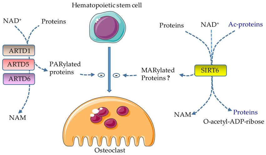
Cells 2019, 8, 1201 2 of 15
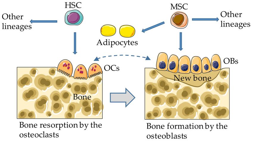
process that preserves bone mass and biomechanical properties (Figure 1). An imbalance between bone
resorption and formation underlies a variety of diseases featured by excessive bone gain or loss [23–25].
While the osteoclasts arise from hematopoietic stem cells, the osteoblasts derive from mesenchymal
stem cells (MSCs), which can also differentiate into adipocytes and chondrocytes under the influence
of specific environmental cues [26,27], as detailed below. Excessive bone marrow adipogenesis at the
expenses of osteogenesis has deleterious effects on bone, a tissue in which various cell types, including
mesenchymal and hematopoietic cells, express a repertoire of ARTs and SIRTs. We review the impact
of ADP-ribosylation on the differentiation of the osteoclasts, osteoblasts, and adipocytes, focusing on
ART and SIRT members with a functional link to bone health and disease.
Figure 1. Bone remodeling. In adults, the osteoclasts (OCs) and other hematopoietic lineages (not
depicted) arise from bone marrow hematopoietic stem cells (HSCs); the osteoblasts differentiate from
mesenchymal stem cells (MSCs), which can also develop into adipocytes and other lineages (not
depicted). Dashed and gray arrows indicate bidirectional regulatory interactions and coupling between
bone resorption and formation, respectively.
2. Osteoclast Differentiation
During development, the emerging ossification centers recruit myeloid progenitors where
they undergo terminal differentiation into the osteoclasts, which resorb the mineralized matrix, an
action that over time results in the formation of the bone marrow cavity [28–30]. Postnatally, the
damaged or old bone matrix is sensed and removed by the osteoclasts, and is evenly replaced by
the osteoblasts. Potential sensors of defective bone matrix components include the innate immune
complex, NOD-like receptor family (NLR), pyrin domain containing 3 (NLRP3) inflammasome,
which is activated by bone matrix degradation products and promotes osteoclast differentiation [31,
32]. While bone marrow myeloid precursors (e.g., CD11blow CD115high CD117high -expressing cells)
differentiate into the osteoclasts in homeostatic conditions, circulating monocytes are capable of
forming osteoclasts or fusing with pre-existing multinucleated osteoclasts in pathological settings,
such as inflammatory arthritis [33–38]. Osteoclast differentiation, activity, and survival depend on
macrophage colony-stimulating factor (M-CSF) and receptor activator of NF-κB ligand (RANKL),
whose expression and signaling outputs are regulated by various factors such as hormones (e.g.,
parathyroid hormone, estrogen, and 1,25α-dihydroxyvitamin D3) and pro-inflammatory cytokines,
including those of the tumor necrosis factor (TNF) and interleukin-1 (IL-1) families [23–25,39]. For
simplicity, this review focuses on osteoclast differentiation, though other biological aspects of these
cells, such as activity and survival, are occasionally described. M-CSF, RANKL, and the majority of
osteoclast-regulating factors are mainly produced by cells of the osteoblast lineage, and immune cells
(e.g., macrophages, T and B lymphocytes) though the osteoclasts themselves produce factors such as
sphingosine-1-phosphate and Wnts, which act not only in autocrine manner, but also paracrine fashion,
regulating the functions of neighboring cells such as the osteoblasts [40–45]. Osteoclast differentiation
is driven by complex interactions among various transcription factors, including the nuclear factor of
activated T cells cytoplasmic 1 (NFATc1), NF-κB, and c-Fos [46]. While the effects of PTMs such as
phosphorylation, methylation, ubiquitination, and SUMOylation on transcriptional regulation and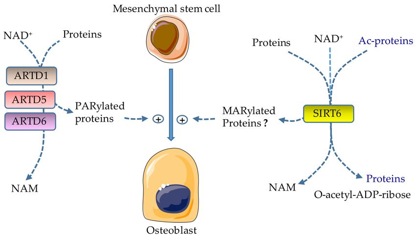
Cells 2019, 8, 1201                                                                               3 of 15

other key osteoclastogenic events have been extensively studied [47–55], only a few studies have
investigated the role of ADP-ribosylation in osteoclast biology.

2.1. Role of ARTD1 in Osteoclast Differentiation
      ARTD1 is the most studied member of the ARTD family in the skeleton. Early studies show that
ARTD1 protein levels decline during in vitro osteoclast differentiation induced by RANKL, a response
that correlates with increased expression of the a3 isoform of the V-ATPase subunit, tartrate-resistant
acid phosphatase, and brain-type creatine kinase [56–59]. In agreement with the proposition that
ARTD1 is a negative regulator of osteoclastogenesis, this protein binds to and represses the activity of
the promoters of the aforementioned genes in the osteoclast precursors [56–59]. Follow up studies
using engineered mice expressing uncleavable ARTD1 or Artd1-deficient mice, not only reinforce the
anti-osteoclastogenic functions of ARTD1, but also shed light into the underlying mechanisms [60–63].
Novel insights include the demonstration that i) ARTD1 inhibits histone3lysine4 trimethylation
(H3K4me3), histone marks of active chromatin, at the promoters of key osteoclastogenic factors such as
B lymphocyte-induced maturation protein 1 (Blimp1), and ii) ARTD1 PARylates itself during osteoclast
formation, a prerequisite modification that targets this protein for destruction through the proteasome
pathway [63]. ARTD1 also inhibits H3K4me3 and H4 acetylation, thereby impeding the recruitment
of the RelA subunit of NF-κB to the IL-1β promoter [61]. Progressive decline in ARTD1 levels also
occurs during the differentiation of myotubes, which are multinucleated fibers that arise from the
fusion of myoblasts [64]. The basis for the apparent inverse correlation between ARTD1 abundance
and multinucleation is unclear, though it is tempting to speculate that the degradation of this enzyme,
whose activity can deplete total intracellular NAD+ levels by 80% may be necessary to prevent energy
collapse during the high energy-demanding differentiation process.
      Mice lacking ARTD1 globally or selectively in myeloid cells indistinguishably exhibit a low bone
mass phenotype associated with an increased number of the osteoclasts on bone surfaces (Wang et al.,
personal communication). Consistent with the view of osteoclast lineage autonomous actions of
ARTD1, in vitro osteoclastogenesis from isolated mouse bone marrow cells is higher in Artd1 null
cells compared to wild-type controls [61]. Potential ARTD1 substrates include the master regulators
of osteoclast differentiation, NF-κB and NFATc1, which are PARylated by this enzyme in T cells and
smooth muscle cells [65–69]. However, such interplay is unlikely in light of the recent study indicating
that PARylated NF-κB and NFATc1 are undetectable in cells of the osteoclast lineage. Instead, ARTD1
consistently PARylates histone H2B among other proteins, and decreases the occupancy of H2B at
the NFATc1 promoter, thereby inhibiting NFATc1 expression and restraining osteoclast differentiation
(Wang et al., personal communication).
      ARTD1 is cleaved at D214 into 89 kDa and 24 kDa fragments, presumably by caspase-7, in response
to activation of the NLRP3 and NLR, CARD containing 4 (NLRC4) inflammasomes [63,70–72].
Consistent with its pro-inflammatory actions, loss of ARTD1 partially protects joints from destruction
in the mouse model of collagen antibody-induced arthritis [73–77]. In line with the ability of ARTD1
and its cleaved fragments to activate signaling platforms such as the NF-κB pathway, knockin mice
expressing uncleavable ARTD1 are resistant to ischemia/reperfusion-induced inflammation in intestine
and kidney [60,65]. Unexpectedly, this ARTD1 mutant does not affect inflammatory outcomes induced
by hyperactive NLRP3 inflammasome [62]. These conflicting results may be explained by the fact that
ARTD1 actions are cell-context-dependent. Indeed, ARTD1 promotes NF-κB PARylation or activity
in cultured smooth muscle cells, neuronal cells, and macrophages, while negatively regulating this
transcription factor in lymphocytic leukemia cells [65,66,68,72]. Despite some gaps in our understanding
of ARTD1 mechanisms of action, evidence overwhelmingly indicates that this enzyme negatively
regulates osteoclast development (Figure 2).
Cells 2019, 8, 1201 4 of 15
Figure 2. Effects of ADP-ribosylation on osteoclast formation. ARTD1, ARTD5, and ARTD6 catalyze the
attachment of ADP-ribose polymers from NAD+ to target proteins (PARylation), releasing nicotinamide
(NAM) in the process; their actions lead to the inhibition of osteoclast differentiation. SIRT6 inhibits
osteoclast development; however, the effects of its MARylating actions in this process are not clear
because this enzyme also has deacetylase activity. Lysine deacetylation is coupled to NAD+ hydrolysis,
yielding a deacetylated targeted protein, O-acetyl-ADP-ribose, and NAM. Ac, acetyl.
2.2. Role of ARTD5 and ARTD6 in Osteoclast Differentiation
ARTD5 (also known as PARP5A or tankyrase 1) and ARTD6 (also referred to as PARP5B or
tankyrase 2) [8] are expressed by many cell types, including the osteoclast lineage [78–81]. ARTD5 and
ARTD6 are implicated in a range of biological processes, including DNA repair, glucose homeostasis
and energy expenditure, and skeletal metabolism (through their interactions with the adaptor protein
SH3 domain-binding protein 2, SH3BP2 and AXIN 1/2) [78,82–86]. PARylation targets SH3BP2 for
ubiquitination by the E3-ubiquitin ligase RNF46, and subsequently for degradation [78,87]. Missense
mutations in SH3BP2 result in SH3BP2 that is stable, as it escapes the destructive actions of ARTD5 and
ARTD6, and are associated with cherubism, a hereditary childhood-onset autoinflammatory disorder,
whose severity regresses after puberty [88]. Focal facial bone lesions and deformities associated with the
destruction of the jaws and dental complications characterize this disease [88]. Knockin mice expressing
the most common disease-associated allele develop systemic inflammation (e.g., excessive TNF-α
production) and bone loss due to massive osteoclast differentiation as a consequence of heightened
sensitivity to M-CSF- and RANKL-induced signals; these events ultimately cumulate in hyperactivation
of osteoclastogenic pathways such as Src, Syk, ERK1/2, and NFATc1 [78,79]. Conversely, Sh3bp2-deficient
osteoclasts exhibit defective bone resorption in vitro [80]. Furthermore, pharmacological inhibition
of ARTD5 and ARTD6, which results in SH3BP2 accumulation, promotes osteoclast differentiation
in vitro and bone resorption in vivo [81,89], findings that are consistent with accelerated in vitro
osteoclastogenesis of osteoclast precursors lacking both ARTD5 and ARTD6 [78]. A recent study
suggests that oral bacteria produce pathogen-associated molecular patterns (PAMPs), which in
conjunction with danger-associated molecular patterns (DAMPs) released during the remodeling of
the jaws, provide tissue-restricted bone lesions in cherubism. Decreased jaw remodeling with age
leading to attenuated levels of DAMPs may underlie the reported regression of this disorder over time
in the affected patients [90]. Thus, ARTD5 and ARTD6 function as negative regulators of osteoclast
differentiation (Figure 2).
2.3. Role of SIRT6 in Osteoclast Differentiation
SIRTs are involved in the regulation of insulin secretion, gluconeogenesis, transcriptional regulation,
and several other biological responses [91–95]. Relevant to this review are SIRT4 and SIRT6, owing
to their MARylation activity that targets numerous proteins including glutamate dehydrogenase
and ARTD1 [91,96–98]. SIRT6 also MARylates itself, a presumed mechanism of self-regulation [99].
Notably, SIRT6, but not SIRT4, has a strong deacetylase activity, a reaction where lysine deacetylation isCells 2019, 8, 1201 5 of 15
coupled to NAD+ hydrolysis yielding O-acetyl-ADP-ribose, nicotinamide, and a deacetylated targeted
protein [100–103].
Consistent with SIRT6 inhibitory effects on the transactivation of NF-κB, an important regulator of
osteoclast development, overexpression of SIRT6 suppresses RANKL-induced OC formation in vitro
and bone destruction in mice with collagen-induced arthritis [25,104–106]. Conversely, SIRT6 deficiency
causes premature aging associated with increased osteoclastogenesis and low bone mass, or osteopenia
associated with low bone turnover [107–110]. Moreover, myeloid-specific deletion of SIRT6 results in a
decrease in estrogen receptor α protein levels and apoptosis of pre-osteoclasts, resulting in massive
bone resorption during aging and following ovariectomy [111]. Accordingly, SIRT6 transgenic mice
are protected from ovariectomy-induced bone loss. Mechanistically, SIRT6 deacetylates estrogen
receptor α at K171 and K299, thereby preventing its proteasomal degradation. In contrast to these
studies, SIRT6 reportedly forms a complex with Blimp1 to negatively regulate the expression of
anti-osteoclastogenic genes such as V-maf musculoaponeurotic fibrosarcoma oncogene homolog B
(Mafb), consistent with the increased bone mass and decreased osteoclast number in mice lacking
SIRT6 in hematopoietic cells [112]. Conversely, retroviral-mediated overexpression of SIRT6 increases
osteoclast formation [112]. Thus, SIRT6 regulates osteoclast differentiation, but the extent to which
SIRT6-driven MARylation affects osteoclastogenesis is unclear given the deacetylase activity of this
enzyme (Figure 2).
3. Osteoblast Differentiation
The osteoblasts differentiate from MSCs when exposed to growth factors such as bone
morphogenetic proteins (BMPs), Wnts, Hedgehog, and Notch, which activate transcription factors such
as RUNX2, osterix (OSX), and β-catenin [113]. Unlike osteoclastogenesis, osteoblast differentiation
and function are regulated negatively by inflammatory signals, some of which are mediated by
ARTs [114,115].
3.1. Role of ARTD1 in Osteoblast Differentiation
Immunohistochemical analysis of human bone samples shows that ARTD1 is mostly expressed
in osteoblasts in the areas of new bone formation, to a lesser extent in osteoclasts, while no
positive staining is detected in osteocytes, suggesting a role for ARTD1 in bone formation [116].
Poly-ADP-ribose (PAR) motifs are unexpectedly detected by nuclear magnetic resonance (NMR) and
immunostaining in the bone extracellular matrix, mostly in the calcifying region of the growth plate,
but to a lesser degree in the adjoining nonmineralized hypertrophic cartilage [117]. Thus, PAR units,
which may be released during cell necrosis, are potentially implicated in bone matrix calcification.
Earlier studies using human MSCs and SAOS-2 cells show that during osteoblast differentiation,
hydrogen peroxide activates ARTD1 and promotes osteoblastogenesis via activation of the p38 MAPK
pathway [118–120]. Consistent with its bone anabolic actions, the recruitment of ARTD1 by the long
non-encoding RNA (lncRNA) STEEL results in increased angiogenesis and fracture healing [121].
Furthermore, the ARTD inhibitor PJ34 suppresses osteogenic differentiation of murine MSCs, but
does not affect chondrocyte or adipocyte differentiation [122]. Thus, ARTD1 promotes osteoblast
differentiation under physiological conditions. However, ARTD1 also interacts with NF-κB in mediating
TNF-induced suppression of phosphate-regulating gene with homologies to endopeptidases on the
X chromosome (Phex), whose important functions in bone mineralization include the inhibition of
the expression of the hypophosphatemic fibroblast growth factor 23 (FGF23) [123,124]. Thus, while
ARTD1 favors osteogenesis in homeostatic conditions (Figure 3), it may compromise this process in
inflammatory states.Cells 2019, 8, 1201 6 of 15
Figure 3. Effects of ADP-ribosylation on osteobast formation. ARTD1, ARTD5, and ARTD6 catalyze the
attachment of ADP-ribose polymers from NAD+ to target proteins (PARylation), releasing nicotinamide
(NAM) in the process; their activities promote osteoblast differentiation. SIRT6 promotes osteoblast
development; however the effects of MARylation driven by SIRT6 in this process are not clear because
this enzyme also has deacetylase activity. Lysine deacetylation is coupled to NAD+ hydrolysis, yielding
a deacetylated targeted protein, O-acetyl-ADP-ribose, and NAM. Ac, acetyl.
3.2. Role of ARTD5 and ARTD6 in Osteoblast Differentiation
ARTD5 and ARTD6 PARylate and destabilize AXIN, a negative regulator of the critical
osteogenic Wnt/β-catenin signaling pathway [125]. Bone formation is impaired both in vivo and
in vitro in Sh3bp2-deficient cells through mechanisms involving the tyrosine kinase ABL, and the
transcription factors TAZ and RUNX2 [80,126]. The inhibitors of ARTD5 and ARTD6 enhance in vitro
osteoblastogenesis as the result of accumulated SH3BP2, promote nuclear translocation of ABL, TAZ,
and RUNX2, but they paradoxically decrease bone mass in mice associated with an increased number
of osteoclasts [81]. Thus, by stabilizing AXIN and SH3BP2, the inhibitors of ARTD5 and ARTD6 have
the potential of inhibiting osteogenesis (Figure 3) and inflicting substantial damage to the skeleton.
3.3. Role of SIRT6 in Osteoblast Differentiation
SIRT6-deficient mice exhibit stunted growth as a result of abnormal development of the growth
plate and impaired bone formation [108–110,127]. This phenotype is consistent with the plethoric actions
of this enzyme. Indeed, SIRT6 regulates the expression of RUNX2 and OSX through deacetylation of
H3K9; its deficiency is associated with hyperacetylation of H3K9 at the promoter of dickkopf-related
protein 1 (Dkk1), a potent negative regulator of osteoblastogenesis [110]. SIRT6 also modulates
the expression of the components of BMP signaling, actions that are p300/CBP-associated factor
(PCAF)-dependent [128]. Finally, SIRT6 promotes osteogenic differentiation of rat bone marrow
MSCs partially via suppression of NF-κB [129]. Thus, the actions of SIRT6 are pro-osteogenic
osteoblastogenesis. However, given the importance of protein deacetylation in osteogenesis, a function
that is also carried out by SIRT6, the role of MARylation mediated by this enzyme in this process
is unclear.
4. Adipocyte Differentiation
Bone marrow adiposity and visceral fat are implicated in the pathogenesis of bone diseases
such as osteoporosis [130,131]. Excessive differentiation of MSCs towards adipocytes in conjunction
with the secretion of adipokines (e.g., adiponectin) and cytokines (e.g., IL-6) adversely impact bone
metabolism. The adipogenic differentiation program of MSCs is controlled by transcription factors
such as CCAAT/enhancer binding protein α (C/EBPα) and peroxisome proliferator-activated receptor
γ (PPARγ) [132,133].Cells 2019, 8, 1201 7 of 15
Role of ARTD1 in Adipocyte Differentiation
ARTD1 activity drops for several hours during the early phase of preadipocytes 3T3-L1 cell
differentiation into adipocytes before returning to baseline levels and subsequently reaching higher
levels, presumably as a result of chromatin modifications [134]. Studies using stromal cells from
the fat pads of ARTD1-deficient mice show that the loss of this enzyme is associated with impaired
adipocyte function and differentiation [135]. Accordingly, when fed with a high-fat diet, Artd1 mice
develop hepatosteatosis and dysregulated glucose metabolism. Using the preadipocyte 3T3-L1 cells,
this group further demonstrates that ARTD1 is recruited to the promoters of PPARγ2 and its target
genes such as CD36 and aP2 in a PAR-dependent manner, responses that correlate with decreased
histone marks of repressed chromatin (H3K9me3), while marks of active chromatin (H3K4me3) are
increased [136]. However, studies based on a different mouse line suggest that lack of ARTD1 increases
energy expenditure through SIRT1 activation [137], findings that are consistent with the browning of
primary white adipocytes in vitro by olaparib, an inhibitor of ARTD1 and ARTD2 [138]. Other studies
also show that ARTD1 PARylates C/EBPβ, thereby inhibiting its DNA binding and transcriptional
activities, and ultimately, adipogenesis [139]. Thus, ARTD1 plays various roles in the differentiation of
adipocytes, acting at different stages to promote or inhibit this process.
5. Therapeutic Implications
ARTs are novel and promising targets for cancer therapies. Numerous studies have shown that
various small-molecule inhibitors of ARTDs are efficacious against various cancers, including ovarian
cancer, breast cancer, colon cancer, lung cancer, prostate cancer, hepatocellular carcinoma, osteosarcoma,
and chordoma [140–151]. More importantly, the US Food and Drug Administration has approved
three different ARTD inhibitors, olaparib, niraparib, and rucaparib for the treatment of BRCA1- or
BRAC2-mutated ovarian cancers. Considering the crucial role that ARTDs play in the pathogenesis
of acute tissue injury or periodontitis, some of these drugs may be indicated for the treatment of
inflammatory osteolysis. However, pre-clinical evidence indicates that genetic or pharmacological
inhibition of ARTD1 or ARTD5 and ARTD6 causes bone loss, a high-risk factor for fracture. Therefore,
comprehensive translational studies may help understand the extent to which ARTD inhibitors may
adversely affect the skeleton.
Author Contributions: C.W. and G.M. conceived and wrote the manuscript.
Funding: This work was funded by NIH/NIAMS AR064755 and AR068972 grants to G.M.
Acknowledgments: We thank Dustin Kress for critical reading of the manuscript. Due to a large amount of
research in the area of ART biology, we are sure to have missed some important papers. We apologize in advance
to authors who have been omitted. The drawings in Figures 1–3 were modified from https://smart.servier.com.
Conflicts of Interest: G.M. is a consultant for Aclaris Therapeutics, Inc. Other authors declare no conflict
of interest.
Abbreviations
ADP: adenosine diphosphate; ARH, ADP-ribosyl hydrolase; ART, ADP-ribosyltransferase; ARTD, diphtheria
toxin-like ART; ARTC, cholera toxin-like ART; Blimp1, B lymphocyte induced maturation protein 1; BMPs,
bone morphogenetic proteins; C/EBPα, CCAAT/enhancer binding protein α; DAMPs, danger-associated
molecular patterns; Dkk1, dickkopf-related protein 1; IL-1, interleukin-1; lncRNA, long non-encoding RNA;
Mafb, V-maf musculoaponeurotic fibrosarcoma oncogene homolog B; MAPK, mitogen-activated protein kinase;
MARylation, mono-ADP-ribosylation; M-CSF, macrophage colony-stimulating factor; MSC, mesenchymal stem
cell; NFATc1, nuclear factor of activated T cells cytoplasmic 1; PAMPs, pathogen-associated molecular patterns;
NAM, nicotinamide; NLRC4, NLR, CARD containing 4; NLRP3, NOD-like receptor family (NLR), pyrin
domain containing 3; PARG, poly(ADP-ribose) glycohydrolase; OSX, osterix; PAR, poly-ADP-ribose; PARP,
poly(ADP-ribose) polymerase; PARylation, poly-ADP-ribosylation; PCAF, p300/CBP-associated factor; Phex,
phosphate-regulating gene with homologies to endopeptidases on the X; PM, post-translational modification;
PPARγ, peroxisome proliferator-activated receptor γ; RANKL, receptor activator of NF-κB ligand; SH3BP2, SH3
domain-binding protein 2; SIRT, sirtuin; TARG1, terminal ADP-ribose protein glycohydrolase 1; TNF, tumor
necrosis factor.Cells 2019, 8, 1201 8 of 15
References
1. Kunze, F.A.; Hottiger, M.O. Regulating Immunity via ADP-Ribosylation: Therapeutic Implications and
Beyond. Trends Immunol. 2019, 40, 159–173. [CrossRef] [PubMed]
2. Bütepage, M.; Eckei, L.; Verheugd, P.; Lüscher, B. Intracellular Mono-ADP-Ribosylation in Signaling and
Disease. Cells 2015, 4, 569–595. [CrossRef] [PubMed]
3. Cohen, M.S.; Chang, P. Insights into the biogenesis, function, and regulation of ADP-ribosylation. Nat. Chem.
Biol. 2018, 14, 236–243. [CrossRef] [PubMed]
4. Gupte, R.; Liu, Z.; Kraus, W.L. PARPs and ADP-ribosylation: Recent advances linking molecular functions to
biological outcomes. Genes Dev. 2017, 31, 101–126. [CrossRef] [PubMed]
5. Vivelo, C.A.; Leung, A.K. Proteomics approaches to identify mono-(ADP-ribosyl)ated and
poly(ADP-ribosyl)ated proteins. Proteomics 2015, 15, 203–217. [CrossRef] [PubMed]
6. Alemasova, E.E.; Lavrik, O.I. Poly(ADP-ribosyl)ation by PARP1: Reaction mechanism and regulatory
proteins. Nucleic Acids Res. 2019, 47, 3811–3827. [CrossRef]
7. Di Girolamo, M.; Fabrizio, G. Overview of the mammalian ADP-ribosyl-transferases clostridia toxin-like
(ARTCs) family. Biochem. Pharmacol. 2019, 167, 86–96. [CrossRef]
8. Hottiger, M.O. Nuclear ADP-Ribosylation and Its Role in Chromatin Plasticity, Cell Differentiation, and
Epigenetics. Annu. Rev. Biochem. 2015, 84, 227–263. [CrossRef]
9. Crawford, K.; Bonfiglio, J.J.; Mikoč, A.; Matic, I.; Ahel, I. Specificity of reversible ADP-ribosylation and
regulation of cellular processes. Crit. Rev. Biochem. Mol. Biol. 2018, 53, 64–82. [CrossRef]
10. Bartlett, E.; Bonfiglio, J.J.; Prokhorova, E.; Colby, T.; Zobel, F.; Ahel, I.; Matic, I. Interplay of Histone Marks
with Serine ADP-Ribosylation. Cell Rep. 2018, 24, 3488–3502.e5. [CrossRef]
11. Abplanalp, J.; Hottiger, M.O. Cell fate regulation by chromatin ADP-ribosylation. Semin. Cell Dev. Boil. 2017,
63, 114–122. [CrossRef] [PubMed]
12. Racz, B.; Hantó, K.; Tapodi, A.; Solti, I.; Kálmán, N.; Jakus, P.; Kovacs, K.; Debreceni, B.; Gallyas, F.; Sumegi, B.
Regulation of MKP-1 expression and MAPK activation by PARP-1 in oxidative stress: A new mechanism for
the cytoplasmic effect of PARP-1 activation. Free. Radic. Boil. Med. 2010, 49, 1978–1988. [CrossRef] [PubMed]
13. Gibson, B.A.; Zhang, Y.; Jiang, H.; Hussey, K.M.; Shrimp, J.H.; Lin, H.; Schwede, F.; Yu, Y.; Kraus, W.L.
Chemical Genetic Discovery of PARP Targets Reveals a Role for PARP-1 in Transcription Elongation. Science
2016, 353, 45–50. [CrossRef] [PubMed]
14. Marjanović, M.P.; Crawford, K.; Ahel, I. PARP, transcription and chromatin modeling. Semin. Cell Dev. Boil.
2017, 63, 102–113. [CrossRef] [PubMed]
15. Kraus, W.L. Transcriptional control by PARP-1: Chromatin modulation, enhancer-binding, coregulation, and
insulation. Curr. Opin. Cell Boil. 2008, 20, 294–302. [CrossRef] [PubMed]
16. Krishnakumar, R.; Kraus, W.L. The PARP side of the nucleus: Molecular actions, physiological outcomes,
and clinical targets. Mol. Cell 2010, 39, 8–24. [CrossRef] [PubMed]
17. Ji, Y.; Tulin, A.V. The roles of PARP1 in gene control and cell differentiation. Curr. Opin. Genet. Dev. 2010, 20,
512–518. [CrossRef]
18. Kraus, W.L. PARPs and ADP-Ribosylation: 50 Years . . . and Counting. Mol. Cell 2015, 58, 902–910. [CrossRef]
19. D’Amours, D.; Desnoyers, S.; D’Silva, I.; Poirier, G.G. Poly(ADP-ribosyl)ation reactions in the regulation of
nuclear functions. Biochem. J. 1999, 342, 249–268. [CrossRef]
20. Kronenberg, H.M. Developmental regulation of the growth plate. Nature 2003, 423, 332–336. [CrossRef]
21. Betts, J.G.; Desaix, P. Bone Tissue and the Skeletal System. Available online: https://courses.lumenlearning.
com/austincc-ap1/chapter/bone-tissue-and-the-skeletal-system/ (accessed on 2 September 2019).
22. Hall, B.K. Earliest Evidence of Cartilage and Bone Development in Embryonic Life. Clin. Orthop. Relat. Res.
1987, 255. [CrossRef]
23. Mbalaviele, G.; Veis, D.J. Inflammasomes in Bone Diseases. Exp. Suppl. 2018, 108, 269–279. [PubMed]
24. Mbalaviele, G.; Novack, D.V.; Schett, G.; Teitelbaum, S.L. Inflammatory osteolysis: A conspiracy against
bone. J. Clin. Investig. 2017, 127, 2030–2039. [CrossRef] [PubMed]
25. Novack, D.V.; Mbalaviele, G. Osteoclasts-Key Players in Skeletal Health and Disease. Microbiol. Spectr. 2016,
4, 4. [CrossRef] [PubMed]
26. Pittenger, M.F. Multilineage Potential of Adult Human Mesenchymal Stem Cells. Science 1999, 284, 143–147.
[CrossRef]Cells 2019, 8, 1201 9 of 15
27. Peister, A.; Mellad, J.A.; Larson, B.L.; Hall, B.M.; Gibson, L.F.; Prockop, D.J. Adult stem cells from bone
marrow (MSCs) isolated from different strains of inbred mice vary in surface epitopes, rates of proliferation,
and differentiation potential. Blood 2004, 103, 1662–1668. [CrossRef]
28. Charbord, P.; Tavian, M.; Humeau, L.; Péault, B. Early ontogeny of the human marrow from long bones: An
immunohistochemical study of hematopoiesis and its microenvironment. Blood 1996, 87, 4109–4119.
29. Chen, L.T.; Weiss, L. The development of vertebral bone marrow of human fetuses. Blood 1975, 46, 389–408.
[CrossRef]
30. Seike, M.; Omatsu, Y.; Watanabe, H.; Kondoh, G.; Nagasawa, T. Stem cell niche-specific Ebf3 maintains the
bone marrow cavity. Genes Dev. 2018, 32, 359–372. [CrossRef]
31. Alippe, Y.; Wang, C.; Ricci, B.; Xiao, J.; Qu, C.; Zou, W.; Novack, D.V.; Abu-Amer, Y.; Civitelli, R.; Mbalaviele, G.
Bone matrix components activate the NLRP3 inflammasome and promote osteoclast differentiation. Sci. Rep.
2017, 7, 6630. [CrossRef]
32. Alippe, Y.; Mbalaviele, G. Omnipresence of inflammasome activities in inflammatory bone diseases. Semin.
Immunopathol. 2019. [CrossRef] [PubMed]
33. Charles, J.F.; Hsu, L.-Y.; Niemi, E.C.; Weiss, A.; Aliprantis, A.O.; Nakamura, M.C. Inflammatory arthritis
increases mouse osteoclast precursors with myeloid suppressor function. J. Clin. Investig. 2012, 122,
4592–4605. [CrossRef] [PubMed]
34. Jacome-Galarza, C.E.; Lee, S.-K.; Lorenzo, J.A.; Aguila, H.L. Identification, characterization, and isolation
of a common progenitor for osteoclasts, macrophages, and dendritic cells from murine bone marrow and
periphery. J. Bone Miner. Res. 2013, 28, 1203–1213. [CrossRef] [PubMed]
35. Jacquin, C.; Gran, D.E.; Lee, S.K.; Lorenzo, J.A.; Aguila, H.L. Identification of multiple osteoclast precursor
populations in murine bone marrow. J. Bone Miner. Res. 2006, 21, 67–77. [CrossRef] [PubMed]
36. Jacome-Galarza, C.E.; Percin, G.I.; Muller, J.T.; Mass, E.; Lazarov, T.; Eitler, J.; Rauner, M.; Yadav, V.K.;
Crozet, L.; Bohm, M.; et al. Developmental origin, functional maintenance and genetic rescue of osteoclasts.
Nature 2019, 568, 541–545. [CrossRef] [PubMed]
37. Gu, R.; Santos, L.L.; Ngo, D.; Fan, H.; Singh, P.P.; Fingerle-Rowson, G.; Bucala, R.; Xu, J.; Quinn, J.M.W.;
Morand, E.F. Macrophage migration inhibitory factor is essential for osteoclastogenic mechanisms in vitro
and in vivo mouse model of arthritis. Cytokine 2015, 72, 135–145. [CrossRef] [PubMed]
38. Romas, E.; Bakharevski, O.; Hards, D.K.; Kartsogiannis, V.; Quinn, J.M.W.; Ryan, P.F.J.; Martin, T.J.;
Gillespie, M.T. Expression of osteoclast differentiation factor at sites of bone erosion in collagen-induced
arthritis. Arthritis Rheum. 2000, 43, 821. [CrossRef]
39. Kanatani, M.; Sugimoto, T.; Takahashi, Y.; Kaji, H.; Kitazawa, R.; Chihara, K. Estrogen via the Estrogen
Receptor Blocks cAMP-Mediated Parathyroid Hormone (PTH)-Stimulated Osteoclast Formation. J. Bone
Miner. Res. 1998, 13, 854–862. [CrossRef] [PubMed]
40. Dirckx, N.; Moorer, M.C.; Clemens, T.L.; Riddle, R.C. The role of osteoblasts in energy homeostasis. Nat. Rev.
Endocrinol. 2019. [CrossRef] [PubMed]
41. Lerner, U.H.; Ohlsson, C. The WNT system: Background and its role in bone. J. Intern. Med. 2015, 277,
630–649. [CrossRef] [PubMed]
42. Sartawi, Z.; Schipani, E.; Ryan, K.B.; Waeber, C. Sphingosine 1-phosphate (S1P) signalling: Role in bone
biology and potential therapeutic target for bone repair. Pharmacol. Res. 2017, 125, 232–245. [CrossRef]
[PubMed]
43. Meshcheryakova, A.; Mechtcheriakova, D.; Pietschmann, P. Sphingosine 1-phosphate signaling in bone
remodeling: Multifaceted roles and therapeutic potential. Expert Opin. Ther. Tar. 2017, 21, 725–737.
[CrossRef] [PubMed]
44. Pederson, L.; Ruan, M.; Westendorf, J.J.; Khosla, S.; Oursler, M.J. Regulation of bone formation by osteoclasts
involves Wnt/BMP signaling and the chemokine sphingosine-1-phosphate. Proc. Natl. Acad. Sci. USA 2008,
105, 20764–20769. [CrossRef]
45. Quint, P.; Ruan, M.; Pederson, L.; Kassem, M.; Westendorf, J.J.; Khosla, S.; Oursler, M.J. Sphingosine
1-Phosphate (S1P) Receptors 1 and 2 Coordinately Induce Mesenchymal Cell Migration through S1P
Activation of Complementary Kinase Pathways. J. Boil. Chem. 2013, 288, 5398–5406. [CrossRef] [PubMed]
46. Park, J.H.; Lee, N.K.; Lee, S.Y. Current Understanding of RANK Signaling in Osteoclast Differentiation and
Maturation. Mol. Cells 2017, 40, 706–713. [PubMed]Cells 2019, 8, 1201 10 of 15
47. Sato, K.; Suematsu, A.; Nakashima, T.; Takemoto-Kimura, S.; Aoki, K.; Morishita, Y.; Asahara, H.; Ohya, K.;
Yamaguchi, A.; Takai, T.; et al. Regulation of osteoclast differentiation and function by the CaMK-CREB
pathway. Nat. Med. 2006, 12, 1410–1416. [CrossRef] [PubMed]
48. Huh, J.-E.; Lee, W.I.; Kang, J.W.; Nam, D.; Choi, D.-Y.; Park, D.-S.; Lee, S.H.; Lee, J.-D. Formononetin
Attenuates Osteoclastogenesis via Suppressing the RANKL-Induced Activation of NF-κB, c-Fos, and Nuclear
Factor of Activated T-Cells Cytoplasmic 1 Signaling Pathway. J. Nat. Prod. 2014, 77, 2423–2431. [CrossRef]
49. Kim, J.H.; Kim, N. Regulation of NFATc1 in Osteoclast Differentiation. J. Bone Metab. 2014, 21, 233–241.
[CrossRef]
50. Yasui, T.; Tsutsumi, S.; Aburatani, H.; Hirose, J.; Nakamura, K.; Tanaka, S. Epigenetic regulation of osteoclast
differentiation: Possible involvement of Jmjd3 in the histone demethylation of Nfatc1. J. Bone Miner. Res.
2011, 26, 2665–2671. [CrossRef]
51. Collins, P.E.; Mitxitorena, I.; Carmody, R.J. The Ubiquitination of NF-κB Subunits in the Control of
Transcription. Cells 2016, 5. [CrossRef]
52. Kim, J.H.; Kim, K.; Jin, H.M.; Song, I.; Youn, B.U.; Lee, S.H.; Choi, Y.; Kim, N. Negative feedback control
of osteoclast formation through ubiquitin-mediated down-regulation of NFATc1. J. Biol. Chem. 2010, 285,
5224–5231. [CrossRef] [PubMed]
53. Nayak, A.; Glöckner-Pagel, J.; Vaeth, M.; Schumann, J.E.; Buttmann, M.; Bopp, T.; Schmitt, E.; Serfling, E.;
Berberich-Siebelt, F. Sumoylation of the Transcription Factor NFATc1 Leads to Its Subnuclear Relocalization
and Interleukin-2 Repression by Histone Deacetylase. J. Boil. Chem. 2009, 284, 10935–10946. [CrossRef]
[PubMed]
54. Chen, N.-M.; Neesse, A.; Dyck, M.L.; Steuber, B.; Koenig, A.O.; Lubeseder-Martellato, C.; Winter, T.; Forster, T.;
Bohnenberger, H.; Kitz, J.; et al. Context-Dependent Epigenetic Regulation of Nuclear Factor of Activated T
Cells 1 in Pancreatic Plasticity. Gastroenterology 2017, 152, 1507–1520.e15. [CrossRef] [PubMed]
55. Yasui, T.; Hirose, J.; Aburatani, H.; Tanaka, S. Epigenetic regulation of osteoclast differentiation. Ann. N. Y.
Acad. Sci. 2011, 1240, 7–13. [CrossRef] [PubMed]
56. Beranger, G.E.; Momier, D.; Rochet, N.; Quincey, D.; Guigonis, J.M.; Samson, M.; Carle, G.F.; Scimeca, J.C.
RANKL Treatment Releases the Negative Regulation of the Poly(ADP-Ribose) Polymerase-1 on Tcirg1 Gene
Expression During Osteoclastogenesis. J. Bone Miner. Res. 2006, 21, 1757–1769. [CrossRef]
57. Beranger, G.E.; Momier, D.; Guigonis, J.-M.; Samson, M.; Carle, G.F.; Scimeca, J.-C.; Guigonis, J.; Scimeca, J.
Differential Binding of Poly(ADP-Ribose) Polymerase-1 and JunD/Fra2 Accounts for RANKL-Induced Tcirg1
Gene Expression During Osteoclastogenesis. J. Bone Miner. Res. 2007, 22, 975–983. [CrossRef] [PubMed]
58. Beranger, G.E.; Momier, D.; Rochet, N.; Carle, G.F.; Scimeca, J.C. Poly(adp-ribose) polymerase-1 regulates
Tracp gene promoter activity during RANKL-induced osteoclastogenesis. J. Bone Miner. Res. 2008, 23,
564–571. [CrossRef]
59. Chen, J.; Sun, Y.; Mao, X.; Liu, Q.; Wu, H.; Chen, Y. RANKL Up-regulates Brain-type Creatine Kinase via
Poly(ADP-ribose) Polymerase-1 during Osteoclastogenesis. J. Boil. Chem. 2010, 285, 36315–36321. [CrossRef]
60. Petrilli, V.; Herceg, Z.; Hassa, P.O.; Patel, N.S.; Paola, R.D.; Cortes, U.; Dugo, L.; Filipe, H.-M.; Thiemermann, C.;
Hottiger, M.O.; et al. Noncleavable poly(ADP-ribose) polymerase-1 regulates the inflammation response in
mice. J. Clin. Investig. 2004, 114, 1072–1081. [CrossRef]
61. Robaszkiewicz, A.; Qu, C.; Wisnik, E.; Ploszaj, T.; Mirsaidi, A.; Kunze, F.A.; Richards, P.J.; Cinelli, P.;
Mbalaviele, G.; Hottiger, M.O. ARTD1 regulates osteoclastogenesis and bone homeostasis by dampening
NF-kappaB-dependent transcription of IL-1beta. Sci. Rep. 2016, 6, 21131. [CrossRef]
62. Wang, C.; Xu, C.-X.; Alippe, Y.; Qu, C.; Xiao, J.; Schipani, E.; Civitelli, R.; Abu-Amer, Y.; Mbalaviele, G.
Chronic inflammation triggered by the NLRP3 inflammasome in myeloid cells promotes growth plate
dysplasia by mesenchymal cells. Sci. Rep. 2017, 7, 4880. [CrossRef] [PubMed]
63. Wang, C.; Qu, C.; Alippe, Y.; Bonar, S.L.; Civitelli, R.; Abu-Amer, Y.; O Hottiger, M.; Mbalaviele, G.
Poly-ADP-ribosylation-mediated degradation of ARTD1 by the NLRP3 inflammasome is a prerequisite for
osteoclast maturation. Cell Death Dis. 2016, 7, e2153. [CrossRef] [PubMed]
64. Oláh, G.; Szczesny, B.; Brunyánszki, A.; López-García, I.A.; Gero, D.; Radak, Z.; Szabo, C.
Differentiation-Associated Downregulation of Poly(ADP-Ribose) Polymerase-1 Expression in Myoblasts
Serves to Increase Their Resistance to Oxidative Stress. PLoS ONE 2015, 10, e0134227. [CrossRef] [PubMed]Cells 2019, 8, 1201 11 of 15
65. Castri, P.; Lee, Y.J.; Ponzio, T.; Maric, D.; Spatz, M.; Bembry, J.H. Poly(ADP-ribose) polymerase-1 and its
cleavage products differentially modulate cellular protection through NF-κB-dependent signaling. BBA-Mol.
Cell Res. 2014, 1843, 640–651.
66. Zerfaoui, M.; Errami, Y.; Naura, A.S.; Suzuki, Y.; Kim, H.; Ju, J.; Liu, T.; Hans, C.P.; Kim, J.G.; Elmageed, Z.Y.A.;
et al. Poly(ADP-ribose) polymerase-1 is a determining factor in Crm1-mediated nuclear export and retention
of p65 NF-kappa B upon TLR4 stimulation. J. Immunol. 2010, 185, 1894–1902. [CrossRef] [PubMed]
67. Valdor, R.; Schreiber, V.; Saenz, L.; Martínez, T.; Muñoz-Suano, A.; Domínguez-Villar, M.; Ramírez, P.;
Parrilla, P.; Aguado, E.; Garcia-Cozar, F. Regulation of NFAT by poly(ADP-ribose) polymerase activity in T
cells. Mol. Immunol. 2008, 45, 1863–1871. [CrossRef] [PubMed]
68. Kameoka, M.; Ota, K.; Tetsuka, T.; Tanaka, Y.; Itaya, A.; Okamoto, T.; Yoshihara, K. Evidence for regulation of
NF-kappaB by poly(ADP-ribose) polymerase. Biochem. J. 2000, 346, 641–649. [CrossRef] [PubMed]
69. Olabisi, O.A.; Soto-Nieves, N.; Nieves, E.; Yang, T.T.C.; Yang, X.; Yu, R.Y.L.; Suk, H.Y.; Macian, F.; Chow, C.-W.
Regulation of Transcription Factor NFAT by ADP-Ribosylation. Mol. Cell. Boil. 2008, 28, 2860–2871.
[CrossRef] [PubMed]
70. Malireddi, R.K.S.; Ippagunta, S.; Lamkanfi, M.; Kanneganti, T.-D. Cutting edge: Proteolytic inactivation of
poly(ADP-ribose) polymerase 1 by the Nlrp3 and Nlrc4 inflammasomes. J. Immunol. 2010, 185, 3127–3130.
[CrossRef]
71. Qu, C.; Bonar, S.L.; Hickman-Brecks, C.L.; Abu-Amer, S.; McGeough, M.D.; Peña, C.A.; Broderick, L.; Yang, C.;
Kading, J. NLRP3 mediates osteolysis through inflammation-dependent and -independent mechanisms.
FASEB. J. 2015, 29, 1269–1279. [CrossRef]
72. Erener, S.; Pétrilli, V.; Kassner, I.; Minotti, R.; Castillo, R.; Santoro, R.; Hassa, P.O.; Tschopp, J.; Hottiger, M.O.
Inflammasome-Activated Caspase 7 Cleaves PARP1 to Enhance the Expression of a Subset of NF-κB Target
Genes. Mol. Cell 2012, 46, 200–211. [CrossRef] [PubMed]
73. García, S.; Bodaño, A.; González, A.; Forteza, J.; Gómez-Reino, J.J.; Conde, C. Partial protection against
collagen antibody-induced arthritis in PARP-1 deficient mice. Arthritis Res. Ther. 2006, 8. [CrossRef]
[PubMed]
74. Kunze, F.A.; Bauer, M.; Komuczki, J.; Lanzinger, M.; Gunasekera, K.; Hopp, A.K.; Lehmann, M.; Becher, B.;
Müller, A.; Hottiger, M.O. ARTD1 in Myeloid Cells Controls the IL-12/18–IFN-γ Axis in a Model of Sterile
Sepsis, Chronic Bacterial Infection, and Cancer. J. Immunol. 2019, 202, 1406–1416. [CrossRef] [PubMed]
75. Oliver, F.J.; Murcia, J.M.; Nacci, C.; Decker, P.; Andriantsitohaina, R.; Muller, S.; de la Rubia, G.; Stoclet, J.C.;
de Murcia, G. Resistance to endotoxic shock as a consequence of defective NF-κB activation in poly
(ADP-ribose) polymerase-1 deficient mice. EMBO J. 1999, 18, 4446–4454. [CrossRef] [PubMed]
76. Burkart, V.; Wang, Z.-Q.; Radons, J.; Heller, B.; Herceg, Z.; Stingl, L.; Wagner, E.F.; Kolb, H. Mice lacking the
poly(ADP-ribose) polymerase gene are resistant to pancreatic beta-cell destruction and diabetes development
induced by streptozocin. Nat. Med. 1999, 5, 314–319. [CrossRef] [PubMed]
77. Mabley, J.G.; Jagtap, P.; Perretti, M.; Getting, S.J.; Salzman, A.L.; Virag, L.; Szabo, E.; Soriano, F.G.; Liaudet, L.;
Abdelkarim, G.E.; et al. Anti-inflammatory effects of a novel, potent inhibitor of poly (ADP-ribose)
polymerase. Inflamm. Res. 2001, 50, 561–569. [CrossRef] [PubMed]
78. Levaot, N.; Voytyuk, O.; Dimitriou, I.; Sircoulomb, F.; Chandrakumar, A.; Deckert, M.; Krzyzanowski, P.M.;
Scotter, A.; Gu, S.; Janmohamed, S.; et al. Loss of Tankyrase-mediated destruction of 3BP2 is the underlying
pathogenic mechanism of cherubism. Cell 2011, 147, 1324–1339. [CrossRef] [PubMed]
79. Ueki, Y.; Lin, C.-Y.; Senoo, M.; Ebihara, T.; Agata, N.; Onji, M.; Saheki, Y.; Kawai, T.; Mukherjee, P.M.;
Reichenberger, E.; et al. Increased Myeloid Cell Responses to M-CSF and RANKL Cause Bone Loss and
Inflammation in SH3BP2 “Cherubism” Mice. Cell 2007, 128, 71–83. [CrossRef] [PubMed]
80. Levaot, N.; Simoncic, P.; Dimitriou, I.; Scotter, A.; Rose, J.L.; Willett, T.; Ng, A.; Wang, C.; Janmohamed, S.;
Grynpas, M.; et al. 3BP2 deficient mice are osteoporotic with impaired osteoblast and osteoclast functions.
J. Clin. Invest. 2011, 121, 3244–3257. [CrossRef]
81. Fujita, S.; Mukai, T.; Mito, T.; Kodama, S.; Nagasu, A.; Kittaka, M.; Sone, T.; Ueki, Y.; Morita, Y. Pharmacological
inhibition of tankyrase induces bone loss in mice by increasing osteoclastogenesis. Bone 2018, 106, 156–166.
[CrossRef] [PubMed]
82. Jiang, Q.; Paramasivam, M.; Aressy, B.; Wu, J.; Bellani, M.; Tong, W.; Seidman, M.M.; Greenberg, R.A.
MERIT40 cooperates with BRCA2 to resolve DNA interstrand cross-links. Genes Dev. 2015, 29, 1955–1968.
[CrossRef] [PubMed]Cells 2019, 8, 1201 12 of 15
83. Nagy, Z.; Kalousi, A.; Furst, A.; Koch, M.; Fischer, B.; Soutoglou, E. Tankyrases Promote Homologous
Recombination and Check Point Activation in Response to DSBs. PLoS Genet. 2016, 12, 1005791. [CrossRef]
[PubMed]
84. Zhong, L.L.; Ding, Y.; Bandyopadhyay, G.; Waaler, J.; Börgeson, E.; Smith, S.; Zhang, M.; Phillips, S.A.;
Mahooti, S.; Mahata, S.K.; et al. The PARsylation activity of tankyrase in adipose tissue modulates systemic
glucose metabolism in mice. Diabetologia 2016, 59, 582–591. [CrossRef] [PubMed]
85. Yeh, T.-Y.J.; Beiswenger, K.K.; Li, P.; Bolin, K.E.; Lee, R.M.; Tsao, T.-S.; Murphy, A.N.; Hevener, A.L.; Chi, N.-W.
Hypermetabolism, Hyperphagia, and Reduced Adiposity in Tankyrase-Deficient Mice. Diabetes 2009, 58,
2476–2485. [CrossRef] [PubMed]
86. Mariotti, L.; Pollock, K.; Guettler, S. Regulation of Wnt/beta-catenin signalling by tankyrase-dependent
poly(ADP-ribosyl)ation and scaffolding. Br. J. Pharmacol. 2017, 174, 4611–4636. [CrossRef]
87. Guettler, S.; LaRose, J.; Petsalaki, E.; Gish, G.; Scotter, A.; Pawson, T.; Rottapel, R.; Sicheri, F. Structural Basis
and Sequence Rules for Substrate Recognition by Tankyrase Explain the Basis for Cherubism Disease. Cell
2011, 147, 1340–1354. [CrossRef] [PubMed]
88. Ueki, Y.; Tiziani, V.; Santanna, C.; Fukai, N.; Maulik, C.; Garfinkle, J.; Ninomiya, C.; Doamaral, C.; Peters, H.;
Habal, M.; et al. Mutations in the gene encoding c-Abl-binding protein SH3BP2 cause cherubism. Nat. Genet.
2001, 28, 125–126. [CrossRef]
89. Mukai, T.; Fujita, S.; Morita, Y. Tankyrase (PARP5) Inhibition Induces Bone Loss through Accumulation of Its
Substrate SH3BP2. Cells 2019, 8, 195. [CrossRef]
90. Yoshitaka, T.; Mukai, T.; Kittaka, M.; Alford, L.M.; Masrani, S.; Ishida, S.; Yamaguchi, K.; Yamada, M.;
Mizuno, N.; Olsen, B.R.; et al. Enhanced TLR-MYD88 signaling stimulates autoinflammation in SH3BP2
cherubism mice and defines the etiology of cherubism. Cell Rep. 2014, 8, 1752–1766. [CrossRef]
91. Haigis, M.C.; Mostoslavsky, R.; Haigis, K.M.; Fahie, K.; Christodoulou, D.C.; Murphy, A.J.; Valenzuela, D.M.;
Yancopoulos, G.D.; Karow, M.; Blander, G.; et al. SIRT4 inhibits glutamate dehydrogenase and opposes the
effects of calorie restriction in pancreatic beta cells. Cell 2006, 126, 941–954. [CrossRef]
92. Moynihan, K.A.; Grimm, A.A.; Plueger, M.M.; Bernal-Mizrachi, E.; Ford, E.; Cras-Méneur, C.; Permutt, M.A.;
Imai, S.I. Increased dosage of mammalian Sir2 in pancreatic beta cells enhances glucose-stimulated insulin
secretion in mice. Cell Metab. 2005, 2, 105–117. [CrossRef] [PubMed]
93. Bordone, L.; Motta, M.C.; Picard, F.; Robinson, A.; Jhala, U.S.; Apfeld, J.; McDonagh, T.; Lemieux, M.;
McBurney, M.; Szilvasi, A.; et al. Sirt1 regulates insulin secretion by repressing UCP2 in pancreatic beta cells.
PLoS Biol. 2006, 4, e31. [CrossRef]
94. Rodgers, J.T.; Lerin, C.; Haas, W.; Gygi, S.P.; Spiegelman, B.M.; Puigserver, P. Nutrient control of glucose
homeostasis through a complex of PGC-1alpha and SIRT1. Nature 2005, 434, 113–118. [CrossRef]
95. Canto, C.; Auwerx, J. Targeting sirtuin 1 to improve metabolism: All you need is NAD(+)? Pharmacol. Rev.
2012, 64, 166–187. [CrossRef] [PubMed]
96. Mao, Z.; Hine, C.; Tian, X.; Van Meter, M.; Au, M.; Vaidya, A.; Seluanov, A.; Gorbunova, V.; Hine, C. SIRT6
promotes DNA repair under stress by activating PARP1. Science 2011, 332, 1443–1446. [CrossRef]
97. Rezazadeh, S.; Yang, D.; Tombline, G.; Simon, M.; Regan, S.P.; Seluanov, A.; Gorbunova, V. SIRT6 promotes
transcription of a subset of NRF2 targets by mono-ADP-ribosylating BAF170. Nucleic Acids Res. 2019, 47,
7914–7928. [CrossRef]
98. Van Meter, M.; Kashyap, M.; Rezazadeh, S.; Geneva, A.J.; Morello, T.D.; Seluanov, A.; Gorbunova, V.
SIRT6 represses LINE1 retrotransposons by ribosylating KAP1 but this repression fails with stress and age.
Nat. Commun. 2014, 5, 5011. [CrossRef]
99. Liszt, G.; Ford, E.; Kurtev, M.; Guarente, L. Mouse Sir2 Homolog SIRT6 Is a Nuclear ADP-ribosyltransferase.
J. Boil. Chem. 2005, 280, 21313–21320. [CrossRef]
100. North, B.J.; Marshall, B.L.; Borra, M.T.; Denu, J.M.; Verdin, E. The human Sir2 ortholog, SIRT2, is an
NAD+-dependent tubulin deacetylase. Mol. Cell 2003, 11, 437–444. [CrossRef]
101. Bhardwaj, A.; Das, S. SIRT6 deacetylates PKM2 to suppress its nuclear localization and oncogenic functions.
Proc. Natl. Acad. Sci. USA 2016, 113, E538–E547. [CrossRef]
102. Pan, P.W.; Feldman, J.L.; Devries, M.K.; Dong, A.; Edwards, A.M.; Denu, J.M. Structure and Biochemical
Functions of SIRT6. J. Boil. Chem. 2011, 286, 14575–14587. [CrossRef] [PubMed]Cells 2019, 8, 1201 13 of 15
103. Dominy, J.E.; Lee, Y.; Jedrychowski, M.P.; Chim, H.; Jurczak, M.J.; Camporez, J.P.; Ruan, H.-B.; Feldman, J.;
Pierce, K.; Mostoslavsky, R.; et al. The deacetylase Sirt6 activates the acetyltransferase GCN5 and suppresses
hepatic gluconeogenesis. Mol. Cell 2012, 48, 900–913. [CrossRef] [PubMed]
104. Kawahara, T.L.; Michishita, E.; Adler, A.S.; Damian, M.; Berber, E.; Lin, M.; Mccord, R.A.; Ongaigui, K.C.;
Boxer, L.D.; Chang, H.Y.; et al. SIRT6 links histone H3 lysine 9 deacetylation to NF-kappaB-dependent gene
expression and organismal life span. Cell 2009, 136, 62–74. [CrossRef] [PubMed]
105. Abu-Amer, Y. NF-κB signaling and bone resorption. Osteoporos. Int. 2013, 24, 2377–2386. [CrossRef]
[PubMed]
106. Lee, H.-S.; Ka, S.-O.; Lee, S.-M.; Lee, S.-I.; Park, J.-W.; Park, B.-H. Overexpression of Sirtuin 6 Suppresses
Inflammatory Responses and Bone Destruction in Mice With Collagen-Induced Arthritis. Arthritis Rheum.
2013, 65, 1776–1785. [CrossRef] [PubMed]
107. Mostoslavsky, R.; Chua, K.F.; Lombard, D.B.; Pang, W.W.; Fischer, M.R.; Gellon, L.; Liu, P.; Mostoslavsky, G.;
Franco, S.; Murphy, M.M.; et al. Genomic Instability and Aging-like Phenotype in the Absence of Mammalian
SIRT6. Cell 2006, 124, 315–329. [CrossRef] [PubMed]
108. Zhang, D.; Jing, J.; Lou, F.; Li, R.; Ping, Y.; Yu, F.; Wu, F.; Yang, X.; Xu, R.; Li, F.; et al. Evidence for excessive
osteoclast activation in SIRT6 null mice. Sci. Rep. 2018, 8, 10992. [CrossRef]
109. Zhang, D.-M.; Cui, D.-X.; Xu, R.-S.; Zhou, Y.-C.; Zheng, L.-W.; Liu, P.; Zhou, X.-D. Phenotypic research on
senile osteoporosis caused by SIRT6 deficiency. Int. J. Oral Sci. 2016, 8, 84–92. [CrossRef]
110. Sugatani, T.; Agapova, O.; Malluche, H.H.; Hruska, K.A. SIRT6 deficiency culminates in low-turnover
osteopenia. Bone 2015, 81, 168–177. [CrossRef]
111. Moon, Y.J.; Zhang, Z.; Bang, I.H.; Kwon, O.K.; Yoon, S.-J.; Kim, J.R.; Lee, S.; Bae, E.J.; Park, B.-H. Sirtuin 6 in
preosteoclasts suppresses age- and estrogen deficiency-related bone loss by stabilizing estrogen receptor α.
Cell Death Differ. 2019. [CrossRef]
112. Park, S.J.; Huh, J.-E.; Shin, J.; Park, D.R.; Ko, R.; Jin, G.-R.; Seo, D.-H.; Kim, H.-S.; Shin, H.-I.; Oh, G.T.;
et al. Sirt6 cooperates with Blimp1 to positively regulate osteoclast differentiation. Sci. Rep. 2016, 6, 26186.
[CrossRef] [PubMed]
113. Long, F. Building strong bones: Molecular regulation of the osteoblast lineage. Nat. Rev. Mol. Cell Boil. 2011,
13, 27–38. [CrossRef] [PubMed]
114. Gibon, E.; Lu, L.; Goodman, S.B. Aging, inflammation, stem cells, and bone healing. Stem Cell Res. Ther.
2016, 7, 44. [CrossRef] [PubMed]
115. Abdelmagid, S.M.; Barbe, M.F.; Safadi, F.F. Role of inflammation in the aging bones. Life Sci. 2015, 123, 25–34.
[CrossRef] [PubMed]
116. Lo, M.L.; Pannone, G.; Santarelli, A.; Lo, R.L.; De, L.A.; Rubini, C.; Bambini, F.; Bufo, P.; Dioguardi, M.;
Procaccini, M. Expression of poly(ADP-ribose) polymerase in bone regeneration. J. Biol. Regul. Homeost.
Agents. 2014, 28, 801–807.
117. Chow, W.Y.; Rajan, R.; Muller, K.H.; Reid, D.G.; Skepper, J.N.; Wong, W.C.; Brooks, R.A.; Green, M.; Bihan, D.;
Farndale, R.W.; et al. NMR Spectroscopy of Native and in Vitro Tissues Implicates PolyADP Ribose in
Biomineralization. Science 2014, 344, 742–746. [CrossRef] [PubMed]
118. Hegedűs, C.; Robaszkiewicz, A.; Lakatos, P.; Szabo, E.; Virág, L. Poly(ADP-ribose) in the bone: From
oxidative stress signal to structural element. Free. Radic. Boil. Med. 2015, 82, 179–186. [CrossRef]
119. Robaszkiewicz, A.; Erdélyi, K.; Kovács, K.; Kovács, I.; Bai, P.; Rajnavölgyi, É.; Virág, L. Hydrogen
peroxide-induced poly(ADP-ribosyl)ation regulates osteogenic differentiation-associated cell death. Free.
Radic. Boil. Med. 2012, 53, 1552–1564. [CrossRef]
120. Robaszkiewicz, A.; Valkó, Z.; Kovács, K.; Hegedűs, C.; Bakondi, E.; Bai, P.; Virág, L. The role of p38 signaling
and poly(ADP-ribosyl)ation-induced metabolic collapse in the osteogenic differentiation-coupled cell death
pathway. Free. Radic. Boil. Med. 2014, 76, 69–79. [CrossRef]
121. Zhang, S.Z.; Lu, Z.F.; Xu, Y.J.; Shi, H.F.; Liu, Y.Z.; Rui, Y.J. STEEL participates in fracture healing through
upregulating angiogenesis-related genes by recruiting PARP 1. Eur. Rev. Med. Pharmacol. Sci. 2018, 22,
3669–3675.
122. Kishi, Y.; Fujihara, H.; Kawaguchi, K.; Yamada, H.; Nakayama, R.; Yamamoto, N.; Fujihara, Y.; Hamada, Y.;
Satomura, K.; Masutani, M. PARP Inhibitor PJ34 Suppresses Osteogenic Differentiation in Mouse
Mesenchymal Stem Cells by Modulating BMP-2 Signaling Pathway. Int. J. Mol. Sci. 2015, 16, 24820–24838.
[CrossRef] [PubMed]Cells 2019, 8, 1201 14 of 15
123. Majewski, P.M.; Thurston, R.D.; Ramalingam, R.; Kiela, P.R.; Ghishan, F.K. Cooperative role of NF-κB and
poly(ADP-ribose) polymerase 1 (PARP-1) in the TNF-induced inhibition of PHEX expression in osteoblasts.
J. Biol. Chem. 2010, 285, 34828–34838. [CrossRef] [PubMed]
124. Majewski, P.M.; K˛edzierska, U.; Banasiak, Ł.; Kiela, P. Significance of NF-κB signaling and PARP1 activity
in the TNF-induced inhibition of PHEX gene expression in human osteoblasts. Acta Biochim. Pol. 2018, 65,
573–580.
125. Huang, S.-M.A.; Mishina, Y.M.; Liu, S.; Cheung, A.; Stegmeier, F.; Michaud, G.A.; Charlat, O.; Wiellette, E.;
Zhang, Y.; Wiessner, S.; et al. Tankyrase inhibition stabilizes axin and antagonizes Wnt signalling. Nature
2009, 461, 614–620. [CrossRef] [PubMed]
126. Matsumoto, Y.; Rose, J.L.; Kent, O.A.; Wagner, M.J.; Narimatsu, M.; Levy, A.D.; Omar, M.H.; Tong, J.;
Krieger, J.R.; Riggs, E.; et al. Reciprocal stabilization of ABL and TAZ regulates osteoblastogenesis through
transcription factor RUNX2. J. Clin. Investig. 2016, 126, 4482–4496. [CrossRef] [PubMed]
127. Piao, J.; Tsuji, K.; Ochi, H.; Iwata, M.; Koga, D.; Okawa, A.; Morita, S.; Takeda, S.; Asou, Y. Sirt6 regulates
postnatal growth plate differentiation and proliferation via Ihh signaling. Sci. Rep. 2013, 3, 3022. [CrossRef]
128. Zhang, P.; Liu, Y.; Wang, Y.; Zhang, M.; Lv, L.; Zhang, X.; Zhou, Y. SIRT6 promotes osteogenic differentiation
of mesenchymal stem cells through BMP signaling. Sci. Rep. 2017, 7, 10229. [CrossRef]
129. Sun, H.; Wu, Y.; Fu, D.; Liu, Y.; Huang, C. SIRT6 Regulates Osteogenic Differentiation of Rat Bone Marrow
Mesenchymal Stem Cells Partially via Suppressing the Nuclear Factor-κB Signaling Pathway. Stem Cells
2014, 32, 1943–1955. [CrossRef]
130. Bredella, M.A.; Torriani, M.; Ghomi, R.H.; Thomas, B.J.; Brick, D.J.; Gerweck, A.V.; Rosen, C.J.; Klibanski, A.;
Miller, K.K. Vertebral bone marrow fat is positively associated with visceral fat and inversely associated with
IGF-1 in obese women. Obesity (Silver Spring) 2011, 19, 49–53. [CrossRef]
131. Bredella, M.A.; Torriani, M.; Ghomi, R.H.; Thomas, B.J.; Brick, D.J.; Gerweck, A.V.; Harrington, L.M.;
Breggia, A.; Rosen, C.J.; Millerb, K.K. Determinants of bone mineral density in obese premenopausal women.
Bone 2011, 48, 748–754. [CrossRef]
132. Hawkes, C.P.; Mostoufi-Moab, S. Fat-bone interaction within the bone marrow milieu: Impact on
hematopoiesis and systemic energy metabolism. Bone 2019, 119, 57–64. [CrossRef] [PubMed]
133. Smink, J.J.; Leutz, A. Instruction of mesenchymal cell fate by the transcription factor C/EBPβ. Gene 2012, 497,
10–17. [CrossRef] [PubMed]
134. Pekala, P.H.; Lane, M.D.; Watkins, P.A.; Moss, J. On the mechanism of preadipocyte differentiation. Masking
of poly(ADP-ribose) synthetase activity during differentiation of 3T3-L1 preadipocytes. J. Boil. Chem. 1981,
256, 4871–4876.
135. Erener, S.; Mirsaidi, A.; Hesse, M.; Tiaden, A.N.; Ellingsgaard, H.; Kostadinova, R.; Donath, M.Y.; Richards, P.J.;
Hottiger, M. ARTD1 deletion causes increased hepatic lipid accumulation in mice fed a high-fat diet and
impairs adipocyte function and differentiation. FASEB J. 2012, 26, 2631–2638. [CrossRef] [PubMed]
136. Erener, S.; Hesse, M.; Kostadinova, R.; Hottiger, M.O. Poly(ADP-ribose)polymerase-1 (PARP1) controls
adipogenic gene expression and adipocyte function. Mol. Endocrinol. 2012, 26, 79–86. [CrossRef] [PubMed]
137. Bai, P.; Cantó, C.; Oudart, H.; Brunyánszki, A.; Cen, Y.; Thomas, C.; Yamamoto, H.; Huber, A.; Kiss, B.;
Houtkooper, R.H.; et al. PARP-1 inhibition increases mitochondrial metabolism through SIRT1 activation.
Cell Metab. 2011, 13, 461–468. [CrossRef] [PubMed]
138. Nagy, L.; Rauch, B.; Balla, N.; Ujlaki, G.; Kis, G.; Abdul-Rahman, O.; Kristóf, E.; Sipos, A.; Antal, M.; Tóth, A.;
et al. Olaparib induces browning of in vitro cultures of human primary white adipocytes. Biochem. Pharmacol.
2019, 167, 76–85. [CrossRef]
139. Luo, X.; Ryu, K.W.; Kim, D.S.; Nandu, T.; Medina, C.J.; Gupte, R.; Gibson, B.A.; Soccio, R.E.; Yu, Y.H.;
Gupta, R.K.; et al. PARP-1 Controls the Adipogenic Transcriptional Program. by PARylating C/EBPbeta and
Modulating Its Transcriptional Activity. Mol. Cell 2017, 65, 260–271. [CrossRef]
140. Mirza, M.R.; Monk, B.J.; Herrstedt, J.; Oza, A.M.; Mahner, S.; Redondo, A.; Fabbro, M.; Ledermann, J.A.;
Lorusso, D.; Vergote, I.; et al. Niraparib Maintenance Therapy in Platinum-Sensitive, Recurrent Ovarian
Cancer. New Engl. J. Med. 2016, 375, 2154–2164. [CrossRef] [PubMed]
141. Marchetti, C.; Imperiale, L.; Gasparri, M.L.; Palaia, I.; Pignata, S.; Boni, T.; Bellati, F.; Panici, P.B. Olaparib,
PARP1 inhibitor in ovarian cancer. Expert Opin. Investig. Drugs 2012, 21, 1575–1584. [CrossRef] [PubMed]
142. Keung, M.Y.T.; Wu, Y.; Vadgama, J.V. PARP Inhibitors as a Therapeutic Agent for Homologous Recombination
Deficiency in Breast Cancers. J. Clin. Med. 2019, 8, 435. [CrossRef] [PubMed]You can also read