Prolactin promotes a partial recovery from the atrophy of both male and female gerbil prostates caused by castration
←
→
Page content transcription
If your browser does not render page correctly, please read the page content below
Zanatelli et al. Reproductive Biology and Endocrinology               (2021) 19:94
https://doi.org/10.1186/s12958-021-00777-2

 RESEARCH                                                                                                                                            Open Access

Prolactin promotes a partial recovery from
the atrophy of both male and female gerbil
prostates caused by castration
Marianna Zanatelli1, Simone Jacovaci Colleta2, Luiz Henrique Alves Guerra2, Fernanda Cristina Alcântara Santos3,
Rejane Maira Góes1,2, Patricia Simone Leite Vilamaior2 and Sebastião Roberto Taboga1,2*

  Abstract
  Background: The male and female prostates are controlled by steroid hormones, suffering important
  morphological and physiological changes after castration. Prolactin is involved in the regulation of the male
  prostate, having already been identified in the tissue, acting through its receptor PRLR. In the Mongolian gerbil, in
  addition to the male prostate, the female prostate is also well developed and active in its secretion processes. The
  aim of the present study was to evaluate the effects of exposure to exogenous prolactin in the prostate of both
  intact and castrated male and female gerbils in order to establish if prolactin administration can sustain prostate
  cell activity in conditions of sexual hormone deprivation.
  Methods: The morphological analyses were performed by biometric analysis, lesion histological analysis and
  morphometric-stereological aspects. In addition, immune-cytochemical tests were performed for prolactin and its
  receptor, as well as for the receptors of androgen and oestrogen and serum prolactin dosage. All data were
  submitted to ANOVA or Kruskal-Wallis tests for comparison between groups. P < 0.05 was considered to be
  statistically significant.
  Results: The results showed a strong influence of prolactin on the morphology of the prostate, with the
  development of important epithelial alterations, after only 3 days of administration, and an expressive epithelial cell
  discard process after 30 days of administration. Prolactin acts in synergy with testosterone in males and mainly with
  oestrogens in females, establishing different steroid hormonal receptor immunoreactivity according to sex. It was
  also demonstrated that prolactin can assist in the recovery from some atrophic effects caused in the gland after
  castration, without causing additional tissue damage.
  Conclusions: The prolactin and its receptor are involved in the maintenance of the homeostasis of male and
  female gerbils, and also cause distinct histological alterations after exogenous exposure for 3 and 30 days. The
  effects of prolactin are related to its joint action on androgens and oestrogens and it can also assist in the recovery
  from the atrophic effects of castration.
  Keywords: Female prostate, Prostate, Gerbil, Castration, Prolactin, Prolactin receptor

* Correspondence: sebastiao.taboga@unesp.br
1
 Department of Structural and Functional Biology, Institute of Biology, State
University of Campinas – UNICAMP, SP, Campinas, Brazil
2
 Laboratory of Microscopy and Microanalysis, Department of Biology, São
Paulo State University – UNESP/IBILCE, Rua Cristóvão Colombo, 2265, Jardim
Nazareth, SP 15054-000 São José do Rio Preto, Brasil
Full list of author information is available at the end of the article

                                         © The Author(s). 2021 Open Access This article is licensed under a Creative Commons Attribution 4.0 International License,
                                         which permits use, sharing, adaptation, distribution and reproduction in any medium or format, as long as you give
                                         appropriate credit to the original author(s) and the source, provide a link to the Creative Commons licence, and indicate if
                                         changes were made. The images or other third party material in this article are included in the article's Creative Commons
                                         licence, unless indicated otherwise in a credit line to the material. If material is not included in the article's Creative Commons
                                         licence and your intended use is not permitted by statutory regulation or exceeds the permitted use, you will need to obtain
                                         permission directly from the copyright holder. To view a copy of this licence, visit http://creativecommons.org/licenses/by/4.0/.
                                         The Creative Commons Public Domain Dedication waiver (http://creativecommons.org/publicdomain/zero/1.0/) applies to the
                                         data made available in this article, unless otherwise stated in a credit line to the data.
Zanatelli et al. Reproductive Biology and Endocrinology   (2021) 19:94                                                    Page 2 of 17

Background                                                               Estrogen promote an increase of prolactin secretion as a
In recent years, in addition to the many studies on pros-                result of diminish of dopamine[24].
tate cancer and other male prostatic diseases, the female                   In mammals, PRL is primarily involved with lactation
prostate has been given much attention due to studies                    and the reproductive process, the mammary gland being
reporting that the gland can suffer pathological changes                 the major target for hormone action [24]. Circulating
such as prostatitis, benign prostatic hyperplasia and ma-                prolactin is also detected in males, but at lower levels
lignant neoplasm [1–3], as well as giving rise to clinical               than in females [25]. Over recent decades, studies in ani-
problems such as para-urethral cystitis [4], urinary tract               mal models have suggested that prolactin participates in
infections [5], female urethral syndrome [6] and urethral                the normal development, growth and function of the
adenocarcinomas [7]. The human female prostate,                          male prostate gland [20, 22]. The discovery that the hu-
historically known as Skene’s para-urethral gland, is a                  man prostate expresses prolactin and prolactin receptors
group of glands arranged in ductal structures situated                   (PRLR) demonstrated that it might be a direct target of
alongside the urethra, on the anterior vaginal wall [8],                 prolactin [26]. Besides, studies with rats demonstrated
while the human male prostate is a walnut-shaped gland                   that the prostate is an extra-pituitary organ of prolactin
situated at the base of the bladder [9].                                 production, and the hormone produced can act lo-
   The rodent Meriones unguiculatus (Mongolian gerbil)                   cally as a growth factor by the autocrine or paracrine
has been used by our research group as an excellent                      route [22, 25].
model for female prostate studies, since the gland in                       Clinical and experimental studies have demonstrated
these animals is homologous to the human female pros-                    the pleiotropic role of prolactin, stimulating cellular pro-
tate, while the male gerbil ventral prostate is highly                   liferation and secretory activity in the prostate, under
frequent and physiologically developed [10–13]. The                      normal or pathological conditions [27]. PRL has been as-
male gerbil presents a multi-lobulated prostate, whose                   sociated with a number of different forms of cancer,
histology and ultrastructure are also comparable to the                  among them human breast and prostate cancer [23, 25].
human prostate, such as the smooth muscle layer                          Besides, hyperprolactinemy is associated with amenor-
around the acini and the cell types constituting the glan-               rhea, galactorrhea, pseudo-pregnancy and infertility in
dular epithelium [14, 15]. The epithelium is composed                    women [28]. Human patients with benign prostatic
of two principal cell types – secretory cells and basal                  hyperplasia or prostate cancer have higher blood levels
cells, and the stroma is composed of fibroblasts and                     of PRL [23]. Hyperprolactinemia caused enlargement
smooth muscular cells [16]. The female prostate is histo-                and inflammation of the lateral rat prostate [29]. In
logically similar to the male prostate. The ducts and                    addition, estrogen seems to be essential for the inflamma-
alveoli are composed of a cubic epithelium. The epithe-                  tory role of PRL in the prostate, since in aromatase-
lium may have apical cilia, a female-only feature. Basal                 deficient mice study, high levels of PRL were not sufficient
cells can be found among the secreting cells. Surround-                  to cause inflammatory effects [30].
ing this epithelium are concentric layers of smooth                         The initial step in PRL action is the binding to a spe-
muscle interspersed with fibroblasts that together consti-               cific membrane receptor, the PRLR, which belongs to
tute the muscle stroma [11, 17]. Although there is no                    the class 1 cytokine receptor superfamily [25, 30] and
clear homology between prostate lobes and human pros-                    has been a therapeutic target in treating cancer [31].
tate zones, many studies involving the manipulation of                   Prolactin receptors have been reported in rodent male
hormones have been directed at the ventral lobe, prob-                   prostates [21, 25, 31, 32], indicating that the gland is re-
ably because it is a large lobe, sensitive to androgens and              sponsive to the hormone.
to lesion development [18, 19].                                             Overall, androgens are required for the normal growth
   There is intriguing evidence of the involvement of                    and functional activities of the male prostate, while
peptide hormones in the regulation of male prostatic tis-                oestrogen and progesterone are primarily responsible for
sue [20, 21]. These hormones might directly influence                    the homeostasis of the female gland [10, 33–35]. Al-
the expression of genes, interact with signal transduction               though it has been shown that the exogenous action of
routes of steroid hormones, and probably mediate some                    prolactin on the prostate of rats has no influence from
properties of steroid actions [22]. The peptide hormone                  androgens [36], studies over the years have demon-
prolactin (PRL) is secreted mainly by the pituitary gland                strated that prolactin can act in synergism with andro-
and, to a lesser extent, by peripheral tissues, such as the              gens and that oestrogens stimulate PRL secretion by the
breast, decidua, prostate, and the brain, and it is involved             anterior pituitary [24, 37–39]. Although the castrated
in a broad spectrum of physiological processes in verte-                 and hyperprolactinemia inducted rat have been used as
brates [23]. The principal stimulatory and inhibitory                    in vivo model for study of the hormonal regulation of
control of prolactin secretion is a hypothalamic hormone                 normal and pathological prostate development [29].There
that inhibits the prolactin secretion the dopamine.                      have still been no studies of the action of prolactin on the
Zanatelli et al. Reproductive Biology and Endocrinology   (2021) 19:94                                                   Page 3 of 17

gerbil prostate, a highly sensitive experimental model for               research group [15, 33, 34], making it possible to use the
endocrine deregulation [40]. In this context, the aim of                 same animals in comparison with 3-day and 30-day
the present study was to evaluate the effect of prolactin                treated groups. All animal-handling procedures were
exposure on the male and female gerbil prostates both                    carried out during the morning (between 8:00 and 10:00
under normal steroid hormone conditions and after cas-                   a.m.). The prostate glands were dissected, together with
tration, in order to establish if prolactin administration               the urethra (prostatic complex), and were weighed and
can sustain prostate cell activity in conditions of sexual               fixed. The pituitary glands were also weighed for bio-
hormone deprivation.                                                     metric analysis (Fig. 1).

Materials and methods                                                    Histochemistry
Experimental design                                                      For light microscopy, three prostatic fragments per
Adult (90 days) gerbils (48 females and 48 males) were                   group were fixed for 24 h in Karnovsky fixative (0.1 M
provided by the breeding centre of São Paulo State                       Sörensen phosphate buffer, pH 7.4, containing 5 % para-
University (UNESP; São José do Rio Preto, SP). The ex-                   formaldehyde and 2.5 % glutaraldehyde) and embedded
periment was performed in accordance with the require-                   in historesin (Leica Historesin Embedding Kit™, Nussloch,
ments of the Ethics Committee of Experimental Animals                    Germany), and the five others in 4 % paraformaldehyde in
of Sao Paulo State University (protocol number: 053/                     phosphate-buffered saline and embedded in paraffin
2011 CEUA). Animals were maintained in plastic cages                     (Histosec™; Merck, Darmstadt, Germany). The historesin-
under conventional conditions (25 °C, 40–70 % relative                   embedded fragments were cut into sections of 3 μm and
humidity, 12 light/12 dark), with water and rodent food                  submitted to staining by hematoxylin–eosin (H&E) and
ad libitum.                                                              Periodic Acid and Schiff (PAS). The tissue sections were
  All animals were euthanized by CO2 inhalation and                      analysed in a Olympus BX60 light microscope (Olympus,
decapitation after 3 or 30 days from the beginning of the                Tokio, Japan) and microscopic fields were digitalized using
administration of the drug. Females from the Co group                    the Image-Pro-Plus software version 4.5 for Windows
were euthanized in the first pro-oestrus phase reached                   (Media Cybernetics, Inc., Bethesda, USA).
between 114 and 141 days of age, in order to prevent
discrepancies in prostate histology relative to the                      Stereological, morphometric and karyometric analysis
oestrous cycle [13]. The pro-oestrus phase was                           For all the measurements, H&E stained slides were used.
determined by vaginal smear, according to Nishino and                    The stereological analyses were carried out using
Totsucawa [32]. The males and females of the Co and                      Weibel’s M130 multipoint test system [35] to calculate
Ca groups were euthanized at 114 or 141 days of age.                     the relative frequency of each component of prostatic
During this period, histo-physiological and morphomet-                   tissue (epithelium, lumen, smooth muscular layer and
ric prostatic patterns remain practically unchanged, as                  non-muscular stroma), as described by Huttunen et al.
had already been observed by previous studies of our                     [36]. For this, 12 random fields were captured, which

 Fig. 1 Illustrated diagram of the experimental design
Zanatelli et al. Reproductive Biology and Endocrinology   (2021) 19:94                                                     Page 4 of 17

covered the entire prostate extension of each animal, to-                methanol) for 20 min, and the blockade of non-specific
talling 36 fields per experimental group. To determine epi-              protein–protein interactions was achieved by incubating
thelium height (µm) and smooth muscle layer thickness                    sections in 5 % powdered skim milk solution diluted in
(µm), besides the nuclear area (µm2) and perimeter (µm)                  washed buffer for 30–60 min. Sections were incubated
of secretory cells, morphometric and karyometric analysis                with the following primary antibodies diluted in 1 % bo-
was performed, respectively. For these analyses, 200                     vine serum albumin (BSA) in washed buffer at 4 °C over-
measurements were obtained for each experimental group.                  night: anti-androgen receptor (AR, rabbit polyclonal,
                                                                         clone N-20, dilution 1:50, Santa Cruz Biotechnology),
Histopathological analysis                                               anti-oestrogen receptor alpha (ERα, rabbit polyclonal,
Prostate lesions and inflammation were assessed by inci-                 clone MC-20, dilution 1:100, Santa Cruz Biotechnology),
dence and multiplicity. Histological sections stained with               anti-oestrogen receptor beta (ERβ, rabbit polyclonal,
H&E were evaluated under light microscopy at 400x mag-                   clone H-150, dilution 1:50, Santa Cruz Biotechnology),
nification using an Olympus BX-60 microscope. The evalu-                 anti-prolactin receptor (PRLR, mouse monoclonal, clone
ated histological sections were evenly spaced along the                  U5, dilution 1:100, Abcam), anti-prolactin (PRL, goat
blocks used for each animal (n = 8). Diagnosis of morpho-                polyclonal, clone C-17, dilution 1:75, Santa Cruz Biotech-
logical changes and inflammatory disorders was performed                 nology). The use of these antibodies was appropriate in
according to Shappell et al. [19], Dema et al. [37] and                  gerbils because of the high level of sequence conservation
Gonçalves et al. [38]. For the quantification of prostate                of these proteins in mammals [24, 39]. Sections were then
lesions, we considered intraepithelial neoplasms (PINs) and              incubated with NovoLink Max Polymer detection system
cribiform intraepithelial neoplasms (PINCs), characterized               (Leica), revealed with diaminobenzidine (DAB; Sigma) and
by regions with stratification of cells with evident nucleoli,           counterstained with Harris Hematoxylin. As a negative
polymorphism and nuclear enlargement. The quantifica-                    control, the primary antibodies were replaced with BSA
tion of inflammation was undertaken by counting inflam-                  1 %. Immunostaining was assessed using an Olympus BX-
matory foci, characterized by the presence of intraluminal               60 light microscope and counts were performed using
or pronounced epiductal cells of the immune system such                  Image-Pro Plus software, Version 4.5 for Windows (Media
as lymphocytes, plasma cells and neutrophils.                            Cybernetics). The PRLR, PRL, Erα, Erβ and AR indices
   For prostate lesions and inflammation, the multiplicity               were expressed as percentages of positive cells from the
was estimated by counting foci of disorders per section                  total cells, counted in 30 microscopic fields randomly
and the results expressed in mean ± SD per experimental                  selected from each experimental group. A minimum of
group, and the incidence was estimated by the number                     1000 cells were counted, and then the percentage of im-
of animals exhibiting each type of disorder and expressed                munoreactivity was calculated as the number of positive
as a percentage.                                                         cells divided by the total number of cells. The intensity of
                                                                         staining was not taken into consideration, and all cells
Serum prolactin concentration                                            with positively stained nuclei or cytoplasm (depending on
Eight animals per group were used for the determination                  the location of each marker) were considered to be
of plasma prolactin. Blood samples were collected from                   positive. All images and quantitative measurements were
the trunk of decapitated gerbils into test tubes with 4mL                performed by the investigators blinded to both the animal
separation gel. Plasma was separated by centrifugation                   identity and experimental condition.
(3000 rpm for 15 min at 4 °C) and stored at − 80 °C for
posterior analysis. Prolactin plasma was assayed by the                  Statistical analyses
double-antibody radio-immunoassay (RIA) method, with                     All quantitative data were subjected to statistical analysis
specific kits provided by the National Hormone and                       with Prism 6.01 software (GraphPad Software, Inc. CA,
Peptide Program (Harbor-UCLA, USA). The antiserum                        USA). All data were assessed for normality and then sub-
and reference preparations were anti-rat PRL-S9 and PRL-                 mitted either to a ANOVA test with a Tukey post-test (for
RP3, respectively. The lower limit of detection was 0.19 ng/             parametric data), or to a Kruskal-Wallis test with a Dunn
mL, and the intra-assay coefficient of variation was 4 %.                post-test (for non-parametric data). The results were pre-
                                                                         sented in terms of the mean ± standard deviation and p
Immunocytochemistry                                                      values ≤ 0.05 were considered to be statistically significant.
For the analysis, paraffin sections were deparaffinized,
and rehydrated through graded alcohols and distilled                     Results
water. Antigen retrieval was performed in citrate buffer                 PRL affected relative prostatic complex weight and
(citric acid monohydrate, 0.21 %, pH 6.0), at 98 °C for                  caused changes in other biometric parameters
20–50 min. The blockade of endogenous peroxidases                        Body weight did not alter in the female group. In males,
was achieved by covering the slides with H2O2 (3 % in                    long administration of prolactin (P30) caused a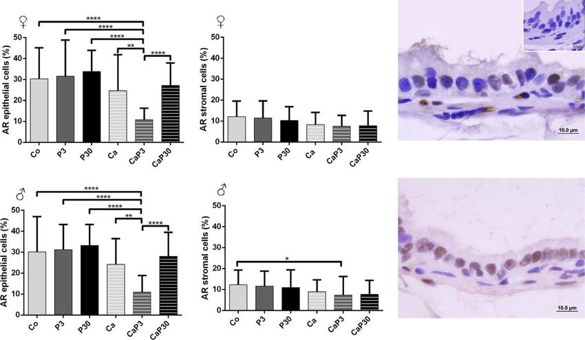
Zanatelli et al. Reproductive Biology and Endocrinology          (2021) 19:94                                                                   Page 5 of 17

reduction in body weight when compared with the Ca                                 In males, castration (Ca) caused a reduction in relative
group (Figs. 1 and 2a, b).                                                       prostatic complex weight compared to uncastrated ani-
  In females, castration (Ca), as well as long treatment                         mals (Co, P3 and P30). The administration of prolactin
with prolactin (P30), caused a decrease in prostate com-                         in the castrated animals did not interfere in the prostatic
plex relative weight when compared to the control group                          relative weight compared to that of the castrated group
(Co). Short treatment with prolactin in castrated animals                        (Ca) (Fig. 2d).
(CaP3), on the other hand, showed weight recovery com-                             In females, the CaP30d group showed a decreased in
pared to the Ca and P30 groups (Fig. 2c).                                        pituitary relative weight in females compared to other

 Fig. 2 Biometric data of female (left graphics) and male (right graphics) gerbils from different experimental groups. Values expressed as mean ± SD. a
 Body weight. b Prostate complex relative weight. c In left ovaries, in right testicles. d Adrenals weight. e Pituitary weight. Different letters represent
 statistically significant differences between the experimental groups, p ≤ 0.05. Statistical analysis based on ANOVA A and Tukey’s tests
Zanatelli et al. Reproductive Biology and Endocrinology       (2021) 19:94                                                                Page 6 of 17

groups, while in males no significant differences were                       other hand, in females and males from the P30 (Fig. 3i, j
found between the groups (Fig. 2e, f).                                       and Table 1) groups, the morphological atypia in the epi-
                                                                             thelium were substantially reduced when compared with
PRL caused significant morphological changes in the                          P3. Furthermore, some epithelial cells showed a clear halo
prostate and recovery from the atrophic effects of                           around the pyknotic nucleus (Fig. 3j inset). Many acini
castration                                                                   showed epithelial cell debris in the lumen (Fig. 3i,j).
When compared to the Co group (Fig. 3a, b), the P3                             After castration, both female and male prostates suf-
group (Fig. 3e, f) showed expressive histological alter-                     fered acinar regression, observed by the decrease in
ations in both the male and female prostates. These al-                      lumens and the epithelium tortuosity (Fig. 3c, d). In gen-
terations included multiple regions with morphological                       eral, animals from the CaP3 (Fig. 3 g, h) and CaP30
atypia in the epithelium, mostly resulting in a cribriform                   (Fig. 3k, l) groups showed the same recovery from atro-
architecture throughout the gland and the presence of                        phic effects, such as more voluminous and rounded
inflammatory cells in the subepithelial and stromal re-                      acini, healthier epithelium, and more organized sml.
gions. In addition to these disorders, only uncastrated
groups showed an incidence of inflammation, with the ex-                     Morphometric-stereological and kariometrical analysis
ception of the CaP3 group for males (Table 1). In acini                      The results obtained with the stereological, morphomet-
with lesions, in females, ciliated cells were observed                       ric and kariometric analyses are presented in Table 2.
grouped and not isolated as is commonly observed in con-
trol females (Fig. 3a inset). In general, there was an appar-                PRL caused opposite effects in the epithelial height and
ent reduction in glycoprotein secretion production, but                      sml thickness of the male and female prostate
this secretion was also observed within the microacini                       The castration of females promoted a decrease in epithe-
formed in altered epithelium (Fig. 3e, f inset). On the                      lial height and sml thickness (Co vs. Ca). The

      a                                   b                                   c                                   d

      e                                   f                                   g                                   h

      i                                   j                                   k                                   l

 Fig. 3 Histological aspects of the gerbil female and male ventral prostates from different experimental groups stained with H&E and Periodic acid
 and Schiff (E and F inset). a and b- Prostate from the Co group showing a normal morphologic pattern of the gland. c and d- Castrated gland
 from the Ca group with intense regression, observed by the decrease in lumens area and the epithelium tortuosity e and f- P3d group showed
 expressive histological alterations, note the cribriform architecture throughout the gland, in details inset note the microacini formed with
 secretion. g and h- CaP3 group recovery of atrophic effects of castration, as acini more voluminous and rounded, epithelium healthier, sml more
 organized compared with the castrated animals. i and j- The morphological atypia in the epithelium were substantially reduced when compared
 with P3, observe epithelial cells debris into the lumen and the inset details in j a epithelial cell show a clear halo around the pyknotic nucleus. k
 and l- CaP30 group presents recovery of atrophic effects of castration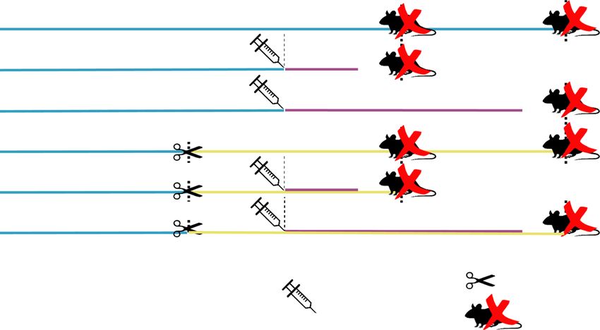
Zanatelli et al. Reproductive Biology and Endocrinology               (2021) 19:94                                                                              Page 7 of 17

Table 1 Incidence and Multiplicity of inflammations and morphological alterations observed in prostates
                                                  Co                         P3                      P30                      Ca             CaP3                      CaP30
                                                  Inflammation
Incidence (%)                         ♀           12.5                       25.0                    25.0                     0              0                         0
                                      ♂           25.0                       50.0                    25.0                     0              13.0                      0
Multiplicity (mean ± SD)              ♀           0.037 ± 0.1                0.061 ± 0.1             0.082 ± 0.1              0              0                         0
                                      ♂           0.227 ± 0.4                0.435 ± 0.6             0.220 ± 0.4              0              0.041 ± 0.1               0
                                                  Morphological alterations
Incidence (%)                         ♀           62.5                       75.0                    12.5                     0              62.5                      0
                                      ♂           12.5                       50.0                    12.5                     0              0                         0
Multiplicity (mean ± SD)              ♀           0.310 ± 0.3 a,b
                                                                             1.333 ± 1.7   a
                                                                                                     0.082 ± 0.2   a,b
                                                                                                                              0   b
                                                                                                                                             0.405 ± 0.5 a,b
                                                                                                                                                                       0b
                                      ♂           0.020 ± 0.05  a,b
                                                                             1.040 ± 1.8   a
                                                                                                     0.020 ± 0.05   a,b
                                                                                                                              0   b
                                                                                                                                             0b
                                                                                                                                                                       0b
Different letters represent statistically significant differences between the experimental groups, p ≤ 0.05

administration of PRL promoted increased epithelial                                    after 30 days of treatment. In the castrated group, PRL
height and sml thickness (P3 and P30 vs. CO). The same                                 promoted a similar decrease in epithelial height in both
effects were observed in the sml thickness of castrated                                the CaP3 and CaP30 groups compared to the Ca group.
groups (Ca vs. CaP3 and CaP30). However, the epithelial
height only increased in ovari-ectomized CaP30 groups.                                 PRL caused changes in the nuclear area and perimeter
   In males, surgical castration caused an increase in epi-                            In females, 30 days of PPL administration caused an in-
thelial height and sml thickness (Co vs. Ca). The admin-                               crease in the nuclear area and perimeter in uncastrated
istration of PRL promoted a decrease in sml thickness in                               and castrated groups. The castrated animals (Ca group)
uncastrated (Co vs. P3 and P30) and in castrated groups                                had a decrease in the nuclear area and perimeter when
(Ca vs. CaP3 and CaP30). With regard to epithelial                                     compared with uncastrated animals (Co group).
height, PRL caused a decrease in the P3 and P30 groups                                   In uncastrated males, the nuclear area had a decrease
in relation to the CO group; this effect was more marked                               in the P3 and P30 groups when compared with the Co

Table 2 Stereological, Morphometric and Kariometric data from different experimental groups (mean ± SD)
Parameters                            Co                     P3                      P30                    Ca                        CaP3                     CaP30
Stereological (%)
Epithelium                     ♀      28.70 ± 7.1 a          29.08 ± 10.3 a          24.22 ± 10.2 a,b       25.60 ± 10.4 a,b          20.92 ± 6.4 b,c          16.30 ± 4.6 c
                               ♂      29.29 ± 7.5            30.75 ± 10.8            25.40 ± 10.1           27.25 ± 8.5               25.58 ± 5.6              28.51 ± 9.0
Lumen                          ♀      34.61 ± 19.2 a         28.96 ± 17.6 a          34.95 ± 16.0 a         28.41 ± 13.4 a            25.69 ± 8.3 a            14.32 ± 6.7 b
                               ♂      36.62 ± 14.4 a,b       34.33 ± 17.4 a,b        44.58 ± 14.1 b         24.30 ± 12.6 c            25.81 ± 10.3 a,c         18.12 ± 11.7 c
SML                            ♀      16.76 ± 6.4 a          18.27 ± 3.2 a,b         17.76 ± 5.4 a,b        21.53 ± 6.4 b,c           25.20 ± 9.3 c            26.9 ± 11.0 c
                               ♂      24.44 ± 6.6 a,b        21.72 ± 7.2 b           19.37 ± 5.9 b          29.25 ± 5.6 a,c           32.76 ± 7.0 c,d          37.29 ± 8.5 d
Non-muscular Stroma            ♀      20.50 ± 9.6 a          23.77 ± 10.0 a,b        23.24 ± 11.5 a,b       24.6 ± 14.0 a,b           28.43 ± 8.3 b,c          43.03 ± 16.9 c
                               ♂      9.82 ± 6.0 a           13.39 ± 8.9 a,b         10.87 ± 5.8 a,c        19.40 ± 9.9 b             16.01 ± 9.6 a,b          16.22 ± 9.2 b,c
Morphometric (µm)
Epithelium height              ♀      13.10 ± 3.1 a          15.81 ± 5.0 b,c         17.79 ± 6.3 c          9.47 ± 4.3 d              10.77 ± 3.4 d            15.44 ± 4.9 b
                               ♂      19.66 ± 3.9 a          16.19 ± 4.5 b           14.07 ± 3.9 c          31.63 ± 10.4 d            14.64 ± 4.3 b,c          13.52 ± 5.3 b,c
SML thickness                  ♀      8.56 ± 3.9 a           10.27 ± 3.0 b           16.13 ± 5.4 c          6.32 ± 2.6 d              9.68 ± 3.1 b             18.00 ± 6.3 c
                               ♂      15.23 ± 4.2 a          10.81 ± 3.9 b           12.10 ± 3.9 b          38.75 ± 12.9 c            14.56 ± 3.8 a            16.15 ± 5.0 a
Kariometric
Nuclear area (µm2)             ♀      12.89 ± 3.0 a          14.75 ± 3.8 a           30.36 ± 5.7 b          8.84 ± 2.4 c              13.12 ± 2.5 a            27.56 ± 5.4 b
                               ♂      31.09 ± 5.2 a          26.37 ± 5.3 b           24.35 ± 5.7 c          22.36 ± 5.7 d             27.07 ± 5.1 b            24.20 ± 5.4 c
Nuclear perimeter (µm)         ♀      15.63 ± 2.3 a,b        15.64 ± 2.0 b           21.32 ± 1.9 c          12.46 ± 1.7 d             14.67 ± 1.6 a            20.73 ± 2.3 c
                               ♂      24.92 ± 3.1 a          20.45 ± 2.7 b           20.38 ± 2.7 b          19.39 ± 2.6 c             21.05 ± 2.2 b            20.11 ± 3.0 b,c
Different letters represent statistically significant differences between the experimental groups, p ≤ 0.05
Zanatelli et al. Reproductive Biology and Endocrinology (2021) 19:94 Page 8 of 17
group; this effect was more marked after 30 days of staining of the cytoplasm. In some acini, an intensive
treatment. On the other hand, in castrated animals, the positive reaction was localized to single epithelial cells
nuclear area showed an increase. With regard to the nu- scattered within the cytoplasm.
clear perimeter, the P3 and P30 groups showed a de- In females, immunostained cell counts data showed
crease in perimeter in relation to the Co group. Among that there was no alteration in immunostained PRLR in
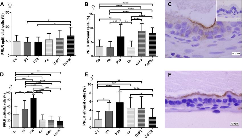
the castrated animals, only the CaP3 group showed an castrated animals (Ca) when compared to control (Co),
increase in nuclear perimeter. but decreased in PRL for stromal cells. When prolactin
Both Ca and Co animals had a decrease in nuclear was administered to castrated females, it increased PRL
area and perimeter. in the epithelial cells and stromal cells of the CaP3
group, but PRLR was only increased in stromal cells of
Stereological analysis the CaP3 group (Fig. 4a, b).
In uncastrated females, the frequency of epithelium, In the uncastrated groups, the P3 group showed an in-
stroma, lumen and sml did not change when treated crease in PRL epithelial cells when compared with the Co
with prolactin. In castrated animals, the epithelium and and P30 groups. The P30 group also showed a decrease in
lumen showed a frequency decrease in the CaP30 group, PRL stromal cells and an increase in PRLR stromal cells in
while the stroma presented a frequency increase, and the comparison with the CO and P3 groups (Fig. 5a, b).
CaP3 and CaP30 groups showed greater sml frequency In males, castration decreased PRLR immunostaining
(Table 3). in epithelial cells and increased it in stromal cells (CO
The comparison of uncastrated animals with their cor- vs. Ca). In uncastrated males, administration of exogen-
responding castrated group showed a decrease in CaP3 ous prolactin increased PRLR immunostaining in both
and CaP30 epithelial frequency; however, the sml com- epithelial and stromal cells, and this effect was more
partment had an increase in frequency. The CaP30 pronounced after 30 days of treatment (Fig. 4d, e). Pro-
group showed an increase in stroma frequency and a de- lactin increased the PRL immunostained cells in stroma
crease in lumen (Table 3). and, after 30 days of treatment, in epithelial cells as well
In males, the epithelial compartment remained unaltered (Fig. 5e). In castrated males, administration of prolactin
in all the groups. A comparison between uncastrated ani- only decreased PRLR in stromal cells in the CaP30
mals and their corresponding castrated group showed an group. However, PRL immunostaining increased in both
increase in stroma Ca vs. Co. Castration increased the size epithelial and stromal cells (Fig. 4d, e).
of the SML compartment and this effect was enhanced with
prolactin treatment. The comparison between uncastrated
animals and their corresponding castrated group showed Treatment with prolactin altered ERα and ERβ
an increase in SML in both CaP3 and CaP30 groups. With immunostaining
regard to lumen, the castrated groups had a diminution in Castration reduced ERα immunostaining in epithelial
frequency when compared to uncastrated groups (Table 3). and stromal cells of the female prostate (Co vs. Ca).
Treatment with prolactin for 3 days in uncastrated fe-
PRLR and PRL presented the same immunostaining pattern males decreased ERα immunostaining in epithelial cells.
The presence of the prolactin receptor (PRLR) and the In castrated females, there was an increased ERα count
soluble prolactin (PRL) in male and female gerbil pros- in stromal cells in CaP3 and CaP30 group and in epithe-
tates was studied by immunocytochemistry. Gerbil pitu- lial cells only in CaP30 group (Fig. 6a, b).
itaries were used as positive control tissues. PRLR and In males, castration increased ERα immunostaining in
PRL both presented the same immunostaining pattern epithelial and stromal cells of the prostate (Co vs. Ca).
(Figs. 4c and f and 5c and f). Both were mainly located The administration of exogenous prolactin in uncas-
on the apical surfaces of the secretory epithelial cells of trated males increased ERα immunostained cells in
prostatic acini, but also appeared scattered throughout stroma in both groups (P3 and P30), but epithelial cells
the cytoplasm. The nucleus of the cells remained un- only increased in the P3 group. In castrated males, the
stained. A low number of stromal cells showed positive CaP3 group showed an increase in immunostained cells
Table 3 Prolactin serum concentrations (ng/mL) in gerbils from different experimental groups
Sex Groups
Co PRL 3d PRL 30d Ca CaPRL 3d CaPRL 30d
Female 6.57 ± 1.1 a 4.64 ± 0.7 b 4.37 ± 0.8 b,c 4.72 ± 0.5 b 4.45 ± 0.5 b 4.59 ± 1.0 c
Male 3.75 ± 0.6 a 4.17 ± 0.6 a,c 3.83 ± 0.5 b,c 5.12 ± 0.5 b 4.86 ± 0.8 b 4.66 ± 0.5 b,c
Different letters represent statistically significant differences between the experimental groups, p ≤ 0.05Zanatelli et al. Reproductive Biology and Endocrinology (2021) 19:94 Page 9 of 17
Fig. 4 Graphics obtained by epithelial and stromal cell immunostaining count for PRLR and their respective immunocytochemistry photomicrographs.
Counter-stained with Harris hematoxylin. Black arrows indicate immunostaining of epithelial cell, white arrows indicate immunostaining of stromal
cells. Inset refer to negative controls for the technique. Values are expressed as mean in percentage ± SD. Different letters represent statistically
significant differences between the experimental groups, p ≤ 0.05. Statistical analysis based on ANOVA A and Tukey’s tests
in the epithelium and a decrease in the stroma in com- of epithelial cells in comparison with the Co and Ca
parison with CaP30 (Fig. 6d, e). groups (Fig. 8d, e).
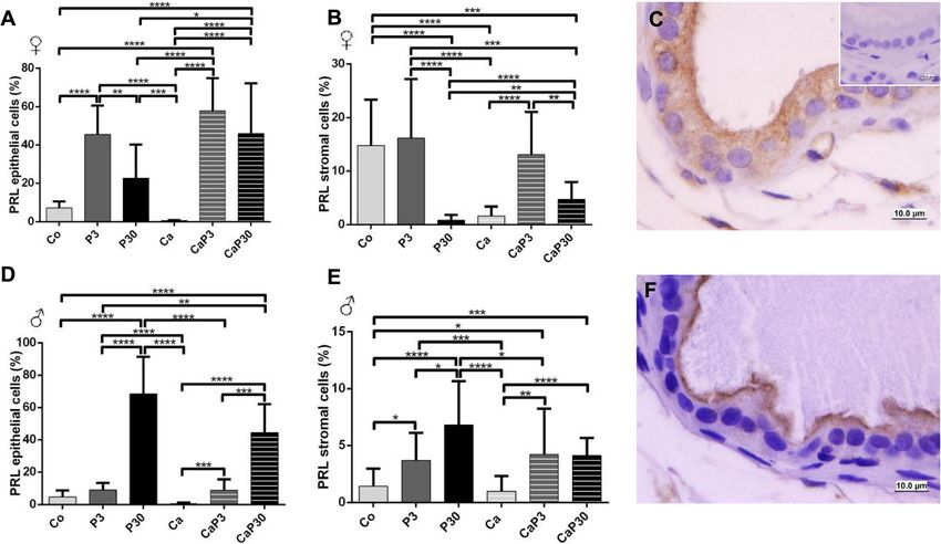
Castration decreased ERβ stromal cell immunostaining
for females. Exogenous prolactin administered in uncas- Discussion
trated females decreased ERβ immunostaining for stromal Understanding the role of prolactin in the prostate and
cells in both groups and for epithelial cells when the ani- the way this hormone acts in homeostasis or promoting
mals were treated for 30 days. When administered for 3 pathologies of the gland is of immense interest. Al-
days in castrated animals, exogenous prolactin increased though some studies have shown that the administration
ERβ immunostaining in both epithelial and stromal cells of prolactin, at the same dosage employed in the present
(Fig. 7a, b). study, produces effects on the rat prostate [40–42], this
In males, castration did not alter male prostatic cell is the first study to examine the high dose effect of pro-
ERβ immunostaining. When administered for 30 days, lactin on the gerbil prostate. In the present study, we
exogenous prolactin increased ERβ epithelial immuno- have investigated the effects of exposure to high doses of
staining in both intact and castrated males (P30 and exogenous prolactin in the ventral lobe of the male pros-
CaP30) and the P30 group showed an increase in immu- tate and in the female prostate of the Mongolian gerbil
nostained stromal cells (Fig. 7d, e). over 2 treatment periods. The effect of hormone admin-
istration was evaluated in intact animals, under the influ-
Castration did not alter AR immunostaining ence of endogenous sex hormones, and in castrated
Castration did not alter AR immunostaining cells in fe- animals, i.e. without the influence of these hormones.
males (Co vs. Ca). Exogenous prolactin did not cause The results demonstrated that prolactin changes the
changes in immunostaining when administered to un- morphology of the prostate, acting on both epithelial
castrated animals, and only reduced epithelial immuno- and stromal cells. The principal morphological alter-
staining when administered for 3 days to castrated ations are presented in Fig. 9.
animals (Fig. 8a, b). We can consider that the dose of prolactin adopted
In males, administration of exogenous prolactin for 3 was high, since the serological level of prolactin was
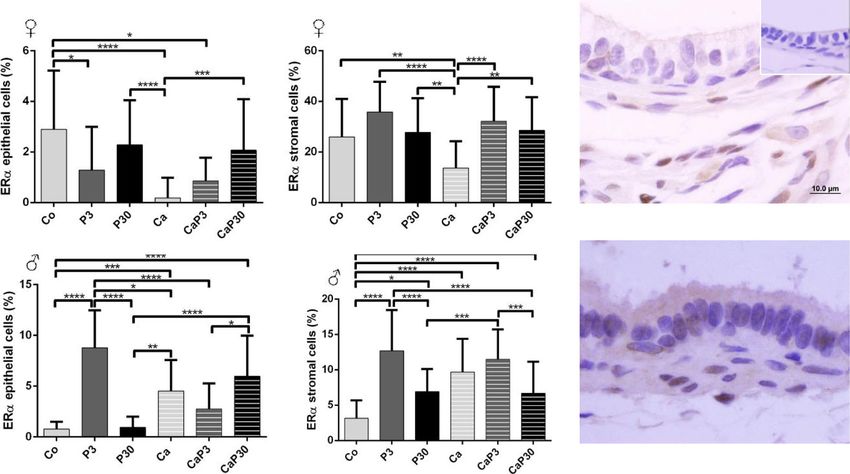
days in castrated animals decreased AR immunostaining around 6.5 ng/mL in the female gerbil in the pre-oestrusZanatelli et al. Reproductive Biology and Endocrinology (2021) 19:94 Page 10 of 17 Fig. 5 Graphics obtained by epithelial and stromal cell immunostaining count for PRL and their respective immunocytochemistry photomicrographs. Counter-stained with Harris hematoxylin. Black arrows indicate immunostaining of epithelial cell, white arrows indicate immunostaining of stromal cell. Inset refer to negative controls for the technique. Values are expressed as mean in percentage ± SD. Different letters represent statistically significant differences between the experimental groups, p ≤ 0.05. Statistical analysis based on ANOVA A and Tukey’s tests phase and 3.7 ng/mL in the male gerbil, according to area, the perimeter of the secretory epithelial cells being our measurements; the pathological condition of hyper- directly related to reduced synthetic activity of these prolactinemia is defined as circulating PRL levels above cells. The accompanying decreases in prostate weight, the normal range, occurring in conditions other than epithelium height and sml thickness are other indica- pregnancy and lactation, when physiological hyperpro- tions of the marked process of acinar regression caused lactinemia occurs. The major cause of pathological by hormonal suppression [51, 52]. The morphometric hyperprolactinemia involves tumours of pituitary lacto- data showed an increase in epithelial height and sml troph cells (prolactinomas), the main source of PRL in thickness in the male Ca group. During the atrophy the organism [43]. process, the prostatic epithelium regresses and can form Exposure to this high dose of prolactin affected the curvatures and undulations, which give the impression morphology of the gerbil prostate in comparison with of having increased in height. In addition, smooth the control animals. The animals in the control groups muscle cells become more synthesizing than contractile, exhibited the general aspect of the gland observed in and, possibly, produce more constituents of extracellular control females and males similar to that previously de- matrix, which can increase in volume or thickness [18]. scribed by other authors [11, 15, 44–46] for the prostate However, the other data obtained for this group corrob- of rodents of the same species as those used in this study orate the effects established for prostatic regression (Meriones unguiculatus). already described in the literature. The morphological effects of castration were also very Castration also leads to the appearance of a specific similar to those already described. The hormone ablation cell phenotype, the spumous cell. This cell type has been caused by castration resulted in a prostate gland in a observed by our research group with more frequency in process of atrophy previously observed [47–50]. As the male and female gerbil prostates which have undergone epithelium regresses, the stromal cells and extracellular the hormonal ablation process due to castration [53–55]. matrix are remodelled to adjust to the reduced organ As regards the effect of prolactin administered for a size [18]. The reduced amount of glycoprotein secretion short duration, the prostates of females and males from within the lumens added to a decrease in the nuclear the PRL 3d group developed several morphological
Zanatelli et al. Reproductive Biology and Endocrinology         (2021) 19:94                                                                  Page 11 of 17

 Fig. 6 Graphics obtained by epithelial and stromal cell immunostaining count for ERα and their respective immunocytochemistry photomicrographs.
 Counter-stained with Harris hematoxylin. Black arrows indicate immunostaining of epithelial cell, white arrows indicate immunostaining of stromal cell.
 Inset refer to negative controls for the technique. Values are expressed as mean in percentage ± SD. Different letters represent statistically significant
 differences between the experimental groups, p ≤ 0.05. Statistical analysis based on ANOVA A and Tukey’s tests

alterations in the epithelium, showing mostly a cribri-                         volume, revealed in the histological sections by deleting
form aspect to the gland, accompanied by an intense in-                         epithelial cell portions within the lumen. Some cell con-
flammation process. The cribriform pattern of epithelial                        stituents of epithelium had a pyknotic nucleus, sur-
cell proliferation was also demonstrated in organ culture                       rounded by a clear halo. This process resembles the
of the human male prostate after administration of pro-                         apoptosis type described by Rosa-Ribeiro et al. [58] for the
lactin for 7 days, when epithelial stratification was ob-                       rat prostate, as the phenomenon of desquamation, a col-
served with the formation of microacini [26]. It is known                       lective epithelial cell deletion particular to androgen
that there is a relationship between the increase of pro-                       deprivation. Further studies with the use of molecular
lactin levels and the increase of prostate diseases in men                      markers for these apoptotic cells will clarify whether it is
[21], and also that prolactin is responsible for the devel-                     the same process. A study performed by Herrera-
opment of inflammation in the male gland [56]. The                              Covarrubias et al. [57] in rats showed that long exposure
present study demonstrated that similar effects can also                        to exogenous prolactin, from 4 weeks, leads to histological
occur in the female and male glands of Mongolian ger-                           alterations in the prostate that may be considered as pre-
bils. The morphological atypia observed in these pros-                          cancerous, even in individuals with low levels of andro-
tates may, in certain cases, develop into prostatic                             gens. In the present study, which evaluated exposure to
intraepithelial neoplasia (PIN), but not necessarily [57].                      prolactin for up to 30 days, the histological changes indi-
A 3-day period of administration is insufficient to be                          cated a more acute effect of prolactin action (3 days) and
able to characterize such a pre-neoplastic lesion, but it is                    subsequent structural recovery through cell discard mech-
important to monitor this effect during this treatment.                         anisms. Further studies involving longer-term prolactin
   The period of prolactin exposure influences the actions                      exposure may clarify whether the prostate gland develops
of this hormone on the prostate, since long-term treat-                         well-established pre-neoplastic lesions, as occurs in the rat
ment has shown different results for both male and female                       gland.
gerbils. Treatment with prolactin for 30 days does not re-                        The presence of ciliated cells in the female prostate is
sult in the same effects as treatment for 3 days. No major                      worthy of note. Ciliated cells are seen with some fre-
foci of inflammation and altered acini were found. The                          quency in the prostate epithelium of normal adult fe-
gland, on the other hand, showed a decrease in epithelial                       male gerbils; however, its role in the physiology of this
Zanatelli et al. Reproductive Biology and Endocrinology         (2021) 19:94                                                                  Page 12 of 17

 Fig. 7 Graphics obtained by epithelial and stromal cell immunostaining count for ERβ and their respective immunocytochemistry photomicrographs.
 Counter-stained with Harris hematoxylin. Black arrows indicate immunostaining of epithelial cell, white arrows indicate immunostaining of stromal cell.
 Inset refer to negative controls for the technique. Values are expressed as mean in percentage ± SD. Different letters represent statistically significant
 differences between the experimental groups, p ≤ 0.05. Statistical analysis based on ANOVA A and Tukey’s tests

gland is not well understood. In the present study, cili-                       castration [20, 59–62]. In addition, it increases prostate
ated cells were found more often in female prostates                            weight, stimulates DNA and RNA synthesis in all lobes
from the PRL groups (3d and 30d), to the extent that, in                        of the gland [63], and increases the levels of zinc and cit-
the PRL 3d group, these cells appeared “in groups”. The                         rate in the lateral lobe (reviewed in Rui and Purvis [64],
presence of this cell type is probably related to an abnor-                     and in Costello and Franklin [20], all of which effects
mal differentiation of basal cells of prostatic epithelium,                     can occur independently of the presence of androgens.
influenced by a hormonal imbalance [17, 55]. In female                            The location of prolactin and its receptor, in the gerbil
prostates treated with androgen and anti-oestrogen                              prostate, was studied by immunocytochemistry, using an
hormones, pre-neoplastic and neoplastic alterations ap-                         anti-PRL antibody or anti-PRLR antibody, respectively.
peared in acini which had ciliated cells, so the appear-                        PRLR undergoes changes in its transmembrane domain
ance of these cells can indicate in advance the                                 after being activated by PRL and can then act as a key
installation of malignant or pre-malignant lesions [17].                        regulator of many biological processes, such as growth
  The administration of prolactin promoted the recovery                         and metabolism [65]. These receptors are mainly located
of some regressive aspects of the gland already after 3                         in the apical region of the secretory epithelium cells. In
days of administration in castrated animals. There was                          an immunocytochemistry reaction for PRLR, the nucleus
an increase in prostatic weight and nuclear area and per-                       of the cells remained unmarked and low immunostain-
imeter, indicating a tendency to secretory activity of                          ing was observed in the stromal cells [61]. This same
returning epithelial cells. In females there was also an in-                    pattern was observed in the gerbil female and male pros-
crease in epithelial height and in sml thickness. Al-                           tates in the present study.
though the acini had regressed, the administration of                             Exogenous prolactin administration in the prostate of
prolactin after castration appears to have mitigated their                      non-castrated and castrated animals increased the im-
atrophic effects, without, however, causing the appear-                         munostaining of PRLR and PRL in both sexes, especially
ance of significant lesions. Some studies have shown that                       after 30 days of administration. In high concentrations,
prolactin acts as a growth factor for prostate tissue with                      prolactin can saturate the receptor and hinders further
an important role in the survival of prostate cells after                       receptor dimerization, serving to explain the frequently
Zanatelli et al. Reproductive Biology and Endocrinology (2021) 19:94 Page 13 of 17
Fig. 8 Graphics obtained by epithelial and stromal cell immunostaining count for AR and their respective immunocytochemistry photomicrographs.
Counter-stained with Harris hematoxylin. Black arrows indicate immunostaining of epithelial cell, white arrows indicate immunostaining of stromal cell.
Inset refer to negative controls for the technique. Values are expressed as mean in percentage ± SD. Different letters represent statistically significant
differences between the experimental groups, p ≤ 0.05. Statistical analysis based on ANOVA A and Tukey’s tests
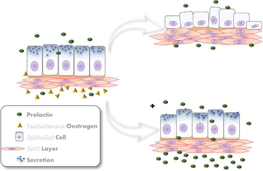
B
A
C
Fig. 9 Illustrative scheme of prolactin action in partial recovery from the atrophy prostate caused by castration. A: Control prostate show cubic/
prismatic simple epithelium and muscular stroma cell layer (SMC); B: Castrated prostates show acinar regression, epithelium height regresses, the
SMC are remodeled to adjust the reduced organ size. C: Castrated with prolactin administration group recovery the atrophic effects of castration,
as more voluminous epithelial cells and more organized and compact SMC compared with the castrated animalsZanatelli et al. Reproductive Biology and Endocrinology (2021) 19:94 Page 14 of 17 observed bell-shaped, dose-dependent curves [28]. The administration of prolactin did not cause changes The present study demonstrated that the dose used in prostate AR in uncastrated females but increased was not yet sufficient to reach the maximum binding them in uncastrated males. It is well established that, in threshold between hormone and receptor, so that the the male prostate, prolactin increases levels of androgen tissue still remained responsive to increased circulat- receptors [72]. Gómes and co-workers [73] showed an ing prolactin. increase in the number of AR-positive cells after treat- Although castration did not cause a change in the ment with prolactin in uncastrated rats. Circulating pro- immunostaining of PRLR, sexual hormonal ablation de- lactin is also detected in males, although it is present at creased PRL immunostaining in female and male pros- lower levels than in females. In the male prostate, andro- tatic tissue alike. In the female this decrease in immuno- gens are related to the differentiation of the secretory marking for PRL is related to decreased serum prolactin epithelium, and growth and regulation of secretory activ- levels. The steroid hormones, particularly oestrogen, ity [74]. These functions are also related to androgens in stimulate the release of prolactin by lactotrophs of the the female gland [10]; however, the female body is more pituitary gland, and the suppression of these hormones, controlled by oestrogens and progesterone, which are caused by castration, interrupts the stimulation of pituit- present in substantially higher levels than testosterone. ary prolactin release [24]. We also observed a decrease This may explain why androgen receptors were not in- in pituitary weight associated with castration. In rats, the fluenced by the manipulation of prolactin levels in fe- weight of the pituitary gland increases with increasing male prostate tissue. body weight of the animal [66], and it is known that fe- In castrated males, the administration of prolactin for male hormones related to reproduction have an influ- 3 days decreased AR immunostaining in epithelial and ence on pituitary gland volume [67]; it is also known stromal cells. Prolactin acts directly on the prostate in that oestrogens influence the expression of the PRL gene synergy with androgens, stimulating its activity and in- in the pituitary gland and can result in pituitary cell creasing tissue sensitivity to androgenic action [75]. hyperplasia [68]. There is an increase in binding testosterone after incu- However, in the male, serum prolactin levels increased bation of the human prostate with exogenous prolactin after castration. The literature shows conflicting data [76]. In addition, the increase in circulating levels of pro- about prolactin levels in orchiectomized males; some lactin is related to the pathogenesis of prostatic tumours studies have shown an increase [69] and others a reduc- (reviewed in Costello and Franklin [20]). The present tion [40, 70]. study demonstrated the synergistic action between pro- Exogenous exposure to prolactin decreases serum pro- lactin and androgenic hormones in the ventral prostate lactin levels in castrated animals. These data are consistent of the Mongolian gerbil, showing the dependence of pro- with those found in the work of Constantino et al. [40], lactin on the presence of testosterone to promote tissue- which also observed a decrease in serum prolactin levels specific effects. in castrated males treated for 10 days with subcutaneous The immunostaining of oestrogen receptors ERα and injections of prolactin, at the same doses as those used in ERβ decreased in the female prostate after castration, the present study. The authors suggested the occurrence whereas AR did not change; these data are consistent of a short loop feedback, where prolactin itself acts in the with the results previously published by our research brain to stimulate the production of dopamine and group for the gerbil female prostate [52]. In males, there thereby inhibit its own secretion. Our data also indicate was an increase in ERα expression after castration. Oes- the same phenomenon in the case of gerbil prostates. In trogens are produced from androgens by the action of intact males, the administration of exogenous prolactin in- aromatase [39]. Castration reduces the levels of circulat- creased PRL serum levels; probably the presence of testos- ing androgens and oestrogens in the body. This hormo- terone maintained high production of endogenous nal fall may have led to increased levels of ERα in the prolactin which, added to the injected prolactin, increased tissue in order to increase sensitivity to oestrogen. ERβ the serum level of the hormone. expression did not change after castration. The an- In animals that presented lesions, the prolactin re- drogenic peak occurring during puberty coincides ceptor did not show increased expression in the af- with a decrease in ERα expression in the prostate, fected acini; however, these acini showed high AR indicating that androgens can suppress the action of immunostaining in males. Androgens regulate the these receptors on the tissue [77]. In contrast, andro- growth and proliferation of prostatic cells. Tumour gens increase ERβ expression in the prostate of development results from a multi-step process that rodents, whereas oestrogens themselves cannot self- initially leads to the formation of low- to high-grade regulate this receptor [78, 79]. prostatic intraepithelial neoplasia, which is primarily In females under normal hormonal conditions, controlled by androgens [71]. exogenous prolactin administration did not alter ERα
Zanatelli et al. Reproductive Biology and Endocrinology           (2021) 19:94                                                                    Page 15 of 17

expression in prostate tissue, and decreased ERβ only in                         PINCs: Cribiform intraepithelial neoplasms; PINs: Intraepithelial neoplasms;
stromal cells. However, in castrated females, prolactin                          PRL: Prolactin; PRLR: Prolactin receptors; RIA: Radioimmunoassay

administration increased ERα and ERβ immunostaining                              Acknowledgements
in epithelial and stromal cells. It is well established in                       The authors are grateful to Luiz Roberto Falleiros Junior for technical
the literature that oestrogens exert a strong influence on                       assistance, as well as to all researchers at the Laboratory of Microscopy and
                                                                                 Microanalysis.
prolactin levels, stimulating prolactin secretion by the pi-
tuitary gland lactotrophs [24]. The present study showed                         Authors’ contributions
that prolactin, in contrast, influences the oestrogen re-                        Marianna Zanatelli conceived, designed and conducted all experimental
                                                                                 methods and contributed to the writing of the manuscript. Sebastião
ceptor levels of the female prostate in conditions of low                        Roberto Taboga was supervisor and reviser of the study. Simone Jacovaci
estradiol serum levels, making the tissue more respon-                           Colleta reviewed the literature research, was a principal author of the
sive to this hormone. PRL appears to be a key up-                                manuscript and prepared the tables, graphs and figures. Luiz Henrique Alves
                                                                                 Guerra reviewed the literature research and statistical analysis, was a principal
regulator of the ER in many reproductive tissues, such as                        author of the manuscript and reviewed tables, graphs and figures. The
the ovarian corpus luteum [80, 81], the mammary gland                            authors read and approved the final manuscript.
and the decidua [82], increasing mRNA and protein
levels of both ER subtypes.                                                      Funding
                                                                                 This work was supported by the National Research Council – CNPq
  In the male, the scenario was the opposite: prolactin                          (fellowship to S R Taboga, Procs. Nr. 301596/2011-5 and Nr. 442630/2014-0)
administration increased ERα and ERβ expression in the                           and São Paulo State Research Foundation – FAPESP (Fellowship to M
prostate of intact animals but did not alter the expres-                         Zanatelli, Proc. Nr. 2012/00695-6).

sion of ERα in the prostate of castrated animals. An                             Availability of data and materials
androgen-dependent relationship of prolactin was                                 This paper is part of the Thesis presented by M Zanatelli to the Institute of
observed in the male organism, where serum levels of                             Biology, UNICAMP, in partial fulfilment of the requirement for a PhD in Cell
                                                                                 Biology. The original text has public access in the repositories of that
testosterone are much higher and dominate the other                              University.
steroid hormones and the actions they control. A com-
parative study between males and females of gerbil                               Declarations
evaluated ERα and ERβ expression in the prostate and                             Ethics approval and consent to participate
demonstrated that testosterone reduced ERα expression                            The research was conducted in accordance with the requirements of the
in the female prostate, while it was necessary for the                           Ethics Committee of Experimental Animals of Sao Paulo State University
                                                                                 (protocol number: 053/2011 CEUA).
expression of ERβ in the male gland [34].
                                                                                 Consent for publication
                                                                                 Not applicable.
Conclusions
The data presented demonstrate that prolactin and its re-                        Competing interests
ceptor are expressed in the female and male prostate                             The authors declare that there are no conflicts of interest.
gland of the Mongolian gerbil and influence its mainten-                         Author details
ance and homeostasis. The administration of exogenous                            1
                                                                                  Department of Structural and Functional Biology, Institute of Biology, State
prolactin to intact animals promotes the appearance of                           University of Campinas – UNICAMP, SP, Campinas, Brazil. 2Laboratory of
                                                                                 Microscopy and Microanalysis, Department of Biology, São Paulo State
important histological alterations in the gland after just 3                     University – UNESP/IBILCE, Rua Cristóvão Colombo, 2265, Jardim Nazareth, SP
days of administration, as well as a subsequent cellular dis-                    15054-000 São José do Rio Preto, Brasil. 3Department of Morphology, Federal
carding process that apparently recovers the morpho-                             University of Goiás -UFG , GO, Goiânia, Brazil.
logical aspects of the tissue, after 30 days of                                  Received: 31 March 2021 Accepted: 3 June 2021
administration. Prolactin has also been involved in the re-
covery of the gland after atrophy caused by castration. A
                                                                                 References
relationship of dependence between prolactin and testos-                         1. Zaviacic M, Sidlo J, Borovský M. Prostate specific antigen and prostate
terone in the male prostate has been demonstrated,                                   specific acid phosphatase in adenocarcinoma of Skene’s paraurethral glands
whereas, in the female gland, prolactin establishes a                                and ducts. Virchows Arch A Pathol Anat Histopathol. 1993;423(6):503–5.
                                                                                 2. Dodson MK, Cliby WA, Pettavel PP, Keeney GL, Podratz KC. Female urethral
greater performance with the oestrogens, taking into ac-                             adenocarcinoma: evidence for more than one tissue of origin? Gynecol
count the differences between the hormonal profiles of                               Oncol. 1995;59(3):352–7.
the sexes.                                                                       3. Whipple B. Book Review: The Human Female Prostate: From Vestigial
                                                                                     Skene’s Paraurethral Glands and Ducts to Woman’s Functional Prostate.By
                                                                                     Milan Zaviacic. Slovak Academic Press, Bratislava, Slovakia, 1999, 171 pp.,
                                                                                     $49.00. Arch Sex Behav. 2002;31(5):457–8. Available from: https://doi.org/10.1
Abbreviations                                                                        023/A:1019852411051.
AR: Androgen receptor; BSA: Bovine serum albumin; Ca: Castrated group;           4. Konecki T, Salagierski M, Sosnowski M. Treatment of paraurethral cysts
CaP3: Castrated - Prolactin 3 days group; CaP30: Castrated; Prolactin 30 days        in female patients - Description of three cases. Cent Eur J Urol. 2009;
group; Co: Control group; DAB: Diaminobenzidine; ERα: Estrogen receptor              62(2):111–3.
alpha; ERβ: Estrogen receptor beta; H&E: Hematoxylin–eosin; P3: Prolactin 3      5. Moalem S, Reidenberg JS. Does female ejaculation serve an antimicrobial
days group; P30: Prolactin 30 days group; PAS: Periodic Acid and Schiff;             purpose? Med Hypotheses. 2009;73(6):1069–71.
You can also read