Lipoprotein Lipase Links Dietary Fat to Solid Tumor Cell Proliferation
←
→
Page content transcription
If your browser does not render page correctly, please read the page content below
Published OnlineFirst January 31, 2011; DOI: 10.1158/1535-7163.MCT-10-0802

                                                                                                                      Molecular
                                                                                                                         Cancer
Therapeutic Discovery                                                                                               Therapeutics

Lipoprotein Lipase Links Dietary Fat to Solid Tumor
Cell Proliferation
Nancy B. Kuemmerle1,2, Evelien Rysman3, Portia S. Lombardo2,4, Alison J. Flanagan2,4, Brea C. Lipe1,2,
Wendy A. Wells2,5, Jason R. Pettus5, Heather M. Froehlich5, Vincent A. Memoli2,5, Peter M. Morganelli2,6,
Johannes V. Swinnen3, Luika A. Timmerman7, Leila Chaychi4, Catherine J. Fricano2,4, Burton L. Eisenberg2,8,
William B. Coleman9, and William B. Kinlaw2,4

Abstract
              Many types of cancer cells require a supply of fatty acids (FA) for growth and survival, and interrupting de
           novo FA synthesis in model systems causes potent anticancer effects. We hypothesized that, in addition to
           synthesis, cancer cells may obtain preformed, diet-derived FA by uptake from the bloodstream. This would
           require hydrolytic release of FA from triglyceride in circulating lipoprotein particles by the secreted enzyme
           lipoprotein lipase (LPL), and the expression of CD36, the channel for cellular FA uptake. We find that selected
           breast cancer and sarcoma cells express and secrete active LPL, and all express CD36. We further show that
           LPL, in the presence of triglyceride-rich lipoproteins, accelerates the growth of these cells. Providing LPL to
           prostate cancer cells, which express low levels of the enzyme, did not augment growth, but did prevent the
           cytotoxic effect of FA synthesis inhibition. Moreover, LPL knockdown inhibited HeLa cell growth. In contrast
           to the cell lines, immunohistochemical analysis confirmed the presence of LPL and CD36 in the majority of
           breast, liposarcoma, and prostate tumor tissues examined (n ¼ 181). These findings suggest that, in addition to
           de novo lipogenesis, cancer cells can use LPL and CD36 to acquire FA from the circulation by lipolysis, and this
           can fuel their growth. Interfering with dietary fat intake, lipolysis, and/or FA uptake will be necessary to
           target the requirement of cancer cells for FA. Mol Cancer Ther; 10(3); 427–36. 2011 AACR.

Introduction                                                              for the 3 enzymes required to produce palmitic acid from
                                                                          cytosolic citrate [ATP citrate-lyase, acetyl CoA-carboxy-
  Many tumors, including those arising in breast, colon,                  lase, and fatty acid synthase (FASN)]. Importantly, lipo-
ovary, and prostate, exhibit a lipogenic phenotype. This                  genic tumor cell growth is slowed in vitro and survival is
features brisk rates of saturated long-chain fatty acid (FA)              reduced by FA synthesis inhibitors, whereas nontrans-
synthesis driven by enhanced expression of genes coding                   formed cells are unaffected (reviewed in refs. 1, 2). More-
                                                                          over, blocking de novo lipogenesis with FASN inhibitors
                                                                          in vivo exerts potent antitumor effects in rodent models of
Authors' Affiliations: 1Section of Hematology and Oncology, Department
of Medicine, Dartmouth-Hitchcock Medical Center, and Dartmouth Med-       breast (3) and prostate (4) cancer. These observations,
ical School; 2Norris Cotton Cancer Center, Dartmouth-Hitchcock Medical    coupled with the low rates of FA synthesis in most
Center, Lebanon, New Hampshire; 3Laboratory for Experimental Medicine     normal human tissues (5), have spurred efforts to develop
and Endocrinology, Katholieke Universiteit Leuven, Leuven, Belgium;
4
 Section of Endocrinology and Metabolism, Department of Medicine,         anticancer therapies based on inhibiting lipogenic
and 5Department of Pathology, Dartmouth-Hitchcock Medical Center,         enzyme activities or silencing the corresponding genes.
and Dartmouth Medical School; 6Department of Immunology and Micro-
biology, V. A. Medical Center, White River Junction, Vermont and Dart-
                                                                             Attempts to exploit the metabolic requirements of
mouth Medical School, Lebanon, New Hampshire; 7Cancer Research            lipogenic cancers have thus far focused solely on disrupt-
Institute, UCSF/Helen Diller Comprehensive Cancer Center, University      ing de novo FA synthesis. Cytotoxicity following inhibi-
of California at San Francisco – San Francisco, California; 8Department
of Surgery, Dartmouth Medical School, Lebanon, New Hampshire; and         tion of lipid synthesis, however, may be obviated by the
9
 Department of Pathology and Laboratory Medicine, Lineberger Compre-      provision of exogenous FA (6–8). This observation, and
hensive Cancer Center, University of North Carolina School of Medicine,   the improved outcome of breast cancer patients ingesting
Chapel Hill, North Carolina
                                                                          a low fat diet (9), led us to hypothesize that triglyceride in
Note: Supplementary material for this article is available at Molecular
Cancer Therapeutics Online (http://mct.aacrjournals.org/).                circulating lipoprotein particles could provide an addi-
                                                                          tional, exogenous source of FA for tumors. This would
N.B. Kuemmerle and E. Rysman contributed equally to this work.
                                                                          require triglyceride-rich chylomicrons or very low den-
Corresponding Author: William B. Kinlaw, 606 Rubin Building, Dart-
mouth-Hitchcock Medical Center, One Medical Center Drive, Lebanon,        sity lipoproteins (VLDL) as substrate, extracellular lipo-
NH 03756. Phone: 603-653-9961; Fax: 603-653-9952. Email: william.         protein lipase (LPL) for hydrolysis, and FA translocase
kinlaw@hitchcock.org                                                      (CD36) for cellular uptake of the free FA (reviewed in ref.
doi: 10.1158/1535-7163.MCT-10-0802                                        10). As LPL is a secreted enzyme that is bound to the
2011 American Association for Cancer Research.                           luminal surface of capillary endothelial cells, it could

www.aacrjournals.org                                                                                                                       427

           Downloaded from mct.aacrjournals.org on February 20, 2021. © 2011 American Association for Cancer
                                                      Research.
Published OnlineFirst January 31, 2011; DOI: 10.1158/1535-7163.MCT-10-0802
Kuemmerle et al.
potentially be supplied by tumor cells or by nonmalig- were from the American Type Culture Collection
nant cells in the tumor microenvironment. except VCaP, which was from ECACC, and these lines
were acquired recently and were of low passage num-
Materials and Methods ber (Published OnlineFirst January 31, 2011; DOI: 10.1158/1535-7163.MCT-10-0802
Lipoprotein Lipase and Cancer Cell Growth
4 TBS washes, membranes were developed with NBT- adipocytes, which express high levels of LPL and FASN.
BCIP (Pierce). We also sorted the breast cancer lines by their global gene
expression signatures (21). These signatures include the
Bacterial expression of human LPL luminal type (estrogen receptor–positive; ERþ), the basal,
The 2.3 kb EcoRI–HindIII fragment coding LPL was or triple-negative type that lacks receptors for estrogen,
excised from pCMV-SPORT6-LPL (Open Biosystems) progestin, and trastuzumab (22), and the type with Her2/
and inserted into the pProEx-HTa His-tag vector. Most neu amplification. Only 6 breast cancer cell lines
of the fusion protein could not be solubilized, but those (HCC2157, HCC1008, HCC1599, Du4475, SUM149, and
recovered showed reactivity with the anti-His4 antibody SUM190) expressed high levels of LPL mRNA, and each
(Invitrogen). For immunodot assays 10 ng protein from of these exhibited the aggressive basal gene expression
cleared lysates of Escherichia coli DH5a transformed with signature (Supplementary Fig. S1). Expression of LPL
empty or LPL plasmid were spotted onto PVDF mem- mRNA by selected cell lines was verified by RT-PCR
branes, blocked, and incubated with antibody as (Fig. 1A), as was expression of CD36 mRNA (Fig. 1B).
described earlier in the text. LiSa-2 liposarcoma cells, which we previously showed to
Affinity isolation of LPL
Human milk and conditioned cell culture media were
fractionated over heparin sepharose (Sigma) by a proce-
dure modified from Hata and colleagues (17).
LPL activity
We used the radiochemical assay of Nilsson-Ehle and
Schotz (18) or a colorimetric assay based on determina-
tion of glycerol production (BioVision). We used a
protocol based on that of Cruz and colleagues (19) for
determination of heparin-releasable LPL. Briefly, 5 106
cells from 75 cm2 flasks were cultured for 72 hours, and
scraped pellets were washed 3 times in PBS with or
without 100 U/mL heparin. Media and lysed cell pellets
were assayed in triplicate for residual LPL activity.
Immunohistochemistry
Immunohistochemistry was done as previously
described (20). Anti-LPL monoclonal antibody clone 43
was used at a dilution of 1:10, with Citra Plus antigen
retrieval (Biogenix). CD36 was assessed by an affinity-
purified rabbit polyclonal antibody (Thermo Scientific)
according to the supplier’s protocol. The Institutional
Review Board (IRB)-approved the use of breast cancer
tissue and the tissue microarray containing 147 primary
breast cancers from postmenopausal women, diagnosed
between 2000 and 2007 at Dartmouth-Hitchcock Medical
Center, Lebanon, NH. Each case was represented by one
tissue core 1.0 mm in diameter. The liposarcoma tissue
microarray, also IRB approved, contained 26 liposarco-
mas diagnosed between 1995 and 2008 at Dartmouth-
Hitchcock Medical Center. Each case was represented Figure 1. LPL, CD36, and FASN gene expression in cancer cells. A–C,
by two to four 1.0-mm tissue cores. Prostate cancer ethidium-stained gel electrophoresis of RT-PCR products. Cell lines
specimens were acquired at the Katholieke Universiteit analyzed are listed above each lane. stds, electrophoretic size standards;
Leuven, Belgium, with IRB approval. LiSa-2, liposarcoma line; Du4475, breast cancer cells lacking receptors for
sex steroids and trastuzumab; T47D, breast cancer cells with receptors for
estrogen and progesterone, but not trastuzumab; BT474, breast cancer
Results cells with receptors for sex steroids and trastuzumab; PC3, LNCaP, and
VCaP, prostate cancer lines; fibro, human fibroblasts. A, primers
We used a cDNA microarray to screen 45 breast can- corresponded to cyclophilin (cyc) or LPL mRNAs. B, primers corresponded
cer–derived cell lines from the dataset of Neve and to the FA translocase CD36. C, primers detected FASN mRNA. D, real-time
RT-PCR quantitation of LPL mRNA (normalized to 18S rRNA, mean
colleagues (11) for LPL gene expression, and for FASN SEM, n ¼ 3 wells/cell line). HeLa adenocarcinoma cells are included as a
mRNA as a marker for de novo FA synthesis. We analyzed positive control, as we previously reported expression of LPL mRNA by
cell lines because breast tumor samples may contain this cell line (25).
www.aacrjournals.org Mol Cancer Ther; 10(3) March 2011 429
Downloaded from mct.aacrjournals.org on February 20, 2021. © 2011 American Association for Cancer
Research.Published OnlineFirst January 31, 2011; DOI: 10.1158/1535-7163.MCT-10-0802
Kuemmerle et al.
exhibit the lipogenic phenotype (23), also expressed LPL neuþ BT474 breast cancer cells, and fibroblasts did not
and CD36, as expected for a tumor cell derived from an secrete detectable lipase activity. Prostate cancer cells
adipocytic lineage. All of the cell types expressed sub- produced low levels of the enzyme. LPL activities in
stantial FASN mRNA (Fig. 1C), and in the breast cancer breast milk and murine striated muscle were substan-
cell lines this did not vary among the gene expression tially greater than those observed in any of the condi-
signatures (Supplementary Fig. S1). Quantitative real- tioned (72 hours) media.
time RT-PCR of representative lines confirmed that We found that available antibodies were not suffi-
LiSa-2 liposarcoma and triple-negative Du4475 breast ciently specific to analyze LPL protein by immunohisto-
cancer cells expressed the highest levels of LPL mRNA chemistry. We therefore raised a mouse monoclonal
(Fig. 1D). In contrast, prostate cancer cells, which are antibody using a peptide representing residues 20 to
highly lipogenic (24), expressed relatively low levels of 36 of the human enzyme as antigen. This antibody is
LPL mRNA, and ERþ T47D and BT474 breast cancer cells highly specific (Supplementary Fig. S2), and permitted
expressed essentially none. detection of heparin sepharose–purified LPL from tissue
We examined conditioned tissue culture media for LPL culture media conditioned by Du4475 breast cancer and
enzyme activity, and it paralleled the levels of LPL LiSa-2 liposarcoma cells (Fig. 2C, top). The band recog-
mRNA (Fig. 2A). LPL activity accumulated over time nized by this antibody in Western analysis of milk was
in culture media of LiSa-2 liposarcoma and Du4475 breast verified to represent LPL by mass spectrometry. We
cancer cells (Fig. 2B). In contrast, ERþ T47D, ERþ Her2/ could not detect LPL protein in media from ERþ breast
Figure 2. Production of LPL activity by breast cancer, liposarcoma, and prostate cancer cells and in a breast cancer tissue sample. A, lipase activity
[mean SEM, 4 samples/group, corrected for cellular protein content and normalized to the value observed in milk (9 103 cpm/2h)]. Human breast
milk (50 mL), mouse gastrocnemius muscle (50 mg protein, 45 103 cpm/2h), or tissue culture media conditioned by the indicated cell lines for 3 days were
assessed for lipase activity (mean SEM, 4 samples/group, corrected for cellular protein content and activity observed in unconditioned media). The dotted
line denotes the LPL activity found in unconditioned culture medium. B, time course of accumulation of lipase activity in conditioned culture media. Media
(50 mL) were removed from cultures at the indicated intervals (mean cpm/mg protein SEM, n ¼ 4 wells/timepoint). C, top, identification of LPL in conditioned
cell culture media. LPL was heparin-sepharose affinity purified from 10 mL fresh culture medium, 1.0 mL human breast milk, or 10 mL culture media
conditioned (72 hours) by LiSa-2 liposarcoma or DU4475 triple-negative breast cancer cells, eluted with 0.6 to 0.8 mol/L NaCl, and analyzed by Western blot
using anti-human LPL clone 43 (1:200). The band from milk was verified to contain LPL by mass spectrometry. Bottom, Western analysis of a breast tumor
homogenate (50 mg protein without affinity purification) and breast milk (10 mL) for LPL. A band of the appropriate size is apparent in the tumor sample. D,
estimation of the heparin-releasable LPL pool in breast cancer tissue and HeLa cells. Left, tumor associated LPL activity is significantly reduced by heparin
treatment (P ¼ 0.0001). Right, heparin reduced LPL activity residing in HeLa cell pellets by 29% (P < 0.04). HR, heparin releasable.
430 Mol Cancer Ther; 10(3) March 2011 Molecular Cancer Therapeutics
Downloaded from mct.aacrjournals.org on February 20, 2021. © 2011 American Association for Cancer
Research.Published OnlineFirst January 31, 2011; DOI: 10.1158/1535-7163.MCT-10-0802
Lipoprotein Lipase and Cancer Cell Growth
or prostate cancer cells. Western analysis of a clinical Our FCS contained 660 mg triglyceride/mL. LPL
breast tumor homogenate (50 mg protein) without affinity secreted by cells is removed when culture media are
purification revealed a single band exhibiting the same replaced, so the enzyme content in tissue culture never
migration as that observed in milk (Fig. 2C, bottom). approaches that observed in tissues. We therefore
It seemed possible that expression of heparanase assessed the functional significance of LPL by adding
could inactivate LPL, and thus could vitiate the meta- the enzyme to media containing 10% FCS and measuring
bolic relevance of LPL expression by tumors. We cell accumulation. LPL activity under these culture con-
assessed expression of the heparanase gene (HPSE) using ditions approximated that observed in mouse muscle.
cDNA microarray data from 45 human breast cancer cell LPL enhanced the growth of T47D breast cancer cells,
lines. This showed that the cells generally express very which do not express LPL, and of LiSa-2 liposarcoma
low levels of heparanase mRNA, as was the general case cells, which express LPL (Fig. 3A and B). This effect of
for LPL mRNA. We were intrigued to note that the LPL was greatly reduced in media containing FCS that
subgroup of triple-negative cell lines exhibiting sub- was nearly depleted of trigyceride (20 mg/mL).
stantial LPL expression also expressed the lowest levels LNCaP prostate cancer cell growth was not accelerated
of heparanase mRNA. Indeed, linear regression of the by LPL addition. The ability of these cells to use exogen-
relationship between LPL and heparanase mRNAs in ous triglyceride-derived FA to maintain growth was
lines with the basal A signature revealed a statistically revealed, however, in the presence of soraphen A, a
significant inverse correlation (P ¼ 1.27 105,, R2 ¼ potent inhibitor of the lipogenic enzyme acetyl CoA-
0.38). Thus, the coupling of high LPL with low hepar- carboxylase (7). The cells were rescued from Soraphen
anase expression seems to provide an advantage to the A–induced cytotoxicity by provision of LPL in the pre-
subset of cells that produce substantial LPL. Our exam- sence, but not in the absence, of lipoproteins (Fig. 3C).
ination of total and heparin-releasable LPL activity in a Experiments using PC3 prostate cancer cells yielded
freshly prepared breast tumor homogenate also reflects similar results (Fig. 3D).
on this question, as heparin-releasable activity was In complementary studies we assessed the impact of
readily detectable, arguing against depletion of a cell siRNA-mediated knockdown of LPL mRNA on the
surface–bound LPL pool in breast tumors (see in the growth of HeLa cells, which we previously reported to
following text). express the LPL gene (25), and its interaction with inhibi-
We carried out 2 experiments to determine whether tion of lipogenesis by soraphen A. Two different siRNAs
cancer-associated LPL is bound to tumor cells by non- each caused greater than 90% disappearance of LPL
covalent interactions with cell surface heparan sulfate mRNA, whereas a nonspecific siRNA was without effect
proteoglycans, using a protocol based on that of Cruz and (Fig. 3E). Soraphen A caused a major inhibition of HeLa
colleagues (19). First, we homogenized freshly resected cell accumulation, and this effect was prevented by pro-
invasive breast cancer tissue shown to contain LPL vision of LPL to the cultures (Fig. 3F). Transfection of LPL
immunoreactivity (Fig. 2C, bottom), and extracted equal siRNA A or B, but not of the nonspecific siRNA, sig-
aliquots with buffer containing or not containing heparin. nificantly inhibited HeLa cell growth, and the anticancer
LPL activity in the control sample was 1,032 8 without effects of the 2 LPL siRNAs were further enhanced by
heparin, 768 4 with heparin treatment (mean SE, exposure to soraphen A.
nmol/L glycerol produced/g tumor/h, measured in tri- We used immunohistochemistry to assess the rele-
plicate; P < 0.0001). This represented a heparin-releasable vance of our findings in cultured cells to human tumors.
fraction of 26% of the total tumor-associated LPL activity We assessed the expression of markers of de novo lipo-
(represented by the portion of the bar labeled HR, Fig. 2D, genesis [FASN, THRSP (Spot 14, S14)], lipolysis (LPL),
left). and exogenous FA uptake (CD36) in a panel of 147 breast,
Second, we determined the heparin-releasable fraction 24 liposarcoma, and 10 prostate tumor tissues (examples
of LPL in HeLa cells, and calculated turnover rates for in Fig. 4). FASN was cytosolic, in agreement with pre-
cellular LPL pools (Fig. 2D, right). Residual LPL activity vious studies. S14, which promotes expression of the
in cell pellets was 13,260 1,080 without, and 9,360 820 FASN gene (26, 27), was primarily nuclear, as reported
with heparin exposure (units are nmol/L glycerol pro- (20).
duced/flask/h, mean SEM, n ¼ triplicate measure- In contrast to our findings in breast cancer cell lines,
ments/group; P < 0.04). We thus estimate that 29% of the LPL immunoreactivity was observed in all of the breast
HeLa cell–associated pool of LPL is heparin-releasable tumors examined, and, also in contrast to the cell lines,
(indicated by HR on the graph), a fraction similar to that expression was not limited to triple-negative tumors.
observed in the breast tumor sample. Measurement of Similarly, all liposarcoma and prostate tumors examined
LPL activity in culture media indicated that 36,000 expressed readily detectable LPL by immunohistochem-
4,000 units of LPL activity were secreted per 24 hours. We istry. Intracellular LPL showed an asymmetric, perinuc-
therefore estimate that the total cellular LPL pool turns lear distribution suggestive of localization to the Golgi
over more than 2.7 times/d, whereas the heparin-labile apparatus, as predicted for a glycosylated and secreted
pool (3,900 units/well) turns, presumably by secretion, protein (Fig. 4C, insets). As expected, extracellular
more than 9.2 times/d. LPL was found on the luminal surfaces of capillaries
www.aacrjournals.org Mol Cancer Ther; 10(3) March 2011 431
Downloaded from mct.aacrjournals.org on February 20, 2021. © 2011 American Association for Cancer
Research.Published OnlineFirst January 31, 2011; DOI: 10.1158/1535-7163.MCT-10-0802
Kuemmerle et al.
Figure 3. LPL stimulates tumor cell growth in the presence of lipoproteins. A, T47D breast cancer cells were grown for 72 hours in media containing
complete or lipoprotein-depleted FCS (triglyceride content 660 and 20 mg/mL, respectively) plus the indicated concentrations of LPL. Media were replaced
at 24-hour intervals. Data in this and other panels are mean SEM, normalized to the control group (seeded in 24-well plates at 20,000 cells/well,
n ¼ 6 wells/group). *, P < 0.05 compared with control. B, LiSa-2 liposarcoma cells were grown for 72 hours in media containing complete or lipoprotein-
depleted FCS plus the indicated concentrations of LPL were replaced at 24-hour intervals. *, P < 0.05 compared with control. C, LnCaP prostate
cancer cells were treated with the indicated concentrations of LPL with or without 100 nmol/L soraphen A to inhibit lipid synthesis. Comparisons are within the
lipoprotein plus and minus groups. *, P < 0.05 compared with no LPL or soraphen A; #, P < 0.05 compared with no LPL, þ soraphen A. D, PC3 prostate
cancer cells were treated as in C. *, P < 0.05 compared with no LPL or soraphen A; #, P < 0.05 compared with no LPL, þ soraphen A. E, 2 LPL siRNAs
(A, B), but not a nonspecific siRNA (NS), cause a substantial decline in LPL mRNA. Data are mean LPL mRNA signal normalized to 18S RNA SEM,
4 wells/group. RNA was harvested 48 hours after transfection. *, P < 0.05 compared with the nonspecific siRNA. F, LPL siRNA impairs the growth of HeLa
cells and augments the antiproliferative effect of soraphen A. Data are viable cells/well, mean SEM (n ¼ 4 wells/group). Cell growth was assessed 96 hours
after siRNA transfection. *, P < 0.05 compared with the no siRNA, no soraphen A, no LPL group; #, P < 0.05 compared with the control siRNA, no soraphen A
group; @, P < 0.05 compared with the respective siRNA groups (A, B) without soraphen A.
(Supplementary Fig. S2C, left). We stained tonsil tissue as lymphoid stroma indeed showed no staining except for
a negative control, based on previous work showing scattered isolated monocytes, whereas the highly prolif-
undetectable LPL mRNA in lymphoid cells (25). The erative basal (stem cell) layer of the mucosal epithelium
432 Mol Cancer Ther; 10(3) March 2011 Molecular Cancer Therapeutics
Downloaded from mct.aacrjournals.org on February 20, 2021. © 2011 American Association for Cancer
Research.Published OnlineFirst January 31, 2011; DOI: 10.1158/1535-7163.MCT-10-0802
Lipoprotein Lipase and Cancer Cell Growth
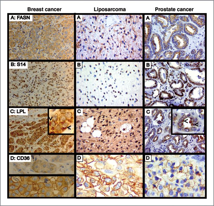
Figure 4. Immunohistochemical
analysis of markers of FA
metabolism in breast,
liposarcoma, and prostate tumors.
Slides from a representative
invasive ductal carcinoma of the
breast (left column), liposarcoma
(middle column), and prostatic
adenocarcinoma (right column)
were immunostained for (A) FASN,
(B) THRSP (Spot 14, S14), (C) LPL,
or (D) CD36. Original
magnification, 40. Detection was
with peroxidase (brown pigment),
and slides were counterstained
with hematoxylin (blue pigment).
FASN staining is cytosolic, and
S14 is nuclear. LPL showed an
asymmetric, perinuclear
distribution (arrows in insets)
compatible with localization to the
Golgi apparatus. Note that the
well-visualized prostate tumor
stroma does not express
detectable LPL. CD36 exhibited 2
distinct patterns of subcellular
localization in breast tumors. Only
a cytosolic signal was seen in 29%
of cases (D, left, top), whereas
prominent cell surface staining
was seen in 69% (D, left, bottom),
and only 2% were devoid of CD36
immunoreactivity. Staining was
also seen in most liposarcoma and
prostate cancers. This was
primarily a cell surface pattern,
and was not uniformly present
across those tumors.
overlying the tonsil unexpectedly showed a strong signal receptor syndecan-1 and the lipoprotein (28). RT-PCR
(Supplementary Fig. S2C, right). revealed readily detectable syndecan-1 mRNA from
The majority of tumors also stained for CD36 (Fig. 4D). DU4475 breast cancer cells, whereas LiSa-2 and T47D
Interestingly, 2 distinct staining patterns were observed exhibited a faint signal (Supplementary Fig. S3A). We
in breast cancer tissue. Of the 144 evaluable cores, 42 incubated fibroblasts and DU4475 cells with fluorescently
exhibited diffuse cytoplasmic staining without accentua- labeled VLDL particles, and assessed for cellular uptake
tion at the plasma membrane (Fig. 4D, left, top), whereas by confocal microscopy. Abundant uptake was observed
100 also showed a strong cell surface signal (Fig. 4D, left, in fibroblasts (Supplementary Fig. S3B) but not in DU4475
bottom). Only two breast cancer cases were devoid of cells (Supplementary Fig. S3C). Occasional fluorescence
staining. A statistically significant difference in the pre- was detected on the cell surface (Supplementary Fig. S3D)
valence of the membranous staining pattern between the but never within the breast cancer cells.
triple-negative and ERþ breast cancers was shown by c2
analysis (42% vs. 76%, P < 0.02). Discussion
Of the 25 liposarcoma cases, 21 stained for CD36,
almost all in a mixed cytoplasmic plus plasma membrane Our data show that cancer cells may use two different
pattern (Fig. 4D, middle), including all 9 cases of well- mechanisms to acquire FA to fuel proliferation. Breast
differentiated liposarcoma. Of the 9 evaluable prostate and liposarcoma tumors are equipped for both lipid
cancers, 4 showed focally positive staining in a mixed synthesis and for LPL-mediated extracellular lipolysis
cytoplasmic and plasma membrane pattern (Fig. 4D, followed by FA uptake via CD36. Prostate cancer cells,
right), whereas 5 cases scored negative for CD36. which have a very high capacity for de novo lipogenesis
Expression of LPL by breast cancer cells suggested the (24), express very little LPL. The low LPL expression
possibility that the cells could use the enzyme not only to could be explained in part by the reported loss of hetero-
hydrolyze extracellular triglyceride, but also for receptor- zygosity at the LPL locus in 47% of prostate tumors,
mediated endocytosis of triglyceride-rich lipoproteins. owing to the presence of a nearby tumor suppressor
This process uses LPL as a bridge between the cell surface gene (29). These cells, however, can acquire sufficient
www.aacrjournals.org Mol Cancer Ther; 10(3) March 2011 433
Downloaded from mct.aacrjournals.org on February 20, 2021. © 2011 American Association for Cancer
Research.Published OnlineFirst January 31, 2011; DOI: 10.1158/1535-7163.MCT-10-0802
Kuemmerle et al.
exogenous FA to maintain growth in the face of FA advanced this area. Previous work established a tight
synthesis inhibition when they are supplied with LPL linkage of enhanced FA synthesis to transformation (35),
and triglyceride-rich lipoprotein particles. and recent studies have defined the role of an intracel-
LPL expression has been shown to be a marker of poor lular lipase, monoacyl glycerol lipase in promoting
prognosis in chronic B-cell lymphocytic leukemia (B- tumorigenesis. Monoacyl glycerol lipase provides, by
CLL; refs. 30, 31). The single reported examination of de-esterification, a stream of intracellular free FA to fuel
the functional significance of LPL in B-CLL was difficult proliferation, growth, and migration (36). This study
to interpret because Orlistat, a compound that inhibits shows a complementary role for LPL, an extracellular
both LPL and FASN (4), was used to inhibit LPL in those lipase, in providing a stream of FA to fuel cancer cell
studies (32). To our knowledge, these are the first experi- proliferation.
ments to show widespread expression of LPL by solid Various hypotheses have been proposed to explain the
tumors. We find that, in contrast to cultured breast cancer dependence of tumors on lipogenesis, but it is clear that
cell lines, where substantial LPL is found only in a subset the primary metabolic fate of FA in proliferating tumor
with a triple-negative gene expression signature, the cells is incorporation into phospholipids destined for
enzyme is a universal component of breast tumors, irre- membrane biosynthesis (37, 38). As mitochondrial pro-
spective of biomarker status. Moreover, we also find that duction and export of citrate are the key steps required to
all liposarcoma and prostate tumors examined also maintain de novo lipogenesis in the cytosol, this begs the
express LPL. question of how such mitochondrial metabolism may be
Several plausible explanations exist for the discrepancy maintained under the hypoxic (but not anoxic) conditions
between cell lines and tumors with respect to LPL expres- that prevail in tumors. Indeed, hypoxia-induced factor-1,
sion. First, the cell lines have been passaged over time in a key mediator of the cellular response to hypoxia,
culture systems lacking vascular endothelium, which is reduces the fractional entry of glucose-derived carbon
the physiologic site for LPL action, or reliably fixed into mitochondria by downregulating pyruvate dehydro-
concentrations of triglyceride-rich lipoprotein substrate, genase, thus driving the increased lactate production that
whereas cell culture media generally contain high con- is the most well-recognized aspect of intermediary meta-
centrations of glucose. Thus, de novo synthesis, rather bolism in tumors (39). However, net flux of carbon
than lipolysis or receptor-mediated endocytosis, may through the glycolytic pathway is substantially elevated
have been selected as the preferred mechanism for FA in glucose-avid tumor cells, because of increased uptake
acquisition in cell culture. Second, it is possible that and trapping. The reduced amount of carbon directed to
interactions with stroma elicit LPL expression. Third, mitochondria is thus sufficient to provide an estimated
each of the breast cancer cell lines that we find to express 60–85% of the ATP generated (40). Brisk citrate export
substantial levels of LPL are not only triple-negative, but from mitochondria seems to be favored by incomplete
are also nonadherent to tissue culture plasticware. In combustion, as a consequence of the truncated Krebs
view of reports that cellular detachment provokes major cycle in tumor mitochondria (41), which also may serve
metabolic adaptations in Her2/neu-expressing breast to reduce oxygen use by reducing carbon flux through
cancer cells (33), we examined the hypothesis that cellular steps downstream from citrate in the cycle. Thus, the
detachment (72 hours) would provoke enhanced LPL competing oxygen-sparing and anabolic demands on
mRNA expression. This proved, however, not to be the tumors are met by a balanced set of metabolic alterations,
case (data not shown). Irrespective of the cause of the the former favored by hypoxia-induced factor-1, and the
discrepancy, it is important to recognize that tissue latter driven by oncogenes (reviewed in ref. 42). Overall,
culture experiments may not faithfully recreate in vivo it seems that the uptake of exogenous FA, for which this
physiology. study shows most tumors to be equipped, would be an
Efficient utilization by cancer cells of FA released by advantageous response to the metabolic dilemma of
extracellular lipolysis would require the expression of hypoxic, proliferating cancer cells.
both LPL and CD36. It was therefore not surprising to Our findings have several implications. First, thera-
find CD36 expression in the majority of tumor tissues peutic efforts aimed solely at inhibition of long-chain FA
examined. CD36 is known to traffic from cytoplasm to the synthesis may not be effective for tumors that are pro-
plasma membrane in response to insulin stimulation of vided with LPL and express CD36. Such tumors may be
adipocytes (34). We observed cell surface localization in sensitive to agents that inhibit the enzymes for both
70% of breast cancers, whereas 30% exhibited only a lipogenesis and lipolysis, such as Orlistat (4) or the diet-
cytoplasmic signal. On the basis of our observation that ary supplement conjugated linoleic acid, which can sup-
cell surface staining was significantly less frequent in press the genes required for both pathways (8, 43). Efforts
triple-negative tumors, we speculate that CD36 traffick- to target LPL will need to take into account the possibility
ing may be driven by cell surface acting growth factors that prolonged systemic suppression of LPL activity
and/or sex steroids in breast cancers. could result in hypertriglyceridemia and consequent
Although further experiments are required to delineate pancreatitis, particularly if dietary fat intake is not cur-
the precise roles of lipogenesis and lipolysis in transfor- tailed. Second, the ability of nearby nonmalignant cells to
mation, proliferation, and metastasis, recent studies have provide LPL to the tumor microenvironment may favor
434 Mol Cancer Ther; 10(3) March 2011 Molecular Cancer Therapeutics
Downloaded from mct.aacrjournals.org on February 20, 2021. © 2011 American Association for Cancer
Research.Published OnlineFirst January 31, 2011; DOI: 10.1158/1535-7163.MCT-10-0802
Lipoprotein Lipase and Cancer Cell Growth
the ability of tumor cells, particularly those with low tionsforschung, Braunschweig, Germany. Triglyceride measurements
were kindly provided by Hong K. Lee, Department of Pathology,
lipogenic potential, to establish metastases in LPL-rich Dartmouth-Hitchcock Medical Center, Lebanon, NH. We specially
tissues such as lung or fatty bone marrow. To benefit from thank Rebecca O’Meara MT (ASCP), Pathology Translational Research
Laboratory at Dartmouth Medical School, for doing the immunohisto-
LPL provided by tumor stroma, the expression of CD36 chemistry.
by the tumor would be required. Third, the presence of
LPL in the tumor vasculature may mediate the reported
Grant Support
effects of dietary fat intake on outcome (9). In addition to
the well-characterized lipogenic tumor phenotype, our
studies indicate the expression of a previously unappre- This work was supported by NIH Grant RO1CA126618 (W.B. Kinlaw),
ciated lipolytic pathway active in cancer cells as well. NIH Training Grant DK07508 (N.B. Kuemmerle), a Howard Hughes
Medical Foundation Fellowship 52005870 (A.J. Flanagan), Norris Cotton
Cancer Center Prouty grants (B.L. Eisenberg, W.B. Kinlaw), Grant
Disclosure of Potential Conflicts of Interest G.0590.08 (J.V. Swinnen), a fellowship (E. Rysman) from the Research
Foundation-Flanders (FWO), the N.C.I. Bay Area Breast Cancer SPORE
No potential conflicts of interest were disclosed. P50 CA58207 (L.A. Timmerman), and the Program in Experimental and
Molecular Medicine at Dartmouth Medical School (C.J. Fricano).
The costs of publication of this article were defrayed in part by the
Acknowledgments payment of page charges. This article must therefore be hereby marked
advertisement in accordance with 18 U.S.C. Section 1734 solely to indicate
MCK-LPL transgenic mice were kindly supplied by Ira Goldberg, this fact.
Columbia School of Medicine, New York, NY. We thank Martin Wabitsch
(University of Ulm, Germany) for the LiSa2 cells. Soraphen A was kindly Received August 25, 2010; revised January 11, 2011; accepted January
provided by Klaus Gerth and Rolf Jansen, Helmholtz-Zentrum f€ ur Infek- 19, 2011; published OnlineFirst January 31, 2011.
References
1. Kuhajda F. Fatty acid synthase and cancer: new application of an old by SREBP-1c, superinduction with progestin, and implication in cell
pathway. Cancer Res 2006;66:5977–80. growth. Exp Cell Res 2005;312:278–88.
2. Menendez J, Lupu R. Fatty acid synthase and the lipogenic phenotype 15. Mosmann T. Rapid colorometric assay for cellular growth and survival:
in cancer pathogenesis. Nat Rev Cancer 2007;7:763–77. application to proliferation and cytotoxicity assays. J Immunol Meth-
3. Alli P, Pinn M, Jaffee E, McFaden J, Kuhajda F. Fatty acid synthesis ods 1983;65:55–63.
inhibitors are chemopreventive for mammary cancer in neu-N trans- 16. Goldstein J, Basu S, Brown M. Receptor-mediated endocytosis of
genic mice. Oncogene 2005;24:39–46. low-density lipoprotein in cultured cells. Methods Enzymol 1983;
4. Kridel S, Axelrod F, Rozenkrantz N, Smith J. Orlistat is a novel inhibitor 98:241–60.
of fatty acid synthase with antitumor activity. Cancer Res 2004; 17. Hata A, Ridinger D, Sutherland S, Emi M, Shuhua Z, Myers R, et al.
64:2070–5. Binding of lipoprotein lipase to heparin. J Biol Chem 1993;268:8447–
5. Weiss L, Hoffman G, Schreiber R, Andres H, Fuchs E, Korber E, et al. 57.
Fatty-acid biosynthesis in man, a pathway of minor importance. J Biol 18. Nilsson-Ehle P, Schotz M. A stable, radioactive substrate emulsion
Chem 1986;367:905–12. assay of lipoprotein lipase. J Lipid Res 1976;17:536–41.
6. Kuhajda F, Jenner K, Wood F, Hennigar R, Jacobs L, Dick J, et al. Fatty 19. Cruz W, Kwon G, Marshall C, McDaniel M, Semenkovich C. Glu-
acid synthesis: a potential selective target for antineoplastic therapy. cose and insulin stimulate heparin-releasable lipoprotien lipase
Proc Natl Acad Sci U S A 1994;91:6279–383. activity in mouse islets and INS-1 cells. J Biol Chem 2001;276:
7. Beckers A, Organe S, Timmermans L, Scheys K, Peeters A, Brussel- 12162–8.
mans K, et al. Chemical inhibition of acteyl-CoA carboxylase induces 20. Wells W, Schwartz G, Morganelli P, Cole B, Chambers J, Kinlaw WB.
growth arrest and cytotoxicity selectively in cancer cells. Cancer Res Expression of "Spot 14" (THRSP) predicts disease free survival in
2007;67:8180–7. invasive breast cancer: immunohistochemical analysis of a new
8. Donnelly C, Olsen A, Lewis L, Eisenberg B, Eastman A, Kinlaw W. molecular marker. Breast Cancer Res Treat 2006;98:231–40.
Conjugated linoleic acid (CLA) inhibits expression of the Spot 14 21. Sorlie T, Perou C, Tibshirani R, Aas T, Geisler S, Johnsen H, et al. Gene
(THRSP) and fatty acid synthase genes and impairs the growth of expression patterns of breast carcinomas distinguish tumor sub-
human breast cancer and liposarcoma cells. Nutr Cancer 2009;61: classes with clinical implications. Proc Natl Acad Sci U S A 2001;
114–22. 98:10896–74.
9. Chlebowski RT, Blackburn GL, Thomson CA, Nixon DW, Shapiro A, 22. Cleator S, Heller W, Coombes R. Triple-negative breast cancer:
Hoy MK, et al. Dietary fat reduction and breast cancer outcome: therapeutic options. Lancet Oncol 2007;8:235–44.
interim efficacy results from the Women's Intervention Nutrition Study. 23. Hughes D, Martel P, Kinlaw W, Eisenberg B. The synthetic triterpenoid
J Natl Cancer Inst 2006;98:1767–76. CDDO-Im inhibits fatty acid synthase expression and has antiproli-
10. Goldberg I, Eckel R, Abumrad N. Regulation of fatty acid uptake into ferative and proapoptotic effects in human liposarcoma cells. Cancer
tissues: lipoprotein lipase- and CD36-mediated pathways. J Lipid Res Invest 2007;26:118–27.
2009;50:S86–90. 24. Swinnen J, Brusselmans K, Verhoeven G. Increased lipogenesis in
11. Neve R, Chin K, Fridlyand J, Yeh J, Baehner F, Fevr T, et al. A cancer cells: new players, novel targets. Curr Opin Clin Nutr Metab
collection of breast cancer cell lines for the study of functionally Care 2006;9:358–65.
distinct cancer subtypes. Cancer Cell 2006;10:515–27. 25. Kinlaw W, Quinn J, Wells W, Roser-Jones C, Moncur J. S14 in breast
12. Irizarry R, Hobbs B, Collin F, Beazer-Barclay Y, Antonellis K, Scherf U, cancer: a marker of aggressive disease and a potential therapeutic
et al. Exploration, normalization, and summaries of high density target. Endocrinology 2006;147:4048–55.
oligonucleotide array probe level data. Biostatistics 1998;4:249–64. 26. Kinlaw W, Church J, Harmon J, Mariash C. Direct evidence for a role of
13. Eisen M, Spellman P, Brown P, Botstein D. Cluster analysis and the "spot 14" protein in the regulation of lipid synthesis. J Biol Chem
display of genome-wide expression patterns. Proc Natl Acad Sci U 1995;270:16615–8.
S A 1998;95:14863–8. 27. Moreau A, Teruel C, Beylot M, Albalea V, Tomasi V, Umbdenstock T,
14. Martel P, Bingham C, McGraw C, Baker C, Morganelli P, Meng M, et al. A novel pregnane X receptor and S14-mediated lipogenic
et al. S14 protein in breast cancer cells: direct evidence for regulation pathway in human hepatocyte. Hepatology 2009;49:2068–79.
www.aacrjournals.org Mol Cancer Ther; 10(3) March 2011 435
Downloaded from mct.aacrjournals.org on February 20, 2021. © 2011 American Association for Cancer
Research.Published OnlineFirst January 31, 2011; DOI: 10.1158/1535-7163.MCT-10-0802
Kuemmerle et al.
28. Obunike J, Edwards I, Rumsey S, Curtiss L, Wagnerf W, Deckelbaum 36. Nomura D, Long J, Niessen S, Hoover H, Ng S, Cravatt B. Mono-
R., et al. Cellular differences in lipoprotein lipase-mediated uptake of acylglycerol lipase regulates a fatty acid network the promotes cancer
low density lipoproteins. J Biol Chem 1994;269:13129–35. pathogenesis. Cell 2010;140:49–61.
29. Bova G, Carter B, Bussemakers M, Emi M, Fujiwara Y, Kyprianou N, 37. Pizer E, Wood F, Pasternack G, Kuhajda F. Fatty acid synthase (FAS):
et al. Homozygous deletion and frequent allelic loss of chromosome a target for cytotoxic antimetabolites in HL60 promyelocytic leukemia
8p22 loci in human prostate cancer. Cancer Res 1993;53:3869–73. cells. Cancer Res 1996;56:745–51.
30. Heintel D, Kienle D, Shehata M, Krober A, Kroemer E, Schwarzinger I, 38. Swinnen J, Van Veldhoven P, Timmermans L, De Schrijver E, Brussel-
et al. High expression of lipoprotein lipase in poor risk B-cell chronic mans K, Vanderhoydonc F, et al. Fatty acid synthase drives the
lymphocytic leukemia. Leukemia 2005;19:1216–23. synthesis of phospholipids partitioning into detergent-resistant mem-
31. van't Veer M, Broojimans A, Langerak A, Verhaaf B, Goudswaard C, brane microdomains. Biochem Biophys Res Commun 2003;302:898–
Graveland W, et al. The predictive value of lipoprotein liase for survival 903.
in chronic lymphocytic leukemia. Haematologica 2006;91:56–63. 39. Kim J, Tchernyshyov I, Semenza G, Dang C. HIF-1-mediated
32. Pallasch C, Schwamb J, Konigs S, Schulz A, Debey S, Kofler D, et al. expression of pyruvate dehydrogenase kinase: a metabolic switch
Targeting lipid metabolism by the lipoprotein lipase inhibitor orlistat required for cellular adaptation to hypoxia. Cell Metab 2006;3:177–
results in apoptosis of B-cell chronic lymphocytic leukemia cells. 85.
Leukemia 2008;22:585–92. 40. Pedersen P. Tumour mitochondria and the bioenergetics of cancer
33. Schafer Z, Grassian A, Song L, Jiang Z, Gerhart-Hines Z, Irie H, et al. cells. Prog Exp Tumor Res 1978;22:190–274.
Antioxidant and oncogene rescue of metabolic defects caused by loss 41. Parlo R, Coleman P. Enhanced rate of citrate export from cholesterol-
of matrix attachment. Nature 2009;461:109–13. rich hepatoma mitochondria. J Biol Chem 1984;259.
34. Luiken J, Dyck D, Han X, Tandon N, Arumugum Y, Glatz J, et al. Insulin 42. Gordan J, Thompson C, Simon M. HIF and c-Myc: sibling rivals for
induces the translocation of the fatty acid transporter FAT/CD36 to the control of cancer cell metabolism and proliferation. Cancer Cell
plama membrane. Am J Physiol Endocrinol Metab 2002;282:E491–5. 2007;12:108–13.
35. Yang Y, Han W, Morin P, Chrest F, Pizer E. Activation of fatty acid 43. Harvatine K, Bauman D. SREBP1 and thyroid hormone responsive
synthase during neoplastic transformation: role of mitogen-activated spot 14 (S14) are involved in the regulation of bovine mammary lipid
protein kinase and phophatidylinositol 3-kinase. Exp Cell Res synthesis during diet-induced milk fat depression and treatment with
2002;279:80–90. CLA. J Nutr 2006;136:2468–74.
436 Mol Cancer Ther; 10(3) March 2011 Molecular Cancer Therapeutics
Downloaded from mct.aacrjournals.org on February 20, 2021. © 2011 American Association for Cancer
Research.Published OnlineFirst January 31, 2011; DOI: 10.1158/1535-7163.MCT-10-0802
Lipoprotein Lipase Links Dietary Fat to Solid Tumor Cell
Proliferation
Nancy B. Kuemmerle, Evelien Rysman, Portia S. Lombardo, et al.
Mol Cancer Ther 2011;10:427-436. Published OnlineFirst January 31, 2011.
Updated version Access the most recent version of this article at:
doi:10.1158/1535-7163.MCT-10-0802
Supplementary Access the most recent supplemental material at:
Material http://mct.aacrjournals.org/content/suppl/2011/01/31/1535-7163.MCT-10-0802.DC1
Cited articles This article cites 42 articles, 15 of which you can access for free at:
http://mct.aacrjournals.org/content/10/3/427.full#ref-list-1
Citing articles This article has been cited by 16 HighWire-hosted articles. Access the articles at:
http://mct.aacrjournals.org/content/10/3/427.full#related-urls
E-mail alerts Sign up to receive free email-alerts related to this article or journal.
Reprints and To order reprints of this article or to subscribe to the journal, contact the AACR Publications
Subscriptions Department at pubs@aacr.org.
Permissions To request permission to re-use all or part of this article, use this link
http://mct.aacrjournals.org/content/10/3/427.
Click on "Request Permissions" which will take you to the Copyright Clearance Center's
(CCC)
Rightslink site.
Downloaded from mct.aacrjournals.org on February 20, 2021. © 2011 American Association for Cancer
Research.You can also read