Establishment of Long-Term Primary Cortical Neuronal Cultures From Neonatal Opossum Monodelphis domestica
←
→
Page content transcription
If your browser does not render page correctly, please read the page content below
ORIGINAL RESEARCH
                                                                                                                                              published: 18 March 2021
                                                                                                                                        doi: 10.3389/fncel.2021.661492

                                            Establishment of Long-Term Primary
                                            Cortical Neuronal Cultures From
                                            Neonatal Opossum Monodelphis
                                            domestica
                                            Antonela Petrović † , Jelena Ban † , Ivana Tomljanović, Marta Pongrac, Matea Ivaničić,
                                            Sanja Mikašinović ‡ and Miranda Mladinic*

                                            Laboratory for Molecular Neurobiology, Department of Biotechnology, University of Rijeka, Rijeka, Croatia

                            Edited by:      Primary dissociated neuronal cultures have become a standard model for studying
                    Valerio Magnaghi,
               University of Milan, Italy
                                            central nervous system (CNS) development. Such cultures are predominantly prepared
                         Reviewed by:
                                            from the hippocampus or cortex of rodents (mice and rats), while other mammals are less
                 Eduardo Martin Lopez,      used. Here, we describe the establishment and extensive characterization of the primary
          Yale University, United States    dissociated neuronal cultures derived from the cortex of the gray South American short-
                        Sergei Antonov,
   Institute of Evolutionary Physiology     tailed opossums, Monodelphis domestica. Opossums are unique in their ability to fully
      and Biochemistry (RAS), Russia        regenerate their CNS after an injury during their early postnatal development. Thus, we
                 *Correspondence:           used cortex of postnatal day (P) 3–5 opossum to establish long-surviving and nearly
                   Miranda Mladinic
                                            pure neuronal cultures, as well as mixed cultures composed of radial glia cells (RGCs)
          mirandamp@biotech.uniri.hr
                                            in which their neurogenic and gliogenic potential was confirmed. Both types of cultures
    †
     These authors have contributed
   equally to this work and share first
                                            can survive for more than 1 month in vitro. We also prepared neuronal cultures from the
                           authorship       P16–18 opossum cortex, which were composed of astrocytes and microglia, in addition
                  ‡
                    Present address:        to neurons. The long-surviving opossum primary dissociated neuronal cultures represent
                     Sanja Mikašinovi´c,
                                            a novel mammalian in vitro platform particularly useful to study CNS development
        Department of Histology and
    Embryology, Faculty of Medicine,        and regeneration.
   University of Rijeka, Rijeka, Croatia
                                            Keywords: opossums, cortex, primary neuron cell culture, radial glia cells, astrocytes, postnatal
                   Specialty section:
        This article was submitted to
             Cellular Neurophysiology,
                a section of the journal    INTRODUCTION
    Frontiers in Cellular Neuroscience
                                            A full understanding of the structure, function, and development of the mammalian central nervous
         Received: 30 January 2021          system (CNS) is necessary to develop treatments that could allow successful regeneration of the
        Accepted: 25 February 2021          adult injured or degenerated tissue. Mammalian developing CNS is challenging to study because
         Published: 18 March 2021           of the extreme complexity of the dynamic events involved, including cell proliferation, migration,
                              Citation:     and differentiation. Moreover, it emerged that neurodegenerative diseases often originate during
    Petrovi´c A, Ban J, Tomljanovi´c I,     development, despite late onset (Schaefers and Teuchert-Noodt, 2016).
Pongrac M, Ivani či´c M, Mikašinovi´c S        Dissociated primary neuronal cultures represent an excellent in vitro tool to study CNS
and Mladinic M (2021) Establishment
                                            development, as well as neuronal maturation and functional activity, at the single-cell level and at
      of Long-Term Primary Cortical
   Neuronal Cultures From Neonatal
                                            the network scale. These cultures allow us to gain mechanistic insights in a simplified but more
  Opossum Monodelphis domestica.            controlled context, compared to in vivo conditions (Beaudoin et al., 2012; Humpel, 2015). The
   Front. Cell. Neurosci. 15:661492.        studies on primary neuronal cultures contributed to many fundamental discoveries regarding
    doi: 10.3389/fncel.2021.661492          development such as neuronal polarization, neurite outgrowth, axon guidance (pathfinding),

Frontiers in Cellular Neuroscience | www.frontiersin.org                            1                                         March 2021 | Volume 15 | Article 661492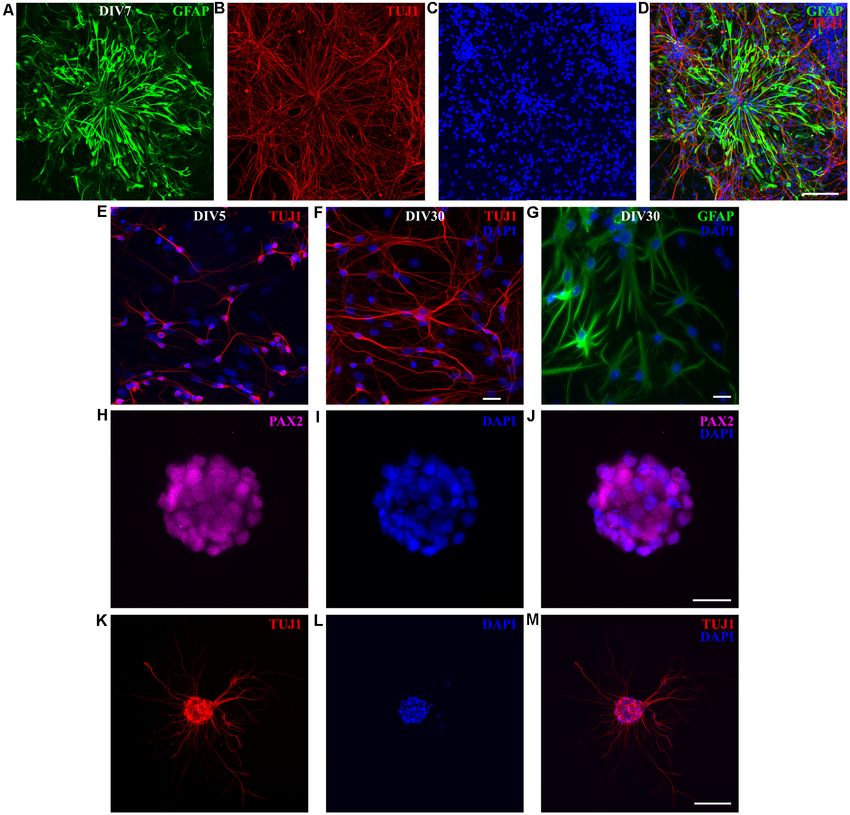
Petrovi´c et al.                                                                                                       M. domestica Primary Neuronal Cultures

synaptogenesis and neuronal network formation, activity and                survival of neuronal cell cultures (up to 1 month), as well
maturation, recapitulating in vitro many aspects that occur                as efficient maintenance and differentiation of RGCs into
in vivo (Cáceres et al., 2012; Kaech et al., 2012).                        neurons in vitro. These M. domestica-derived primary
    The most frequently used source for culturing primary                  CNS cultures are novel and robust in vitro platforms
neurons is the cortex or hippocampus of late embryonic or                  to use in a variety of studies, including development
early postnatal rodents (i.e., rats and mice; Kaech and Banker,            and regeneration.
2006; Beaudoin et al., 2012). Primary neurons derived from other
mammalian species have been less frequently used. For instance,            MATERIALS AND METHODS
cortical primary neuronal cultures were recently obtained
from developing porcine telencephalon (Aubid et al., 2019),                Animals
sheep (Reddy et al., 2015), and marsupials such as opossum                 In this work, South American gray short-tailed opossum
(Bartkowska et al., 2013), with the ability to maintain them               (Monodelphis domestica) pups of both sexes at postnatal day
in vitro for not more than 7, 5 or 2 days, respectively, suggesting        (P)3–5 and 16–18 were used. The M. domestica colony was
their relatively limited period. Thus, in addition to rodents, there       maintained at the animal house facility of the University of
is a need to employ other mammalian species as a source for                Trieste, following the guidelines of the Italian Animal Welfare
primary neuronal cultures, to identify the differences among               Act, and their use was approved by the Local Veterinary
mammals, and to avoid mistakes in translating the knowledge to             Service, the Ethics Committee board, and the National
humans (Bonfanti and Peretto, 2011; Rodemer et al., 2020).                 Ministry of Health (Permit Number: 1FF80.N.9Q3), following
    In this work, we have established and extensively                      the European Union guidelines for animal care (d.1.116/92;
characterized long-term primary cortical dissociated cultures,             86/609/C.E.). The animals are housed in standard laboratory
prepared from the neonatal gray South American short-                      cages in a temperature- and humidity-controlled environment
tailed opossums (Monodelphis domestica). Opossums are                      (27–28◦ C; 50–60% humidity) with a 12/12 h light/dark
marsupials (Metatheria), which are mammalian infraclass that               cycle and ad libitum access to food and water. According
diverged from placental mammals (Eutheria) between 170                     to recently reported developmental transcriptome analysis
and 190 M years ago (Kumar and Hedges, 1998; Goodstadt                     (Cardoso-Moreira et al., 2019), P3–5 opossums correspond to
et al., 2007; Dooley et al., 2013). Unlike the other marsupials,           E15.5–18.5 rat or E14–16 mice embryos, while P16–18 opossums
opossums such as M. domestica lack a pouch and the pups                    are developmentally similar to neonatal (P1–P2) rat or mice. The
(usually from 1 to 12) are born after 14 days of gestation at              body weight and size of animals through postnatal ages used in
a very immature state (Nicholls et al., 1990; Mladinic et al.,             this study are shown in Table 1 and Supplementary Figure 1.
2009). Postnatal day 0 (P0) opossums are comparable to
embryonic day 12 (E12) rat or E11.5 mice embryo, according to              Primary Dissociated Neuronal Cultures
recent transcriptome analysis that matches previous estimates              The dissociation protocol was developed following the
(Smith, 2001; Cardoso-Moreira et al., 2019). During the                    procedures previously described for rodents (Kaech and
first two postnatal weeks, M. domestica neonates can fully                 Banker, 2006; Beaudoin et al., 2012; Pozzi et al., 2017) and
regenerate the spinal cord after injury (Nicholls et al., 1994;            all efforts were made to minimize suffering and to reduce the
Saunders et al., 1995, 1998; Varga et al., 1995; Nicholls and              number of animals used. Primary neurons were isolated from
Saunders, 1996). This ability to fully regenerate the spinal               the cortex of neonatal opossums from two developmentally
cord after injury abruptly stops in opossums at the postnatal              significant postnatal age groups: P3–5 and P16–18, respectively.
day 12 in the cervical part of the spinal cord and at the                  Both left and right hemispheres from each animal were used
postnatal day 17 in less mature lumbar segments of the                     while olfactory bulbs and remaining subcortical structures
spinal cord (Wintzer et al., 2004; Mladinic et al., 2005, 2010).           were removed. Dissection was performed in the ice-cold
Moreover, fundamental brain development events such as                     oxygenated (95% O2 /5% CO2 ) dissection solution, consisted of
the formation of hexalaminar cortex, cerebellum formation,                 113 mM NaCl, 4.5 mM KCl, 1 mM MgCl2 × 6H2 O, 25 mM
and gliogenesis occur during the first month in opossums                   NaHCO3 , 1 mM NaH2 PO4 , 2 mM CaCl2 × 2H2 O, 11 mM
(Saunders et al., 1989; Molnár et al., 1998; Puzzolo and                   glucose and 0.5% w/v Penicillin/Streptomycin/Amphotericin
Mallamaci, 2010; Seelke et al., 2013; Tepper et al., 2020),                B, pH 7.4 (all from Sigma–Aldrich, St. Louis, MO, USA).
offering the unique possibility to obtain developing (embryonic-
like) CNS cells from postnatal animals, at a wide range of
postnatal days.                                                            TABLE 1 | Age, body weight and size of opossums used in this study.
    Here, we describe the protocols to successfully develop and            Age                        Body weight (g)                           Body size (mm)
establish different in vitro primary dissociated cultures from
                                                                           P3                            0.14 ± 0.01                               9.14 ± 0.48
developing cortex of postnatal opossums (M. domestica) of
                                                                           P4                            0.19 ± 0.02                              11.50 ± 1.73
different ages, in which different CNS cell types predominate,             P5                            0.22 ± 0.02                              12.60 ± 0.42
including neurons, radial glia cells (RGCs), and astrocytes.               P16                           1.43 ± 0.04                              31.20 ± 2.05
We have chosen P3–5 and P16–18 opossums since those                        P17                           1.63 ± 0.05                              33.33 ± 1.15
ages differ essentially in their capacity to regenerate spinal             P18                           1.71 ± 0.11                              33.00 ± 1.41

cord tissue after injury. Our protocols allow long-term                    For each age 10–17 pups from at least three different litters were measured.

Frontiers in Cellular Neuroscience | www.frontiersin.org               2                                            March 2021 | Volume 15 | Article 661492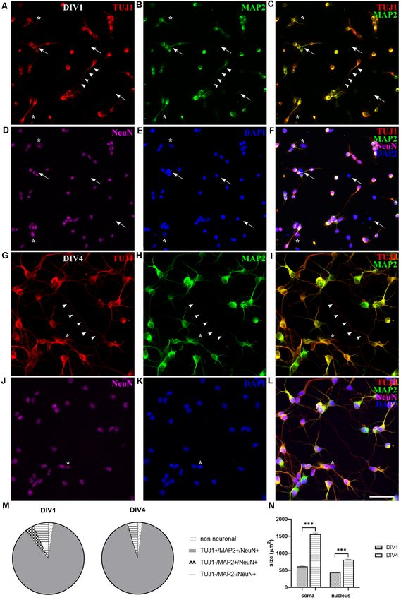
Petrovi´c et al.                                                                                                  M. domestica Primary Neuronal Cultures

The meninges were removed, and the tissue was chopped                        media changes were done once per week changing only a half
into small pieces. Before enzymatic digestion, the cortices                  of the medium with the fresh neuronal medium. The cortical
were washed three times with the sterile phosphate-buffered                  cultures were maintained in an incubator at 32◦ C, 5% CO2 , and
saline (PBS) containing 137 mM NaCl, 2.7 mM KCl, 10 mM                       95% relative humidity.
Na2 HPO4 , 2 mM KH2 PO4 (all from Sigma–Aldrich). Cortices
were digested with prewarmed trypsin in PBS (Santa Cruz                      Radial Glial Cell Cultures
Biotechnology, SCBT, Dallas, TX, USA) by incubating at 32.5◦ C.              Radial glial cells (RGCs) were prepared using the same protocol
The tissue from P3 to P5 pups was incubated in 0.5% w/v                      for dissection and dissociation as described for P3–5 neuronal
trypsin for 10 min, while that of P16–18 pups was incubated                  cultures. Unlike neuronal cultures, RGCs were plated on poly-
in 2.5% w/v trypsin for 15 min. After three washes with                      L-ornithine-coated dishes or coverslips without laminin at
PBS, cells were dissociated using a 1 ml tip by pipetting up                 the density of 5 × 10⁴ cells per well in a 24-well plate.
and down 15–20 times in a trituration solution containing                    DMEM with stable glutamine supplemented with 10% w/v
10 µg/ml DNAse I (Sigma–Aldrich), 1 mg/ml trypsin inhibitor                  FBS and 1% w/v Penicillin/Streptomycin was used both for
(SCBT), and 1% w/v bovine serum albumin (BSA; Pan-Biotech                    plating and culture. Cell passaging was done at 90%–100%
GmbH, Aidenbach, Germany) in Hank’s Balanced Salt Solution                   confluence using 0.5% trypsin in PBS for 5 min at 32◦ C
(HBSS) solution, w/o Ca2 + and Mg2 + (Pan-Biotech). The                      and subsequent replating on the poly-L-ornithine-coated glass
supernatant containing dissociated cells was collected and                   coverslips in plating medium. To allow the formation of
layered on top of the 5% w/v BSA cushion in HBSS in the                      neurospheres, dissociated cells from the opossum cortex were
5 ml tube to remove the cell debris. The trituration step was                cultured in a plating medium on a non-adherent tissue culture
repeated two times for P3–5 and three times for P16–18 digested              flask at the density of 6 × 10⁵ cells per 25 cm2 flask. The
cortices. Cells were collected by centrifugation for 5 min at                cells were transferred to a new tissue culture flask the next
100 g and then resuspended in a plating medium consisting of                 day to remove residual adherent cells such as fibroblasts.
Dulbecco’s minimum essential medium (DMEM) with stable                       A quarter of the media was changed at DIV1 and DIV4.
glutamine supplemented with 10% w/v fetal bovine serum (FBS)                 Neurospheres were grown until DIV7 (when they reached
and 1% w/v Penicillin/Streptomycin (all from Pan-Biotech)                    approximately 100 µm in diameter) and then transferred to
and purified by preplating the cell suspension for 5 min on                  poly-L-ornithine and laminin-coated glass coverslips in neuronal
the plastic tissue culture dishes. Cells were counted using                  medium (Neurobasal medium supplemented with B27, see
the hemocytometer and plated on glass coverslips (12 mm                      ‘‘Neuronal Cortical Cultures of P16-18 Opossums’’ section) to
diameter) precoated with 50 µg/ml poly-L-ornithine and                       differentiate over the next 7 days.
2 µg/ml laminin (all from Sigma–Aldrich) at the density of
1 × 10⁵ cells per well in a 12-well plate or 5 × 10⁴ cells per               Immunofluorescence
well in a 24-well plate. The next day, 2/3 of the medium was                 Cells were fixed for 20 min at room temperature (RT, 20–22◦ C)
changed with the neuronal medium containing Neurobasal                       with 4% paraformaldehyde (PFA, Sigma–Aldrich) containing
medium supplemented with B27 (both from Thermo Fisher                        200 mM sucrose in PBS, pH 6.9. After fixation, cells were
Scientific, Waltham, MA, USA), 1 mM L-glutamine, and 1%                      washed with PBS, saturated with 0.1 M glycine, permeabilized
Penicillin/Streptomycin (both from Pan-Biotech). Subsequent                  with 0.1% Triton X-100 (all from Sigma–Aldrich) in PBS, and

TABLE 2 | Primary antibodies used in this work.

Antibody                          Host and isotype         Dilution   Immunogen                             Producer Cat #               UniProt
                                                                                                            RRID                         Identity

Glutamic acid                     Mouse monoclonal IgG1    1:200      Rat brain GAD, purified using         Sigma–Aldrich                86.6%
decarboxylase 65 (GAD65)                                              Protein A chromatography              SAB4200232
                                                                                                            AB_10762670
Glial fibrillary acidic protein   Mouse monoclonal IgG1    1:200      GFAP from porcine spinal cord         Sigma–Aldrich G3893          84.9%
(GFAP)                                                                                                      AB_477010
Ionized calcium-binding           Rabbit polyclonal        1:100      Synthetic peptide corresponding to    Abcam ab15580                63.4%
adaptor molecule 1 (Iba1)                                             Human Iba1 (C terminal)               AB_10862652
Microtubule-associated            Mouse monoclonal IgG1    1:200      Bovine MAP2                           Sigma–Aldrich M1406          82.8%
protein 2 (MAP2)                                                                                            AB_477171
Neuronal nuclei (NeuN)            Rabbit monoclonal        1:200      Synthetic peptide within Human        Abcam ab177487               84%
                                                                      NeuN aa 1–100 (Cysteine residue)      AB_2532109
Paired box gene 2 (PAX2)          Rabbit monoclonal IgG    1:50       Synthetic peptide within Human        Abcam ab79389                94.5%
                                                                      Pax2 aa 1–100                         AB_1603338
Synapsin 1                        Rabbit polyclonal        1:100      Synapsin I (a mixture of Ia and Ib)   Millipore AB1543             72.3%
                                                                      purified from bovine brain            AB_2200400
β-Tubulin III (TUJ1)              Mouse monoclonal IgG2a   1:200      Microtubules derived from rat brain   Biolegend 801201             99.8%
                                                                                                            AB_2313773
Vesicular Glutamate               Guinea pig polyclonal    1:200      Synthetic peptide from rat            Chemicon AB5907              96.4%
Transporter 2 (vGLUT2)                                                VGLUT2 protein                        AB_2301731

Frontiers in Cellular Neuroscience | www.frontiersin.org                 3                                      March 2021 | Volume 15 | Article 661492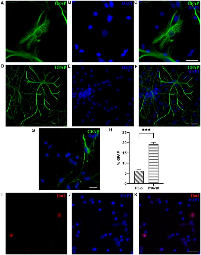
Petrovi´c et al.                                                                                        M. domestica Primary Neuronal Cultures

lastly washed with PBS, each step lasting 5 min. Cells were            Statistical Analysis
blocked with 0.5% w/v BSA (Pan-Biotech) in PBS for 30 min.             All results have been obtained from at least three independent
Incubation with the primary antibodies (Table 2) was done              experiments and are presented as a bar graph with mean ± SEM.
in a wet chamber for 1 h, followed by the washing steps in             Statistical analysis was performed using GraphPad Prism 8.4
PBS and incubation with the secondary antibodies. For every            (GraphPad Software Inc., La Jolla, CA, USA). To test the
primary antibody used, the protein sequence similarity between         normality of data, depending on the number of values tested,
opossum and immunogen was compared using the Universal                 either D’Agostino–Pearson or Shapiro–Wilk normality test was
Protein Resource (UniProt1 ). The secondary antibodies were            used. Brown–Forsythe test was used to test the equality of
goat anti-mouse Alexa Fluorr 555 (Thermo Fisher Scientific,            variances. One-way ANOVA was used to compare three or
Cat# A32732, RRID: AB_2633281, 1:400), goat anti-mouse Alexa           more data groups when data followed Gaussian distribution.
Fluorr 488 (Thermo Fisher Scientific, Cat# A32723, RRID:               Following the one-way ANOVA, the Holm–Šídák test was
AB_2633275, 1:400), goat anti-rabbit Alexa Fluorr 555 (Thermo          performed for multiple comparisons between data groups.
Fisher Scientific, Cat# A32732, RRID: AB_2633281, 1:400), goat         Data groups with different variances that fail a normality test
anti-rabbit Alexa Fluorr 647 (Abcam, Cat# ab150083, RRID:              were compared using the Kruskal–Wallis test. Following the
AB_2714032, 1:300), goat anti-mouse immunoglobulin (Ig) G1             Kruskal–Wallis test, Dunn’s multiple comparisons test was
Alexa Fluorr 488 (Thermo Fisher Scientific, Cat# A-21121,              performed for multiple comparisons between data groups.
RRID: AB_2535764, 1:300) and goat anti-mouse IgG2a Alexa               T-test was used when comparing two normally distributed
Fluorr 555 (Thermo Fisher Scientific, Cat# A-21137, RRID:              data groups with equal variances, and in case of unequal
AB_2535776, 1:300) and the incubation time was 30 min in               variances, Welch’s t-test was used. The accepted level of
the dark. For F-actin staining, Phalloidin-iFluor 488 (Abcam,          significance was p < 0.05. p < 0.001 very significant ∗∗∗ ,
Cambridge, UK) was incubated for 30 min together with the              0.001–0.01 very significant ∗∗ , 0.01–0.05 significant ∗ , ≥0.05
secondary antibody. Cell nuclei were stained with a 300 nM             not significant.
nuclear stain 40 ,6-diamidino-2-phenylindole (DAPI, Thermo
Fisher Scientific) diluted in PBS for 5 min. Finally, coverslips       RESULTS
were washed with PBS and in dH2 O. Coverslips were mounted
on a glass slide using the mounting medium (Vectashield,               Establishment of Neuronal Cultures From
Vector Laboratories, Burlingame, CA, USA) and sealed with nail
                                                                       P3 to P5 Opossum Cortex
polish. All the incubations were performed at RT. Proliferation
                                                                       As described in ‘‘Materials and Methods’’ section in detail,
assay was done using Click-iTTM 5-ethynyl-20 -deoxyuridine
                                                                       the dissection protocol was adapted from the standard and
(EdU) Cell Proliferation Kit, Alexa FluorTM 488 dye (Thermo
                                                                       well-established protocols for rodent embryonic or postnatal
Fisher Scientific). EdU was added to the cells according to
                                                                       neuronal cultures from cortex or hippocampus (Kaech and
manufacturer instruction immediately after the plating and
                                                                       Banker, 2006; Beaudoin et al., 2012), with some modifications.
incubated for 24 h. Cells were fixed at DIV1 and immunolabeled
                                                                       In particular, to preserve cell viability during the dissection
for 30 min at RT in Click-iTr reaction cocktail. The cells
                                                                       procedure, an ice-cold oxygenated dissection medium was used.
were analyzed using an Olympus IX83 inverted fluorescent
                                                                       We introduced two-step enzymatic digestion, bovine serum
microscope (Olympus, Tokyo, Japan) equipped with differential
                                                                       albumin (BSA) cushion, and preplating steps (see ‘‘Materials
interference contrast (DIC) and fluorescence optics (mirror
                                                                       and Methods’’ section). Importantly, since opossums have lower
units: U-FUNA: EX360–370, DM410, EM420–460, U-FBW:
                                                                       body temperature than most placental mammals and slightly
EX460–495, DM505, EM510IF, and U-FGW: EX530–550,
                                                                       lower than other marsupials (Harder et al., 1996), their primary
DM570, EM575IF (Olympus) and Cy5 (EX620/60, DM660,
                                                                       cultures have been maintained at 32◦ C, as previously described
EM700/75, Chroma, Irvine, CA, USA). Fluorescence images were
                                                                       for organotypic cultures derived from developing opossum
acquired with Hamamatsu Orca R2 CCD camera (Hamamatsu
                                                                       cortex (Puzzolo and Mallamaci, 2010).
Photonics, Hamamatsu, Japan) and CellSens software (Olympus,
Japan). 10× 0.3 numerical aperture (NA) air, 20× 0.5 NA                M. domestica-Derived Neuronal Growth Cones (GCs)
air, and 40× 1.4 NA oil immersion objectives were used.                Cortical dissociated neurons from P3 to P5 opossums attached
For each image 15–30 slices were acquired with slice spacing           and survived well after one day in vitro (DIV1) and were positive
of 1 µm (10× and 20× objectives) and 0.3–0.5 µm (40×                   for β-tubulin III marker (TUJ1, Figure 1A), specific for early
objective). For each image, a maximum intensity projection             postmitotic neurons (Menezes and Luskin, 1994; von Bohlen
was used. CellSens and ImageJ by W. Rasband (developed                 Und Halbach, 2007). Growth cones (GCs), highly dynamic
at the U.S. National Institutes of Health2 ) were used for             actin-rich structures that play an essential role in axon guidance
image processing and analysis. For GC, soma, and nuclei                (Dent et al., 2011; Vitriol and Zheng, 2012), were formed at the
surface area analysis, the contour of each region of interest          tips of the growing neurites, as shown in Figure 1B. We analyzed
(ROI) was manually traced, and the surface was measured                the size of M. domestica-derived GCs using phalloidin (F-actin)
using ImageJ/Fiji.                                                     fluorescence stain (Figure 1B). The GCs had variable sizes with
                                                                       an average value of 39.50 ± 3.09 µm2 (n = 40). To the best
1 http://www.uniprot.org                                               of our knowledge, these are the first experimental observations
2 http://rsbweb.nih.gov/ij/                                            showing that neuronal GCs derived from the opossum cortex

Frontiers in Cellular Neuroscience | www.frontiersin.org           4                                  March 2021 | Volume 15 | Article 661492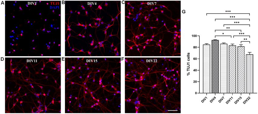
Petrovi´c et al. M. domestica Primary Neuronal Cultures
made up by TUJ1-negative neurons, expressing MAP2 and
NeuN markers (TUJ1− /MAP2+ /NeuN+ , 0.26 ± 0.54%, n = 442).
Neurons positive only to NeuN marker (TUJ1− /MAP2− /NeuN+
cells) were reduced to 4.53 ± 0.55% (n = 442). However,
at DIV4 the total number of neurons (TUJ1, MAP2, and
NeuN triple-positive cells) exceeded hugely the number of
non-neuronal cells, which was further reduced to around
2% (TUJ1− /MAP2− /NeuN− cells, 2.11 ± 0.48%, n = 442),
indicating the progression of neuronal differentiation in
culture (Figure 2M).
Since TUJ1 and MAP2 are also cytoskeletal markers, we
used them to follow in vitro neurite outgrowth in opossum
primary cultures. Similar to rodents (Dotti et al., 1988;
Cáceres et al., 2012), at DIV1 in M. domestica-derived cortical
neuronal cultures it was not possible to distinguish axons from
dendrites as TUJ1 and MAP2 were coexpressed in all neurites
(Figures 2A–C). In contrast, at DIV4, axons were identified as
TUJ1+ /MAP2− neurites (Figures 2G–L, arrowheads). At DIV4,
we counted on average 4.70 ± 0.13 neurites per neuron (the
total number of TUJ1-positive neurites emerging from soma;
170 neurons analyzed). This result is similar to rat hippocampal
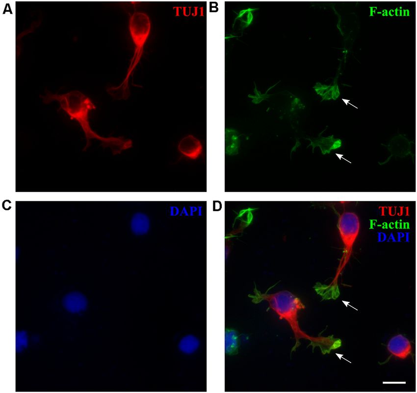
FIGURE 1 | M. domestica-derived neurons and neuronal growth cones neuronal cultures cultured in Neurobasal/B27-based medium
(GCs) at DIV1. Primary neuronal cultures prepared from P5 opossums were
(Pozzi et al., 2017), where also the enhanced branching was
fixed 24 h after plating and stained for (A) β-tubulin III (TUJ1, red), (B)
filamentous (F)-actin (green), (C) nuclear stain 40 ,6-diamidino-2-phenylindole observed in the first 4 days in vitro.
(DAPI; blue) and (D) merged. Arrows indicate GCs identified with F-actin Moreover, the 2.5-fold increase of neuronal cell bodies
immunostaining. Scale bar, 10 µm. was also observed at DIV4: at DIV1 average soma surface
area was 198.92 ± 4.62 µm2 (n = 83) while at DIV4 it
can be efficiently obtained in vitro, with a size comparable to rat reached 503.10 ± 11.15 µm2 (n = 95). Nuclei surface
hippocampal GCs (Pozzi et al., 2017). increased 1.8-fold: from 140.93 ± 3.01 µm2 (n = 83) at
DIV1–261.03 ± 4.77 µm2 (n = 95) at DIV4. This analysis was
Neuronal Outgrowth and Expression of Neuronal done using MAP2 and DAPI staining for soma and nuclei,
Markers respectively (see ‘‘Materials and Methods’’ section). Our in vitro
Next, we explored the expression of different neuronal markers observations correlate with the increase in average neuron size
in opossum cortical primary cultures. For instance, NeuN, observed in vivo, during the first postnatal week in the rat brain
neuron-specific nuclear protein widely expressed in vertebrates (Bandeira et al., 2009).
(Mullen et al., 1992) has been previously detected on cortical
sections of developing opossums by immunohistochemistry In vitro Neuronal Maturation and Synapse Formation
(Seelke et al., 2013; Bartkowska et al., 2014), while microtubule- We analyzed in vitro maturation and long-term survival of
associated protein (MAP)2 is a conventional somatodendritic opossum cortical cultures, as well as progressive neuronal
neuronal marker (Binder et al., 1986; Menezes and Luskin, 1994; network formation. Figure 3 shows neuronal network formation
Dehmelt and Halpain, 2005). during the 3 weeks in vitro. At DIV1 (Figures 2A–F, 3A,
At DIV1, NeuN, MAP2, and TUJ1 neuronal markers were Supplementary Figure 2), following dissociation and plating,
already detected in cultures of dissociated P3–5 opossums’ neurons started to regrow their processes and established
neurons (Figure 2). The majority of the cells (86.83 ± 0.98%, first connections with the neighboring cells. We counted
n = 367) were triple positive for TUJ1, MAP2, and NeuN the percentage of TUJ1-positive neurons among total cells
(TUJ1+ /MAP2+ /NeuN+ , Figure 2F). The lower portion at DIV1 (84.82 ± 4.87%, n = 663) and for all time
of neurons (6.86 ± 0.76%, n = 367) was expressing points considered (Figure 3G). TUJ1 was the neuronal
only NeuN (TUJ1− /MAP2− /NeuN+ , Figures 2A–F, marker of choice because it stains both dendrites and axons
asterisks) and TUJ1-negative neurons were also found (for better visualization of neurites, the grayscale images
(TUJ1− /MAP2+ /NeuN+ , 4.02 ± 0.85%, n = 367). Finally, are available in Supplementary Figure 2). After 4 days
triple-negative non-neuronal cells (TUJ1− /MAP2− /NeuN− ) in culture in serum-free Neurobasal/B27-based medium, the
were 2.28 ± 0.35% of all cells (n = 367, Figures 2A–F, arrows). highest percentage of neurons (92.66 ± 2.02%, n = 603) was
These results suggest that at DIV1 the expression of NeuN reached (Figures 3B,G), resulting in nearly pure neuronal
slightly precedes MAP2 and TUJ1, with the vast majority of cultures. At DIV7, 86.28 ± 4.25% (n = 980) of cells were
neurons expressing all three neuronal markers (Figure 2M). TUJ1+ neurons (Figure 3C) and virtually all neurons have
At DIV4 virtually all (>99%) neurons present in culture established connections with each other. At DIV11 and DIV15,
coexpressed TUJ1 and MAP2, while only a minor portion was the proportion of neurons remained above 80% (DIV11:
Frontiers in Cellular Neuroscience | www.frontiersin.org 5 March 2021 | Volume 15 | Article 661492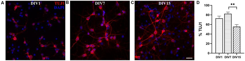
Petrovi´c et al.                                                                                                           M. domestica Primary Neuronal Cultures

  FIGURE 2 | Expression of neuronal markers in cortical cultures derived from P5 opossum. Cells were fixed at DIV1 and stained for (A) TUJ1 (red), (B) MAP2
  (green), (D) NeuN (magenta), and (E) DAPI nuclear stain (blue). (C) Merged image of TUJ1 and MAP2. (F) Merged image of all markers. (G–L) Same as (A–F) but for
  DIV4 cultures. Asterisks indicate TUJ1− /MAP2− /NeuN+ and arrows indicate non-neuronal (TUJ1− /MAP2− /NeuN− ) cells. Arrowheads indicate TUJ1+ /MAP2− axon.
  (M) Percentage of expression for TUJ1, MAP2, and NeuN at DIV1 and DIV4, respectively. (N) Neuronal cell bodies (soma) and neuronal nuclei size increased from
  DIV1 to DIV4. Unpaired t-test with Welch’s correction. DIV1 soma vs. DIV4 soma p < 0.001∗∗∗ , DIV1 nucleus vs. DIV4 nucleus p < 0.001∗∗∗ . Scale bar, 50 µm.

83.28 ± 2.50, n = 637 and DIV15: 81.53 ± 2.99%, n = 560,                             efficient neuronal survival during the first 2 weeks in vitro, results
Figures 3D,E), while at DIV22 it decreased to 67.68 ± 3.45%                          very similar to those obtained with E18 rat cortical neurons (Kim
(n = 633, Figure 3F). Our results have shown the striking and                        et al., 2007; Cullen et al., 2010).

Frontiers in Cellular Neuroscience | www.frontiersin.org                         6                                       March 2021 | Volume 15 | Article 661492
Petrovi´c et al. M. domestica Primary Neuronal Cultures
FIGURE 3 | Primary neuronal cultures derived from the P3 to P5 cortex of M. domestica. Neurons were stained for β-tubulin III (TUJ1, red) and nuclei were stained
with DAPI (blue). The cells were fixed and stained at (A) DIV1, (B) DIV4, (C) DIV7, (D) DIV11, (E) DIV15, and (F) DIV22, respectively. For better visualization of
neurites, the grayscale images are available in Supplementary Figure 2. (G) Histogram showing percentage of neurons at the times indicated in panels (A–F).
One-way ANOVA followed by Holm–Šídák multiple comparisons test. DIV4 vs. DIV11 p = 0.0395∗ , DIV4 vs. DIV15 p = 0.00644∗∗ , DIV1 vs. DIV22 p < 0.001∗∗∗ ,
DIV4 vs. DIV22 p < 0.001∗∗∗ , DIV7 vs. DIV22 p < 0.001∗∗∗ , DIV15 vs. DIV22 p = 0.0013∗∗ . Scale bar, 50 µm.
We were able to maintain opossum cortical cultures up and hippocampal cultures are well characterized (Beaudoin
to 1 month in vitro (Supplementary Figure 3), although the et al., 2012). The cortex of older animals is in general more
cell survival decreased significantly after 3 weeks. Starting difficult to dissociate and therefore few additional modifications
from DIV15, the formation of cell clusters was observed, and regarding the protocol for P3–5 opossums were introduced:
this became progressively more evident in the following days trypsin concentration and incubation time were increased with
(Supplementary Figure 3). These clusters were organized in additional pipetting during dissociation.
spherical aggregates of neuronal cell bodies from which neurites Neuronal cultures derived from P16 to P18 opossums
grew with parallel orientation. To form the aggregates, the followed similar dynamics of network formation to those
cells necessarily migrated in vitro, since the tissue was initially observed in P3 to P5 cortical cultures, during the first 2 weeks
dissociated and plated as single-cell suspension (see Figure 3A). in vitro (Figure 5A). At DIV1, 72.80 ± 4.67% cells were TUJ+
Similar structures were previously described in long-term (n = 2,070, Figures 5A,D) and their proportion increased to
(DIV67) rat hippocampal cultures (Todd et al., 2013). We 82.37 ± 3.67% (n = 704, Figures 5B,D) at DIV7. At DIV15, the
confirmed the expression of TUJ1 as well as MAP2 throughout percentage of neurons was significantly reduced to 55.59 ± 4.15%
the observation period, including DIV30, using several different (n = 1,514, Figures 5C,D). Although it was possible to keep
antibodies for each marker (Supplementary Figures 4, 5). neuronal cultures derived from P16 to P18 opossum in vitro up
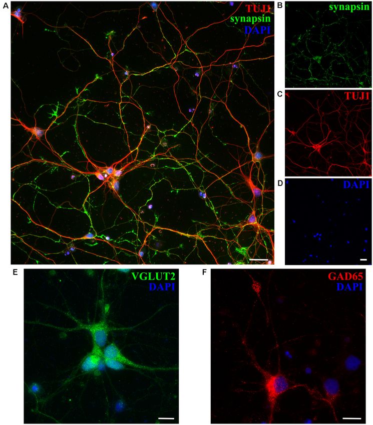
Next, we checked the expression of synaptic markers such to 1 month (Supplementary Figure 5), their lower long-term
as synapsin in opossum neuronal cultures, using previously survival was observed, compared to cultures derived from P3 to
described protocols (Cullen et al., 2010; Todd et al., 2013; P5 animals. The lower percentage of neurons present in cultures
Petrovic et al., 2019) and confirming the formation of synaptic derived from older animals (P16–18) reflects the situation in vivo,
connections and the formation of in vitro neuronal networks as well as it is probably related to the higher susceptibility of
at DIV15 (Figures 4A–D). Moreover, both excitatory and P16–18 neurons to the dissociation process, as shown in mice
inhibitory neuronal subtypes were found, using as markers hippocampal or cortical primary cultures (Beaudoin et al., 2012).
vesicular glutamate transporter 2 (VGLUT2) and glutamic acid
decarboxylase (GAD65), respectively (Figures 4E,F).
Non-neuronal Cells
Neuronal Cortical Cultures of Gliogenesis occurs at late embryonic and postnatal age in rodents
P16–18 Opossums (Kaech and Banker, 2006; Malatesta et al., 2008), while in
In addition to neuronal cultures derived from P3 to P5 neonates, opossums it presumably starts around P18 when the switch
we also established primary cortical cultures from P16 to from radial glia to astrocytes morphology is first observed
P18 opossums. At this age, pups are around 3 cm long and (Puzzolo and Mallamaci, 2010). Thus, we analyzed the content
their body weight is around 1.5 g (Table 1, Supplementary of non-neuronal cells in opossum cortical cultures, derived from
Figure 1). They are developmentally equivalent to P0–3 rats both P3–5 and P16–18 opossums, in which the percentage of
or mice (Cardoso-Moreira et al., 2019), whose primary cortical neurons is much smaller.
Frontiers in Cellular Neuroscience | www.frontiersin.org 7 March 2021 | Volume 15 | Article 661492Petrovi´c et al. M. domestica Primary Neuronal Cultures
FIGURE 4 | Expression of synaptic and neuronal subtype markers at DIV15 of cortical cultures obtained from P5 opossums. (A) Merged image of staining for
(B) synapsin (green; C) β-tubulin III (TUJ1, red) and (D) DAPI nuclear stain (blue). (E) Excitatory VGLUT2-positive (green), (F) GABAergic inhibitory GAD65-positive
(red) neurons, and DAPI-positive nuclei (blue), respectively. Scale bar, 25 µm.
To identify astrocytes, we used glial fibrillary acidic to P16–18 cultures, primary astrocytes prepared from the
protein (GFAP), the most commonly used marker of P3–5 cortex showed less elaborated, thinner, and elongated
astrocytes, which in M. domestica was previously used on shapes, indicating a lower degree of maturity (Figure 6G). The
tissue slices (Puzzolo and Mallamaci, 2010). As shown in GFAP-positive cells were less abundant in cultures derived
Figure 6, at DIV7, GFAP-positive cells were present, as both from P3 to P5 opossums, presenting only 5.71 ± 0.53%
protoplasmic-like (Figures 6A–C) and stellate astrocytes of total cells (n = 1,948, Figure 6H), vs. 19.35 ± 0.83%
(Figures 6D–F), as previously observed in rodent cultures (n = 1,085) of GFAP+ cells in cultures derived from
(Verstraelen et al., 2014; Wolfes et al., 2017). In contrast P16 to P18 cortex. In addition to GFAP, we confirmed
Frontiers in Cellular Neuroscience | www.frontiersin.org 8 March 2021 | Volume 15 | Article 661492Petrovi´c et al. M. domestica Primary Neuronal Cultures
FIGURE 5 | Primary dissociated cortical cultures derived from P16 to P18 opossums. (A) DIV1, (B) DIV7, and (C) DIV15 neuronal cultures stained for β-tubulin III
(red). Nuclei were stained with DAPI (blue). (D) Histogram showing percentage of neurons at the time points indicated in panels (A–C). Kruskal–Wallis test followed
by Dunn’s multiple comparisons test. DIV7 vs. DIV15 p = 0.0071∗∗ . Scale bar, 25 µm.
the expression of intermediate filament protein vimentin resembling RGCs because of their bipolar and elongated
(Eliasson et al., 1999; de Pablo et al., 2019) in developing morphology, with processes that extended for several hundreds
astrocytes derived from both P3–5 and P16–18 opossums of micrometers. As in previously described neuronal cultures,
(Supplementary Figure 6). M. domestica-derived astrocytes’ GFAP and TUJ1 had exclusive staining. The presence of
morphology is strikingly similar to those derived from rodents postmitotic TUJ1-positive neurons showed that effective
(Verstraelen et al., 2014; Ulloa Severino et al., 2016; Pozzi et al., neuronal differentiation occurred despite the presence of FBS in
2017; Wolfes et al., 2017), offering the new source of mammalian the culture medium (Figure 7B). Overall cell density increased
astrocytes for further in vitro investigations. with time (compare DIV7 cultures shown in Figure 3C with
In addition to astrocytes, Iba1-positive microglial cells Figures 7A–D). We, therefore, assumed that new RGC and
were found at DIV1 in cultures derived from the P18 cortex, new neurons were generated in vitro, in addition to preexisting
accounting for 2.15 ± 0.71% of cells (n = 531, Figures 6G–I). neurons (plated at ‘‘DIV0’’).
We were not able to identify any microglia in cultures To verify the in vitro neurogenic potential of RGCs, we
derived from P3 to P5 cortex, nor oligodendrocytes, replated the cultures maintained for 1 month in vitro. Cells
neither in P3–5 or P16–18-derived cultures, likely because were trypsinized and replated on poly-L-ornithine-coated glass
the oligodendrogenesis in opossums occurs around P40 coverslips in a plating medium (see ‘‘Materials and Methods’’
(Puzzolo and Mallamaci, 2010). section). Five days after replating, TUJ1-positive neurons were
found (Figure 7E). These cultures were able to survive for an
Radial Glia Cells additional 1 month and they were composed of both neurons
RGCs represent the main population of neural progenitors (Figure 7F) and astrocytes (Figure 7G). We have successfully
in the developing cortex (Malatesta et al., 2008). Opossum replated these cultures for up to four passages (unpublished
neonates are equivalent to rodent embryos and most of observations). These data showed that opossum-derived RGCs
the cortical neurogenesis occurs after the birth with first have both neurogenic and gliogenic in vitro potential. Moreover,
cortical layering observed around P4 (Saunders et al., the fact that cells can be maintained and expanded in culture
1989; Puzzolo and Mallamaci, 2010). Therefore, in the for more than two months significantly reduces the number of
primary cultures derived from postnatal opossum brains, sacrificed animals.
we expected the presence of proliferative progenitor/radial Finally, to additionally assess the in vitro neurogenic potential
glial cells. To evaluate the proliferative state of P3–5-derived of opossum-derived RGCs, we performed a neurosphere assay
cultures, we performed 5-ethynyl-20 -deoxyuridine (EdU)- (Ferrari et al., 2010; Pastrana et al., 2011) by culturing
based proliferation assay at DIV1 (see ‘‘Materials and dissociated cortical cells in suspension, using non-adherent
Methods’’ section) and obtained 18.15 ± 5.36% EdU+ cells tissue culture surfaces in proliferative FBS-based medium.
(n = 575, Supplementary Figure 7). TUJ1 and EdU showed Dissociated cortical cells formed floating spherical aggregates
exclusive staining confirming that TUJ1 is expressed only in of approximately 100 µm in diameter. These neurospheres
post-mitotic neurons while double-positive GFAP+ /EdU+ cells were positive for paired box gene 2 (PAX2, Figures 7H–J), an
confirmed the proliferative state of RGCs (Supplementary early marker of neural progenitors during CNS development
Figure 8). (Terzić et al., 1998; Vinci et al., 2016). When plated on
To maintain proliferative cell state, instead of using the adhesive substrate (poly-L-ornithine- and laminin-coated
serum-free Neurobasal/B27 medium (Brewer et al., 1993), coverslips) in differentiation promoting, serum-free medium,
used to obtain almost pure neuronal cultures (see Figures 2, 3), neurospheres extended long processes and expressed TUJ1
we kept the cultures in FBS-based medium, for 1 week. At DIV7, (Figures 7K–M). These results confirmed that RGCs derived
RGC-like GFAP-positive cells were observed (Figures 7A–D), from opossums have neurogenic properties and opened the
Frontiers in Cellular Neuroscience | www.frontiersin.org 9 March 2021 | Volume 15 | Article 661492Petrovi´c et al. M. domestica Primary Neuronal Cultures FIGURE 6 | Glial cells in cortical cultures prepared from P16 to P18 opossums. (A) Astrocytes at DIV7 primary cultures derived from the P17 cortex with protoplasmic-like morphology were stained for glial fibrillary acidic protein (GFAP, green). (B) Cell nuclei were stained with DAPI (blue). (C) Merged images of panels (A,B). (D–F) Same as panels (A–C) showing astrocytes with stellate morphology. (G) DIV7 primary astrocytes derived from the P4 cortex and stained for GFAP (green) and DAPI (blue). (H) Histogram showing percentage of GFAP-positive cells among total cells, stained with DAPI, from primary cultures derived from different postnatal ages (P3–5 and P16–18, respectively). Unpaired t-test. P3–5 vs. P16–18 p < 0.001∗∗∗ . (I) Microglia in cortical cultures derived from P18 opossum, fixed at DIV1 and immunostained for microglial marker Iba1 (red). (J) DAPI nuclear stain and (K) merged image. Scale bar, 25 µm. Frontiers in Cellular Neuroscience | www.frontiersin.org 10 March 2021 | Volume 15 | Article 661492
Petrovi´c et al.                                                                                                                 M. domestica Primary Neuronal Cultures

  FIGURE 7 | P3 opossum-derived radial glial cells (RGCs) and neurospheres. (A) DIV7 primary cultures derived from P3 cortex stained for glial fibrillary acidic protein
  (GFAP, green), (B) TUJ1 (red), and (C) nuclear stain DAPI (blue). (D) Merged image. Scale bar, 100 µm. Neuronal cultures (E) 5 and (F) 30 days after replating,
  stained for TUJ1 (red) and DAPI (blue). (G) Same as (F) but for GFAP (green) and DAPI (blue). (H–M) Immunocytochemical characterization and development of
  neurospheres. (H–J) DIV7 neurospheres in proliferation-promoting medium, stained for PAX2 (magenta), DAPI (blue), and merge, respectively. Scale bar, 25 µm.
  (K) DIV7 neurospheres attached on poly-L-ornithine- and laminin-coated glass coverslips and cultured in the serum-free neuronal medium for additional 7 days
  extend long processes and express TUJ1 (red). (L) Nuclear stain (DAPI, blue) and (M) merge. Scale bar, 100 µm.

perspective to further investigations, including those aiming to                         shown that both neuronal and astrocyte differentiation, the
develop successful cell replacement therapies after CNS injury                           formation of GCs, synapses, and neurospheres as well as RGCs
or degeneration.                                                                         proliferation occurs in vitro, which makes opossum cortical
                                                                                         cultures a favorable platform for investigating CNS development
DISCUSSION                                                                               and regeneration.

In this study, we have shown that long-term primary neuronal                             M. domestica as a Favorable Source of
cultures can be successfully obtained from neonatal opossums                             CNS Cells
(M. domestica) and that with the use of the animals of                                   There are several advantages in using opossums as the CNS
different postnatal age, as well of different procedures and                             cell source (Saunders et al., 1989; Nicholls et al., 1990; Seelke
media, the enrichment of different CNS cell types, such                                  et al., 2013). First, the relatively short gestation (1 week shorter
as neurons or RGCc, can be obtained. Moreover, we have                                   than rodents) allows easier and faster breeding. Second, they

Frontiers in Cellular Neuroscience | www.frontiersin.org                            11                                        March 2021 | Volume 15 | Article 661492
Petrovi´c et al. M. domestica Primary Neuronal Cultures lack pouch making pups easily accessible. Third, opossum positive only to NeuN marker (and negative to TUJ1 and neonates are born very immature, offering embryonic-like tissue MAP2 markers) were further reduced to 90% neurons We successfully established primary cultures from the (Kaech and Banker, 2006). Starting from DIV1, the cells P16–18 opossum cortex. Unlike cortical cultures prepared from expressing different neuronal markers were detected, with P3 to P5 opossums, P16–18 cultures had a lower proportion of the majority of the cells (>86%) being triple positive for neurons throughout the observation period and in particular TUJ1, MAP2, and NeuN markers. A lower portion of at DIV15 (around 55%). We showed that this was due to the neurons (around 7%) was expressing only NeuN marker, increased ratio of non-neuronal cells, by using specific markers while non-neuronal cells (triple-negative for TUJ1, MAP2, for astrocytes and microglia. These results are comparable to and NeuN) represented only around 2% of all cells. At postnatal (P0–3) rodent cultures, where the percentage of glia DIV4 the percentage of neurons further increased, with the can vary roughly between 10% and 20% (Beaudoin et al., 2012; number of non-neuronal cells stable around 2%. Neurons Todd et al., 2013; Ulloa Severino et al., 2016). The morphology Frontiers in Cellular Neuroscience | www.frontiersin.org 12 March 2021 | Volume 15 | Article 661492
Petrovi´c et al.                                                                                                                       M. domestica Primary Neuronal Cultures

of M. domestica-derived astrocytes was strikingly similar to                                 DATA AVAILABILITY STATEMENT
primary rat astrocytes, cultured using similar experimental
protocols (Verstraelen et al., 2014; Pozzi et al., 2017; Wolfes                              The original contributions presented in the study are included
et al., 2017). The response of opossum-derived astrocytes to                                 in the article/Supplementary Material, further inquiries can be
specific growth factors or 3D materials (Puschmann et al.,                                   directed to the corresponding author.
2014; Ulloa Severino et al., 2016) could be investigated in
the future.                                                                                  ETHICS STATEMENT
RGCs                                                                                         The animal study was reviewed and approved by Ethical
RGCs are highly dynamic cells that are actively involved                                     Committee of the Department of Biotechnology of the University
in cortical histogenesis, representing the main progenitor                                   of Rijeka.
population of all CNS cell lineages (Malatesta et al., 2008; Borrell
and Götz, 2014). We have successfully obtained and propagated                                AUTHOR CONTRIBUTIONS
primary RGCs cultures derived from P3 to P5 opossums that
                                                                                             JB and MM designed and supervised the research. AP, IT, MP,
supported both RGCs and neuronal survival in vitro. RGCs can
                                                                                             MI, and SM prepared primary cultures. AP, JB, MP, MI, and SM
be passaged and replated several times and for several weeks
                                                                                             performed immunofluorescence and imaging. AP, JB, MP, and
or even months, confirming both the neurogenic and gliogenic
                                                                                             MI analyzed the data. All authors contributed to the article and
in vitro potential of RGCs. M. domestica-derived RGCs can
                                                                                             approved the submitted version.
therefore be used as a source of mammalian neurons, glia, and
progenitor cells.
                                                                                             FUNDING
Neuronal Connections and 3D                                                                  The experimental work has been conducted on equipment
Organoid-Like Structures in Opossum                                                          financed by the European Regional Development Fund (ERDF)
Cortical Cultures                                                                            within the project ‘‘Research Infrastructure for Campus-based
We have shown that primary M. domestica cortical neurons                                     Laboratories at University of Rijeka’’ (RC.2.2.06-0001), the
follow events and dynamics involved in the formation of                                      Croatian Science Foundation (Hrvatska Zaklada za Znanost;
functional neuronal networks, very similar to the well-known                                 CSF) grant IP-2016-06-7060, the financial support from
rodent model. These events involve polarization (i.e., formation                             the University of Rijeka (18.12.2.1.01, 18-258-6427 and
of GCs, axon, and dendrite specification; Dotti et al.,                                      18-290-1463), and the International Centre for Genetic
1988) and expression of synaptic, as well as excitatory or                                   Engineering and Biotechnology (ICGEB), Grant/Award
inhibitory neuronal subtypes markers. We have previously                                     Number: CRP/CRO14-03.
used synapsin, vGLUT2, and GAD65 on the opossum
spinal cord (Petrovic et al., 2019), and here we additionally                                ACKNOWLEDGMENTS
confirmed their expression in developing M. domestica primary
cortical cultures.                                                                           We are profoundly grateful to Prof. John G. Nicholls for his
    Interestingly, in long-term (DIV30) cultures, the formation                              precious advices and useful discussions. We thank the animal
of 3D- and organoid-like structures (Supplementary Figure 3)                                 house facility at the University of Trieste for the housing of the
opens the possibility to establish more developed neural                                     opossum colony.
systems that could more closely mimic in vivo complexity
(Todd et al., 2013).                                                                         SUPPLEMENTARY MATERIAL
    The high degree of protein sequence homology (Mikkelsen
et al., 2007) between M. domestica and other mammalian species,                              The Supplementary Material for this article can be found
especially human proteins, should allow, facilitate and encourage                            online at: https://www.frontiersin.org/articles/10.3389/fncel.
neuroscientists for future investigations on opossums.                                       2021.661492/full#supplementary-material.

REFERENCES                                                                                   Bartkowska, K., Gajerska, M., Turlejski, K., and Djavadian, R. L. (2013). Expression
                                                                                                of trkc receptors in the developing brain of the monodelphis opossum
Aubid, N. N., Liu, Y., Vidal, J. M. P., and Hall, V. J. (2019). Isolation and culture           and its effect on the development of cortical cells. PLoS One 8:e74346.
   of porcine primary fetal progenitors and neurons from the developing dorsal                  doi: 10.1371/journal.pone.0074346
   telencephalon. J. Vet. Sci. 20:e3. doi: 10.4142/jvs.2019.20.e3                            Beaudoin, G. M. J. III, Lee, S.-H., Singh, D., Yuan, Y., Ng, Y.-G., Reichardt, L. F.,
Bandeira, F., Lent, R., and Herculano-Houzel, S. (2009). Changing numbers of                    et al. (2012). Culturing pyramidal neurons from the early postnatal mouse
   neuronal and non-neuronal cells underlie postnatal brain growth in the rat.                  hippocampus and cortex. Nat. Protoc. 7, 1741–1754. doi: 10.1038/nprot.
   Proc. Natl. Acad. Sci. U S A 106, 14108–14113. doi: 10.1073/pnas.0804650106                  2012.099
Bartkowska, K., Aniszewska, A., Turlejski, K., and Djavadian, R. L. (2014).                  Binder, L. I., Frankfurter, A., and Rebhun, L. I. (1986). Differential
   Distribution and function of trkb receptors in the developing brain                          localization of MAP-2 and tau in mammalian neurons in situ.
   of the opossum Monodelphis domestica. Dev. Neurobiol. 74, 707–722.                           Ann. N Y Acad. Sci. 466, 145–166. doi: 10.1111/j.1749-6632.1986.
   doi: 10.1002/dneu.22165                                                                      tb38392.x

Frontiers in Cellular Neuroscience | www.frontiersin.org                                13                                           March 2021 | Volume 15 | Article 661492
Petrovi´c et al. M. domestica Primary Neuronal Cultures Bonfanti, L., and Peretto, P. (2011). Adult neurogenesis in mammals—a theme telencephalon. J. Neurosci. 14, 5399–5416. doi: 10.1523/JNEUROSCI.14-09- with many variations. Eur. J. Neurosci. 34, 930–950. doi: 10.1111/j.1460-9568. 05399.1994 2011.07832.x Mikkelsen, T. S., Wakefield, M. J., Aken, B., Amemiya, C. T., Chang, J. L., Borrell, V., and Götz, M. (2014). Role of radial glial cells in cerebral cortex folding. Duke, S., et al. (2007). Genome of the marsupial Monodelphis domestica Curr. Opin. Neurobiol. 27, 39–46. doi: 10.1016/j.conb.2014.02.007 reveals innovation in non-coding sequences. Nature 447, 167–177. Brewer, G. J., Torricelli, J. R., Evege, E. K., and Price, P. J. (1993). doi: 10.1038/nature05805 Optimized survival of hippocampal neurons in B27-supplemented neurobasal, Mladinic, M., Lefèvre, C., Del Bel, E., Nicholls, J., and Digby, M. (2010). a new serum-free medium combination. J. Neurosci. Res. 35, 567–576. Developmental changes of gene expression after spinal cord injury in neonatal doi: 10.1002/jnr.490350513 opossums. Brain Res. 1363, 20–39. doi: 10.1016/j.brainres.2010.09.024 Cáceres, A., Ye, B., and Dotti, C. G. (2012). Neuronal polarity: demarcation, Mladinic, M., Muller, K. J., and Nicholls, J. G. (2009). Central nervous growth and commitment. Curr. Opin. Cell Biol. 24, 547–553. doi: 10.1016/j.ceb. system regeneration: from leech to opossum. J. Physiol. 587, 2775–2782. 2012.05.011 doi: 10.1113/jphysiol.2009.169938 Cardoso-Moreira, M., Halbert, J., Valloton, D., Velten, B., Chen, C., Shao, Y., et al. Mladinic, M., Wintzer, M., Del Bel, E., Casseler, C., Lazarevic, D., Crovella, S., (2019). Gene expression across mammalian organ development. Nature 571, et al. (2005). Differential expression of genes at stages when regeneration can 505–509. doi: 10.1038/s41586-019-1338-5 and cannot occur after injury to immature mammalian spinal cord. Cell. Mol. Cullen, D. K., Gilroy, M. E., Irons, H. R., and Laplaca, M. C. (2010). Synapse- Neurobiol. 25, 407–426. doi: 10.1007/s10571-005-3150-z to-neuron ratio is inversely related to neuronal density in mature neuronal Molnár, Z., Knott, G. W., Blakemore, C., and Saunders, N. R. (1998). Development cultures. Brain Res. 1359, 44–55. doi: 10.1016/j.brainres.2010.08.058 of thalamocortical projections in the south american gray short-tailed opossum de Pablo, Y., Marasek, P., Pozo-Rodrigálvarez, A., Wilhelmsson, U., Inagaki, M., (Monodelphis domestica). J. Comp. Neurol. 398, 491–514. Pekna, M., et al. (2019). Vimentin phosphorylation is required for normal cell Mullen, R. J., Buck, C. R., and Smith, A. M. (1992). NeuN, a neuronal specific division of immature astrocytes. Cells 8:1016. doi: 10.3390/cells8091016 nuclear protein in vertebrates. Development 116, 201–211. Dehmelt, L., and Halpain, S. (2005). The MAP2/tau family of microtubule- Nicholls, J. G., Stewart, R. R., Erulkar, S. D., and Saunders, N. R. (1990). Reflexes, associated proteins. Genome Biol. 6:204. doi: 10.1186/gb-2004-6-1-204 fictive respiration and cell division in the brain and spinal cord of the newborn Dent, E. W., and Gertler, F. B. (2003). Cytoskeletal dynamics and transport opossum, Monodelphis domestica, isolated and maintained in vitro. J. Exp. Biol. in growth cone motility and axon guidance. Neuron 40, 209–227. 152, 1–15. doi: 10.1016/s0896-6273(03)00633-0 Nicholls, J. G., Vischer, H., Varga, Z., Erulkar, S., and Saunders, N. R. (1994). Dent, E. W., Gupton, S. L., and Gertler, F. B. (2011). The growth cone cytoskeleton Repair of connections in injured neonatal and embryonic spinal cord in vitro. in axon outgrowth and guidance. Cold Spring Harb. Perspect. Biol. 3:a001800. Prog. Brain Res. 103, 263–269. doi: 10.1016/s0079-6123(08)61141-3 doi: 10.1101/cshperspect.a001800 Nicholls, J., and Saunders, N. (1996). Regeneration of immature mammalian Dooley, J. C., Franca, J. G., Seelke, A. M. H., Cooke, D. F., and Krubitzer, L. A. spinal cord after injury. Trends Neurosci. 19, 229–234. doi: 10.1016/0166- (2013). A connection to the past: Monodelphis domestica provides insight into 2236(96)10021-7 the organization and connectivity of the brains of early mammals. J. Comp. Pastrana, E., Silva-Vargas, V., and Doetsch, F. (2011). Eyes wide open: a critical Neurol. 521, 3877–3897. doi: 10.1002/cne.23383 review of sphere-formation as an assay for stem cells. Cell Stem Cell 8, 486–498. Dotti, C. G., Sullivan, C. A., and Banker, G. A. (1988). The establishment doi: 10.1016/j.stem.2011.04.007 of polarity by hippocampal neurons in culture. J. Neurosci. 8, 1454–1468. Petrovic, A., Veeraraghavan, P., Olivieri, D., Nistri, A., Jurcic, N., and Mladinic, M. doi: 10.1523/JNEUROSCI.08-04-01454.1988 (2019). Loss of inhibitory synapses causes locomotor network dysfunction of Eliasson, C., Sahlgren, C., Berthold, C. H., Stakeberg, J., Celis, J. E., Betsholtz, C., the rat spinal cord during prolonged maintenance in vitro. Brain Res. 1710, et al. (1999). Intermediate filament protein partnership in astrocytes. J. Biol. 8–21. doi: 10.1016/j.brainres.2018.12.029 Chem. 274, 23996–24006. doi: 10.1074/jbc.274.34.23996 Pozzi, D., Ban, J., Iseppon, F., and Torre, V. (2017). An improved method for Ferrari, D., Binda, E., De Filippis, L., and Vescovi, A. L. (2010). Isolation of neural growing neurons: comparison with standard protocols. J. Neurosci. Methods stem cells from neural tissues using the neurosphere technique. Curr. Protoc. 280, 1–10. doi: 10.1016/j.jneumeth.2017.01.013 Stem Cell Biol. 15, 2D.6.1–2D.6.18. doi: 10.1002/9780470151808.sc02d06s15 Puschmann, T. B., Zandén, C., Lebkuechner, I., Philippot, C., de Pablo, Y., Goodstadt, L., Heger, A., Webber, C., and Ponting, C. P. (2007). An analysis Liu, J., et al. (2014). HB-EGF affects astrocyte morphology, proliferation, of the gene complement of a marsupial, Monodelphis domestica: evolution differentiation and the expression of intermediate filament proteins. of lineage-specific genes and giant chromosomes. Genome Res. 17, 969–981. J. Neurochem. 128, 878–889. doi: 10.1111/jnc.12519 doi: 10.1101/gr.6093907 Puzzolo, E., and Mallamaci, A. (2010). Cortico-cerebral histogenesis in the Harder, J. D., Hsu, M. J., and Garton, D. W. (1996). Metabolic rates and body opossum Monodelphis domestica: generation of a hexalaminar neocortex temperature of the gray short-tailed opossum (Monodelphis domestica) during in the absence of a basal proliferative compartment. Neural Dev. 5:8. gestation and lactation. Physiol. Zool. 69, 317–339. doi: 10.1086/physzool.69.2. doi: 10.1186/1749-8104-5-8 30164187 Reddy, R. C., Amodei, R., Estill, C. T., Stormshak, F., Meaker, M., and Roselli, C. E. Humpel, C. (2015). Organotypic brain slice cultures: a review. Neuroscience 305, (2015). Effect of testosterone on neuronal morphology and neuritic growth of 86–98. doi: 10.1016/j.neuroscience.2015.07.086 fetal lamb hypothalamus-preoptic area and cerebral cortex in primary culture. Kaech, S., and Banker, G. (2006). Culturing hippocampal neurons. Nat. Protoc. 1, PLoS One 10:e0129521. doi: 10.1371/journal.pone.0129521 2406–2415. doi: 10.1038/nprot.2006.356 Ren, Y., and Suter, D. M. (2016). Increase in growth cone size correlates Kaech, S., Huang, C.-F., and Banker, G. (2012). General considerations for live with decrease in neurite growth rate. Neural Plast. 2016:3497901. imaging of developing hippocampal neurons in culture. Cold Spring Harb. doi: 10.1155/2016/3497901 Protoc. 2012, 312–318. doi: 10.1101/pdb.ip068221 Rodemer, W., Gallo, G., and Selzer, M. E. (2020). Mechanisms of axon elongation Kim, M.-J., Oh, S.-J., Park, S.-H., Kang, H.-J., Won, M. H., Kang, T.-C., following cns injury: what is happening at the axon tip? Front. Cell. Neurosci. et al. (2007). Neuronal loss in primary long-term cortical culture involves 14:177. doi: 10.3389/fncel.2020.00177 neurodegeneration-like cell death via calpain and p35 processing, but Russell, W. M. S., and Burch, R. L. (1960). The principles of humane experimental not developmental apoptosis or aging. Exp. Mol. Med. 39, 14–26. technique. Med. J. Australia 1:500. doi: 10.5694/j.1326-5377.1960.tb doi: 10.18632/aging.202548 73127.x Kumar, S., and Hedges, S. B. (1998). A molecular timescale for vertebrate Saunders, N. R., Adam, E., Reader, M., and Møllgård, K. (1989). Monodelphis evolution. Nature 392, 917–920. doi: 10.1038/31927 domestica (gray short-tailed opossum): an accessible model for studies Malatesta, P., Appolloni, I., and Calzolari, F. (2008). Radial glia and neural stem of early neocortical development. Anat. Embryol. 180, 227–236. cells. Cell Tissue Res. 331, 165–178. doi: 10.1007/s00441-007-0481-8 doi: 10.1007/BF00315881 Menezes, J. R., and Luskin, M. B. (1994). Expression of neuron-specific tubulin Saunders, N. R., Deal, A., Knott, G. W., Varga, Z. M., and Nicholls, J. G. defines a novel population in the proliferative layers of the developing (1995). Repair and recovery following spinal cord injury in a neonatal Frontiers in Cellular Neuroscience | www.frontiersin.org 14 March 2021 | Volume 15 | Article 661492
You can also read