CXCR6 deficiency impairs cancer vaccine efficacy and CD8+ resident memory T- cell recruitment in head and neck and lung tumors
←
→
Page content transcription
If your browser does not render page correctly, please read the page content below
Open access Original research
CXCR6 deficiency impairs cancer
J Immunother Cancer: first published as 10.1136/jitc-2020-001948 on 10 March 2021. Downloaded from http://jitc.bmj.com/ on October 22, 2021 by guest. Protected by copyright.
vaccine efficacy and CD8+ resident
memory T-cell recruitment in head and
neck and lung tumors
Soumaya Karaki,1,2 Charlotte Blanc,1,2 Thi Tran,1,2 Isabelle Galy-Fauroux,1,2
Alice Mougel,1,2 Estelle Dransart,3 Marie Anson,1,2 Corinne Tanchot,1,2
Lea Paolini,1,2 Nadege Gruel,4,5 Laure Gibault,6 Francoise Lepimpec-Barhes,7
Elizabeth Fabre,8 Nadine Benhamouda,9 Cecile Badoual,6 Diane Damotte,10
Emmanuel Donnadieu ,11 Sebastian Kobold,12,13 Fathia Mami-Chouaib,14
Rachel Golub,15 Ludger Johannes,3 Eric Tartour 1,2,9
To cite: Karaki S, Blanc C, ABSTRACT explains why the intranasal route of vaccination is the
Tran T, et al. CXCR6 deficiency Background Resident memory T lymphocytes (TRM) most appropriate strategy for inducing these cells in the
impairs cancer vaccine efficacy are located in tissues and play an important role in head and neck and pulmonary mucosa, which remains a
and CD8+ resident memory major objective to overcome resistance to anti-PD-1/PD-
immunosurveillance against tumors. The presence of TRM
T-cell recruitment in head and
prior to treatment or their induction is associated to the L1, especially in cold tumors.
neck and lung tumors. Journal
for ImmunoTherapy of Cancer response to anti-Programmed cell death protein 1 (PD-
2021;9:e001948. doi:10.1136/ 1)/Programmed death-ligand 1 (PD-L1) immunotherapy
jitc-2020-001948 and the efficacy of cancer vaccines. Previous work by our INTRODUCTION
group and others has shown that the intranasal route of Different subpopulations of memory CD8+
►► Additional material is vaccination allows more efficient induction of these cells in T cells have been characterized: (1) central
published online only. To view, head and neck and lung mucosa, resulting in better tumor memory CD8+ T cells, expressing CCR7
please visit the journal online protection. The mechanisms of in vivo migration of these and CD62L lymph node-homing receptors,
(http://dx.doi.org/10.1136/jitc- cells remains largely unknown, apart from the fact that are mainly found in secondary lymphoid
2020-001948). they express the chemokine receptor CXCR6. organs; (2) effector- memory CD8+ T cells,
SK, CB and TT contributed
Methods We used CXCR6-deficient mice and an not expressing the receptors for migration to
equally. intranasal tumor vaccination model targeting the Human lymphoid organs, circulate in the periphery
Papillomavirus (HPV) E7 protein expressed by the TC-1 and in tissues and play an effector role (ie,
RG, LJ and ET are joint senior lung cancer epithelial cell line. The role of CXCR6 and
authors.
release of cytotoxic granules and cytokine
its ligand, CXCL16, was analyzed using multiparametric
secretion); (3) CD8+ memory stem cells are
cytometric techniques and Luminex assays.
Accepted 24 January 2021
Human biopsies obtained from patients with lung cancer poorly differentiated with a high capacity for
were also included in this study. self-renewal and proliferation and are able to
Results We showed that CXCR6 was preferentially reconstitute the whole spectrum of memory
expressed by CD8+ TRM after vaccination in mice and CD8+ T- cell populations; and (4) resident
also on intratumoral CD8+ TRM derived from human lung memory CD8+ T cells (TRM) expressing reten-
cancer. We also demonstrate that vaccination of Cxcr6- tion receptors such as CD103, CD49a and
deficient mice induces a defect in the lung recruitment CD69, are found in non- lymphoid periph-
of antigen-specific CD8+ T cells, preferentially in the TRM eral tissues and do not recirculate except
subsets. In addition, we found that intranasal vaccination for possible retrograde transport in lymph
with a cancer vaccine is less effective in these Cxcr6- nodes.1 Recent studies have also reported
© Author(s) (or their deficient mice compared with wild-type mice, and this that CD8+ TRM cells can give rise to circulating
employer(s)) 2021. Re-use loss of efficacy is associated with decreased recruitment
effector and memory T cells, but they remain
permitted under CC BY-NC. No of local antitumor CD8+ TRM. Interestingly, intranasal, but
commercial re-use. See rights predisposed to migrate back to their tissue of
not intramuscular vaccination induced higher and more
and permissions. Published by sustained concentrations of CXCL16, compared with
origin.2 These cells are among the first cells
BMJ. able to act in healthy or tumor tissues, and
other chemokines, in the bronchoalveolar lavage fluid and
For numbered affiliations see pulmonary parenchyma. because of their local mobility, they play a
end of article.
Conclusions This work demonstrates the in vivo role of major role in tissue immune surveillance.3 4
Correspondence to CXCR6-CXCL16 axis in the migration of CD8+ resident After their activation, these cells also allow the
Professor Eric Tartour; memory T cells in lung mucosa after vaccination, resulting recruitment of circulating immune cells via
eric.tartour@aphp.fr in the control of tumor growth. This work reinforces and the secretion of cytokines and chemokines
Karaki S, et al. J Immunother Cancer 2021;9:e001948. doi:10.1136/jitc-2020-001948 1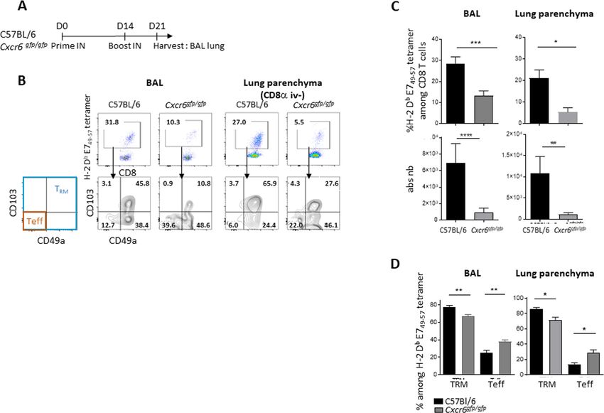
Open access
J Immunother Cancer: first published as 10.1136/jitc-2020-001948 on 10 March 2021. Downloaded from http://jitc.bmj.com/ on October 22, 2021 by guest. Protected by copyright.
in order to amplify the local immune response.5 These CXCR6-deficient mice, in which the coding region of the
TRM cells appear to be more cytotoxic than other CD8+ T chemokine receptor CXCR6 has been substituted with
effector cells, as CD103 expression is correlated with the the coding region of the Enhanced Green Fluorescent
levels of granzymes.6 Protein (eGFP). These mice were obtained from Jackson
The role of TRM in antitumor immunity has recently Laboratory (cat# JAX: 005693) and bred in our animal
been highlighted. For example, it has been shown in mice facility. Male CXCR6gfp/gfp mice were crossed with female
that these lymphocytes delay tumor progression.7 8 Our C57BL/6J mice to generate CXCR6gfp/+ mice. B6.SJL-
team has shown that TRM specifically induced by intra- PtprcaPepcb/BoyJ (CD45.1) were purchased from Charles
nasal vaccination plays an essential role in controlling River. CD3 knockout (KO) mice were obtained from B
the growth of orthotopic murine head and neck or lung Malissen’s laboratory (CIML, Marseille, France) and bred
tumors.9 10 Other studies have identified TRM as a key medi- in our animal facility.
ator of cancer vaccines targeting mucosal tumors.11 12 Mice were used in experiments at 8–10 weeks of age.
In humans, TRM has been found in different types of All mice were housed in INSERM U970-PARCC animal
cancer, such as melanoma, urothelial carcinoma, endome- facility under specific pathogen-free conditions.
trial adenocarcinoma, and particularly lung cancer.9 13 14
Our team and other groups have shown that a larger Tumor cells
CD103+CD8+ T-cell infiltrate was associated with better TC-1 cells expressing the Human Papillomavirus
survival in lung cancer, even in multivariate analysis.6 9 13 (HPV)16 E6–E7 proteins were obtained from the labo-
In the present study, we wanted to analyze the mechanism ratory of T C Wu (Department of Pathology, School of
by which intranasal vaccination promotes preferential Medicine, Johns Hopkins University, Baltimore, Mary-
intratumoral infiltration of TRM in head and neck cancer land, USA). Cells were cultured in RPMI 1640 (Life Tech-
and lung tumors, compared with systemic vaccination. nologies) supplemented with 10% heat-inactivated fetal
Previous results, based on phenotypical and transcrip- calf serum (FCS, GE Healthcare), 1 mM sodium pyruvate
tomic analysis, from our team and other groups, have (Life Technologies), 1 mM non- essential amino acids
identified CXCR6 chemokine receptor as a core marker (Life Technologies), 100 U/mL penicillin and 100 µg/
of TRM present in lung or head and neck tumors, but its mL streptomycin (Life Technologies), and 0.5 mM 2-β
function has not been investigated.6 9 15–18 mercaptoethanol (Life Technologies), and incubated at
CXCR6 binds a unique ligand, CXCL16, which can 37°C in 5% CO2. They were regularly tested for myco-
exist in transmembrane and soluble forms,19 the latter plasma contamination.
requiring cleavage from the membrane by a disintegrin
and metalloproteinase domain-10, or a disintegrin and Vaccine and adjuvant
metalloproteinase domain-17.20 CXCL16 is produced by The STxB- E7 vaccine was produced by the chemical
epithelial and immune cells and can serve as an alarmin coupling of the N- bromoacetylated E743–57 peptide to
to recruit cells to the site of inflammation.21 22 the sulfhydryl group of a recombinant nontoxic Shiga
In contrast with TRM, CXCR6 is detected at very low levels toxin B-subunit variant according to previously described
on naive CD8+ T cells19 and is upregulated by priming by procedures.33 After purification, endotoxin concen-
dendritic cells,23 or after T-cell receptor activation.24 trations determined by the Limulus assay test (Lonza,
CXCR6 promotes homing of lymphocytes to non- Aubergenville, France) were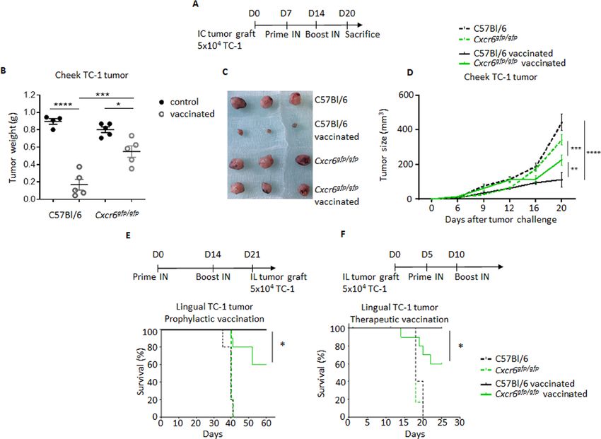
Open access
J Immunother Cancer: first published as 10.1136/jitc-2020-001948 on 10 March 2021. Downloaded from http://jitc.bmj.com/ on October 22, 2021 by guest. Protected by copyright.
10 after tumor graft for tongue tumor and at D7 and D14 BV711 (clone SA051D1) (all Abs from Biolegend). Then
for cheek tumor. staining was performed as described previously. All the
The mean survival time was calculated using the Kaplan– cells were labeled using the live/dead cell aqua blue
Meier method and statistical analysis was performed viability (Life Technologies).
using a log-rank test. Analysis of differences in tumor Non-circulating CD8+ T cells were defined as CD3+C-
volume were performed with two-way analysis of variance D8anegCD8b+. Acquisitions were performed on BD
(ANOVA) and Bonferroni or Tukey post hoc test. Fortessa X20 (Becton Dickinson), and data were analyzed
on live singulet cells with FlowJo Software (BD).
Isolation of lymphocytes from bronchoalveolar lumen fluid,
lung parenchyma, tumors and spleen Chemokines multiplex assays and ELISA
Intravascular staining was performed to discrimi- Supernatants from the first BAL’s washes were collected.
nate between tissue- localized and blood- borne cells as Serum samples were prepared from blood collected
described by Anderson et al.34 Briefly, 5 µg of anti-CD8a from retro-orbital sinus. Proteins were extracted from
APC-efluo780 (clone 53-6-7, ebioscience/Thermofisher) perfused lungs and from spleen by using Bioplex lysis
was injected intravenously 3 min prior to bronchoalveolar buffer (Bio-Rad), and total proteins were determined by
lavage (BAL) and tissue harvest. bicinchoninic acid (BCA) assay (Pierce) according to the
Bronchoalveolar lavage (BAL) was obtained by manufacturer’s protocol.
flushing the lungs with phosphate-buffered saline(PBS)- Chemokines was measured by bead- based multi-
EDTA 0.5 mM via a cannula inserted in the trachea (5 plex immunoassay: CXCL16, MIP1a/CCL3,MIP1b/
washes×1 mL). CCL4,RANTES/CCL5,IP10/CXCL10, CCL20 (R&D
Lungs were perfused with PBS- EDTA 0.5 mM and Systems Biotechne), according to manufacturer protocol
digested in RPMI-1640 medium supplemented with and were analyzed on Bio- Plex 200 (Bio-Rad). The
1 mg/mL collagenase type IV (Life Technologies/Ther- analyte concentration was calculated using a standard
mofisher) and 30 µg/mL DNase I (Roche). Lungs were
curve (5 Parameter logistic (PL) regression), with Bio-
dissociated using the GentleMACS (Miltenyi Biotec,
Plex manager software. When indicated, CXCL16 was
France) lung programs 1 and 2, with gentle shaking
measured by ELISA (R&D Systems Biotechne) according
at 37°C for 30 min in between both steps. Then, the
to manufacturer protocol.
obtained single‐cell suspensions were filtered through a
70 μm strainer washed with PBS–FBS 2%, suspended in
40% Percoll solution and layered over 75% Percoll solu- Transcriptomic analysis
tion (Sigma-Aldrich), and centrifuged at 600×g for 20 min C57BL/6 mice were immunized with STxB-E7 (20 µg)+α-
at room temperature (RT). Interface cells were collected GalCer (2 µg) by intranasal or intramuscular route and
and washed. Tumors were harvested, minced and placed then immunized a second time with STxB- E7 (20 µg)
into GentleMACS C-tube with PBS-FCS 2%, dissociated 14 days later (prime-boost). Seven days after the second
mechanically with GentleMACS dissociator (Miltenyi) immunization, the BAL or spleen of the mice were recov-
according to the manufactor’s standard protocol, then ered, and the CD44hitetramer+CD8+T cells were sorted
filtered on a 70 μm strainer. by BD FACSAria II cell sorter. Then, RNA of the cells
Spleens were dissected and pressed through a 40 µM was extracted using the QIAGEN RNeasy Plus Micro Kit
cell strainer, red blood cells were lysed with osmotic lysis and then genotyped by DNA chip (Affymetrix) by the
buffer. genomics and transcriptomics platform of the Cochin
Cheek tumors were harvested and mechanically dissoci- Institute.
ated by using the GentleMACS (Miltenyi, Bergisch Glad- Data were normalized using Robust Multiarray Aver-
bach, Germany) (program m- imp-tumor-01–01), then aging (RMA) algorithm in Bioconductor with the custom
filtered on a 70 µm strainer. CDF vs 18 (Dai M et al Nucleic Acids Res 2005). Statis-
tical analysis was carried out with the use of Partek GS.
Flow cytometry All the data have been deposited in NCBI’s Gene Expres-
After FcR blocking with CD16/32 Ab (clone 93, ebiosci- sion Omnibus44 and are accessible through GEO Series
ence/ Life Technologies), cells were first incubated for accession number GSE77366 (http://www.ncbi.nlm.nih.
30 min at RT with PE-conjugated DbE749–57 (R9F) tetramer gov/ g eo/ q uery/ a cc. c gi? t oken= g hsrcweibruzvsj& a cc=
(Immudex). Then, cells were washed and stained 20 min GSE77366).
at 4°C with the following Abs : anti-mouse CD8b AF700 First, variations in gene expression were analyzed
or BUV495 (clone YTS156), CD3 PercpCy5.5 (clone 145 using unsupervised hierarchical clustering and PCA to
2C11, ebioscience/Life Technologies), CD103 Pacific assess data from technical bias and outlier samples. To
Blue (clone 2E7, Biolegend), and CD49a Alexa 647 find differentially expressed genes, we applied one-way
(clone Ha31/8, BD Biosciences). For chemokine receptor ANOVA for each gene and made pairwise Tukey’s post
analysis, cells were stained 30 min at RT with anti-mouse hoc tests between groups. Then, we used p values and
CCR5 PE-CY7 (clone HM-CCR5), CCR6 BV785 (clone fold changes to filter and select differentially expressed
29–2 L17), CXCR3 BV650 (clone CXCR3-173), CXCR6 genes. Differentially expressed genes (DEG) enrichment
Karaki S, et al. J Immunother Cancer 2021;9:e001948. doi:10.1136/jitc-2020-001948 3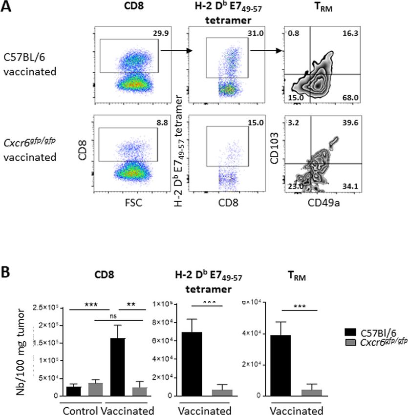
Open access
J Immunother Cancer: first published as 10.1136/jitc-2020-001948 on 10 March 2021. Downloaded from http://jitc.bmj.com/ on October 22, 2021 by guest. Protected by copyright.
analysis was carried out using Ingenuity (Ingenuity
Systems, USA; www.ingenuity.com).
Data availability
All data were deposited in NCBI’s Gene Expression
Omnibus and are accessible through GEO Series acces-
sion number GSE77366 (http://www.ncbi.nlm.nih.
gov/ g eo/ q uery/ a cc. c gi? t oken= g hsrcweibruzvsj& a cc=
GSE77366).
Human lung cancer
Lung cancer biopsies were obtained from non-treated
patients with non-small-cell lung carcinoma who under-
went a lobectomy at the thoracic surgery department of
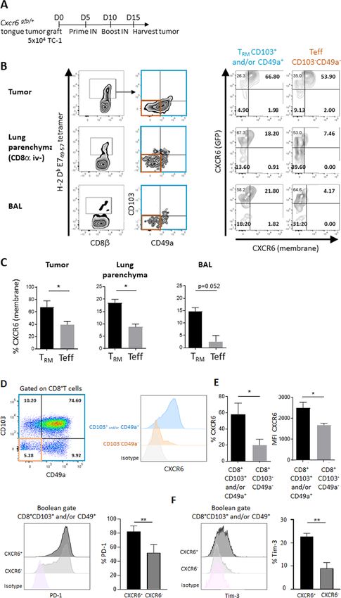
European Georges Pompidou Hospital. Briefly, biopsies Figure 1 Cxcr6 upregulation on CD8+ T cells with a TRM
were digested for 45 min at 37°C with DNAse I (30 IU/ phenotype induced by intranasal vaccination revealed by
mL, Roche) and collagenase type IV (1 mg/mL, Life heat MAP analysis. Mice were vaccinated via intranasal or
Technologies/Thermofisher), then filtered through a intramuscular routes (n=4 mice per group) with STxB-E7
70 µm strainer washed with PBS–FCS 2%. Flow cytometry and the adjuvant α-GalCer (prime D0) and boosted at D14
with STxB-E7. BAL from in vaccinated mice and spleens
analysis of tumor-infiltrating lymphocytes was performed.
from intramuscularly vaccinated mice were collected at
: Receptors for the Fc region (FcRs) were first blocked day 21. H2-Db E749-57 tetramer+ CD44hi CD8+ T cells were
with Human TruStain FcX (Biolegend) 5 min at RT, then sorted and the RNA was extracted. Whole gene expression
stained with anti-human CD3 APC-CY7 (clone HIT-3a, microarray analysis was performed according to the
Biolegend), CD8 BUV395 (clone RPA- T8, BD Biosci- procedure described in the methods section. Left: total gene
ences), CD103 Percpcy5.5 (clone Ber-ACT8, Biolegend), analysis already reported in Nizard et al9 and not detailed
CD49a PE (clone TS2-7, Biolegend), PD1 BV650 (clone here. Rright: focus on the expression of CXCR6 by TRM
EH12.2H7, Biolegend), CXCR6 biotin (clone K041E5, cells. Top: BAL CD8+ T cells (lanes 1–3) showed a typical
Biolegend) and steptavidin BV711 (Biolegend). All cells TRM gene expression profile (CD103+ and CD49a+), which
was not observed in splenic CD8+ T cells (lanes 4–6) (red
were labeled using the live/dead cell aqua blue viability
square means overexpressed, while green square means
(Life Technologies). Acquisitions were performed on BD underexpressed). Bottom: table of the transcriptomic
Fortessa X20 (Becton Dickinson), and data were analyzed characteristics of the Itga1, Itgb1, Itgae and Cxcr6 RNAs, with
on live singulet cells with FlowJo Software (BD). their p value and ‘fold change’. The fold change represents
the difference in quantity between the genes expressed
Statistical analysis by CD8+ T cells from BAL after intranasal immunization
Data are presented as mean±SEM. Statistical compari- and from the spleen after intramuscular immunization.
sons were done with Prism V.8 GraphPad Software (San These extractions were repeated at least three times. BAL,
Diego, California, USA). Analysis of difference between bronchoalveolar lavage.
two groups was performed with Mann- Whitney t-
test;
analysis of difference between more than two groups
express CXCR6 compared with those induced in the
was performed with a one-way ANOVA and Holm-Sidak
spleen after intramuscular immunization (fold change
or Tukey post hoc. Mice survival was estimated using the
6.2, p=0.00035) (figure 1, right). As we have previously
Kaplan-Meier method and log-rank test. P values lower
reported, intramuscular vaccination did not induce TRM
than 0.05 (*) were considered significant.
in the BAL and we could not check for their expression
of CXCR6 after this route of immunization.9
RESULTS CXCR6 is preferentially expressed on intranasal vaccine-
CXCR6 expression is upregulated on CD8+ T cells with a TRM induced E7-specific CD8+ T cells displaying resident memory
phenotype induced by ntranasal vaccination T cell (TRM) phenotype
Our previous study showed that in contrast to the spleen- To investigate the potential role of CXCR6 in the estab-
derived E739–47-specific CD8+ T cells induced by intramus- lishment of lung- homing CD8+ T- cell populations, we
cular immunization, the BAL’s E749-57-specific CD8+ T cells used heterozygous Cxcr6 gfp/+ mice, in which one allele of
induced by intranasal immunization express the core gene the Cxcr6 gene has been replaced by the coding region of
defining TRM (Itgae encoding CD103, Itga1 encoding gfp.35 We also examined the expression of CXCR6 on lung
CD49a and Itgb1, which associates with Itga1 to form the resident CD8+ T cells at the protein levels using a recently
VLA-1 integrin).9 Here, we have completed this analysis available anti-CXCR6 antibody. In the first series of exper-
by a transcriptomic analysis, showing that E749–57-specific iments, Cxcr6gfp/+ mice were first grafted with TC-1 cells
CD8+ T cells with induced by intranasal immunization in in the tongue, then intranasally immunized with STxB-E7
the BAL and displaying a TRM phenotype preferentially at D5 and D10, and then tumor, BAL, lung and spleen
4 Karaki S, et al. J Immunother Cancer 2021;9:e001948. doi:10.1136/jitc-2020-001948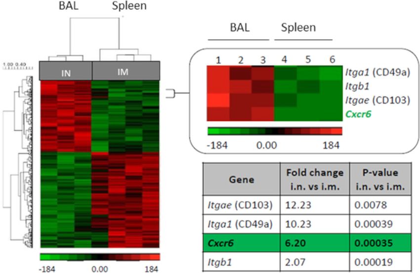
Open access
J Immunother Cancer: first published as 10.1136/jitc-2020-001948 on 10 March 2021. Downloaded from http://jitc.bmj.com/ on October 22, 2021 by guest. Protected by copyright.
were harvested at D15 (figure 2A). We then analyzed in the absolute number of anti-E749-57 CD8+ T cells in the
CXCR6 expression on E749-57-specific CD8+ T cells with BAL compared with mice which had received T cells
a TRM phenotype, defined as CD8+ T cells expressing derived from wild-type mice. A similar trend exists in the
CD103 and/ or CD49a as previously reported,9 or with pulmonary parenchyma. This result confirms the intrinsic
an effector phenotype without the expression of these role of Cxcr6 deficiency in T cells on the decrease of the
markers. CXCR6 was analyzed by flow cytometry based local TRM after vaccination by the intranasal route (online
on GFP expression or with the use of anti-CXCR6 mAb supplemental figure S3).
(figure 2B).9 In tumor, lung parenchyma and BAL, we
found a statistically significant increase in membrane CXCR6 deficiency partially reverses cancer vaccine control of
CXCR6 expression on E7-specific CD8+ T cells with a TRM tumor growth in orthotopic head and neck tumor models
phenotype, when compared with cells with an effector To assess the consequence of Cxcr6 deficiency on the
phenotype (figure 2C). recruitment of CD8+ T cells in the tumor microenvi-
Surprisingly no difference in the expression of GFP ronment and on the control of tumor growth, we set up
could be detected between the TRM and the effector T two orthotopic models using TC-1 tumor cells that were
cells in the various organs (online supplemental figure grafted in the cheek or in the tongue.
S1). Differences in the expression of GFP and membrane C57BL/6 wild- type or Cxcr6 gfp/gfp mice were chal-
CXCR6 had already been observed with these mice.36 We lenged with TC-1 cells in the submucosal lining of
considered that the membrane expression of CXCR6 was the cheek (intracheek) then vaccinated or not with
the most relevant cellular localization for the function of STxB-E7 by intranasal route at D7 and D14, and the
a chemokine receptor.
tumor volume was monitored (figure 4A). Vaccinated
Next, we validated this preferential expression of
C57BL/6 wild-type mice showed significant inhibition
CXCR6 on human TRM derived from lung tumors.
of tumor growth based on the measurement of tumor
Remarkably, 58% of intratumoral TRM expressed CXCR6,
weight or tumor size in comparison to non-vaccinated
while it is detected on only 20% of intratumoral effector
mice (figure 4B–D). In contrast, we observed a partial
CD8+ T cells (figure 2D,E). CXCR6 Mean fluorescence
loss of tumor control by the vaccine in Cxcr6 gfp/gfp mice
intensity (MFI) expression was also higher on TRM than
(figure 4B–D).
on effector cells (figure 2E). Phenotype characterization
To confirm these results in a second orthotopic tumor
of these CXCR6+ TRM showed that they express PD-1 more
model, C57BL/6 wild- type mice and Cxcr6gfp/gfp mice
strongly than CXCR6 negative TRM (figure 2F). These
were engrafted in the sublingual mucosa with TC-1 cells
results show that both mouse and human tumor TRM cells
and were vaccinated prophylactically (before the graft)
preferentially expressed CXCR6 compared with effector
(figure 4E) or therapeutically (after the graft) (figure 4F)
T cells.
with STxB-E7 by intranasal route.
CXCR6 deficiency impairs induction of antigen-specific CD8+ In both settings, all vaccinated C57BL/6 wild-type mice
T cells and TRM in the airway after intranasal vaccination were alive at D60 for the prophylactic model (figure 4E)
We then investigated the role of CXCR6 in the induction and at D25 for the therapeutic model (figure 4F). In
of E749–57-specific CD8+ T and TRM cells in BAL and spleen Cxcr6gfp/gfp, only 60% of vaccinated mice were alive in both
using mice lacking this chemokine receptor (Cxcr6gfp/gfp) models at the same time points. Survival curves indicate
(figure 3A). We observed a statistically significant reduc- that vaccinated Cxcr6gfp/gfp mice showed decreased survival
tion in the total number of E749–57-specific CD8+ T and compared with vaccinated C57BL/6 wild- type mice.
TRM cells in BAL and lung parenchyma of Cxcr6-deficient These results indicate that impairment of CXCR6 expres-
mice, when compared with C57BL/6 wild- type mice sion has a strong negative impact on antitumor immunity.
(figure 3B,C). This decrease is more pronounced on E7 We then addressed whether this partial loss of tumor
specific T cells with a TRM phenotype than with an effector growth control after vaccination is associated with a
phenotype (figure 3D). The latter population is even reduction of intratumoral infiltration by CD8+ T cells.
increased in Cxcr6-deficient mice compared with wild- Tumor- infiltrating lymphocytes were analyzed by flow
type mice (figure 3D). After vaccination, we did not find cytometry in C57BL/6 wild- type or Cxcr6ggfp/gfp mice
any difference in the E749-57 tetramer levels in the spleen engrafted with TC-1 tumor cells in the submucosal lining
between wild- type and Cxcr6gfg/gfpmice (online supple- of the cheek, and vaccinated or not with STxB-E7 at D7
mental figure S2A,B). Notably, CXCR6 membrane expres- and D14. A significant decrease in the absolute numbers
sion on E749-57 specific CD8+ T cells was not detected in of intratumoral CD8+ T cells, total H-2Db-E749-57 CD8+ T
the spleen of Cxcr6gfp/+ mice (online supplemental figure cells, and H-2Db-E749-57 TRM cells was observed in Cxcr6gfp/
gfp
S2C). mice, when compared with C57BL/6 wild-type mice
As Cxcr6 deficiency is not restricted to T cells only, we (figure 5A,B). To reinforce the role of this impaired
performed a transfer experiment, in CD3-deficient mice, migration on CD8+ T cells in Cxcr6-deficient mice on anti-
with T cells derived from wild- type mice (CD45.1) or tumor immunity, we showed that these specific CD8+ T
Cxcr6-deficient mice (CD45.2). We show that mice which cells did not exhibit defaults in cytotoxicity mechanisms
had received the Cxcr6-deficient T cells had a decrease (online supplemental figure S4).
Karaki S, et al. J Immunother Cancer 2021;9:e001948. doi:10.1136/jitc-2020-001948 5Open access
J Immunother Cancer: first published as 10.1136/jitc-2020-001948 on 10 March 2021. Downloaded from http://jitc.bmj.com/ on October 22, 2021 by guest. Protected by copyright.
Figure 2 Cxcr6 (membrane expression) is preferentially expressed on CD8+ resident memory T cells in mice and humans.
(A) Cxcr6gfp/+ mice were grafted with TC-1 cells (5×10⁴) in the tongue at D0, then vaccinated via the in route with STxB-E7 and
poly-ICLC at D5 and D10 and sacrificed at D15. (B) Gating strategy for TRM (in blue: Boolean gate CD103+ and/or CD49a+) and
Teff (in orange: CD103−CD49a−). Representative flow plots showing GFP expression versus surface expression of CXCR6 on
TRM and Teff. (C) Percentage of CXCR6 membrane expression in tumor, lung parenchyma (CD8a IV−) and BAL. Mean±SEM,
n=4/group. Results are representative of two experiments. (D–F) Fresh biopsies from a patient with lung cancer (n=4) were
dissociated and digested, and flow cytometry analysis of tumor-infiltrating lymphocytes was then performed. (D) Representative
flow plot of CD103 and CD49a expression and gating strategy on live CD8. (E) Representative histograms of CXCR6
expression (left), percentage (middle) and MFI (right) of CXCR6 with TRM phenotype (Boolean gate CD103+ and/or CD49a+) and
CD103−CD49a−CD8+ T cells. (F) Representative histograms (left) and percentage of PD1 (right) and Tim-3 (left) among CXCR6+
and CXCR6− TRM. Mean±SEM paired t-test. Poly-ICLC, polyinosinic-polycytiylic acid-poly-l-lysine carboxymethylcellulose. GFP,
Green Fluorescent Protein. *POpen access
J Immunother Cancer: first published as 10.1136/jitc-2020-001948 on 10 March 2021. Downloaded from http://jitc.bmj.com/ on October 22, 2021 by guest. Protected by copyright.
Figure 3 Induction of specific CD8+ T cells and TRM in the lung and airway is impaired in Cxcr6-deficient mice (A) C57BL/6 or
Cxcr6gfg/gfp mice were vaccinated with STxB-E7 via the in route at days 0 and 14, then sacrificed at day 21. (B) Representative
flow plots of H-2 Db E749-57 tetramer gated on CD8+ T cells (top), and TRM (Boolean gate CD103+ and/or CD49a+) and effector T
cells (teff) (CD103−CD49a−) in BAL and lung parenchyma gated on H-2 Db E749-57 tetramer (bottom). (C) Percentage and absolute
number of H-2 Db E749-57 tetramer in BAL and lung parenchyma. (D) Percentage of TRM and teff in BAL (n=21–22 mice/group) and
lung parenchyma (n=5–7 mice/group). these experiments were repeated three times. Mean±SEM Mann-Whitney t-test. *POpen access
J Immunother Cancer: first published as 10.1136/jitc-2020-001948 on 10 March 2021. Downloaded from http://jitc.bmj.com/ on October 22, 2021 by guest. Protected by copyright.
Figure 4 Cxcr6 deficiency impairs tumor control in orthotopic tumors. (A) C57BL/6 or Cxcr6gfg/gfp mice were grafted in the
submucosal lining of the cheek (IC) with TC-1 tumor cells, then vaccinated or not with STxB-E7 via the in route at days 7 and
14, and sacrificed at day 20. (B–D) Tumor weight (B,C) and tumor size (D) were measured at day 20. (D,E) In a second orthotopic
model, TC-1 was grafted in the sublingual mucosa (IL); mice were then vaccinated following a prophylactic (E) or therapeutic
protocol (F), and survival was monitored. All data are representative from two independent experiments. Five mice/group (B–D)
and 10 mice/group (E,F). Mean±SEM analysis of within-group differences was performed with a two-way analysis of variance
and post hoc Tukey test (B–D), and survival was compared between groups with a Kaplan-Meier curve (log-rank test) (E,F).
*POpen access
J Immunother Cancer: first published as 10.1136/jitc-2020-001948 on 10 March 2021. Downloaded from http://jitc.bmj.com/ on October 22, 2021 by guest. Protected by copyright.
Figure 5 Cxcr6 deficiency impairs CD8+ T-cell infiltration in cheek tumor after in vaccination analysis of CD8 and H-2 Db E749-57
specific cells infiltrating a TC-1 cheek tumor in C57BL/6 and Cxcr6gfg/gfp mice, vaccinated or not with STxB-E7 via the intranasal
route (D7 and D14) at day 20 after tumor graft. (A) Representative flow plots and (B) total number of CD8 T cells, H-2 Db E749-57
tetramer and TRM (Boolean gate CD103+ and/or CD49a+). Data are representative of two independent experiments. Five mice/
group. Mean±SEM analysis of difference within groups was performed with a one-way analysis of variance and post hoc Tukey
test, and between two groups with Mann-Whitney t-test. **POpen access
J Immunother Cancer: first published as 10.1136/jitc-2020-001948 on 10 March 2021. Downloaded from http://jitc.bmj.com/ on October 22, 2021 by guest. Protected by copyright.
Figure 6 Recombinant CXCL16 did not amplify recruitment of TRM likely due to endogenous CXCL16 induced after
vaccination. (A) C57BL/6 mice were vaccinated with STxB-E7 via the in route at days 0 and 14 in the presence or absence of
recombinant CXCL16 (2 mg), then sacrificed at day 21. (B) Percentage and absolute number of specific H-2 Db E749-57 tetramer
within CD8 T cells (left) and TRM within tetramer (right) in the BAL are shown. n=8 mice/group. (C,D) C57BL/6 mice were
vaccinated with STxB-E7 (20 µg)+poly ICLC (10 µg) via the intranasal or intramuscular route. CXCL16 was measured by ELISA
at the indicated times after vaccination in BAL (C) and lysate from the lung (D). n=4 mice/group. The experiments were repeated
twice. **POpen access
J Immunother Cancer: first published as 10.1136/jitc-2020-001948 on 10 March 2021. Downloaded from http://jitc.bmj.com/ on October 22, 2021 by guest. Protected by copyright.
LMU Munich, Germany, Member of the German Center for Lung Research (DZL), 4 Mami-Chouaib F, Blanc C, Corgnac S, et al. Resident memory T
Munchen, Germany cells, critical components in tumor immunology. J Immunother
13
German Center for Translational Cancer Research (DKTK), partner site, Munchen, Cancer 2018;6:87.
5 Schenkel JM, Masopust D. Tissue-resident memory T cells. Immunity
Germany
14 2014;41:886–97.
INSERM UMR 1186, Institut Gustave Roussy, Faculté de Médecine-Université 6 Ganesan A-P, Clarke J, Wood O, et al. Tissue-resident memory
Paris-Sud, Université Paris-Saclay, 94805 Villejuif, France features are linked to the magnitude of cytotoxic T cell responses in
15
Unit for Lymphopoiesis, Department of Immunology, Institut Pasteur, INSERM human lung cancer. Nat Immunol 2017;18:940–50.
U1223, 75006 Paris, France 7 Dadi S, Chhangawala S, Whitlock BM, et al. Cancer
immunosurveillance by tissue-resident innate lymphoid cells and
innate-like T cells. Cell 2016;164:365–77.
Acknowledgements We thank the staff of the European Georges Pompidou 8 Park SL, Buzzai A, Rautela J, et al. Tissue-resident memory CD8+
Hospital tumor banks (D Geromin and M Largeau) for providing sample materials T cells promote melanoma-immune equilibrium in skin. Nature
and the PARCC Histology (C Lesaffre) and cytometry (C Knosp) platform as well as 2019;565:366–71.
animal facilities (C Suldac and N Perez) 9 Nizard M, Roussel H, Diniz MO, et al. Induction of resident memory
T cells enhances the efficacy of cancer vaccine. Nat Commun
Contributors IG-F, AM, ED, MA, and LP performed experimental work and analyzed
2017;8:15221.
and interpreted the data. SK, CB, and TT performed experimental work, analyzed 10 Sandoval F, Terme M, Nizard M, et al. Mucosal imprinting of vaccine-
and interpreted the data, and designed the figures. EF, LG, FL-B, CB, DD, and NB induced CD8⁺ T cells is crucial to inhibit the growth of mucosal
contributed to sample collection and analyzed and interpreted the data. NG, CT, ED, tumors. Sci Transl Med 2013;5:172ra20.
SK, and FM-C analyzed and interpreted the data. LJ and RG designed the study and 11 Çuburu N, Graham BS, Buck CB, et al. Intravaginal immunization
revised the manuscript. TE designed the study, supervised the project, and wrote with HPV vectors induces tissue-resident CD8+ T cell responses. J
the manuscript. All authors were involved in the final approval of the manuscript. Clin Invest 2012;122:4606–20.
12 Sun Y-Y, Peng S, Han L, et al. Local HPV recombinant vaccinia boost
Funding This work has been funded by Fondation ARC, INCA PLBio, Labex following priming with an HPV DNA vaccine enhances local HPV-
Immuno-Oncology, Site intégré de recherche en cancérologie (SIRIC CARPEM, SIRIC Specific CD8+ T-cell-mediated tumor control in the genital tract. Clin
Curie), Cancéropole d’Ile de France, Carnot Curie Cancer, FONCER. SK is supported Cancer Res 2016;22:657–69.
by the European Research Council (grant 756017, ARMOR-T). 13 Djenidi F, Adam J, Goubar A, et al. CD8+CD103+ tumor-infiltrating
lymphocytes are tumor-specific tissue-resident memory T cells and
Competing interests None declared. a prognostic factor for survival in lung cancer patients. J Immunol
Patient consent for publication Not required. 2015;194:3475–86.
14 Blanc C, Hans S, Tran T, et al. Targeting resident memory T cells for
Ethics approval Experimental protocols in mice were approved by Paris Descartes cancer immunotherapy. Front Immunol 2018;9:9.
University ethical committee (CEEA 34) in accordance with European guidelines 15 Morgan AJ, Guillen C, Symon FA, et al. CXCR6 identifies a
(EC2010/63). The human study was approved by the local ethics committee (CPP putative population of retained human lung T cells characterised
Tours. CNRIPH n°18.11.21.67518. 05.03.2019). Informed consent was obtained by co-expression of activation markers. Immunobiology
from all subjects. 2008;213:599–608.
16 Kumar BV, Ma W, Miron M, et al. Human tissue-resident memory T
Provenance and peer review Not commissioned; externally peer reviewed. cells are defined by core transcriptional and functional signatures in
Data availability statement Data are available in a public, open access repository. lymphoid and mucosal sites. Cell Rep 2017;20:2921–34.
17 Mackay LK, Minnich M, Kragten NAM, et al. Hobit and Blimp1
The data are available upon request from ET (eric.tartouraphp.fr).
instruct a universal transcriptional program of tissue residency in
Supplemental material This content has been supplied by the author(s). It has lymphocytes. Science 2016;352:459–63.
not been vetted by BMJ Publishing Group Limited (BMJ) and may not have been 18 Guo X, Zhang Y, Zheng L, et al. Global characterization of T cells
peer-reviewed. Any opinions or recommendations discussed are solely those in non-small-cell lung cancer by single-cell sequencing. Nat Med
of the author(s) and are not endorsed by BMJ. BMJ disclaims all liability and 2018;24:978–85.
19 Matloubian M, David A, Engel S, et al. A transmembrane CXC
responsibility arising from any reliance placed on the content. Where the content
chemokine is a ligand for HIV-coreceptor Bonzo. Nat Immunol
includes any translated material, BMJ does not warrant the accuracy and reliability 2000;1:298–304.
of the translations (including but not limited to local regulations, clinical guidelines, 20 Abel S, Hundhausen C, Mentlein R, et al. The transmembrane
terminology, drug names and drug dosages), and is not responsible for any error CXC-chemokine ligand 16 is induced by IFN-gamma and TNF-alpha
and/or omissions arising from translation and adaptation or otherwise. and shed by the activity of the disintegrin-like metalloproteinase
ADAM10. J Immunol 2004;172:6362–72.
Open access This is an open access article distributed in accordance with the
21 Day C, Patel R, Guillen C, et al. The chemokine CXCL16 is highly
Creative Commons Attribution Non Commercial (CC BY-NC 4.0) license, which and constitutively expressed by human bronchial epithelial cells. Exp
permits others to distribute, remix, adapt, build upon this work non-commercially, Lung Res 2009;35:272–83.
and license their derivative works on different terms, provided the original work is 22 Tabata S, Kadowaki N, Kitawaki T, et al. Distribution and kinetics of
properly cited, appropriate credit is given, any changes made indicated, and the use SR-PSOX/CXCL16 and CXCR6 expression on human dendritic cell
is non-commercial. See http://c reativecommons.org/licenses/by-nc/4.0 /. subsets and CD4+ T cells. J Leukoc Biol 2005;77:777–86.
23 Kim CH, Kunkel EJ, Boisvert J, et al. Bonzo/CXCR6 expression
ORCID iDs defines type 1-polarized T-cell subsets with extralymphoid tissue
Emmanuel Donnadieu http://orcid.org/0000-0002-4985-7254 homing potential. J Clin Invest 2001;107:595–601.
Eric Tartour http://orcid.org/0000-0002-7 323-468X 24 Heesch K, Raczkowski F, Schumacher V, et al. The function of the
chemokine receptor CXCR6 in the T cell response of mice against
Listeria monocytogenes. PLoS One 2014;9:e97701.
25 Zaid A, Hor JL, Christo SN, et al. Chemokine receptor-dependent
control of skin tissue-resident memory T cell formation. J Immunol
2017;199:2451–9.
REFERENCES 26 Billerbeck E, Kang Y-H, Walker L, et al. Analysis of CD161
1 Stolley JM, Johnston TS, Soerens AG, et al. Retrograde migration expression on human CD8+ T cells defines a distinct functional
supplies resident memory T cells to lung-draining ln after subset with tissue-homing properties. Proc Natl Acad Sci U S A
influenza infection. J Exp Med 2020;217:e20192197. doi:10.1084/ 2010;107:3006–11.
jem.20192197 27 Nanki T, Shimaoka T, Hayashida K, et al. Pathogenic role of the
2 Fonseca R, Beura LK, Quarnstrom CF, et al. Developmental plasticity CXCL16-CXCR6 pathway in rheumatoid arthritis. Arthritis Rheum
allows outside-in immune responses by resident memory T cells. Nat 2005;52:3004–14.
Immunol 2020;21:412–21. 28 Fukumoto N, Shimaoka T, Fujimura H, et al. Critical roles of
3 Beura LK, Mitchell JS, Thompson EA, et al. Intravital mucosal CXC chemokine ligand 16/scavenger receptor that binds
imaging of CD8+ resident memory T cells shows tissue-autonomous phosphatidylserine and oxidized lipoprotein in the pathogenesis
recall responses that amplify secondary memory. Nat Immunol of both acute and adoptive transfer experimental autoimmune
2018;19:173–82. encephalomyelitis. J Immunol 2004;173:1620–7.
Karaki S, et al. J Immunother Cancer 2021;9:e001948. doi:10.1136/jitc-2020-001948 11Open access
J Immunother Cancer: first published as 10.1136/jitc-2020-001948 on 10 March 2021. Downloaded from http://jitc.bmj.com/ on October 22, 2021 by guest. Protected by copyright.
29 Hombrink P, Helbig C, Backer RA, et al. Programs for the 40 Lee LN, Ronan EO, de Lara C, et al. CXCR6 is a marker for protective
persistence, vigilance and control of human CD8+ lung-resident antigen-specific cells in the lungs after intranasal immunization
memory T cells. Nat Immunol 2016;17:1467–78. against Mycobacterium tuberculosis. Infect Immun 2011;79:3328–37.
30 Mackay LK, Rahimpour A, Ma JZ, et al. The developmental pathway 41 Kohlmeier JE, Reiley WW, Perona-Wright G, et al. Inflammatory
for CD103(+)CD8+ tissue-resident memory T cells of skin. Nat chemokine receptors regulate CD8(+) T cell contraction and memory
Immunol 2013;14:1294–301. generation following infection. J Exp Med 2011;208:1621–34.
31 Sowell RT, Goldufsky JW, Rogozinska M, et al. Il-15 complexes 42 Jiang JQ, He X-S, Feng N, et al. Qualitative and quantitative
induce migration of resting memory CD8 T cells into mucosal tissues. characteristics of rotavirus-specific CD8 T cells vary depending on
J Immunol 2017;199:2536–46. the route of infection. J Virol 2008;82:6812–9.
32 Agostini C, Cabrelle A, Calabrese F, et al. Role for CXCR6 and its 43 Takamura S, Kato S, Motozono C, et al. Interstitial-resident memory
ligand CXCL16 in the pathogenesis of T-cell alveolitis in sarcoidosis. CD8+ T cells sustain frontline epithelial memory in the lung. J Exp
Am J Respir Crit Care Med 2005;172:1290–8. Med 2019;216:2736–47.
33 Pere H, Montier Y, Bayry J, et al. A CCR4 antagonist combined with 44 Ashhurst AS, Flórido M, Lin LCW, et al. CXCR6-Deficiency Improves
vaccines induces antigen-specific CD8+ T cells and tumor immunity the Control of Pulmonary Mycobacterium tuberculosis and Influenza
against self antigens. Blood 2011;118:4853–62. Infection Independent of T-Lymphocyte Recruitment to the Lungs.
34 Anderson KG, Mayer-Barber K, Sung H, et al. Intravascular staining Front Immunol 2019;10:339.
45 Matsumura S, Wang B, Kawashima N, et al. Radiation-induced
for discrimination of vascular and tissue leukocytes. Nat Protoc
CXCL16 release by breast cancer cells attracts effector T cells. J
2014;9:209–22.
Immunol 2008;181:3099–107.
35 Unutmaz D, Xiang W, Sunshine MJ, et al. The primate lentiviral
46 Geissmann F, Cameron TO, Sidobre S, et al. Intravascular immune
receptor Bonzo/STRL33 is coordinately regulated with CCR5 and
surveillance by CXCR6+ NKT cells patrolling liver sinusoids. PLoS
its expression pattern is conserved between human and mouse. J Biol 2005;3:e113.
Immunol 2000;165:3284–92. 47 Takamura S, Yagi H, Hakata Y, et al. Specific niches for lung-resident
36 Wein AN, McMaster SR, Takamura S, et al. CXCR6 regulates memory CD8+ T cells at the site of tissue regeneration enable CD69-
localization of tissue-resident memory CD8 T cells to the airways. J independent maintenance. J Exp Med 2016;213:3057–73.
Exp Med 2019;216:2748–62. 48 Hu-Lieskovan S, Lisberg A, Zaretsky JM, et al. Tumor characteristics
37 Jeyanathan M, Afkhami S, Khera A, et al. Cxcr3 signaling is associated with benefit from pembrolizumab in advanced non-small
required for restricted homing of parenteral tuberculosis vaccine- cell lung cancer. Clin Cancer Res 2019;25:5061–8.
induced T cells to both the lung parenchyma and airway. J Immunol 49 Schmidt ME, Varga SM. The CD8 T cell response to respiratory virus
2017;199:2555–69. infections. Front Immunol 2018;9:678.
38 Kohlmeier JE, Miller SC, Smith J, et al. The chemokine receptor 50 Stary G, Olive A, Radovic-Moreno AF, et al. Vaccines. A mucosal
CCR5 plays a key role in the early memory CD8+ T cell response to vaccine against Chlamydia trachomatis generates two waves of
respiratory virus infections. Immunity 2008;29:101–13. protective memory T cells. Science 2015;348:aaa8205.
39 Tse S-W, Radtke AJ, Espinosa DA, et al. The chemokine receptor 51 Edwards J, Wilmott JS, Madore J, et al. CD103+ Tumor-
CXCR6 is required for the maintenance of liver memory Resident CD8+ T Cells Are Associated with Improved Survival in
CD8⁺ T cells specific for infectious pathogens. J Infect Dis Immunotherapy-Naïve Melanoma Patients and Expand Significantly
2014;210:1508–16. During Anti-PD-1 Treatment. Clin Cancer Res 2018;24:3036–45.
12 Karaki S, et al. J Immunother Cancer 2021;9:e001948. doi:10.1136/jitc-2020-001948You can also read