Functional Characterization of Multiple Ehrlichia chaffeensis Sodium (Cation)/Proton Antiporter Genes Involved in the Bacterial pH Homeostasis
←
→
Page content transcription
If your browser does not render page correctly, please read the page content below
International Journal of
               Molecular Sciences

Article
Functional Characterization of Multiple Ehrlichia chaffeensis
Sodium (Cation)/Proton Antiporter Genes Involved in the
Bacterial pH Homeostasis
Lanjing Wei, Huitao Liu, Kimia Alizadeh, Maria D. Juarez-Rodriguez and Roman R. Ganta *

                                           Center of Excellence for Vector-Borne Diseases, Department of Diagnostic Medicine/Pathobiology,
                                           College of Veterinary Medicine, Kansas State University, New York, NY 66506, USA; lanjingwei@ku.edu (L.W.);
                                           hliu@vet.k-state.edu (H.L.); kimia@vet.k-state.edu (K.A.); mdjuarezro@icloud.com (M.D.J.-R.)
                                           * Correspondence: rganta@vet.k-state.edu; Tel.: +1-785-532-4612

                                           Abstract: Ehrlichia chaffeensis causes human monocytic ehrlichiosis. Little is known about how this
                                           and other related tick-borne rickettsia pathogens maintain pH homeostasis in acidified phagosomes
                                           and the extracellular milieu. The membrane-bound sodium (cation)/proton antiporters are found in
                                           a wide range of organisms aiding pH homeostasis. We recently reported a mutation in an antiporter
                                           gene of E. chaffeensis (ECH_0379) which causes bacterial in vivo attenuation. The E. chaffeensis genome
                                           contains 10 protein coding sequences encoding for predicted antiporters. We report here that nine
                                           of these genes are transcribed during the bacterial growth in macrophages and tick cells. All E.
                                 chaffeensis antiporter genes functionally complemented antiporter deficient Escherichia coli. Antiporter

                                           activity for all predicted E. chaffeensis genes was observed at pH 5.5, while gene products of ECH_0179
Citation: Wei, L.; Liu, H.; Alizadeh,      and ECH_0379 were also active at pH 8.0, and ECH_0179 protein was complemented at pH 7.0.
K.; Juarez-Rodriguez, M.D.; Ganta,
                                           The antiporter activity was independently verified for the ECH_0379 protein by proteoliposome
R.R. Functional Characterization of
                                           diffusion analysis. This is the first description of antiporters in E. chaffeensis and demonstrates that the
Multiple Ehrlichia chaffeensis Sodium
                                           pathogen contains multiple antiporters with varying biological functions, which are likely important
(Cation)/Proton Antiporter Genes
                                           for the pH homeostasis of the pathogen’s replicating and infectious forms.
Involved in the Bacterial pH
Homeostasis. Int. J. Mol. Sci. 2021, 22,
8420. https://doi.org/10.3390/
                                           Keywords: functional genomics; rickettsial diseases; tick-borne diseases; antiporters; pH homeostasis
ijms22168420

Academic Editors: Anne M. Alvarez
and Mohammad Arif                          1. Introduction
                                                 Ehrlichia chaffeensis is an obligate intracellular Gram-negative rickettsial pathogen
Received: 2 July 2021                      and is the causal agent of monocytic ehrlichiosis in people and dogs [1–3]. The Lone-star
Accepted: 2 August 2021                    tick, Amblyomma americanum, is identified as the biological vector for transmitting E. chaf-
Published: 5 August 2021
                                           feensis [4], while white-tailed deer is the major reservoir host [5]. E. chaffeensis has two
                                           developmental forms in infected vertebrate and tick host cells [6,7]: the smaller dense-
Publisher’s Note: MDPI stays neutral       cored cells (DCs) and the larger and pleomorphic reticulate cells (RCs). The DC form is
with regard to jurisdictional claims in
                                           released from the infected host cells and infects naïve host cells by phagocytosis. The
published maps and institutional affil-
                                           DC form then transforms to the RC form, which replicates within the acidified phago-
iations.
                                           somes. RCs transform to DCs and are released from host cells by complete cell lysis or by
                                           exocytosis [6,7]. It is unclear how E. chaffeensis and other related rickettsia maintain pH
                                           homeostasis both within the acidified phagosomes and in the extracellular milieu where
                                           the pH is slightly basic [8].
Copyright: © 2021 by the authors.                Sodium/proton (Na+ /H+ ) antiporters are integral membrane proteins required for
Licensee MDPI, Basel, Switzerland.         maintaining the intracellular pH of cells in all living organisms, including in both Gram-
This article is an open access article
                                           positive and Gram-negative bacteria [9,10]. We previously described a function disruption
distributed under the terms and
                                           mutation within the ECH_0379 gene of E. chaffeensis encoding for a Na+ /H+ antiporter [11].
conditions of the Creative Commons
                                           The mutation caused attenuated growth of the organism within its vertebrate hosts (deer
Attribution (CC BY) license (https://
                                           and dogs), although the pathogen is not completely cleared from the hosts [11]. Further, we
creativecommons.org/licenses/by/
                                           demonstrated the antiporter activity of the ECH_0379 gene product by utilizing a functional
4.0/).

Int. J. Mol. Sci. 2021, 22, 8420. https://doi.org/10.3390/ijms22168420                                          https://www.mdpi.com/journal/ijms
Int. J. Mol. Sci. 2021, 22, 8420                                                                                                            2 of 18

                                    complementation assay in an E. coli strain with mutations in its antiporter genes [12]. In the
                                    current study, we extended our investigations to test the hypothesis that the E. chaffeensis
                                    genome contains multiple antiporters to facilitate the pathogen’s pH homeostasis during
                                    its intracellular growth in acidified phagosomes and the extracellular milieu.
                                          The biochemical characterization of E. chaffeensis proteins is challenging due to its
                                    obligate lifecycle and due to the lack of natural plasmids in the organism. Despite the
                                    recent substantial progress made so far in developing genetic approaches by our research
                                    team [11,12], biochemical characterization remains very limited for this and other related
                                    pathogens. In this study, we described the characterization of 10 predicted E. chaffeensis
                                    antiporter proteins by utilizing an E. coli mutant strain (EP432) with deficiency in its
                                    antiporter activity [12]. We used this E. coli strain in complementation experiments and
                                    defined the functions of all predicted E. chaffeensis antiporters. We further described
                                    differences in the functional E. chaffeensis antiporters, which may aid the pathogen’s pH
                                    homeostasis in acidic, neutral, and basic environments likely encountered during its growth
                                    in a vertebrate host.

                                    2. Results
                                    2.1. The E. chaffeensis Genome Has 10 Protein Coding Sequences Distributed across 6 Loci
                                          Disruption mutation within the E. chaffeensis antiporter gene (ECH_0379) coding
                                    sequence caused reduced growth of the organism within its vertebrate hosts [11]. We
                                    hypothesized that the mutation caused the bacterial attenuation due to its inefficiency in
                                    regulating the pH homeostasis, while its survival at a low level in a host is the result of
                                    the functional redundancy of additional antiporters. Our analysis revealed the presence
                                    of several additional antiporter genes within the E. chaffeensis genome; we found 10 open
                                    reading frames (ORFs) coding for polypeptides identified as sodium (cation)/proton
                                    antiporter proteins or protein subunits (Table 1 and Figure 1). They are distributed across
                                    six loci. Five of the genes are found clustered in one locus. Based on the predicted
                                    annotation as antiporter protein subunits, we reasoned that the five-gene locus encoding
                                    subunits for one antiporter protein were made from a polycistronic message (Table 1). Thus,
                                    we considered the 10 ORFs as encoding six functional antiporter proteins. Homologs for
                                    all 10 genes are also present in other related Anaplasmataceae organisms of the genera,
                                    Anaplasma, Ehrlichia and Wolbachia. Secondary structure predictions analysis revealed that
                                    all 10 predicted antiporter polypeptides contain transmembrane domains (Figure S1, see
                                    Supplementary Materials).

                                                Table 1. Putative antiporters in E. chaffeensis.

         Old_Locus_Tag *                   New_locus_Tag $                  Length of AA #                     Function Annotation
              ECH_0179                       ECH_RS00755                          491                        cation/proton antiporter
              ECH_0328                       ECH_RS01335                          489                  cation/proton antiporter subunit D
                                                                                                      hypothetical protein (sodium/proton
              ECH_0379                       ECH_RS01545                          353
                                                                                                          antiporter; Cheng et al. 2013)
              ECH_0466                       ECH_RS01920                           89                  cation/proton antiporter subunit F
              ECH_0467                       ECH_RS01925                           99                  cation/proton antiporter subunit G
                                                                                                          DUF4040 domain-containing
           ECH_0468a (50 )                   ECH_RS01930                          181                  protein/sodium/proton antiporter
                                                                                                            subunit B (E. ruminantium)
          ECH_0468b (30 )                    ECH_RS01935                          139                  cation/proton antiporter subunit B
            ECH_0469                         ECH_RS01940                          111                  cation/proton antiporter subunit C
            ECH_0474                         ECH_RS01960                          519                  cation/proton antiporter subunit D
            ECH_0944                         ECH_RS03855                          163                 sodium/proton antiporter subunit E
      * E. chaffeensis older genome accession number, CP000236. $ E. chaffeensis newer genome accession number, NC_007799. # Predicted length
      in amino acids.
Int. J. Mol. Sci. 2021, 22, x FOR PEER REVIEW                                                                                     3 of 19

         ECH_0944                   ECH_RS03855                       163                  sodium/proton antiporter subunit E
   Int.* J.
         E.Mol.
            chaffeensis
                Sci. 2021,older genome accession number, CP000236. $ E. chaffeensis newer genome accession number, NC_007799.
                          22, 8420                                                                                                   3 of 18
     #   Predicted length in amino acids.

                                       Figure 1. Antiporter genes identified from the E. chaffeensis genome. Gene names and GenBank
                                       accession
                                    Figure       numbers
                                           1. Antiporter  for the
                                                         genes    genomefrom
                                                               identified werethe
                                                                               included  in Table
                                                                                  E. chaffeensis   1.
                                                                                                 genome. Gene names and GenBank ac-
                                    cession numbers for the genome were included in Table 1.
                                        2.2. E. chaffeensis Antiporter Transcription Assessed In Vitro
                                             To assessAntiporter
                                    2.2. E. chaffeensis  if the antiporter  ORFs were
                                                                     Transcription      actively
                                                                                   Assessed      transcribed, E. chaffeensis RNAs recov-
                                                                                            In Vitro
                                       ered during the bacterial active in vitro replication within a macrophage cell line (DH82)
                                          To assess if the antiporter ORFs were actively transcribed, E. chaffeensis RNAs re-
                                       and tick cell line (ISE6) was evaluated by semi-quantitative RT-PCR using primer pairs
                                    covered during the bacterial active in vitro replication within a macrophage cell line
                                       targeting all 10 ORF (Figure 2). Except for the ECH_0179 ORF, all other ORFs tested
                                    (DH82) and tick cell line (ISE6) was evaluated by semi-quantitative RT-PCR using primer
                                       positive for transcripts in RNA recovered from macrophage cultures. The RNA expres-
                                    pairs targeting all 10 ORF (Figure 2). Except for the ECH_0179 ORF, all other ORFs tested
                                       sion for ECH_0466, ECH_0467, and ECH_0469 was very similar and remained constant
                                    positive for transcripts in RNA recovered from macrophage cultures. The RNA expression
                                       during bacterial growth in the vertebrate macrophage cultures. The remaining genes had
                                    for ECH_0466, ECH_0467, and ECH_0469 was very similar and remained constant during
                                       varying levels of expression, which increased with bacterial growth over time, and higher
                                    bacterial growth in the vertebrate macrophage cultures. The remaining genes had varying
                                       transcription was observed between 54 h and 84 h. As the replicating form (RC) peaks at
                                    levels of expression, which increased with bacterial growth over time, and higher tran-
                                       around the mid-infection phase, we reasoned that the increased transcription for some of
                                    scription was observed between 54 h and 84 h. As the replicating form (RC) peaks at
                                       the antiporters represents the active demand for the proteins during bacterial replication
                                    around   the mid-infection phase, we reasoned that the increased transcription for some of
                                       within infected phagosomes. The gene expression patterns were similar for the E. chaffeensis
                                    thecultured
                                         antiporters   represents
                                                  in tick           the active
                                                          cells, except that thedemand  for the
                                                                                 expression     proteins
                                                                                            levels       during bacterial
                                                                                                   for ECH_0328,  ECH_0379, replication
                                                                                                                               ECH_0474,
                                    within   infected    phagosomes.     The   gene  expression   patterns were   similar
                                       and ECH _0944 were lower compared to the gene expression in macrophage cultures.     for the AsE. in
                                    chaffeensis    cultured
                                       the macrophage          in tickRNA
                                                            cultures,   cells,
                                                                            for except that the
                                                                                the ECH_0179   ORFexpression  levels for
                                                                                                     was undetectable       ECH_0328,
                                                                                                                       in tick cell culture.
                                    ECH_0379, ECH_0474, and ECH _0944 were lower compared to the gene expression in
                                    macrophage cultures. As in the macrophage cultures, RNA for the ECH_0179 ORF was
                                    undetectable in tick cell culture.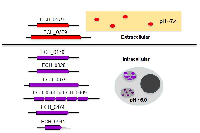
Int. J. Mol. Sci. 2021, 22, x FOR PEER REVIEW 4 of 19
Int. J. Mol. Sci. 2021, 22, 8420 4 of 18
Figure 2. (A) Transcription assessed for the E. chaffeensis antiporter genes during the replication in macrophage (DH82) and
Figure 2. (A) Transcription assessed for the E. chaffeensis antiporter genes during the replication in macrophage (DH82)
(B)
andtick
(B)cell
tick(ISE6) cultures.
cell (ISE6) RNARNA
cultures. concentration was was
concentration measured by qRT-PCR
measured by qRT-PCRtargeted to the
targeted to16S
the rRNA
16S rRNAand and
input RNARNA
input for
each sample
for each was was
sample normalized based
normalized on the
based on16S
therRNA concentrations.
16S rRNA The transcript
concentrations. levelslevels
The transcript for the
fortargeted genesgenes
the targeted were then
were
determined by semi-quantitative
then determined PCR with
by semi-quantitative PCR varying numbers
with varying of amplification
numbers cyclescycles
of amplification per target, whichwhich
per target, rangedranged
from 25 to
from
40
25cycles. The transcript
to 40 cycles. levels were
The transcript levelsmeasured for different
were measured post-infection
for different times: 0,times:
post-infection 6, 12, 0,
24,6,30,
12,36,
24,48,
30,54,
36,60,
48,72,
54,84,
60,96,
72,and
84,
96, h.
108 and 108 h. Positive
Positive controlscontrols
(PC) for(PC) for theincluded
the assays assays included E. chaffeensis
E. chaffeensis genomic genomic
DNA as DNA as the template.
the template. NegativeNegative
controls controls
(NC)
(NC)
had had nuclease-free
nuclease-free water aswater as the template.
the template. The number
The lowest lowest number of temperature
of temperature cycleswe
cycles where where
notedwe thenoted the expression
expression for each
for each
gene gene are presented:
are presented: ECH_0179ECH_0179
(45 cycles),(45 cycles), ECH_0328
ECH_0328 (35 cycles (35
for cycles
DH82 andfor DH82 andfor
45 cycles 45 ISE6),
cyclesECH_0379
for ISE6), ECH_0379
(35 cycles for (35
cycles for DH82 and 45 cycles for ISE6), CH_0466 (35 cycles for DH82 and 40 cycles for ISE6), ECH_0467 (35 cycles for
DH82 and 45 cycles for ISE6), CH_0466 (35 cycles for DH82 and 40 cycles for ISE6), ECH_0467 (35 cycles for DH82 and
DH82 and 45 cycles for ISE6), ECH_0468a (5′) (45 cycles for DH82 and 35 cycles for ISE6), ECH_0468b (3′) (35 cycles for
45 cycles for ISE6), ECH_0468a (50 ) (45 cycles for DH82 and 35 cycles for ISE6), ECH_0468b (30 ) (35 cycles for DH82 and
DH82 and 45 cycles for ISE6), ECH_0469 (30 cycles for DH82 and 45 cycles for ISE6), ECH_0474 (35 cycles for DH82 and
45
45cycles
cyclesfor
forISE6),
ISE6),ECH_0469
and ECH_0944(30 cycles for DH82 and 45 cycles for ISE6), ECH_0474 (35 cycles for DH82 and 45 cycles for
(45 cycles).
ISE6), and ECH_0944 (45 cycles).
2.3. Transcription of E. chaffeensis Antiporter Genes Assessed in the Antiporter Function
2.3. Transcription
Deficient of E. chaffeensis
E. coli Mutant (EP432) Antiporter Genes Assessed in the Antiporter Function Deficient
E. coli Mutant (EP432)
Since molecular tools are still limited for use in the Anaplasmataceae family patho-
Since molecular tools are still limited for use in the Anaplasmataceae family pathogens,
gens, including for E. chaffeensis, it is challenging to study gene/protein function in this
including for E. chaffeensis, it is challenging to study gene/protein function in this pathogenic
pathogenic bacterium in vivo. To overcome this limitation, we used E. coli as a surrogate
bacterium in vivo. To overcome this limitation, we used E. coli as a surrogate system to
system to study the functions of genes and proteins of E. chaffeensis [12,13]. Recently, we
study the functions of genes and proteins of E. chaffeensis [12,13]. Recently, we reported
reported an E. coli function complementation system to define E. chaffeensis antiporter pro-
an E. coli function complementation system to define E. chaffeensis antiporter protein gene
tein gene transcription and protein function [12] (Figure S2). We described that the func-
transcription and protein function [12] (Figure S2). We described that the functional
tional complementation studies in the E. coli EP432 strain are ideally suited for defining
complementation studies in the E. coli EP432 strain are ideally suited for defining the
the function of E. chaffeensis antiporter genes as it has mutations in two of its native anti-
function of E. chaffeensis antiporter genes as it has mutations in two of its native antiporter
genes [12,14]. The E. coli EP432 strain growth is significantly inhibited in the presence ofporter genes [12,14]. The E. coli EP432 strain growth is significantly inhibited in the pres-
Int. J. Mol. Sci. 2021, 22, 8420 ence of sodium chloride [14] and the growth can be restored with its native 5gene of 18 (nhaA)
expressed from a recombinant plasmid or with another bacterial gene homolog that it can
functionally complement [12,15–17]. To define the functions of E. chaffeensis antiporter
sodium
genes, chloride [14] and
we transformed thethe growth
EP432 can be
strain restored
with with its native
recombinant gene (nhaA)
plasmids expressed
containing E. chaffeen-
from a recombinant plasmid or with another bacterial gene homolog that
sis antiporter genes cloned along with their predicted promoter segments; this was con-it can functionally
complement [12,15–17]. To define the functions of E. chaffeensis antiporter genes, we trans-
ducted one gene at a time. The E. coli nhaA gene was similarly cloned to serve as a positive
formed the EP432 strain with recombinant plasmids containing E. chaffeensis antiporter
control.
genesAll transformed
cloned along withEP432 containing
their predicted E. chaffeensis
promoter segments;antiporter gene recombinant
this was conducted one gene plas-
midsatwere
a time.then
Theassessed forgene
E. coli nhaA transcripts by RT-PCR
was similarly cloned toanalysis
serve as(Figure
a positive 3).control.
PredictedAll ampli-
constransformed
were detectedEP432incontaining
the transformed EP432
E. chaffeensis organisms
antiporter gene containing
recombinantthe E. chaffeensis
plasmids were anti-
then
porter assessed for transcripts by RT-PCR analysis (Figure 3). Predicted amplicons were
genes.
detected in the transformed EP432 organisms containing the E. chaffeensis antiporter genes.
3. E. chaffeensis antiporter gene transcripts assessed by RT-PCR for the respective gene recombinant plasmids
Figure 3.Figure
E. chaffeensis antiporter gene transcripts assessed by RT-PCR for the respective gene recombinant plasmids trans-
transformed into the E. coli strain EP432. The assays for each gene included four reactions: 1, RNA sample with reverse
formed into the E. coli strain EP432. The assays for each gene included four reactions: 1, RNA sample with reverse tran-
transcriptase and Taq polymerase; 2, nuclease-free water with reverse transcriptase and Taq polymerase (to serve as
scriptase and Taq polymerase; 2, nuclease-free water with reverse transcriptase and Taq polymerase (to serve as the neg-
the negative control); 3, RNA template with Taq polymerase and without reverse transcriptase (to serve as the negative
ative control); 3, RNA template with Taq polymerase and without reverse transcriptase (to serve as the negative control
control and to eliminate false positives resulting from the residual DNA contamination; 4, E. chaffeensis genomic DNA with
and to eliminate false positives resulting from the residual DNA contamination; 4, E. chaffeensis genomic DNA with Taq
Taq polymerase (to serve as the positive control). (Note; weaker amplicons found in lane 3 for ECH_0328, ECH_0466-9,
polymerase (to serve as the positive control). (Note; weaker amplicons found in lane 3 for ECH_0328, ECH_0466-9,
ECH_0474 and ECH_0944 represent the incomplete elimination of DNA contamination).
ECH_0474 and ECH_0944 represent the incomplete elimination of DNA contamination.)
2.4. E. chaffeensis Antiporter Proteins Functionally Complement Antiporter Deficiency in E. coli
2.4. E. chaffeensis Antiporter Proteins Functionally Complement Antiporter Deficiency in E. coli
We then evaluated if E. chaffeensis antiporter proteins are synthesized in EP432 and
We thentheevaluated
reversed if E. chaffeensis
growth inhibition of EP432 antiporter proteins
in the presence of 200 are
mMsynthesized
NaCl at varyingin EP432
pH and
conditions of the culture media. The complementation at pH 5.5 was
reversed the growth inhibition of EP432 in the presence of 200 mM NaCl at varying pH observed for all
6 genes (Figure
conditions of the 4culture
A; greenmedia.
lines). The
TheECH_0179, ECH_0328, at
complementation andpH
ECH_0944
5.5 wasproteins
observed com-for all 6
plemented E. coli growth very similarly to that observed with its native nhaA gene (positive
genes (Figure 4 A; green lines). The ECH_0179, ECH_0328, and ECH_0944 proteins com-
control) (Figure 4; red lines in each image). Complementation with the ECH_0466-0469
plemented E. coli
gene cluster wasgrowth
slightly very similarly
less compared to to that observed
ECH_0179, with
ECH_0328, anditsECH_0944
native nhaA gene (posi-
proteins,
tive and
control) (Figure 4; red lines in each image). Complementation with
weaker for the ECH_0379 and ECH_0474 proteins. The E. coli growth restoration was the ECH_0466-
0469also
gene cluster
observed forwas slightly
ECH_0179 less at
protein compared
both neutraltoand
ECH_0179, ECH_0328,
basic pH conditions, and ECH_0944
whereas the
ECH_0379 gene complemented at pH 8.0 with no change at neutral
proteins, and weaker for the ECH_0379 and ECH_0474 proteins. The E. coli growth pH (Figure 4 B and C). resto-
In contrast, genes ECH_0328, ECH_0466-0469, ECH_0474, and ECH_0944
ration was also observed for ECH_0179 protein at both neutral and basic pH conditions made the E. coli
more sensitive to NaCl growth at pH 7.0 and pH 8.0.
whereas the ECH_0379 gene complemented at pH 8.0 with no change at neutral pH (Fig-
ure 4 B and C). In contrast, genes ECH_0328, ECH_0466-0469, ECH_0474, and ECH_0944
made the E. coli more sensitive to NaCl growth at pH 7.0 and pH 8.0.Int. J. Mol. Sci. 2021, 22, x FOR PEER REVIEW 6 of 19
Int. J. Mol. Sci. 2021, 22, 8420 6 of 18
Figure 4. E. chaffeensis antiporters restored the growth of a salt-sensitive sodium/proton antiporter deficient strain of E. coli.
Figure 4. E. chaffeensis antiporters restored the growth of a salt-sensitive sodium/proton antiporter deficient strain of E.
The growth
coli. The curves
growth were
curvesmeasured by monitoring
were measured the the
by monitoring ODOD
at at
600 nm
600 nmover
overaa period from1515min
period from minto to
13 13 h. The
h. The salt-stress
salt-stress
was induced by the inclusion of 200 mM NaCl in LB media at varying pH conditions—5.5, 7.0, or 8.0—for all E. chaffeensis
antiporter gene-containing EP432 strains (green lines). The E. coli transformed with the non-recombinant plasmid (blue line)
or the plasmid-containing E. coli antiporter gene (nhaA) (red line) served as negative or positive controls, respectively. E. coli
growth curves assessed at pH 5.5 (A), 7.0 (B) and 8.0 (C) are presented.was induced by the inclusion of 200 mM NaCl in LB media at varying pH conditions—5.5, 7.0, or 8.0—for all E. chaffeensis
antiporter gene-containing EP432 strains (green lines). The E. coli transformed with the non-recombinant plasmid (blue
line) or the plasmid-containing E. coli antiporter gene (nhaA) (red line) served as negative or positive controls, respectively.
Int. J.E. coliSci.
Mol. growth curves
2021, 22, 8420 assessed at pH 5.5 (A), 7.0 (B) and 8.0 (C) are presented. 7 of 18
2.5. Native ECH_0379 Formed a Dimer and Expressed in both Replicating (RC) and Infectious
(DC) Forms and Similarly It Forms Dimers When Expressed as a Recombinant Protein
2.5. Native ECH_0379 Formed a Dimer and Expressed in Both Replicating (RC) and Infectious
(DC)As ECH_0379
Forms protein
and Similarly It had
FormstheDimers
antiporter
Whenactivity
Expressed in as
a broad pH range,
a Recombinant we hypothe-
Protein
sized that it is expressed in both RC and DC forms of E. chaffeensis.
As ECH_0379 protein had the antiporter activity in a broad pH range, we hypothesized
To test this hypothesis, we first cloned and expressed the ECH_0379 recombinant
that it is expressed in both RC and DC forms of E. chaffeensis.
protein in E. coli using a pET expression system and purified to homogeneity (Figure 5).
To test this hypothesis, we first cloned and expressed the ECH_0379 recombinant
The recombinant protein migrated as two different molecular masses: about 42 kDa and
protein in E. coli using a pET expression system and purified to homogeneity (Figure 5). The
85 kDa with His-tag.
recombinant The 42 kDa
protein migrated size different
as two was the molecular
expected size for the
masses: His-tagged
about 42 kDa and recombi-
85 kDa
nant protein, while the second larger protein is likely the self-association
with His-tag. The 42 kDa size was the expected size for the His-tagged recombinant protein, of ECH_0379
protein
while theforming
seconddimers. The presence
larger protein is likelyofthe
both bands as partofofECH_0379
self-association the recombinant protein
protein forming
was confirmed by Western blot analysis using His-tag antibodies. Further,
dimers. The presence of both bands as part of the recombinant protein was confirmed by mass-spec-
trometry
Western analysis by MALDI-TOF
blot analysis using His-tag (Matrix-Assisted Laser Desorption/Ionization
antibodies. Further, mass-spectrometry analysis Time-of-by
Flight) confirmed that both protein bands contained the ECH_0379 protein
MALDI-TOF (Matrix-Assisted Laser Desorption/Ionization Time-of-Flight) confirmed that sequence (not
shown). The dimer
both protein bandsformation
contained of the
ECH_0379
ECH_0379 antiporter
proteinissequence
similar to(not
othershown).
known antiporter
The dimer
proteins [16,18]. Antisera was then raised against the recombinant
formation of ECH_0379 antiporter is similar to other known antiporter proteins ECH_0379 antiporter
[16,18].
and subsequently
Antisera was thenused to assess
raised againstprotein expressionECH_0379
the recombinant in the RC and DC forms
antiporter andof E. chaffeen-
subsequently
sis (Figure
used 6). One
to assess protein
protein band of 80
expression inkDa
the RCsizeand
thatDCis equivalent
forms of E.tochaffeensis
a dimer was recognized
(Figure 6). One
by the ECH_0379
protein band of 80antisera
kDa sizein that
the proteins recovered
is equivalent fromwas
to a dimer bothrecognized
the RC andbyDC theforms of E.
ECH_0379
chaffeensis.
antisera in the proteins recovered from both the RC and DC forms of E. chaffeensis.
A B
KDa M 1 1
130
95
85 kDa
72
55
43
42 kDa
34
26
17
10
Figure
Figure5.5.ECH_0379
ECH_0379recombinant
recombinantprotein
proteinpurified
purifiedtotohomogeneity
homogeneityusing
usingthe
thepET
pETexpression
expressionsystem
system
ininE.E.coli.
coli. (A) The purified recombinant protein was resolved by 12% SDS-PAGEand
(A) The purified recombinant protein was resolved by 12% SDS-PAGE andstained
stainedwith
with
Coomassie blue G250 stain. (B) Western-blot analysis performed for the SDS-PAGE resolved pro-
Coomassie blue G250 stain. (B) Western-blot analysis performed for the SDS-PAGE resolved proteins
teins using the N-terminal expressed His-tag-specific polyclonal sera. (M, protein molecular weight
using the N-terminal expressed His-tag-specific polyclonal sera. (M, protein molecular weight
markers; 1, resolved recombinant protein).
markers; 1, resolved recombinant protein).Int. J. Mol. Sci. 2021, 22, x FOR PEER REVIEW 8 of 19
Int. J. Mol. Sci. 2021, 22, 8420 8 of 18
Figure 6. ECH_0379 antiporter expression assessed in RC and DC forms of E. chaffeensis cultured
in vitro.
Figure Density antiporter
6. ECH_0379 gradient-purified fractions
expression of RC
assessed andand
in RC DCDCforms were
forms of used to recover
E. chaffeensis E. chaffeensis
cultured in
proteins
vitro. Density and were subjected to
gradient-purified Westernofblot
fractions RCanalysis
and DCusing
formspolyclonal
were usedsera
to raised
recoveragainst ECH_0379
E. chaffeensis
proteins and wereprotein.
recombinant subjected
(M,to Western
protein blot analysis
molecular weight using polyclonal
markers; DC andsera
RCraised against
refer to ECH_0379
purified E. chaffeensis
recombinant protein. (M,
proteins recovered protein
from molecular
DC and weightrespectively).
RC fractions, markers; DC and RC refer to purified E. chaffeen-
sis proteins recovered from DC and RC fractions, respectively.)
2.6. Proteoliposome Prepared with the Recombinant ECH_0379 Protein Assessed in Diffusion Analysis
2.6. Proteoliposome Prepared verify
To independently With the
theRecombinant ECH_0379
antiporter activity Protein
of E. Assessed
chaffeensis in Diffusion
ECH_0379 antiporter
Analysis
protein, we performed in vitro proteoliposome diffusion assays using the recombinant
protein. The recombinant
To independently protein
verify the was used
antiporter to constitute
activity proteoliposomes
of E. chaffeensis and tested in
ECH_0379 antiporter
proteoliposome assays (Figure 7). A steady decline of the absorbance
protein, we performed in vitro proteoliposome diffusion assays using the recombinant at 400 nm was
observed
protein. in the presence
The recombinant of NaCl
protein waswhen
usedtested at two different
to constitute concentrations—100
proteoliposomes and tested inmM
or 200 mM—and
proteoliposome assaysat(Figure
three different pH conditions
7). A steady decline of the(pHabsorbance
5.5, 7.0, and 8.0) nm
at 400 (Figure
was 7;
ob-blue
lines).
served In presence
in the control experiments
of NaCl when where theatrecombinant
tested two differentprotein was not included
concentrations—100 mM inor the
200 mM—and at three different pH conditions (pH 5.5, 7.0, and 8.0) (Figure 7; blue lines). the
proteoliposome preparations, there was no change in the absorbance at 400 nm in
solutions
In control containing
experiments either
where 100
the mM or 200protein
recombinant mM NaCl was independent
not included inof the
the proteolipo-
pH variations
(negative controls) (Figure 7; green lines).
some preparations, there was no change in the absorbance at 400 nm in the solutions con-
taining either 100 mM or 200 mM NaCl independent of the pH variations (negative con-
trols) (Figure 7; green lines).Int. J. Mol. Sci. 2021, 22, x FOR PEER REVIEW 9 of 19
Int. J. Mol. Sci. 2021, 22, 8420 9 of 18
Figure 7. The NaCl diffusion assays performed using the proteoliposomes constituted with the ECH_0379 recombinant
Figure 7. The NaCl diffusion assays performed using the proteoliposomes constituted with the ECH_0379 recombinant
protein
protein in
in lipid vesicles. The
lipid vesicles. Thediffusion
diffusionrates
rateswere
weremeasured
measuredbybymonitoring
monitoringthethe decrease
decrease in in
ODOD400400
nmnm absorbance
absorbance in
in the
the presence
presence of NaCl
of NaCl at 100
at 100 mMmM (A) (A) or 200
or 200 mMmM (B) concentrations
(B) concentrations and and at three
at three different
different pH conditions.
pH conditions. Blue Blue and green
and green lines
lines represent
represent liposomes
liposomes prepared
prepared with with and without
and without the purified
the purified ECH_0379
ECH_0379 recombinant
recombinant protein,
protein, respectively.
respectively. The The
datadata
rep-
resent thethe
represent average
averagevalues from
values three
from independent
three independent experiments.
experiments.
3. Discussion
3. Discussion
Sodium (cation)/H+
Sodium (cation)/H+ antiporters
antiporters facilitate
facilitate the exchange of protons for sodium sodium or or other
other
cations [19–21]. Antiporters are critical in regulating regulating intracellular
intracellular pH,pH, sodium
sodium levels,
levels, and
and
volume [22,23].
cell volume [22,23]. Antiporters
Antiporters are are found
found in in diverse
diverse organisms
organisms [24–29].
[24–29]. E.E. chaffeensis
chaffeensis is
is an
an
important tick-transmitted
important tick-transmittedobligateobligatepathogenic
pathogenic bacterium
bacterium causing
causing diseases
diseases in humans
in humans and
and dogs
dogs [30,31].
[30,31]. This intraphagosomal
This intraphagosomal pathogen pathogen replicates
replicates withinwithin acidified
acidified vacuoles,vacuoles,
while
while
the the infectious
infectious form is form is exposed
exposed to a physiological
to a physiological pH of pH of ~7.0.
~7.0. TheThe pathogen’s
pathogen’s ability
ability to
to survive
survive in ainphagosome
a phagosome environment
environment is remarkable,
is remarkable, as itasrequires
it requires efficient
efficient antiporters
antiporters for
for pH
pH homeostasis
homeostasis to maintain
to maintain itsits physiological
physiological pH.Currently,
pH. Currently,no nostudies
studieshavehavedescribed
described
how the pathogen and other related intraphagosomal intraphagosomal rickettsialrickettsial pathogens
pathogens maintain
maintain their
their
cytoplasmic pH homeostasis.
homeostasis.
E. chaffeensis
chaffeensis transitions between the infectious form (DC) outside a host host cell
cell where
where
the pH of of the
the environment
environment is is expected
expected to to bebe ~7,
~7, while
while its
its replication
replication takes
takes place
place within
within
acidified
acidified phagosomes
phagosomes of of monocytes/macrophages.
monocytes/macrophages. Thus, both the intracellular intracellular replicative
replicative
form (RC) and the extracellular
the extracellular DC form E. chaffeensis
form of E. chaffeensis require adaptation
of require adaptation to to rapidly
rapidly
shifting ionic strengths and pH. pH. This
This may may be be best
best accomplished
accomplished by by multiple
multiple antiporters
antiporters
with diverse biological
with diverse biologicalfunctions
functionsininsupportsupportofof thethe pathogen’s
pathogen’s lifecycle
lifecycle within
within a host.
a host. We
We recently
recently reported
reported thatthat a functional
a functional disruption
disruption mutation
mutation in aninE.an E. chaffeensis
chaffeensis antiporter
antiporter gene
gene (ECH_0379)
(ECH_0379) causes causes considerable
considerable decline decline
of theofpathogen
the pathogengrowth growth
within within vertebrate
vertebrate hosts
hosts [11,32]. Despite the antiporter gene function deficiency,
[11,32]. Despite the antiporter gene function deficiency, the mutant remained in the hosts, the mutant remained in
the hosts, although the rickettsemia levels fell below detectable
although the rickettsemia levels fell below detectable levels in circulation as compared to levels in circulation as
compared
wild type E. wild type E. chaffeensis-infected
to chaffeensis-infected animals [11,32]. animals [11,32].
These data These
suggest data
that suggestfunc-
antiporter that
antiporter functional
tional redundancy redundancy
exists exists in
in the organism tothe organism
support its pH tohomeostasis.
support its pH homeostasis.
Bacterial patho-
Bacterial
gens are known to contain different types of antiporters expressed on the membranesthe
pathogens are known to contain different types of antiporters expressed on to
membranes
support their to growth
supportin their growth
diverse in diverse environmental
environmental conditionsThe
conditions [17,33–35]. [17,33–35].
results The
pre-
results
sented inpresented
the current in the
studycurrent studythe
represent represent the first
first detailed detailed description
description of multiple
of multiple antiporters.
antiporters. Our initial search in the E. chaffeensis genome allowed
Our initial search in the E. chaffeensis genome allowed the identification of 10 gene open the identification of
10 gene open reading frames encoding for sodium (cation)/proton antiporter proteins
reading frames encoding for sodium (cation)/proton antiporter proteins or protein subu-
or protein subunits. Homologs for all antiporter genes are also present in the genomesInt. J. Mol. Sci. 2021, 22, 8420 10 of 18
of other E. chaffeensis strains, as well as in all Anaplasmataceae family organisms of the
genera Ehrlichia, Anaplasma, and Wolbachia for which the genome sequences are available in
the GenBank. Further, analysis of the transmembrane topology revealed that all encoded
antiporter proteins from the genes possess transmembrane domains. Except for one, all
genes were also transcribed in E. chaffeensis during its replication in macrophage and
tick cell cultures. We then assessed the antiporter genes in E. coli mutant with functional
deficiency for antiporters; we presented data demonstrating that all predicted antiporters
are functionally active. Specifically, our study demonstrated that all E. chaffeensis genes can
complement the E. coli antiporter function at pH 5.5, while some genes can also provide
complementation at physiological pH conditions. The data establish that all putative
E. chaffeensis antiporter genes code for functional sodium/proton antiporters.
Transcriptional-level expression analysis of RNA transcripts of the putative antiporter
protein genes of E. chaffeensis were detected when cultured in the macrophage cell line and
tick cells, suggesting that they are functional genes and that the translated proteins of the
antiporters may have a biological function during its lifecycle within its vertebrate and
tick hosts. In the E. coli surrogate system, recombinant proteins made from all predicted
antiporters provided functional complementation in relieving the inhibition of its growth
in the presence of NaCl at pH 5.5. In contrast, the protein expression at neutral pH was
unfavorable for all gene products except for ECH_0179 and ECH_0379, thus making the
EP432 strain of E. coli more sensitive to NaCl. ECH_0379 provided the most complementa-
tion at pH 8.0, with minor impact at acidic pH and no major impact at neutral pH. These
data suggest that the ECH_0379 protein may be critical for the bacterium during its growth
within the phagosomes and after its release as DC. Indeed, our data demonstrate that this
protein is expressed in both RC and DC forms of E. chaffeensis. Antiporters synthesized
from the ECH_0179 and ECH_0379 genes, therefore, are likely to support the bacterial
homeostasis for RC and DC forms. ECH_0179 RNA expression was undetectable when
the organism was grown in either macrophage or tick cell cultures, while the ECH_0379
expression was observed at all time points assessed. Consistent with this observation, the
ECH_0379 protein was detected in both the RC and DC forms of E. chaffeensis. The lack
of detectable RNA transcripts for the gene ECH_0179 may indicate that it may have low
expression under in vitro culture conditions, while it is likely expressed in vivo. Together,
ECH_0379 and ECH_0179 gene products may help the bacterium to maintain the pH home-
ostasis for its growth within a phagosome and during its presence outside a host cell. More
studies are necessary to define how such functional diversity contributes to bacterial in vivo
growth. Our studies are consistent with prior reports on antiporters of other Gram-negative
bacteria, where researchers reported that the sodium/proton antiporters have characteristic
individual pH-dependent activity profiles, critical for their homeostatic functions [36,37].
SDS-PAGE and the Western blot analysis of the purified recombinant protein ECH_0379
revealed two resolved proteins with different molecular weights. The fastest migrating
protein was about 42 kDa and the higher molecular mass protein was about 85 kDa, which
is twice the size of the predicted molecular weight, suggesting that it represents a dimer-
ized protein. When assessed for the native protein using the ECH_0379 polyclonal sera,
we observed only one larger dimerized protein band, which migrated at ~80 kDa. A
similar dimer formation has been reported previously for antiporters of E. coli [18] and
Salmonella [16]. The purified recombinant ECH_0379 antiporter protein reconstituted as
lipid vesicles (proteoliposomes) demonstrated antiporter activity under the three differ-
ent pH conditions and at two different NaCl concentrations. These data validate that the
ECH_0379 antiporter indeed has the antiporter function and may be critical for the bacterial
pH homeostasis in a host.
The current study is the first and most detailed description of antiporters in the
rickettsial organism: E. chaffeensis. The pathogen contains multiple genes encoding for
functional antiporters which fall under two sub-groups based on their biological properties
(Figure 8). While all encoded antiporters support the growth of the organism within the
acidified phagosomes, a subset of proteins may also aid the organism’s pH homeostasisInt. J. Mol. Sci. 2021, 22, x FOR PEER REVIEW 11 of 19
Int. J. Mol. Sci. 2021, 22, 8420 11 of 18
(Figure 8). While all encoded antiporters support the growth of the organism within the
acidified phagosomes, a subset of proteins may also aid the organism’s pH homeostasis
during its exposure to the cell-free environment where the infectious DC form may mi-
during
grate to its
enterexposure to the cell-free
naïve macrophages environment
in vivo where
via systemic theThus,
spread. infectious DC formofmay
the expression all migrate
E. chaffeensis antiporters is likely critical in supporting the bacterial ion balance to regulate of all E.
to enter naïve macrophages in vivo via systemic spread. Thus, the expression
chaffeensis
its internal antiporters is likelyDC
pH. The pathogen’s critical
forminis supporting
released out the
of abacterial ion balance
host cell where to regulate its
pH is likely
internal
to be closepH. The pathogen’s
to between DC [8],
7.35 and 7.45 form is ECH_0179
and released out
andof a host cell
ECH_0379 wheremay
proteins pH facil-
is likely to be
itate
closepH to homeostasis
between 7.35 of the
andorganism.
7.45 [8], and ECH_0179 and ECH_0379 proteins may facilitate
pH homeostasis of the organism.
Model
Figure8.8.Model
Figure cartoon
cartoon outlining
outlining E. chaffeensis
E. chaffeensis antiporters
antiporters forpossible
for their their possible roles
roles in pH in pH homeostasis
homeostasis
in
in the
theEhrlichia-containing acidified
Ehrlichia-containing phagosomes
acidified and during
phagosomes andthe releasethe
during of the DC form
release of in
thetheDC
extra-
form in the
cellular milieu.milieu.
extracellular
This
Thisresearch
research marks
marksthe the
beginning the advancement
beginning the advancementof our understanding of how E. of how
of our understanding
chaffeensis and other related rickettsia regulate their intracellular pH under diverse host
E. chaffeensis and other related rickettsia regulate their intracellular pH under diverse
environments, where the pathogens are exposed to acidic and physiological pH condi-
host environments, where the pathogens are exposed to acidic and physiological pH
tions. Notably, we discovered, following BLAST search analysis, that the multiple anti-
conditions.
porter Notably, are
gene homologs we also
discovered, following
well conserved BLAST
in other search
related analysis,
Anaplasma andthat the multiple
Ehrlichia
species pathogens. Considering that therapeutics are limited to only one class of antibiot- Ehrlichia
antiporter gene homologs are also well conserved in other related Anaplasma and
species
ics pathogens.
(tetracycline Considering
derivatives) that therapeutics
for treating are limited
rickettsial diseases [38,39],toproteins
only one class
that of antibiotics
mediate
(tetracycline derivatives)
the pH homeostasis can serve for
as treating rickettsial
ideal targets diseases
for developing [38,39],drug
alternate proteins
choices.that
In- mediate
the pH
deed, homeostasis
antiporters can serve as ideal
are well-established as drugtargets
targetsfor
fordeveloping alternate
bacterial infections drug
[40,41]. choices. In-
Thus,
defining the contributions
deed, antiporters of antiporters as
are well-established in drug
Ehrlichia species
targets for and other infections
bacterial related obligate
[40,41]. Thus,
rickettsial
defining thepathogens will alsoof
contributions aidantiporters
in advancing in the research
Ehrlichia on their
species andinterference
other relatedwith obligate
bacterial
rickettsialgrowth in vivo. will
pathogens Studies canaid
also nowinbe extended tothe
advancing further characterize
research on their E. interference
chaffeensis with
antiporters for theirinrole
bacterial growth in Studies
vivo. the pathogen’s
can now lifebe
cycle and result
extended in the prospects
to further for devel-
characterize E. chaffeensis
oping novel treatment options for the HME and other tick-borne rickettsial diseases in
antiporters for their role in the pathogen’s life cycle and result in the prospects for devel-
humans and animals.
oping novel treatment options for the HME and other tick-borne rickettsial diseases in
humans
4. andand
Materials animals.
Methods
4.1. Bioinformatic
4. Materials and Analysis
Methods
The identity ofAnalysis
4.1. Bioinformatic 9 new antiporter gene open reading frames was established based on
the existing annotation data for the E. chaffeensis genomes entries: GenBank accession
The identity of 9 new antiporter gene open reading frames was established based
on the existing annotation data for the E. chaffeensis genomes entries: GenBank acces-
sion numbers NC_00779 and CP000236. We previously reported that ECH_0379 gene
coding sequence has a significant homology with bacterial antiporter protein subunits
(Cheng et al. 2013). The BLAST search analysis identified homologs for all 10 E. chaffeensis
antiporters in other related Anaplasmataceae family bacteria of the genera Anaplasma,
Ehrlichia, and Wolbachia, for which whole genome sequences are available in the Gen-
Bank database. The secondary structure predictions of the E. chaffeensis putative an-Int. J. Mol. Sci. 2021, 22, 8420 12 of 18
tiporter proteins were performed using the online sequence analysis tools on 2–6 April
2018: YASPIN (http://www.ibi.vu.nl/programs/yaspinwww/) and Smart (http://smart.
embl-heidelberg.de/). TMpred was used for transmembrane domain predictions (https:
//www.ch.embnet.org/software/TMPRED_form.html). Searches for homologous pro-
tein sequences were performed using the SIB BLAST+ Network Service (https://web.
expasy.org/blast/). Conserved domains within the putative antiporter proteins were
performed at https://www.ncbi.nlm.nih.gov/Structure/cdd/wrpsb.cgi. Multiple se-
quence alignments for all the identified E. chaffeensis predicted antiporter proteins and
homologous antiporter protein sequences were conducted using Clustal Omega (https:
//www.ebi.ac.uk/Tools/msa/clustalo/).
4.2. Cultivation of E. chaffeensis
E. chaffeensis Arkansas isolate (ATCC # CRL-10389) was cultured in the canine
macrophages cell line (DH82) or in the Ixodes scapularis embryonic cell line (ISE6) as
previously described [12,42]. For the RNA expression analysis experiments, infected cul-
tures were recovered at various time points post-infection from 0 to 108 h: 0, 6, 12, 24, 30,
36, 48, 54, 60, 72, 84, 96 and 108 h. The harvested cells from each flask were concentrated
by centrifugation at 12,000 g for 10 min. Supernatants were discarded, and the final pellets
were re-suspended in 1 mL of TRI-Reagent (Sigma-Aldrich, St. Louis, MO) and stored at
−80 ◦ C until RNA isolation was completed.
4.3. RNA Isolation
Total RNA was extracted using the TRI-Reagent method (MilliporeSigma, St. Louis,
MO, USA), as per the manufacturer’s recommended protocol, with the following minor
modifications. The cell pellets dissolved in TRI-Reagent were thawed at room temperature
and 0.2 mL of chloroform was added per 1 mL in each of the TRI-Reagent solutions.
Then, the mixture was vortexed vigorously for 15 s, left to stand for 15 min at room
temperature, and then centrifuged for 15 min at 12,000 g at 4 ◦ C. The upper aqueous phase
was transferred to a clean RNase-free tube, to which 0.5 mL of pre-cooled 2-propanol was
added per ml of TRI reagent, then the solution was incubated for 10 min at 4 ◦ C. RNA was
recovered by centrifugation for 10 min at 12,000 g at 4 ◦ C. The supernatant was discarded,
and the pellets were rinsed with 1 mL of 75% pre-cooled ethanol per each ml of TRI reagent
solution containing RNA. The solutions were mixed by pipetting up and down and then
centrifuged at 12,000 g for 5 min at 4 ◦ C. Supernatants were discarded; the RNA pellets
were air-dried and resuspended in 50 µL of nuclease-free water. Residual genomic DNAs
from each RNA were eliminated by adding RQ1 RNase-Free DNase (Promega, Madison,
WI, USA) as per the manufacturer’s protocol and stored at −80 ◦ C until use.
4.4. Gene Expression Levels Determined by Semi-Quantitative PCR
RNA samples from the different time points post-infection from in vitro cultures were
normalized following defining the 16S rRNA levels by TaqMan probe-based RT-PCR assay,
as described in a previous study [43], using the gene-specific rimers and probe, and using
the commercial reagent kit; SuperScript® III Platinum® One-Step Quantitative RT-PCR
System (Invitrogen, Carlsbad, CA, USA). Additional details pertaining to the assay condi-
tions, including the primers and probe used for the assay and standard curve assessment,
are described in [43]. The amplification protocol included a reverse transcription step for
30 min at 50 ◦ C, followed by 5 min of denaturation at 95 ◦ C, and 40 cycles of amplification
performed for 15 s at 95 ◦ C, 40 s at 50 ◦ C, and 45 s at 60 ◦ C with the optics setting turned on
in the StepOnePlus real-time PCR system (Thermo Fisher, Waltham, MA, USA). Based on
the 16S qRT-PCR results, all the RNA concentrations were adjusted to have an equal copy
number of 16 S rRNA per unit volume of RNA. The nucleotide sequences of the antiporter
genes are available at the GenBank {available online: https://www.ncbi.nlm.nih.gov/
(28 July 2021)}. Primers for the one-step RT-qPCR were designed from within the coding
sequences and synthesized by Integrated DNA Technologies (Coralville, IA, USA). TheInt. J. Mol. Sci. 2021, 22, 8420 13 of 18
nucleotide sequences of the primers are listed in Table 2. Primers targeted to E. chaffeensis
antiporter genes were then used to perform one-step reverse transcription PCR (RT-PCR)
using 2 µL of each of normalized RNA templates with the SuperScript® III Platinum®
One-Step Quantitative RT-PCR System commercial kit (Invitrogen, Carlsbad, CA, USA).
The amplification conditions for the assays were as follows: reverse transcription for 1 h at
50 ◦ C, then amplification cycles set at 30 s at 94 ◦ C and 30 s at 51 ◦ C for genes ECH_0328,
ECH_0466, ECH_0467, ECH_0468a, EHC_0468b, EHC_0469, and ECH_0944) or 54 ◦ C
for genes ECH_0179, ECH_0474, and ECH_0379, then 30 s at 72 ◦ C followed by a final
extension step for 5 min at 72 ◦ C, and then storage at 4 ◦ C until analysis. The amplification
cycles varied from 25 to 45 cycles to estimate the variations of gene transcription levels. The
amplicons were resolved in 1.5% agarose gel containing ethidium bromide, and images of
the DNA resolved in the gels were captured using a Kodak Gel Logic 200 imaging system.
Table 2. Primers used for the semi qRT-PCR.
Gene Primer Name Sequence Size
RRG2107 Forward: 50 -GGCTATACAAGTTGGGTTGTTGT-30 672 bp
ECH_0179
RRG2108 Reverse: 50 -CACACATACACCACAGATAGACCT-30
RRG2068 Forward: 50 -GCATGCGATATCATTTGGAA-30 370 bp
ECH_0328
RRG2069 Reverse: 50 -GAATTGGAAAAGCCGCATTA-30
ECH_0379 RRG1276 Forward: 50 -CTAAGGTTGTAGGGAATGCAACC-30 374 bp
RRG1277 Reverse: 50 -ACAAGGTAAGTACCTTGCTTGCTC-30
RRG2054 Forward: 50 -TGCTGCAAATTTGTTTGGAA-30 165 bp
ECH_0466
RRG2055 Reverse: 50 -TCTCCAAAAGAACCATGAAGA-30
RRG2056 Forward: 50 -TGGACTTGCTATGCGATCTG-30 151 bp
ECH_0467
RRG2057 Reverse: 50 -TCAGTCAGCATCATCTTACCTTT -30
RRG2062 Forward: 50 -TTTGATGGCATTGTTGCATT-30 332 bp
ECH_0468a
RRG2063 Reverse: 50 -CTCGAAAACTTGCTAAAATTGC-30
RRG2060 Forward: 50 -CAGGCTGGTGTTATTGTTGC-30 259 bp
ECH_0468b
RRG2061 Reverse: 50 -TACACACAGTCATGCCCACA-30
RRG2058 Forward: 50 -GGTTATAGGTTTGTATGTTACTACTGC-30 213 bp
ECH_0469
RRG2059 Reverse: 50 -ATTGCAACCCCAACAACAAT-30
RRG2109 Forward: 50 -GGCATCTGGTGGGTTTTTAGG -30 525 bp
ECH_0474
RRG2110 Reverse: 50 -GCAGAACATACTGCCTCTACTG-30
RRG2064 Forward: 50 -GGTTTGCCCTATCAGGGTATC-30 426bp
ECH_0944
RRG2065 Reverse: 50 -CACCAGACATTGACTCTTCATCT-30
4.5. Constructing the Recombinant Plasmids
Based on the location of the predicted antiporter genes of the E. chaffeensis genome,
five ORFs spanning from ECH_0466 to ECH_0469 were considered as encoding for one
antiporter protein transcript made from one promoter segment located upstream to the first
gene (ECH_0466), thus reducing the total number of predicted genes to 6 (Figure 1). The
coding sequence of each gene along with the respective promoter sequence was amplified
by PCR using a high-fidelity DNA polymerase (Invitrogen, Carlsbad, CA) with the primer
sequences listed in Table 3, and using E. chaffeensis genomic DNA as the template. PCR
products were then cloned into the plasmid, pBluescript II SK (+) (Stratagene, San Diego,
CA) by directional cloning into BamHI and SalI restriction enzyme sites generated as part
of the amplicons. All the restriction enzymes used in the experiment were bought from
NEB (New England Biolabs, Ipswich, MA). The coding sequence of the E. coli antiporter
gene (nhaA; GenBank accession number NJ74_RS08715) with its promoter sequence was
similarly cloned into pBluescript II SK(+) to serve as a positive control. The presence of
specific inserts in each of the recombinant plasmids was confirmed by DNA sequence
analysis. The recombinant plasmids were named pBSK-0179, pBSK-0328, pBSK-0379, pBSK-
0466-0469, pBSK-0474, and pBSK-0944 for E. chaffeensis antiporter genes, and the E. coli
nhaA gene recombinant plasmid was named pBSK-NhaA.Int. J. Mol. Sci. 2021, 22, 8420 14 of 18
Table 3. Primers used for the recombinant plasmids.
Amplicon
Target Primer Sequence # Vector
Size
Forward:
RRG2097 1976 bp pBluescript II SK(+)
ECH-0179 50 -tgacg-GGATCC-TAGTGCCATTGGAGTATATGTAAG-30
Reverse:
RRG2098
50 -tgacg-GTCGAC-TTTACTTTAATTTTAATAAAGCTGCTG-30
Forward:
RRG2099 1893 bp pBluescript II SK(+)
ECH_0328 50 -tgacg-GTCGAC-TCTTAATCTATAAGCGGCATATGC-30
Reverse:
RRG2100
50 -tgacg-GGATCC-AATATAGGAACAACTAAACAAACTAC-30
Forward:
ECH_0379 PRG2131 1543 bp pBluescript II SK(+)
50 -tgacg-GGATCC-GTTTTTTAGCATCCTTTGTGTTAAAAG-30
Reverse:
PRG2132
50 -tgacg-GTCGAC-ATATCGACAAGCAATTGATACAGAG-30
Forward: 50 -tgacg-GGATCC-
ECH_0466, RRG2101 2291 bp pBluescript II SK(+)
ATGTAGAATTCACAGAGCTTTAGC-30
to 0469 Reverse:
RRG2102
50 -tgacg-GTCGAC-ATTATGATTTGACACTAGACTTACAC-30
Forward:
RRG2103 1883 bp pBluescript II SK(+)
ECH_0474 50 -tgacg-GGATCC-TGTTGTTGATATATATGTTCAGTATG-30
RRG2104 Reverse: 50 -tgacg-GTCGAC -TTACTCTGCATCTTTTGGATTACA-30
Forward:
RRG2105 828 bp pBluescript II SK(+)
ECH-0944 50 -tgacg-GGATCC-GTAATACAACCCCAGTTATACAGA-30
Reverse:
RRG2106
50 -tgacg-GTCGAC-TAATCACTTGTCAGATTTAATGACA-30
Forward:
PRG2158 1817 bp pBluescript II SK(+)
nhaA 50 -tgacg-GTCGAC-GCTCATTTCTCTCCCTGATAACA-30
PRG2159 Reverse: 50 -tgacg-CTGCAG-TGCTCTCTTCTCCTTGACCTT AC-30
Forward: 50 -gag-GCTAGC-
ECH_0379 RRG1397 1056 bp pET-28a(+)
ATGATAATAAAGTTTGGGGTTGAAAATCTGATC-30
Reverse: 50 -cga-CTCGAG-
RRG1398
CTATAAATCTACACTTTCTTCAACAATATTAATAC-30
# Underline indicated the enzyme site.
The pET-28(+) vector (Novagen, Darmstadt, Germany) was used to clone the ECH_0379
gene coding sequence for its protein expression and purification by including the N-
terminal His-tag as part of the recombinant protein. The entire protein-coding sequence of
ECH_0379 was amplified by PCR using E. chaffeensis genomic DNA as the template and
using the proofreading enzyme, pfu DNA polymerase (Promega, Madison, WI). ECH_0379
ORF-specific PCR primers were designed to include NheI and XhoI restriction sites on the
forward and reverse primers, respectively. The PCR products were cloned into pET28
plasmid at NheI and XhoI to generate the recombinant expression plasmid by following
standard molecular cloning procedures, with initial cloning performed utilizing the E.
coli TOP10 strain to generate the recombinant plasmid (pET28-ECH_0379). Following
verification of the insert orientation by restriction enzyme digestion analysis and also by
DNA sequence analysis, the plasmid was retransformed into the E. coli BL21 (DE3) strain
(Invitrogen, Carlsbad, CA) for use in recombinant protein synthesis.
4.6. Growth Complementation under NaCl Stress
Antiporter activities of the putative E. chaffeensis antiporters were assessed by per-
forming functional complementation assays in the E. coli strain, EP432. The antiporter
gene-containing recombinant plasmids and the E. coli nhaA gene-carrying plasmid were
transformed into the EP432 strain; this was conducted one at a time. The nhaA gene-
carrying plasmid transformed E. coli served as the positive control, while non-recombinant
pBluescript II SK+ plasmid transformed E. coli served as the negative control. Before inves-
tigating the antiporter activities of the E. chaffeensis genes, we verified the RNA expression
from each transformed E. coli; total RNA was recovered from 3 mL each of overnight bacte-
rial cultures of recombinant E. coli grown in LB media (10 g/L tryptone, 5 g/L yeast extract,
and 200 mM NaCl) and was used for the analysis. After the DNase treatment, RNAs were
assessed by one-step RT-PCR following the same protocol described in the previous sectionYou can also read