PI4Kg2 Interacts with E3 Ligase MIEL1 to Regulate Auxin Metabolism and Root Development1 OPEN
←
→
Page content transcription
If your browser does not render page correctly, please read the page content below
PI4Kg2 Interacts with E3 Ligase MIEL1 to Regulate Auxin
Metabolism and Root Development1[OPEN]

Chun-Yan Zhao,a,b and Hong-Wei Xue a,b,2,3
a
 School of Agriculture and Biology, Shanghai Jiao Tong University, Shanghai 200240, China
b
 National Key Laboratory of Plant Molecular Genetics, CAS Center for Excellence in Molecular Plant Sciences,
Chinese Academy of Sciences, Shanghai 200032, China
ORCID ID: 0000-0002-7641-5320 (H.-W.X.).

Root development is important for normal plant growth and nutrient absorption. Studies have revealed the involvement of
various factors in this complex process, improving our understanding of the relevant regulatory mechanisms. Here, we
functionally characterize the role of Arabidopsis (Arabidopsis thaliana) phosphatidylinositol 4-kinase g2 (PI4Kg2) in root
elongation regulation, which functions to modulate stability of the RING-type E3 ligase MYB30-INTERACTING E3 LIGASE1
(MIEL1) and auxin metabolism. Mutant plants deficient in PI4Kg2 (pi4kg2) exhibited a shortened root length and elongation
zone due to reduced auxin level. PI4Kg2 was shown to interact with MIEL1, regulating its degradation and furthering the
stability of transcription factor MYB30 (which suppresses auxin metabolism by directly binding to promoter regions of GH3.2
and GH3.6). Interestingly, pi4kg2 plants presented altered hypersensitive response, indicating that PI4Kg2 regulates synergetic
growth and defense of plants through modulating auxin metabolism. These results reveal the importance of protein interaction
in regulating ubiquitin-mediated protein degradation in eukaryotic cells, and illustrate a mechanism coordinating plant growth
and biotic stress response.

   The phosphatidylinositol (PI) signaling pathway is a                        PI4KIIIb1, and PI4KIIIb2; Mueller-Roeber and Pical,
vital regulator of biological function in eukaryotes.                          2002). PI4Ks are essential for many aspects of de-
Phosphoinositides have long been recognized as major                           velopmental processes including root hair growth
components of eukaryotic membranes essential for                               (Preuss et al., 2006), chloroplast division (Okazaki
many functions in eukaryotes, including cell signaling,                        et al., 2015), and pollen growth (Chapman and
membrane dynamics, and trafficking (Ischebeck et al.,                           Goring, 2011). Moreover, a previous study showed
2010). The functions of the PI signaling pathway are                           that RabA4b and PI4Kb1 localize to budding secre-
less understood in plants compared with mammals                                tory vesicles in the trans-Golgi network and regulate
and yeast.                                                                     secretory vesicle size (Kang et al., 2011). Phospha-
   Phosphatidylinositol-4-kinases (PI4Ks) phosphory-                           tidylinositol 4-phosphate interacts specifically with
late PIs at the 49 position of inositol ring to produce                        PLASTID DIVISION1 and PLASTID DIVISION2
phosphatidylinositol-4-phosphate. A BLAST program                              proteins to regulate chloroplast division (Okazaki
analysis showed that there are 12 distinct loci of PI4K                        et al., 2015).
homologs in Arabidopsis (Arabidopsis thaliana): eight                             Although physiological roles of type-III PI4Ks have
belonging to the type-II group (PI4Kg1–8; none have                            been reported, most of the eight type-II PI4Ks in Ara-
been demonstrated as a functional PI4K) and four be-                           bidopsis have not been functionally characterized.
longing to the type-III group (PI4KIIIa1, PI4KIIIa2,                           Type-II PI4Ks contain PI3/PI4 kinase domains and
                                                                               variable numbers (none, one, or two) of ubiquitin-like
                                                                               domains, whereas they lack the PI-binding domains
    1
     The work was supported by the National Science Foundation of              such as pleckstrin homolog (in PI4Ka) or novel ho-
China (grant no. 91954206), the China Postdoctoral Science Foundation          mology domains (in PI4Kb; Mueller-Roeber and Pical,
(grant no. 2018M642002), and the Ten-Thousand Talent Program.                  2002). Lipid kinase activity has not previously been
   2
     Author for contact: hwxue@sjtu.edu.cn.                                    detected in type-II PI4Ks, whereas Arabidopsis PI4Kg3,
   3
     Senior author.                                                            PI4Kg4, and PI4Kg7 were shown to possess autophos-
   The author responsible for distribution of materials integral to the        phorylation activity. AtPI4Kg4 phosphorylates Ser/Thr
findings presented in this article in accordance with the policy de-            residues of ubiquitin fusion degradation1 and regulatory
scribed in the Instructions for Authors (www.plantphysiol.org) is:
Hong-Wei Xue (hwxue@sjtu.edu.cn).
                                                                               particle non-ATPase10 (Galvão et al., 2008). TaPI4KIIg
   H.-W.X. designed experiments; C.-Y.Z. performed experiments;                has been confirmed as a stress-inducible gene in wheat
C.-Y.Z. and H.-W.X. analyzed and interpreted the data, and wrote               (Triticum aestivum), which interacts with and phosphor-
the article.                                                                   ylates wheat ubiquitin fusion degradation protein (Liu
   [OPEN]
         Articles can be viewed without a subscription.                        et al., 2013); however, the effects of ubiquitin fusion
   www.plantphysiol.org/cgi/doi/10.1104/pp.20.00799                            degradation1 and regulatory particle non-ATPase10
Plant PhysiologyÒ, October 2020, Vol. 184, pp. 933–944, www.plantphysiol.org Ó 2020 American Society of Plant Biologists. All Rights Reserved.   933

                                Downloaded on January 28, 2021. - Published by https://plantphysiol.org
                               Copyright (c) 2020 American Society of Plant Biologists. All rights reserved.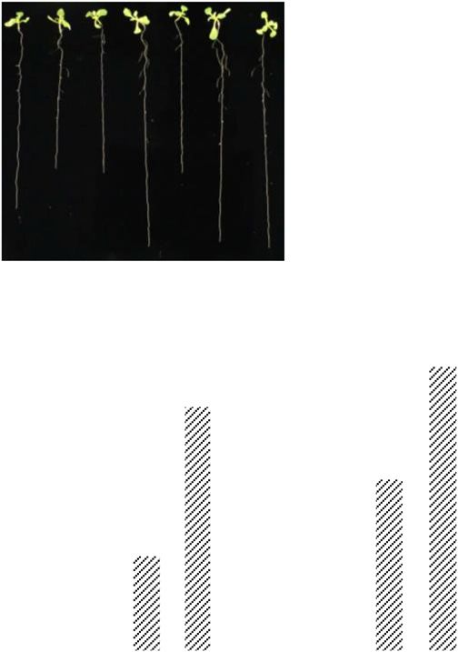
Zhao and Xue

phosphorylation remain unknown. AtPI4Kg3 selec-                       phosphatidylinositol monophosphate 5-kinase1 are
tively binds to a few PIs and is important for reinforcing            involved in root development regulation through reg-
plant responses to environmental stresses and in the                  ulation of PIN proteins (Mei et al., 2012) and PIN-
delay of floral transition (Akhter et al., 2016). AtPI4Kg5             mediated auxin efflux (Barbosa et al., 2016).
interacts with the membrane-bound transcription fac-                     Recent genetic studies have revealed important roles
tor ANAC078 to regulate auxin biosynthesis and leaf                   of distinct E3 ligases in phytohormone signaling path-
margin development (Tang et al., 2016). These studies                 ways (Kelley, 2018), operating via the degradation of
indicate that type-II PI4Ks participate in the regulation             various components, including receptors, positive or
of specific physiological processes through different                  negative regulators, or those related to hormone me-
mechanisms.                                                           tabolisms. Through systematic biochemical and genetic
   The functionality of proteins is tightly regulated                 studies, we here report the functional characterization
by various kinds of posttranslational modifications.                   of an Arabidopsis type-II PI4K, PI4Kg2, that regulates
Ubiquitination is an essential regulatory mechanism                   root growth through its interaction with the ubiquitin
that alters protein stability by mediating the proteaso-              E3 ligase MYB30-INTERACTING E3 LIGASE1 (MIEL1).
mal degradation of target proteins (Schwechheimer,                    MIEL1 is a negative regulator of hypersensitive response
2018). Ubiquitination involves E1 ubiquitin-activating                (HR) cell death by promoting MYB30 turnover in Ara-
enzymes, E2 ubiquitin-conjugating enzymes, and E3                     bidopsis (Marino et al., 2013). MIEL1 protein level is
ligases (Rape, 2018). E3 ligases recognize and bind                   significantly reduced in pi4kg2 mutant plants and
specifically to the substrate (Vierstra, 2009), and thus               MYB30 directly binds to promoter regions of GH3.2
play key roles in protein degradation. Although an in-                and GH3.6, thereby suppressing auxin metabolism and
creasing number of reports show the crucial roles of E3               hence root growth. Our data demonstrate that PI4Kg2 is
ligases in plants, only a few reports have focused on the             critical for MIEL1 stability and MYB30 turnover, and
regulation of E3 ligases at the post-translational level.             elucidate how MIEL1 coordinates auxin-dependent
Indeed, in addition to transcriptional regulation, E3 li-             plant growth and defensive HR in Arabidopsis.
gases are subjected to various post-translational modi-
fications. Tyr phosphorylation of the E3 ligase GARU at
Tyr-321 inhibits the GARU–GID1A interaction and
hence degradation of GID1 (Nemoto et al., 2017). Cop9                 RESULTS
Signalosome, which mediates the de-neddylation of cull-               Deficiency of PI4Kg2 Results in Short Roots
ins, is required to inactivate Cullin-RING E3 ubiquitin–
Ligase complexes, and then deter substrate recognition                   Type-II PI4Ks, which lack the PI-binding domains,
and prevent Cullin-RING E3 ubiquitin–Ligase subunit                   play roles in plant development via protein–protein
instability by counteracting their autoubiquitination and             interaction or protein phosphorylation (Mueller-Roeber
subsequent degradation (Merlet et al., 2009). Pirh2 protein           and Pical, 2002; Galvão et al., 2008; Liu et al., 2013). Our
is a p53-induced E3 ligase regulated by different proteins:           previous study showed that Arabidopsis PI4Kg5 reg-
CaMK II and Cdk9 phosphorylate Pirh2, then abrogate its               ulates auxin biosynthesis and leaf margin development
E3 ligase activity toward p53 (Duan et al., 2007; Bagashev            by interacting with and regulating the cleavage of the
et al., 2013); Tat-interactive protein of 60 kD interacts with        membrane-bound transcription factor ANAC078 (Tang
Pirh2 to alter the subcellular localization of Pirh2 (Logan           et al., 2016). To expand our understanding of type-II
et al., 2004); and PLAGL2 interacts with Pirh2 to prevent             PI4K functions, we further functionally characterized
the proteasomal degradation of Pirh2 (Zheng et al., 2007).            AtPI4Kg2. Analysis by reverse-transcription quantita-
These findings indicate that posttranslational modifica-                tive PCR (RT-qPCR) revealed PI4Kg2 expression in
tion is critical in E3 functions.                                     various tissues (Fig. 1A). Histochemical analysis of
   Many signaling pathways control root growth and                    transgenic lines harboring a PI4Kg2 promoter-reporter
development, particularly the phytohormone auxin,                     construct (Glucuronidase, pPI4Kg2::GUS) showed
which regulates both cell proliferation and elongation                that PI4Kg2 is expressed in young seedlings, floral tis-
of root cells (Street et al., 2016). Previous studies                 sue, rosette leaf, cauline leaf, stem, and inflorescence
showed that AUX1 mediates the inhibitory effects of                   (Fig. 1B). GUS signals were predominantly observed in
cytokinin on root cell elongation (Péret et al., 2012;                the root apical region of young seedlings (Fig. 1B, 1), sug-
Street et al., 2016). YDK1, a GH3 gene, might function as             gesting a potential role of PI4Kg2 in root development.
a negative regulator of root cell elongation by control-                 A transfer DNA (T-DNA) insertional mutant pi4kg2
ling the level of the active form of auxin (Takase et al.,            (Alonso et al., 2003) was identified, which we used to
2004). In rice (Oryza sativa), OsARF12, a transcrip-                  investigate the physiological role of PI4Kg2. T-DNA
tion activator of auxin-response genes, mediates root                 was inserted in the first exon of PI4Kg2 (Supplemental
cell elongation by inhibiting most OsYUCCAs auxin-                    Fig. S1, A and B) and RT-qPCR analysis confirmed the
synthesis genes (Qi et al., 2012). In addition, phospho-              deficient transcription of PI4Kg2 in homozygous mu-
lipase D-derived phosphatidic acid promotes root hair                 tant plant (Supplemental Fig. S1C), indicating that
development under phosphorus deficiency by sup-                        pi4kg2 is a knockout mutant.
pressing vacuolar degradation of PIN2 (Lin et al., 2020).                Phenotypic observation showed that growth of
Phosphatidylinositol monophosphate 5-kinase2 and                      the pi4kg2 mutant is similar to wild type; however,
934                                                                                                           Plant Physiol. Vol. 184, 2020

                               Downloaded on January 28, 2021. - Published by https://plantphysiol.org
                              Copyright (c) 2020 American Society of Plant Biologists. All rights reserved.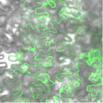
PI4Kg2 Regulates Root Development

                                                                              interestingly, it presents short roots (Fig. 1C). Consid-
                                                                              ering that primary root growth is dependent on both
                                                                              cell proliferation and cell elongation, detailed analysis
                                                                              revealed a decreased length of root elongation zone and
                                                                              decreased root epidermal cell length in the maturation
                                                                              zone for pi4kg2 (Fig. 1D). Considering that there was no
                                                                              change of length of meristematic zone of pi4kg2 roots
                                                                              (Supplemental Fig. S1D), these results suggest that
                                                                              PI4Kg2 regulates root growth by modulating cell length.
                                                                              As expected, complementation studies demonstrated
                                                                              that reinstated PI4Kg2 expression (Supplemental Fig. S2)
                                                                              resulted in recovered root growth, confirming the role of
                                                                              PI4Kg2 in root development.

                                                                              PI4Kg2 Interacts with MIEL1

                                                                                 To investigate the functional mechanism of how
                                                                              PI4Kg2 regulates root cell elongation, yeast two-hybrid
                                                                              screening was performed using whole PI4Kg2 protein
                                                                              as bait to search for PI4Kg2-interacting partners. In-
                                                                              terestingly, MIEL1, a zinc finger RING-type protein
                                                                              localized to the cytoplasm and nucleus (Marino et al.,
                                                                              2013), was identified (Fig. 2A). Subcellular localization
                                                                              studies by observing fluorescence showed the distri-
                                                                              bution of yellow fluorescent protein (YFP)-PI4Kg2 fu-
                                                                              sion protein in cytoplasm and nucleus of Arabidopsis
                                                                              cells, which is similar to that of MIEL1 (Fig. 2B, upper).
                                                                              Transient expression of YFP-PI4Kg2 and cyan fluores-
                                                                              cent protein (CFP)-MIEL1 fusion proteins in Arabi-
                                                                              dopsis protoplasts confirmed that PI4Kg2 colocalizes
                                                                              with MIEL1 (Fig. 2B, lower), further suggesting a pos-
                                                                              sible interaction between them, which was investigated
                                                                              by using bimolecular fluorescence complementation
                                                                              (BiFC) assays. Indeed, strong fluorescence was detected
                                                                              in the nucleus and cytoplasm of cells co-expressing
                                                                              cYFP-MIEL1 and nYFP-PI4Kg2 (Fig. 2C), confirming
                                                                              the PI4Kg2-MIEL1 interaction in vivo.

Figure 1. Deficiency of PI4Kg2 results in shortened roots. A, RT-qPCR         Deficiency of MIEL1 and MYB30 Results in Shorter or
analysis revealed PI4Kg2 expression in various tissues including leaf,
                                                                              Longer Roots, Respectively
stem, flower, root, and silique. ACTIN7 gene was used as an internal
reference. Analysis were biologically repeated three times and data are         Previous studies showed that MIEL1 interacts with
presented as means 6 SE (n 5 3). B, Promoter–GUS fusion analysis              and ubiquitinates MYB30, leading to MYB30 protea-
revealed that PI4Kg2 is expressed in young seedlings (1), rosette leaf (2),
                                                                              somal degradation and suppressed plant defense re-
cauline leaf (3), stem (4), inflorescence (5), and floral tissue (6). Scale
bars 5 200 mm (1 and 6) and 2 mm (2–5). C, Observation (left) and
                                                                              sponses (Marino et al., 2013). Considering that PI4Kg2
measurement (right) of root growth of 10-d–old wild-type (WT), pi4kg2,        interacts with MIEL1, we thus investigated the function
and pi4kg2 seedlings expressing p35S::PI4Kg2-mCherry. Scale bar 5             of MIEL1 and MYB30 in root growth. Observation
1 cm. Experiments were repeated three times and data are presented as         of growth of miel1 (Marino et al., 2013) and myb30
means 6 SD (n . 30). Statistical analysis revealed significant difference     knockout lines (Liu et al., 2014) showed that, similar to
(**P , 0.01). D, pi4kg2 plants exhibited a decreased root elongation          pi4kg2 mutant, miel1 exhibited shortened roots and
zone length and root epidermal cell length in the maturation zone             decreased length of root elongation zone and epidermal
compared to those of wild type. Roots of 7-d–old seedlings were               cells of the root maturation zone, whereas myb30
measured and statistically analyzed (**P , 0.01). Experiments were            showed longer root length and increased length of root
repeated three times and data are presented as means 6 SD (n 5 30).
                                                                              elongation zone and epidermal cells of root maturation
                                                                              zone (Fig. 3), which is consistent with MIEL1-facilitated
                                                                              MYB30 degradation and indicates a specific role for
                                                                              MIEL1 and MYB30 in regulating root growth.
Plant Physiol. Vol. 184, 2020                                                                                                         935

                                 Downloaded on January 28, 2021. - Published by https://plantphysiol.org
                                Copyright (c) 2020 American Society of Plant Biologists. All rights reserved.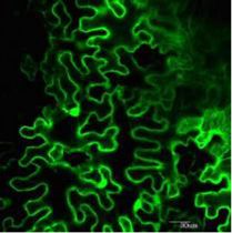
Zhao and Xue

                                                                         PI4Kg2 Regulates MIEL1 Stability and Accelerates MYB30
                                                                         Turnover In Planta

                                                                            Many reports in animals have shown that functions
                                                                         of E3 ubiquitin ligase can be regulated at the post-
                                                                         translational level, including de-neddylation, phos-
                                                                         phorylation, and interaction with other proteins. To test
                                                                         whether MIEL1 is regulated by PI4Kg2 through direct
                                                                         interaction in planta, MIEL1 fused with mCherry was
                                                                         expressed in wild type and pi4kg2 to study the effects of
                                                                         PI4Kg2 on MIEL1 in vivo.
                                                                            A quick cross was performed to obtain the isogenic
                                                                         MIEL1-mCherry plants. Pollens from MIEL1-mCherry
                                                                         (in pi4kg2) homozygous lines were respectively spread
                                                                         on the stigmas of pi4kg2 and wild type, and analysis
                                                                         of F1 plants with comparable MIEL1 expression
                                                                         levels by immunoblot analysis showed that MIEL1-
                                                                         mCherry accumulated less in pi4kg2 compared to that
                                                                         in PI4Kg2/pi4kg2 (Fig. 4A), suggesting that PI4Kg2

Figure 2. PI4Kg2 interacts with E3 ligase MIEL1. A, Yeast two-hybrid
assays revealed a direct interaction between PI4Kg2 and MIEL1.
Transformed yeast cells were grown on SD-Leu-Trp or SD-Leu-Trp-His-
Ade. AD, Activation domain; BD, binding domain. B, Observation of
transiently expressed fusion proteins in Arabidopsis protoplasts
showed that PI4Kg2 colocalizes with MIEL1. Scale bars 5 10 mm. C,
BiFC assays confirmed the MIEL1-PI4Kg2 interaction in planta. A
model no. FV1000 confocal microscope (Olympus) was used and
collection wavelength was set at 510 to 550 nm to strictly filter out
the autofluorescence (details in “Materials and Methods” section),
resulting in two control images (upper, middle and right) that are
blank. Scale bars 5 30 mm.                                               Figure 3. miel1 and myb30 mutants have shortened or elongated roots.
                                                                         A, Root length of 12-d-old wild type (WT), pi4kg2, miel1, myb30,
                                                                         pi4kg2 miel1, pi4kg2 myb30, and miel1 myb30 seedlings were ob-
  Genetic analysis showed that miel1 pi4kg2 double
                                                                         served (left) and measured (right). Scale bar 5 1 cm. B, Length of root
mutants displayed similar root growth as miel1 and
                                                                         elongation zone of 7-d-old wild type, pi4kg2, miel1, and myb30
pi4kg2, whereas pi4kg2 myb30 or miel1 myb30 double                       seedlings. C, Length of epidermal cell of root maturation zone of 7-d-old
mutants presented root growth similar to myb30 (longer                   wild type, pi4kg2, miel1, and myb30 seedlings. Experiments were re-
roots), indicating the genetic epistasis of MYB30 and                    peated three times and data are presented as means 6 SD (n . 30).
that root growth regulation by PI4Kg2 and MIEL1 is                       Statistical analysis was performed using a two-tailed Student’s t test
achieved via the effect of MIEL1 on MYB30.                               compared with wild type (**P , 0.01).

936                                                                                                                 Plant Physiol. Vol. 184, 2020

                                  Downloaded on January 28, 2021. - Published by https://plantphysiol.org
                                 Copyright (c) 2020 American Society of Plant Biologists. All rights reserved.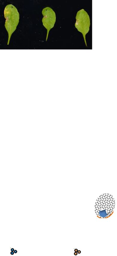
PI4Kg2 Regulates Root Development
functions in regulating MIEL1 stability. Consistently, and the resultant precipitates were subsequently ana-
transient expression of FLAG-tagged MIEL1 alone or lyzed by immunoblot assays with anti-Ub or anti-
with Myc-tagged PI4Kg2 in Nicotiana benthamiana leaves mCherry antibody (Fig. 4B, left bottom). Indeed, anal-
further showed greater accumulation of MIEL1 protein ysis revealed increased ubiquitination of MIEL1 in the
after the co-expression of PI4Kg2 (Supplemental Fig. pi4kg2 mutant (Fig. 4B, upper; Supplemental Fig. S3B),
S3A). These results indicate that PI4Kg2 positively reg- confirming that PI4Kg2 affects MIEL1 ubiquitination
ulates MIEL1 stability through direct interaction. via their interaction.
MIEL1 is degraded by the ubiquitin-26S proteasome Several type-II PI4K members (PI4Kg1, PI4Kg4,
pathway (Lee and Seo, 2016). Considering the PI4Kg2 PI4Kg5, and PI4Kg7) are Ser/Thr protein kinases
effects on MIEL1 stability, whether PI4Kg2 affects the (Galvão et al., 2008; Tang et al., 2016); however, in vitro
ubiquitination of MIEL1 in vivo and hence, degrada- phosphorylation assay by autoradiograph showed that
tion, was investigated by immunoblot analysis. Total there was no autophosphorylation of PI4Kg2 and that
protein extracted from 35S::MIEL1-mCherry (pi4kg2) PI4Kg2 does not phosphorylate MIEL1 (Supplemental
and 35S::MIEL1-mCherry (PI4Kg2/pi4kg2) seedlings Fig. S3C), further indicating that PI4Kg2 affects MIEL1
were immunoprecipitated with an anti-mCherry antibody, ubiquitination and stability via their interaction.
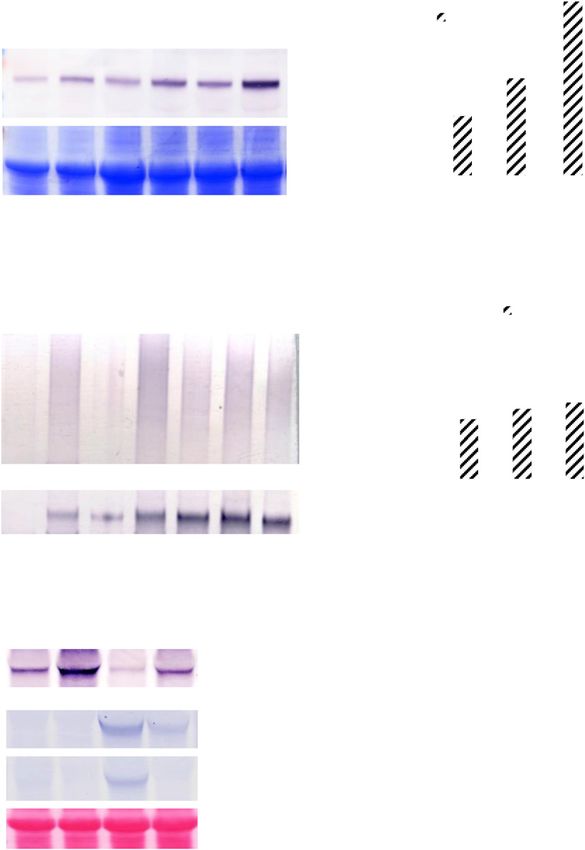
Figure 4. PI4Kg2 regulates MIEL1 stability and
accelerates MYB30 turnover in planta. A, Im-
munoblot analysis showed that deficiency
of PI4Kg2 leads to reduced accumulation of
MIEL1-mCherry. Wild-type (WT) lines express-
ing MIEL1 (PI4Kg2/pi4kg2) were generated by
crossing homozygous pi4kg2 lines expressing
MIEL1. Seven-day-old seedlings were used to
extract proteins and analyzed with an mCherry
antibody (leaf upper). Coomassie Brilliant Blue
staining showed the protein loading (leaf bot-
tom). Band density of MIEL1-mCherry was
measured using the software ImageJ (https://
imagej.en.softonic.com/) by setting band den-
sity in pi4kg2 (L6) as “1” (right). B, PI4Kg2
regulates the ubiquitination of MIEL1. Immu-
noprecipitation (IP) assays showed that defi-
ciency of PI4Kg2 leads to increased ubiquitination
of MIEL1. Wild-type lines expressing MIEL1
(PI4Kg2/pi4kg2) were generated by crossing
homozygous pi4kg2 lines expressing MIEL1.
Seven-day-old seedlings were used to extract
proteins and analyzed using the a-Ubiquitin
(a-UB) antibody (left upper). Loading proteins
were quantified by using an mCherry antibody
(left bottom). Band density of ubiquitinated
MIEL1 was measured using the software ImageJ
by setting band density in PI4Kg2/pi4kg2 (L6) as
“1” (right). C, PI4Kg2 accelerated the degrada-
tion of MYB30. FLAG-tagged MYB30 was
expressed in N. benthamiana in the absence or
presence of mCherry-tagged MIEL1 or Myc-
tagged PI4Kg2, or treated with proteasome in-
hibitor MG132. Levels of MYB30, MIEL1, and
PI4Kg2 were analyzed by immunoblotting with
relevant antibodies. Ponceau-S staining con-
firmed the equal loading of proteins. Band
density is measured by the software ImageJ and
relative density of MYB30-FLAG was calculated
by dividing the density of MYB30-Flag by that
of Rubisco. Data are presented as means 6 SD
(n 5 3).
Plant Physiol. Vol. 184, 2020 937
Downloaded on January 28, 2021. - Published by https://plantphysiol.org
Copyright (c) 2020 American Society of Plant Biologists. All rights reserved.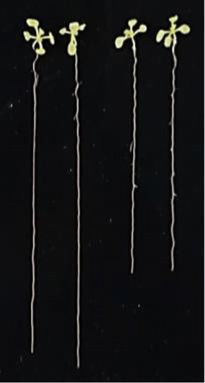
Zhao and Xue

        Figure 5. PI4Kg2 deficiency results in reduced auxin level and MYB30 deficiency results in altered expression levels of GH3 or
        YUC genes in planta. A, Quantification of free IAA content by LC-MS revealed a reduced IAA level in pi4kg2. Roots of 5-d-old
        wild-type (WT) and pi4kg2 seedlings were used for analysis and data are presented as means 6 SD (n 5 5). Statistical analysis was

938                                                                                                                   Plant Physiol. Vol. 184, 2020

                                 Downloaded on January 28, 2021. - Published by https://plantphysiol.org
                                Copyright (c) 2020 American Society of Plant Biologists. All rights reserved.
PI4Kg2 Regulates Root Development
MIEL1 mediates the proteasomal degradation of suppress auxin synthesis; He et al., 2011) was utilized to
MYB30 (Marino et al., 2013). We thus examined whether test whether the phenotype of myb30 was the result of
PI4Kg2-MIEL1 interaction influences the degradation increased auxin accumulation. Indeed, the longer root
and turnover of MYB30. Analysis of N. benthamiana of myb30 was suppressed under L-Kyn treatment (sim-
leaves transiently expressing FLAG-tagged MYB30 ilar root length was observed at 2 mM of L-Kyn; Fig. 5C),
showed that MYB30 protein accumulated after treat- suggesting the longer roots of myb30 might be due to
ment with proteasome inhibitor MG132 and was re- increased auxin content. This was confirmed by in-
duced after the co-expression of MIEL1 (Fig. 4C), which creased free IAA level of myb30 roots demonstrated by
is consistent with the previous reports (Marino et al., LC-MS (Fig. 5D). As expected, RT-qPCR analysis of
2013). Interestingly, MYB30 protein level was signifi- expression of some IAA metabolism-related genes
cantly reduced when co-expressed with PI4Kg2 and showed that GH3 members (GH3.1, GH3,2, GH3.4,
MIEL1 (Fig. 4C), indicating that PI4Kg2 regulates the GH3.6, GH3.7, GH3.13, GH3.17, GH3.18, and GH3.19,
turnover of MYB30 through interacting with and stabi- encoding auxin-metabolism enzymes) presented de-
lizing MIEL1. creased expression levels, whereas YUC4 and YUC7
(encoding key enzymes in auxin biosynthesis) pre-
PI4Kg2 Regulates Auxin Metabolism through MYB30 sented increased expression levels in myb30 roots
(Fig. 5E). Further analysis showed that expression
Root growth is a complex process and regulated by levels of GH3 members (GH3.1, GH3.2, GH3.4, GH3.17,
various developmental signals and environmental fac- GH3.18, and GH3.19) were increased in pi4kg2 and
tors, particularly phytohormones. Auxin level, signal- miel1 roots (Fig. 5F). GH3 proteins synthesize IAA-
ing, and polar transport are involved in root growth amino acid conjugates, either the intermediate des-
regulation. Considering that free indole-3-acetic acid tined for IAA metabolism or the inactive storage form
(IAA) plays main roles in auxin effects, we first inves- of IAA, to reduce the free IAA level. Increased (or de-
tigated whether IAA level is altered in pi4kg2 by liquid creased) expression levels of GH3s are consistent with
chromatography-mass spectrometry (LC-MS) analysis the reduced auxin contents in pi4kg2 and miel1 (or
and results revealed decreased free IAA content in myb30), and confirmed that PI4Kg2 regulates auxin
pi4kg2 roots (Fig. 5A). Consistently, the short-root metabolism and root growth by interacting with MILE1
phenotype of pi4kg2 and miel1 was recused by exoge- to modulate MYB30.
nous IAA and both mutants presented similar root MYB30 is a R2R3-MYB transcriptional activation
length under 0.01 mM of IAA (Fig. 5B), confirming that factor involved in the very first step of HR (Vailleau
reduced IAA level leads to the short-root phenotype of et al., 2002). Interestingly, prediction analysis (The
pi4kg2 and miel1. Arabidopsis Gene Regulatory Information Server,
Considering the genetic epistasis of MYB30 on http://arabidopsis.med.ohio-state.edu/) revealed the
PI4Kg2 and MILE1, and that MIEL1 mediates the pro- presence of putative binding sequences of MYB mem-
teasomal degradation of MYB30, it was hypothesized bers (Supplemental Table S1). A chromatin immuno-
that MYB30 negatively regulates auxin metabolism precipitation (ChIP)-qPCR analysis with an anti-Flag
and hence, cell elongation. The chemical compound antibody was subsequently performed to investigate
L-Kynurenine (L-Kyn, an inhibitor of TAA1/TAR to whether MYB30 directly binds the promoter of these
Figure 5. (Continued.)
performed using a two-tailed Student’s t test (*P , 0.05, compared to wild type). B, Wild-type, miel1, and pi4kg2 seedlings were
grown on Murashige-and-Skoog medium supplemented with IAA for 15 d, and root lengths were measured. Experiments were
repeated three times and data are presented as means 6 SD (n . 30). Statistical analysis revealed significant differences compared
to wild type (*P , 0.05 and **P , 0.01). C, Wild-type and myb30 seedlings were grown on Murashige-and-Skoog medium
supplemented with IAA biosynthesis inhibitor L-Kynurenine (L-Kyn) for 10 d, and root lengths were measured. Experiments were
repeated three times and data are presented as means 6 SD (n . 30). Statistical analysis revealed the significant differences
compared to wild type (*P , 0.05 and **P , 0.01). D, Quantification of free IAA content by LC-MS revealed the increased IAA
level in myb30. Roots of 5-d-old wild type and myb30 seedlings were used for analysis and data are presented as means 6 SD
(n 5 5). Statistical analysis was performed using a two-tailed Student’s t test compared to wild type (*P , 0.05). E, Relative
expression levels of YUCCA and GH3 genes in myb30 and wild type (expression of examined genes in wild type was set as “1”).
Roots of 7-d–old seedlings were used for RNA extraction and RT-qPCR analysis. ACTIN7 gene was used as an internal reference.
Experiments were repeated three times and data are presented as means 6 SE (n 5 3). Statistical analysis was performed using a
two-tailed Student’s t test compared to wild type (*P , 0.05 and **P , 0.01). F, Relative expression levels of GH3 genes in wild
type, pi4kg2, miel1, and myb30 (expression of examined genes in wild type was set as “1”). Roots of 7-d-old seedlings were used
for RNA extraction and RT-qPCR analysis. ACTIN7 gene was used as an internal reference. Experiments were repeated three times
and data are presented as means 6 SE (n 5 3). Statistical analysis was performed using a two-tailed Student’s t test compared to
wild type (*P , 0.05 and **P , 0.01). G, ChIP-qPCR analysis showed that MYB30 directly binds the promoter regions of GH3.2
and GH3.6 to regulate their expression. Two independent lines expressing MYB30-FLAG (L30 and L36) were used for analysis.
Input DNA (from the un-immunoprecipitated DNA) was used as a positive control (100%). Immunoprecipitated DNA from IgG
chromatin was used as a negative control. P1, P2, and P3 are the putative MYB30 binding sequences in promoters of GH3.2 and
GH3.6. Statistical analysis was performed using a two-tailed Student’s t test compared to Input DNA (*P , 0.05 and **P , 0.01).
Plant Physiol. Vol. 184, 2020 939
Downloaded on January 28, 2021. - Published by https://plantphysiol.org
Copyright (c) 2020 American Society of Plant Biologists. All rights reserved.Zhao and Xue
genes. A MYB30-Flag fusion protein (driven by CaMV35S
promoter) was expressed in wild-type seedlings and two
transgenic lines with comparable protein expression
levels were selected for a ChIP assay. qPCR analysis
revealed that distinct DNA fragments of GH3.2 (P2;
Fig. 5G) and GH3.6 (P3; Fig. 5G) promoters were more
enriched, indicating that MYB30 regulates the expression
levels of GH3.2 and GH3.6 genes by directly binding their
promoters.
PI4Kg2 Negatively Regulates Resistance and HR After
Bacterial Inoculation
MIEL1 attenuates HR to bacterial infection, consis-
tent with the enhanced or reduced responses of myb30
or MYB30-overexpression lines (Marino et al., 2013).
Observation showed that compared to chlorosis symp-
toms of wild type with Pseudomonas syringae pv. tomato
AvrRpm1 (Pst AvrRpm1), miel1 plants showed clear HR
cell death symptoms (Fig. 6A). Similar to miel1, pi4kg2
presented HR cell death symptoms as well at 64-h post
infection (hpi) with Pst AvrRpm1 (Fig. 6A), indicating the
important function of PI4Kg2 in plant response to bac-
terial inoculation. Consistent with the faster HR, pi4kg2
showed increased resistance in response to inocula-
tion with Pst AvrRpm1 compared to wild-type plants
(Fig. 6B), confirming that PI4Kg2 functions as a negative
regulator of plant defense.
RT-qPCR analysis of expression of MYB30 very long
chain fatty acid-related target genes, specifically KCS1
and FDH (Marino et al., 2013), showed their increased
expression in pi4kg2 and miel1 plants compared to wild-
type plants after bacterial inoculation (Fig. 6C), further
indicating that PI4Kg2 negatively mediates Arabi-
dopsis defense response, possibly by regulating MYB30
stability.
Figure 6. PI4Kg2 is a negative regulator of HR responses in response to
bacterial inoculation. A, Symptoms developed by pi4kg2, miel1, and
wild-type (WT) 64 hpi with Pst AvrRpm1 (2 3 106 cfu mL21). Experi-
DISCUSSION ments were repeated three times (.5 seedlings each time) and repre-
sentative images were shown. Scale bar 5 1 cm. B, Growth of Pst
Large numbers of E3 ubiquitin ligases (;1,500 E3- AvrRpm1 in pi4kg2, miel1, and wild type. Bacterial growth at 3 d was
encoding genes are predicted in the Arabidopsis ge- measured after inoculation (5 3 105 cfu mL21). Experiments were re-
nome; Guzmán, 2014) regulate multiple cellular and peated three times and data are presented as means 6 SD (n . 5). Sta-
biological processes by interacting with and degrading tistical analysis revealed significant differences compared to wild type
(**P , 0.01). C, Relative expression levels of KCS1 and FDH, the target
distinct target proteins (Merlet et al., 2009). Many re-
genes for MYB30. Wild-type, pi4kg2, and miel1 seedlings were used for
ports have shown that E3 ligases can be regulated extracting RNA after inoculation with Pst AvrRpm1 (5 3 107 cfu mL21)
through posttranslational modifications in animals, for 1 h. ACTIN7 gene was used as an internal reference. Experiments
which remain relatively undocumented in plants. Our were repeated three times and data are presented as means 6 SE (n 5 3).
studies functionally characterized the role of PI4Kg2, D, Model illustrating the regulation of PI4Kg2-MIEL1-MYB30 and the
an Arabidopsis atypical type-II PI4K, in root elonga- effects on plant growth and HR. In wild type, PI4Kg2 interacts with
tion and indicated that PI4Kg2 is an E3 ligase mediator MIEL1 to suppress its ubiquitination and degradation, leading to the
that inhibits ubiquitination of E3 (MIEL1) through proteasomal degradation of MYB30 by MIEL1. Reduced MYB30 protein
protein–protein interaction. These findings provide results in increased auxin content/signaling, and in turn promoted
further insights into Arabidopsis type-II PI4Ks and growth and suppressed HR to avoid superfluous activation. Under
PI4Kg2 deficiency, enhanced MIEL1 ubiquitination and degradation
help to elucidate the regulatory mechanism of plant
results in the suppressed ubiquitination and degradation of MYB30.
growth/development control, particularly the balance Accumulated MYB30 thereby activates the expression of target genes,
between plant resistance and development, at a post- resulting in decreased auxin level, suppressed growth, and stimulated HR.
translational level (Fig. 6D).
940 Plant Physiol. Vol. 184, 2020
Downloaded on January 28, 2021. - Published by https://plantphysiol.org
Copyright (c) 2020 American Society of Plant Biologists. All rights reserved.PI4Kg2 Regulates Root Development
MIEL1 is a C3H2C3 canonical RING-type E3 ligase superfluous activation of HR. Under PI4Kg2 deficiency,
and plays important roles in abscisic acid signaling and MIEL1 is ubiquitinated and degraded, thereby result-
plant defense responses; however, the upstream regu- ing in accumulated MYB30 protein, and hence acti-
lation of MIEL1 remains poorly understood. A previous vated HR and decreased auxin level/suppressed growth
study showed that MIEL1 can be ubiquitinated (Lee (Fig. 6D). Increased auxin enhances plant growth and
and Seo, 2016). We found that PI4Kg2 ensures the re- auxin-mediated susceptibility, whereas decreased auxin
duced ubiquitination and enhanced stability of MIEL1 results in growth inhibition and enhanced disease re-
(Fig. 4) through their interactions, leading to stimulated sistance. Considering the positive effect of PI4Kg2 on
MYB30 degradation, providing additional evidence as MIEL1 stability and negative effect of MIEL1 on MYB30
to how type-II PI4Ks confer their functions in plants. activity, these results manifest that PI4Kg2 may con-
Similarly, in mammalian cells, interaction between tribute to synergistic regulation of plant defense and
PLAGL2 and Pirh2 (Pirh2 encodes a RING-H2 domain- growth.
containing protein with intrinsic ubiquitin–protein
ligase activity) prevents the proteasomal degrada-
tion of Pirh2 (Zheng et al., 2007). These results sug-
gest that protein–protein interaction is a general MATERIALS AND METHODS
regulatory mechanism for ubiquitination and stabil-
Plant Materials and Growth Conditions
ity of E3 ligases; however, the exact function of
ubiquitin-like domains of type-II PI4Ks and the de- All Arabidopsis (Arabidopsis thaliana) plants used in this study were in the
tailed mechanisms behind this regulation require Columbia-0 (Col-0) background. The T-DNA insertion mutants, pi4kg2
(SAIL_11_B03), miel1 (SALK_097638), and myb30 (SALK_027644C) were
further investigation. obtained from the Arabidopsis Biological Resource Center (Li et al., 2009;
MYB30 is a pleiotropic mediator that regulates a Marino et al., 2013).
variety of physiological processes and signaling re- Seeds were surface-sterilized and sown on plates containing Murashige-
sponses, including pathogen-induced HR, flowering and-Skoog medium (Duchefe Biochemie). After stratification at 4°C for 3 d,
seedlings were grown in a phytotron with a 16-h light/8-h dark cycle (22°C) for
time, and brassinosteroid and abscisic acid signaling
normal growth and seed harvesting.
(Raffaele et al., 2006, 2008; Li et al., 2009; Zheng et al.,
2012; Raffaele and Rivas, 2013; Liu et al., 2014). How-
ever, its function in root cell elongation is unclear. Our Identification of T-DNA Mutants
studies show that MYB30 deficiency results in the
promoted cell elongation of roots by altering auxin Mutant pi4kg2 carrying a T-DNA insertion in the first exon was confirmed
metabolism, which is consistent with the decreased by PCR amplification using the primers PI4Kg2-L and PI4Kg2-R. Transcription
level of PI4Kg2 was examined by RT-qPCR using the primers PI4Kg2-3 and
expression of many GH3s and reveals a further function PI4Kg2-4. T-DNA insertion of mutant miel1 was confirmed by PCR amplifica-
of MYB30 in regulating auxin metabolism. tion using the primers MIEL1-1 and MIEL1-2. T-DNA insertion of mutant
Understanding control of the balance between plant myb30 was confirmed by PCR amplification using the primers MYB30-1 and
resistance and development is fundamental, and it was MYB30-2. All primers used in this study are listed in Supplemental Table S2.
recently shown that GA–abscisic acid and auxin–
jasmonic acid synergistically regulate resistance and
growth (Yuan et al., 2018). Interestingly, we found Constructs and Plant Transformation
that PI4Kg2-MIEL1-MYB30 regulates auxin-mediated For expression of PI4Kg2 in wild type or pi4kg2, the coding sequence of
growth and HR, and MYB30 may act in crosstalk of PI4Kg2 (1–903 bp) was amplified with primers (PI4Kg2-5 and PI4Kg2-6) and
growth and defense signaling. MYB30 positively reg- subcloned into the mCherry vector with C-terminal fusion. For expression of
cMyc-PI4Kg2 in Nicotiana benthamiana, the coding sequence of PI4Kg2 was
ulates HR cell death in response to pathogen attack
amplified (primers PI4Kg2-7 and PI4Kg2-8) and subcloned into a modified
(Vailleau et al., 2002) while inhibiting growth by de- pEGAD-4XcMyc vector with N-terminal fusion. For expression of MIEL1-Flag
creasing auxin level. Indeed, auxin is an important in N. benthamiana, the coding sequence of MIEL1 was amplified by PCR
plant growth regulator and also influences plant– (primers MIEL1-3 and MIEL1-4) and subcloned into 1306-Flag vector with
pathogen interactions, and previous studies showed C-terminal fusion. For expression of MIEL1-mCherry in wild type, pi4kg2, or
N. benthamiana, the coding sequence of MIEL1 was amplified by PCR (primers
that elevated IAA biosynthesis in plants led to en- MIEL1-5 and MIEL1-6) and subcloned into an mCherry vector with C-terminal
hanced susceptibility to DC3000 (Mutka et al., 2013). fusion. For expression of MYB30-Flag in N. benthamiana, the coding sequence of
Considering its myriad of regulatory targets and dif- MYB30 was amplified by PCR (primers MYB30-3 and MYB30-4) and subcloned
ferential expression of PI4Kg2-MIEL1-MYB30 after in- into the 1306-Flag vector with C-terminal fusion.
oculation with Pst AvrRpm1, i.e. PI4Kg2 and MYB30 Arabidopsis transformation was performed by the floral-dip method
(Clough and Bent, 1998).
expression was specifically induced at 4 and 1 hpi, re-
spectively, whereas MIEL1 expression was repressed
after inoculation at 1 hpi (Supplemental Fig. S4), it Promoter-Reporter Gene Fusion Studies
would seem that MYB30 acts on upstream effectors of
auxin signaling and HR progress. The promoter of PI4Kg2 (22,491 bp upstream of ATG) was amplified
In sum, PI4Kg2 interacts with MIEL1, thus inhibiting (primers PI4Kg2-P1/PI4Kg2–P2), then cloned into a modified pCAMBIA1300
vector including a GUS reporter. A histochemical assay of GUS activities was
its turnover and leading to increased proteasomal performed according to Tang et al. (2016). Arabidopsis transformation samples
degradation of MYB30, which attenuates auxin me- were observed using differential interference contrast microscopy (SMZ1500;
tabolism, and hence increases auxin level to avoid Nikon).
Plant Physiol. Vol. 184, 2020 941
Downloaded on January 28, 2021. - Published by https://plantphysiol.org
Copyright (c) 2020 American Society of Plant Biologists. All rights reserved.Zhao and Xue
RT-qPCR Analysis vector pA7 (N terminus fusion with YFP or CFP, respectively), resulting in the
YFP-PI4Kg2 and CFP-MIEL1 fusion constructs, which were transiently
RT-qPCR analysis was performed to examine the PI4Kg2 transcription in expressed in Arabidopsis protoplasts by PEG/CaCl2 methods (Yoo et al., 2007).
various tissues, expression of MIEL1 in N. benthamiana (primers MIEL1–7 and The fluorescence was observed by confocal laser scanning microscopy (model
MIEL1–8), and expression of MYB30, GH3.1, GH3.2, GH3.4, GH3.6, GH3.7, no. FV1000; Olympus). For YFP/CFP, we used excitation/emission combina-
GH3.13, GH3.17, GH3.18, GH3.19, YUC4, YUC7, GH3.2-P1, GH3.2-P2, GH3.6- tions of 514 nm/530 to 580 nm for YFP and 458 nm/470 to 500 nm for CFP. The
P1, GH3.6-P2, GH3.6-P3, KCS1, and FDH in Arabidopsis (primers are listed in laser power was set at 20% for the 458-nm laser and 5% for the 514-nm lasers.
Supplemental Table S2). Total RNA was extracted from various tissues and All images were collected using a 203 aperture objective.
used to synthesize complementary DNA by reverse transcription. The ACTIN7
(AT5G09810) and Elongation Factor1a (EF1a) genes were amplified (primers
ACTIN7-1, ACTIN7-2, EF1a-1, and EF1a-2; Geng et al., 2017) and used as in- Recombinant Expression of PI4Kg2 and MIEL1, and In
ternal reference. Vitro Kinase Assay
The coding region of PI4Kg2 was amplified (primers PI4Kg2-15 and PI4Kg2-
Measurement of Free IAA Contents by LC-MS 16) and subcloned into the pGEX-4T-1 vector (Novagen). The coding region of
MIEL1 was amplified (primers MIEL1-15 and MIEL1-16) and subcloned into
Approximately 200 mg of roots from 10-d-old seedlings was frozen in liquid
the vector pET28a (Novagen). All constructs were confirmed by sequencing.
nitrogen and ground to a fine powder for free IAA content measurement using
PI4Kg2 and MIEL1 proteins were expressed in Escherichia coli (strain BL21)
a TSQ Quantum Ultra LC-MS-MS system (Thermo Fisher Scientific) according
supplemented with 1 mM of isopropyl-b-D-thiogalactoside (28°C, 3 h), or
to the description in Tang et al. (2016).
expressed in E. coli (strain Rosetta) supplemented with 0.1 mM of isopropyl-
b-D-thiogalactoside (16°C, 15 h). Proteins were then purified using Ni-NTA His-
Immunoblot Analysis binding resin (Novagen) or Glutathione Sepharose (Novagen) according to the
manufacturer protocols.
Isogenic MIEL1-mCherry plants were first prepared by crossing MIEL1- The assay of kinase activity was performed according to a description in Tan
mCherry (in pi4kg2) homozygous lines (as the male parent) with wild type et al. (2013) with a few modifications.
and pi4kg2, respectively. Protein extracted from transgenic plants was resus-
pended in extraction buffer (20 mM of Tris-HCl at pH 7.5, 150 mM of NaCl, 0.5%
[v/v] TWEEN 20, 1 mM of EDTA, and 1 mM of dithiothreitol) containing a Bacterial Materials
protease inhibitor cocktail (Roche). After addition of an equal volume of 23 SDS
buffer, the samples were boiled for 5 min, fractionated by 10% SDS-PAGE, and Arabidopsis 4-week-old plants were kept at high humidity for 12 h before
transferred to a polyvinylidene fluoride membrane by semidry blotting. The inoculation and injected with a bacterial suspension of Pst AvrRpm1 at the in-
blots were incubated with a mouse anti-mCherry antibody (Abcam) and then dicated bacterial densities using a blunt syringe on the abaxial side of the leaves.
with a bovine anti-mouse IgG alkaline phosphatase-conjugated secondary an- For measure of in planta bacterial growth, injected leaves samples were har-
tibody (Santa Cruz Biotechnology). Alkaline phosphatase activity was detected vested 0 and 3 d. A predetermined dilution for each sample was plated on
by BCIP/NBT Detection Reagents (Invitrogen). King’s B medium and incubated at 28°C for 2 d. Data were submitted to a
For mCherry immunoprecipitation assays, the de-ubiquitination inhibitor PR- statistical analysis (Marino et al., 2013).
619 (Sigma-Aldrich) was added into the extraction buffer. Supernatants were pre-
cleared for 1 h at 4°C with Dynabeads (Life Technology). Then samples were in-
cubated with Anti-mCherry Dynabeads (Life Technology) overnight. All steps were ChIP-qPCR Assay
performed at 4°C. Pulled-down proteins were eluted by 23 SDS buffer, then boiled
for 5 min. MIEL1-immunoprecipitated fractions and ubiquitination of MIEL1 were Ten-day-old wild-type seedlings expressing 35S::MYB30-FLAG were used
analyzed by using anti-mCherry or anti-Ubiquitin antibody, respectively. for ChIP analysis. The ChIP assay was performed according to the manufac-
turer’s protocol of the EpiQuik Plant ChIP Kit (catalog no. P-2014; Epigenetics).
qPCR analysis using Input DNA, immunoprecipitated DNA from IgG anti-
Yeast Two-Hybrid Analysis body, and Flag antibody as templates, respectively, was performed to examine
the fraction of DNA fragments including MYB30 binding sequence in GH3.2
The coding sequence of PI4Kg2 was amplified (primers PI4Kg2-9 and and GH3.6 promoters. Input DNA from un-immunoprecipitated DNA was
PI4Kg2-10) and subcloned into pGBKT7 or pGADT7 vectors. For yeast two- used as a positive control (100%). The negative control was immunoprecipi-
hybrid screening, BD-PI4Kg2 was used as bait to screen the candidate inter- tated DNA from IgG antibody that binds with a nonspecific target and the
acting proteins on synthetic dropout (SD) medium (-Leu/-Trp/-His/-Ade; associated DNA fragments were immunoprecipitated.
Clontech) plates. The pGADT7-MIEL1 construct was generated using primers
MIEL1-9 and MIEL1-10.
For auxotroph assays, candidate clones were streaked onto SD medium Accession Numbers
(-Leu/-Trp/-His/-Ade) and grown at 30°C for 4 d.
Sequence data from this article can be found in the GenBank/EMBL data
libraries under accession numbers listed in Supplemental Tables S1 and S2.
BiFC Assays
The coding regions of PI4Kg2 (primers PI4Kg2-11 and PI4Kg2-12) and
Supplemental Data
MIEL1 (primers MIEL1-11 and MIEL1-12) were fused with nYFP or cYFP, re-
spectively. Resultant constructs PI4Kg2-nYFP and MIEL1-cYFP were trans- The following supplemental materials are available.
formed into Agrobacterium tumefaciens and observed after 48 h according to
Fang and Spector (2010). Considering YFP was excited at 488 nm and emission Supplemental Figure S1. Characterization of pi4kg2 mutant.
at 510 to 580 nm, and chlorophyll autofluorescence was emission at 644 to 714 Supplemental Figure S2. Analysis of pi4kg2 lines expressing PI4Kg2.
nm (Vermeer et al., 2006; Body et al., 2019), BiFC results were imaged with a
model no. FV1000 confocal microscope (Olympus) using the 488-nm argon laser Supplemental Figure S3. PI4Kg2 regulates the stability and ubiquitination
and a dichroic filter to visualize YFP. The laser power was set at 5% for the 488- of MIEL1.
nm lasers. All images were collected using a 203 aperture objective. Collection Supplemental Figure S4. PI4Kg2, MYB30, and MIEL1 genes are spatially
wavelength was set at 510 to 550 nm. and temporally accumulated under inoculation.
Supplemental Table S1. Candidate binding sequences of MYB proteins in
Colocalization Studies of PI4Kg2 and MIEL1 promoter of GH3 and YUC genes.
The coding regions of PI4Kg2 (primers PI4Kg2-13 and PI4Kg2-14) and Supplemental Table S2. Primers used for mutant genotyping and plasmid
MIEL1 (primers MIEL1-13 and MIEL1-14) were amplified and subcloned into construction.
942 Plant Physiol. Vol. 184, 2020
Downloaded on January 28, 2021. - Published by https://plantphysiol.org
Copyright (c) 2020 American Society of Plant Biologists. All rights reserved.PI4Kg2 Regulates Root Development
ACKNOWLEDGMENTS Liu L, Zhang J, Adrian J, Gissot L, Coupland G, Yu D, Turck F (2014)
Elevated levels of MYB30 in the phloem accelerate flowering in Arabi-
We thank Lang-Tao Xiao (Hunan Agricultural University) for assistance
dopsis through the regulation of FLOWERING LOCUS T. PLoS One 9:
with quantification of IAA contents by LC-MS, and Xiao-Shu Gao (Shanghai
e89799
Institute of Plant Physiology and Ecology, Chinese Academy of Sciences) for
Liu P, Xu ZS, Pan-Pan L, Hu D, Chen M, Li LC, Ma YZ (2013) A wheat
microscopy observation.
PI4K gene whose product possesses threonine autophophorylation ac-
Received June 19, 2020; accepted July 20, 2020; published August 11, 2020. tivity confers tolerance to drought and salt in Arabidopsis. J Exp Bot 64:
2915–2927
Logan IR, Sapountzi V, Gaughan L, Neal DE, Robson CN (2004) Control
LITERATURE CITED of human PIRH2 protein stability: involvement of TIP60 and the pro-
teasome. J Biol Chem 279: 11696–11704
Akhter S, Uddin MN, Jeong IS, Kim DW, Liu XM, Bahk JD (2016) Role of Marino D, Froidure S, Canonne J, Ben Khaled S, Khafif M, Pouzet C,
Arabidopsis AtPI4Kg3, a type II phosphoinositide 4-kinase, in abiotic Jauneau A, Roby D, Rivas S (2013) Arabidopsis ubiquitin ligase MIEL1
stress responses and floral transition. Plant Biotechnol J 14: 215–230 mediates degradation of the transcription factor MYB30 weakening
Alonso JM, Stepanova AN, Leisse TJ, Kim CJ, Chen H, Shinn P,
plant defence. Nat Commun 4: 1476
Stevenson DK, Zimmerman J, Barajas P, Cheuk R, et al (2003) Mei Y, Jia WJ, Chu YJ, Xue HW (2012) Arabidopsis phosphatidylinositol
Genome-wide insertional mutagenesis of Arabidopsis thaliana. Science monophosphate 5-kinase 2 is involved in root gravitropism through
301: 653–657
regulation of polar auxin transport by affecting the cycling of PIN
Bagashev A, Fan S, Mukerjee R, Claudio PP, Chabrashvili T, Leng RP,
proteins. Cell Res 22: 581–597
Benchimol S, Sawaya BE (2013) Cdk9 phosphorylates Pirh2 protein and
Merlet J, Burger J, Gomes JE, Pintard L (2009) Regulation of cullin-RING
prevents degradation of p53 protein. Cell Cycle 12: 1569–1577
E3 ubiquitin-ligases by neddylation and dimerization. Cell Mol Life Sci
Barbosa IC, Shikata H, Zourelidou M, Heilmann M, Heilmann I,
66: 1924–1938
Schwechheimer C (2016) Phospholipid composition and a polybasic
Mueller-Roeber B, Pical C (2002) Inositol phospholipid metabolism in
motif determine D6 PROTEIN KINASE polar association with the
Arabidopsis. Characterized and putative isoforms of inositol phospho-
plasma membrane and tropic responses. Development 143: 4687–4700
lipid kinase and phosphoinositide-specific phospholipase C. Plant
Body MJA, Dave DF, Coffman CM, Paret TY, Koo AJ, Cocroft RB, Appel
Physiol 130: 22–46
HM (2019) Use of yellow fluorescent protein fluorescence to track OPR3
Mutka AM, Fawley S, Tsao T, Kunkel BN (2013) Auxin promotes
expression in Arabidopsis thaliana responses to insect herbivory. Front
susceptibility to Pseudomonas syringae via a mechanism independent
Plant Sci 10: 1586
of suppression of salicylic acid-mediated defenses. Plant J 74:
Chapman LA, Goring DR (2011) Misregulation of phosphoinositides in
746–754
Arabidopsis thaliana decreases pollen hydration and maternal fertility.
Nemoto K, Ramadan A, Arimura GI, Imai K, Tomii K, Shinozaki K,
Sex Plant Reprod 24: 319–326
Sawasaki T (2017) Tyrosine phosphorylation of the GARU E3 ubiquitin
Clough SJ, Bent AF (1998) Floral dip: A simplified method for
ligase promotes gibberellin signalling by preventing GID1 degradation.
Agrobacterium-mediated transformation of Arabidopsis thaliana. Plant J
Nat Commun 8: 1004
16: 735–743
Okazaki K, Miyagishima SY, Wada H (2015) Phosphatidylinositol 4-
Duan S, Yao Z, Hou D, Wu Z, Zhu WG, Wu M (2007) Phosphorylation of
phosphate negatively regulates chloroplast division in Arabidopsis.
Pirh2 by calmodulin-dependent kinase II impairs its ability to ubiq-
Plant Cell 27: 663–674
uitinate p53. EMBO J 26: 3062–3074
Péret B, Li G, Zhao J, Band LR, Voß U, Postaire O, Luu DT, Da Ines O,
Fang Y, Spector DL (2010) BiFC imaging assay for plant protein-protein
interactions. Cold Spring Harb Protoc 2: pdb prot5380 Casimiro I, Lucas M, et al (2012) Auxin regulates aquaporin function to
Galvão RM, Kota U, Soderblom EJ, Goshe MB, Boss WF (2008) Charac- facilitate lateral root emergence. Nat Cell Biol 14: 991–998
terization of a new family of protein kinases from Arabidopsis con- Preuss ML, Schmitz AJ, Thole JM, Bonner HK, Otegui MS, Nielsen E
taining phosphoinositide 3/4-kinase and ubiquitin-like domains. (2006) A role for the RabA4b effector protein PI-4Kbeta1 in polarized
Biochem J 409: 117–127 expansion of root hair cells in Arabidopsis thaliana. J Cell Biol 172:
Geng C, Wang HY, Liu J, Yan ZY, Tian YP, Yuan XF, Gao R, Li XD (2017) 991–998
Transcriptomic changes in Nicotiana benthamiana plants inoculated with Qi Y, Wang S, Shen C, Zhang S, Chen Y, Xu Y, Liu Y, Wu Y, Jiang D (2012)
the wild-type or an attenuated mutant of Tobacco vein banding mosaic OsARF12, a transcription activator on auxin response gene, regulates
virus. Mol Plant Pathol 18: 1175–1188 root elongation and affects iron accumulation in rice (Oryza sativa). New
Guzmán P (2014) ATLs and BTLs, plant-specific and general eukaryotic Phytol 193: 109–120
structurally-related E3 ubiquitin ligases. Plant Sci 215-216: 69–75 Raffaele S, Rivas S (2013) Regulate and be regulated: Integration of de-
He W, Brumos J, Li H, Ji Y, Ke M, Gong X, Zeng Q, Li W, Zhang X, An F, fense and other signals by the AtMYB30 transcription factor. Front Plant
et al (2011) A small-molecule screen identifies L-kynurenine as a com- Sci 4: 98
petitive inhibitor of TAA1/TAR activity in ethylene-directed auxin bi- Raffaele S, Rivas S, Roby D (2006) An essential role for salicylic acid in
osynthesis and root growth in Arabidopsis. Plant Cell 23: 3944–3960 AtMYB30-mediated control of the hypersensitive cell death program in
Ischebeck T, Seiler S, Heilmann I (2010) At the poles across kingdoms: Arabidopsis. FEBS Lett 580: 3498–3504
Phosphoinositides and polar tip growth. Protoplasma 240: 13–31 Raffaele S, Vailleau F, Léger A, Joubès J, Miersch O, Huard C, Blée E,
Kang BH, Nielsen E, Preuss ML, Mastronarde D, Staehelin LA (2011) Mongrand S, Domergue F, Roby D (2008) A MYB transcription
Electron tomography of RabA4b- and PI-4Kb1-labeled trans Golgi net- factor regulates very-long-chain fatty acid biosynthesis for activation
work compartments in Arabidopsis. Traffic 12: 313–329 of the hypersensitive cell death response in Arabidopsis. Plant Cell
Kelley DR (2018) E3 ubiquitin ligases: Key regulators of hormone signaling 20: 752–767
in plants. Mol Cell Proteomics 17: 1047–1054 Rape M (2018) Ubiquitylation at the crossroads of development and dis-
Lee HG, Seo PJ (2016) The Arabidopsis MIEL1 E3 ligase negatively regu- ease. Nat Rev Mol Cell Biol 19: 59–70
lates ABA signalling by promoting protein turnover of MYB96. Nat Schwechheimer C (2018) NEDD8—its role in the regulation of Cullin-RING
Commun 7: 12525 ligases. Curr Opin Plant Biol 45(Pt A): 112–119
Li L, Yu X, Thompson A, Guo M, Yoshida S, Asami T, Chory J, Yin Y Street IH, Mathews DE, Yamburkenko MV, Sorooshzadeh A, John RT,
(2009) Arabidopsis MYB30 is a direct target of BES1 and cooperates with Swarup R, Bennett MJ, Kieber JJ, Schaller GE (2016) Cytokinin acts
BES1 to regulate brassinosteroid-induced gene expression. Plant J 58: through the auxin influx carrier AUX1 to regulate cell elongation in the
275–286 root. Development 143: 3982–3993
Lin DL, Yao HY, Jia LH, Tan JF, Xu ZH, Zheng WM, Xue HW (2020) Takase T, Nakazawa M, Ishikawa A, Kawashima M, Ichikawa T,
Phospholipase D-derived phosphatidic acid promotes root hair devel- Takahashi N, Shimada H, Manabe K, Matsui M (2004) ydk1-D, an
opment under phosphorus deficiency by suppressing vacuolar degra- auxin-responsive GH3 mutant that is involved in hypocotyl and root
dation of PIN-FORMED2. New Phytol 226: 142–155 elongation. Plant J 37: 471–483
Plant Physiol. Vol. 184, 2020 943
Downloaded on January 28, 2021. - Published by https://plantphysiol.org
Copyright (c) 2020 American Society of Plant Biologists. All rights reserved.Zhao and Xue
Tan ST, Dai C, Liu HT, Xue HW (2013) Arabidopsis casein kinase1 pro- Vierstra RD (2009) The ubiquitin-26S proteasome system at the nexus of
teins CK1.3 and CK1.4 phosphorylate cryptochrome2 to regulate blue plant biology. Nat Rev Mol Cell Biol 10: 385–397
light signaling. Plant Cell 25: 2618–2632 Yoo SD, Cho YH, Sheen J (2007) Arabidopsis mesophyll protoplasts: A
Tang Y, Zhao CY, Tan ST, Xue HW (2016) Arabidopsis type II phospha- versatile cell system for transient gene expression analysis. Nat Protoc
tidylinositol 4-kinase PI4Kg5 regulates auxin biosynthesis and leaf 2: 1565–1572
margin development through interacting with membrane-bound tran- Yuan P, Du L, Poovaiah BW (2018) Ca21/calmodulin-dependent AtSR1/
scription factor ANAC078. PLoS Genet 12: e1006252 CAMTA3 plays critical roles in balancing plant growth and immunity.
Vailleau F, Daniel X, Tronchet M, Montillet JL, Triantaphylidès C, Roby Int J Mol Sci 19: E1764
D (2002) A R2R3-MYB gene, AtMYB30, acts as a positive regulator of the Zheng G, Ning J, Yang YC (2007) PLAGL2 controls the stability of Pirh2, an
hypersensitive cell death program in plants in response to pathogen E3 ubiquitin ligase for p53. Biochem Biophys Res Commun 364: 344–350
attack. Proc Natl Acad Sci USA 99: 10179–10184 Zheng Y, Schumaker KS, Guo Y (2012) Sumoylation of transcription factor
Vermeer JE, van Leeuwen W, Tobeña-Santamaria R, Laxalt AM, Jones MYB30 by the small ubiquitin-like modifier E3 ligase SIZ1 mediates
DR, Divecha N, Gadella TW Jr., Munnik T (2006) Visualization of abscisic acid response in Arabidopsis thaliana. Proc Natl Acad Sci USA
PtdIns3P dynamics in living plant cells. Plant J 47: 687–700 109: 12822–12827
944 Plant Physiol. Vol. 184, 2020
Downloaded on January 28, 2021. - Published by https://plantphysiol.org
Copyright (c) 2020 American Society of Plant Biologists. All rights reserved.You can also read