Applications of Advanced Ultrasound Technology in Obstetrics

Page created by Duane Douglas
 
CONTINUE READING
Applications of Advanced Ultrasound Technology in Obstetrics
diagnostics

Review
Applications of Advanced Ultrasound Technology in Obstetrics
Kwok-Yin Leung

                                          Obstetrics and Gynaecology, Gleneagles Hong Kong, Hong Kong, China; ky@kyleung.org

                                          Abstract: Over the years, there have been several improvements in ultrasound technologies including
                                          high-resolution ultrasonography, linear transducer, radiant flow, three-/four-dimensional (3D/4D)
                                          ultrasound, speckle tracking of the fetal heart, and artificial intelligence. The aims of this review are
                                          to evaluate the use of these advanced technologies in obstetrics in the midst of new guidelines on and
                                          new techniques of obstetric ultrasonography. In particular, whether these technologies can improve
                                          the diagnostic capability, functional analysis, workflow, and ergonomics of obstetric ultrasound
                                          examinations will be discussed.

                                          Keywords: obstetrics; ultrasound; 3D; 4D; Doppler; artificial intelligence

                                          1. Introduction
                                                Ultrasound is widely used in obstetric practice to detect fetal abnormalities with a
                                          view to provide prenatal opportunities for further investigations including genetic testing
                                          and discussion of management options. In 2010, International Societies of Ultrasound in
                                Obstetrics and Gynecology (ISUOG) published the practice guidelines on the minimal
         
                                          and optional requirements for a routine mid-trimester ultrasound scan [1]. Recently, The
Citation: Leung, K.-Y. Applications       American Institute of Ultrasound in Medicine (AIUM) suggests a detailed diagnostic
of Advanced Ultrasound Technology         second/third trimester scan for high-risk pregnancies [2], and fetal echocardiography for
in Obstetrics. Diagnostics 2021, 11,      at-risk pregnancies [3]. ISUOG has published recent guidelines on indications and practice
1217. https://doi.org/10.3390/            of targeted neurosonography [4,5]. Although the introduction of prenatal cell-free DNA-
diagnostics11071217                       based screening for Down syndrome has changed the role of the first trimester scan, the
                                          latter should still be offered to women [6]. Around 50% of major structural abnormalities
Academic Editor: Ritsuko                  can be detected in the first trimester [7]. In addition, a recent study showed that a routine
Kimata Pooh
                                          scan at around 36 weeks’ gestation can detect around 0.5% of previously undetected fetal
                                          abnormalities, as well as fetal growth restriction (FGR) [8].
Received: 7 June 2021
                                                The detection rate of fetal abnormalities varies, depending on anatomy survey pro-
Accepted: 3 July 2021
                                          tocol, ultrasound equipment and setting, among other factors [9]. A high-resolution
Published: 6 July 2021
                                          ultrasound can facilitate a detailed diagnostic scan and a first-trimester scan and allow the
                                          detection of a small or subtle abnormality [10–12]. Although a detailed diagnostic scan is
Publisher’s Note: MDPI stays neutral
                                          not required for all pregnant women, the indications include family history of congenital
with regard to jurisdictional claims in
                                          malformation, maternal age 35 or above, gestational diabetes mellitus, artificial reproduc-
published maps and institutional affil-
iations.
                                          tion technology, body mass index >= 30, teratogen, fetal nuchal translucency >= 3mm, and
                                          many other conditions [2]. In the midst of such increasing standards of obstetric ultrasound
                                          examination, there is a demand on improving the diagnostic capability, functional anal-
                                          ysis, workflow, and ergonomics. Over the years, there have been several improvements
                                          in ultrasound technologies including high-resolution ultrasonography, linear transducer,
Copyright: © 2021 by the author.
                                          radiant flow, three/four-dimensional (3D/4D) ultrasound, speckle tracking of the fetal
Licensee MDPI, Basel, Switzerland.
                                          heart, and artificial intelligence. The aim of this review is to evaluate the use of these
This article is an open access article
                                          advanced technologies in obstetrics.
distributed under the terms and
conditions of the Creative Commons
                                          2. High-Resolution Ultrasonography
Attribution (CC BY) license (https://
creativecommons.org/licenses/by/              High-resolution ultrasonography includes the use of a high-frequency transducer,
4.0/).                                    and the means of enhancing image and signal processing including harmonic imaging

Diagnostics 2021, 11, 1217. https://doi.org/10.3390/diagnostics11071217                                https://www.mdpi.com/journal/diagnostics
Applications of Advanced Ultrasound Technology in Obstetrics
2. High-Resolution Ultrasonography
                                                    High-resolution ultrasonography includes the use of a high-frequency tra
Diagnostics 2021, 11, 1217                    and the means of enhancing image and signal processing including                   2 ofharmonic
                                                                                                                                     18
                                              (HI), spatial compound imaging (SCI), and speckle reduction imaging (SRI). Com
                                              a transducer with the low-frequency range (2 to 5 MHz), a transducer with the
                                    (HI), spatial compound
                                              quency    range imaging     (SCI), can
                                                                (5 to 9 MHz)     and allow
                                                                                     speckle  reduction
                                                                                            for improved imaging   (SRI). though
                                                                                                             resolution    Compared  with limit
                                    to a transducer with the low-frequency range (2 to 5 MHz), a transducer with the high-
                                              penetration. HI, utilizing the physics of non-linear propagation of ultrasound thr
                                    frequency range (5 to 9 MHz) can allow for improved resolution though with limited tissue
                                              body tissues, can produce high-resolution images with few artifacts. SCI, combin
                                    penetration. HI, utilizing the physics of non-linear propagation of ultrasound through
                                    the body tiple  lines
                                              tissues, canof  sight to
                                                           produce       form a singleimages
                                                                      high-resolution     composite   image
                                                                                                with few       at real-time
                                                                                                          artifacts.          frame rates, ca
                                                                                                                     SCI, combining
                                              angle-dependent
                                    multiple lines of sight to formartifacts.   The use of
                                                                     a single composite     SRIatcan
                                                                                          image      reduceframe
                                                                                                  real-time    speckles
                                                                                                                    rates, or
                                                                                                                           candisturbances
                                                                                                                               reduce        th
                                              from theartifacts.
                                    angle-dependent      echo, which
                                                                  The useis projected   from an
                                                                            of SRI can reduce    ultrasound
                                                                                               speckles         transducer.
                                                                                                         or disturbances   that result
                                    from the echo, which is projected from an ultrasound transducer.
                                               2.1. Fetal Echocardiography and Targeted Neurosonography
                                    2.1. Fetal Echocardiography and Targeted Neurosonography
                                                     ISUOG recommends the use of the highest possible transducer frequency to
                                          ISUOG recommends the use of the highest possible transducer frequency to perform
                                              fetal echocardiography with a view to improve the likelihood of detecting sub
                                    fetal echocardiography with a view to improve the likelihood of detecting subtle heart de-
                                              defects,
                                    fects, albeit        albeit atofthe
                                                  at the expense        expense
                                                                     reduced         of reduced
                                                                                acoustic          acoustic
                                                                                          penetration       penetration
                                                                                                      [10] (Figure 1a–d and[10] (Figure
                                                                                                                              Video S1). 1a–d an
                                    The use ofS1).
                                                 HI The   use of HI
                                                     can improve   thecan improve
                                                                       quality          the quality
                                                                                 of ultrasound      of ultrasound
                                                                                                images,               images,
                                                                                                        especially when        especially when
                                                                                                                           the maternal
                                    abdominal ternal
                                                  wall abdominal     wall
                                                       is thick during  theisthird
                                                                               thick   duringof
                                                                                    trimester the  third trimester
                                                                                                 pregnancy   [11,13]. of pregnancy [11,13].

                                                     (a)                                                          (b)

                                                     (c)                                                          (d)
               Figure 1. High-resolution
                                  Figure 1.ultrasonography      of the fetal heartofatthe
                                              High-resolution ultrasonography          20 fetal
                                                                                           weeks’    gestation
                                                                                                  heart          showing
                                                                                                        at 20 weeks’        (a) a four-chamber
                                                                                                                       gestation  showing (a) a view sh
               ing right atrium (RA),   left atrium
                                  four-chamber    view(LA),  right
                                                       showing      ventricle
                                                                 right          (RV),left
                                                                       atrium (RA),    and    left (LA),
                                                                                          atrium   ventricle
                                                                                                         right (LV),  (b)(RV),
                                                                                                               ventricle  five-chamber      view showing
                                                                                                                                and left ventricle
               cending aorta (AAo)   arising
                                  (LV),        from the left
                                        (b) five-chamber  viewventricle,    the right aorta
                                                                 showing ascending     and left    superior
                                                                                               (AAo)  arising pulmonary
                                                                                                               from the left veins   (RSPV,
                                                                                                                             ventricle,        LSPV) ente
                                                                                                                                         the right
               left atrium (LA), and
                                  anddescending
                                      left superioraorta   (DAo)veins
                                                      pulmonary     behind    the LSPV)
                                                                         (RSPV,   LA (c)enter
                                                                                           Three-vessel      view(LA),
                                                                                                 the left atrium   showing     the PA dividing
                                                                                                                         and descending     aorta into the
               (LPA) and right (RPA)
                                  (DAo)PA,    AAo,
                                          behind   theand
                                                       LA the  superior vena
                                                          (c) Three-vessel   viewcava  (SVC),
                                                                                   showing     the(d)
                                                                                                    PAthree-vessel
                                                                                                       dividing intoandthe trachea
                                                                                                                           left (LPA) view
                                                                                                                                        and showing
                                                                                                                                             right    PA
               the ductal branch (DA)
                                  (RPA)joining
                                         PA, AAo, theand
                                                       DAo,
                                                         the AAo,   SVC,
                                                             superior   venaand  trachea
                                                                               cava (SVC),(T);
                                                                                             (d) Thymus     is anterior
                                                                                                 three-vessel            to the
                                                                                                                and trachea  viewthree   vessels.
                                                                                                                                    showing    PA
                                    with the ductal branch (DA) joining the DAo, AAo, SVC, and trachea (T); Thymus is anterior to the
                                    three vessels. For a targeted neurosonographic examination, ISUOG recommends       the use
                                               resolution transvaginal transducers whenever possible [5]. An alternative is to u
Applications of Advanced Ultrasound Technology in Obstetrics
Diagnostics 2021, 11, 1217                                                                                                                   3 of 18

Diagnostics 2021, 11, x FOR PEER REVIEW                                                                                                     3 of 18
                                        For a targeted neurosonographic examination, ISUOG recommends the use of high-
                                   resolution transvaginal transducers whenever possible [5]. An alternative is to use high-
                                   resolution transabdominal
                                   resolution  transabdominal transducers
                                                                   transducers with
                                                                                 with high
                                                                                       high frequency
                                                                                             frequency reaching
                                                                                                        reaching 8–9
                                                                                                                  8–9 MHz
                                                                                                                      MHz [5].
                                                                                                                             [5]. The
                                                                                                                                  The
                                   anatomy of
                                   anatomy   of the
                                                 thefetal
                                                     fetalbrain
                                                           brainisisexamined
                                                                      examinedin in
                                                                                  details on on
                                                                                    details  a continuum  of transverse,
                                                                                                a continuum              sagittal
                                                                                                              of transverse,      and
                                                                                                                              sagittal
                                   coronal
                                   and     planes
                                       coronal      (Figure
                                                 planes      2a–d,2a–d,
                                                         (Figure     Video S2a,b).
                                                                         Video  S2a,b).

                                     (a)                                                             (b)

                                     (c)                                                             (d)
      Figure 2. High-resolution ultrasonography
                                   ultrasonography of of the fetal brain at 20 weeks’ gestation: transverse views showing (a) posterior
      horn of
      horn   of the
                the lateral
                    lateral ventricle
                            ventricle (Vp),
                                      (Vp), (b)
                                            (b) cavum
                                                cavum septi
                                                       septi pellucidi
                                                              pellucidi (C.S.P.),
                                                                        (C.S.P.), (c)
                                                                                  (c) cerebellum
                                                                                      cerebellum (Cereb),
                                                                                                  (Cereb), Cisterna
                                                                                                           Cisterna magna
                                                                                                                    magna (CM),
                                                                                                                           (CM), nuchal
                                                                                                                                  nuchal
      fold  (NF),  and  sagittal view  showing   (d) corpus   callosum   (CC), thalamus    (TH), brain stem  (BS), and cerebellar
      fold (NF), and sagittal view showing (d) corpus callosum (CC), thalamus (TH), brain stem (BS), and cerebellar vermis (CV).  vermis
      (CV).
                                   2.2. Face and Neck
                                   2.2. Face and Neck
                                         While the prenatal detection of cleft lip is high, the detection rate of subtle abnor-
                                         While
                                   malities      the prenatal
                                              of face            detection
                                                       such as low-set     orof  cleft lip is rotating
                                                                              posteriorly     high, theear,detection    rateface,
                                                                                                               triangular     of subtle  abnormal-
                                                                                                                                   down-slanting
                                   ities of face  such  as  low-set  or  posteriorly    rotating    ear, triangular     face,
                                   palpebral fissures, or a long and marked philtrum remains low [14,15]. These subtle abnor- down-slanting      pal-
                                   pebral   fissures,  or a  long  and   marked     philtrum     remains     low   [14,15].
                                   malities may be features of rare but severe genetic disorders such as 5p deletion syndromeThese    subtle  abnor-
                                   malities   may be which
                                   or RASopathy,       features   of rarechromosome
                                                               require     but severe genetic      disorders
                                                                                            microarray           such as
                                                                                                            analysis     or5p  deletionsequencing
                                                                                                                            targeted     syndrome
                                   or
                                   forRASopathy,
                                        RASopathywhich genes.require
                                                                As such,chromosome        microarray
                                                                            it is important     to performanalysis   or targeted
                                                                                                                a detailed          sequencing
                                                                                                                              ultrasound     scanfor
                                                                                                                                                   to
                                   RASopathy
                                   evaluate fetal genes.
                                                     face As   such, it especially
                                                           in fetuses    is important     to perform
                                                                                      if they   have largea detailed    ultrasound
                                                                                                                NT, heart    defects,scan    to eval-
                                                                                                                                        or unusual
                                   uate  fetal[14,15].
                                   findings    face in fetuses   especiallyultrasonography
                                                        High-resolution       if they have largeallows
                                                                                                     NT, heartthe defects,    or unusual findings
                                                                                                                    clear visualization     of facial
                                   [14,15].
                                   profile, lens, nostrils, lips, maxilla, and ears (Figure 3a–d, Video S3a,b). of
                                             High-resolution      ultrasonography        allows    the  clear   visualization         facial profile,
                                                                                                                                  Recently,   a new
                                   lens,  nostrils,sign,
                                   sonographic       lips, the
                                                             maxilla,  and ears (Figure
                                                                ‘superimposed       line’ sign,3a–d,    Video S3a,b).
                                                                                                   is suggested             Recently, aof new
                                                                                                                      for evaluation              so-
                                                                                                                                             the sec-
                                   nographic
                                   ondary palatesign,bythe  ‘superimposed
                                                         assessment     of theline’   sign, is suggested
                                                                                 vomeromaxillary              for evaluation
                                                                                                         junction                 of the secondary
                                                                                                                     in the midsagittal     view of
                                   palate  by assessment
                                   the palate   [16] (Figureof3a).
                                                                 the vomeromaxillary junction in the midsagittal view of the palate
                                   [16] (Figure 3a).
Applications of Advanced Ultrasound Technology in Obstetrics
Diagnostics 2021, 11, x FOR PEER REVIEW                                                                                                  4
 Diagnostics2021,
Diagnostics  2021,11,
                   11,1217
                       x FOR PEER REVIEW                                                                                                   44 of
                                                                                                                                              of 18
                                                                                                                                                  18

                                         (a)            (a)                                         (b)            (b)

                                         (c)            (c)                                         (d)            (d)
                     Figure 3. High-resolution
      Figure 3. High-resolution     ultrasonographyultrasonography
                                    Figure 3. High-resolution
                                                       of the fetal    of the
                                                                     face at 20fetal
                                                                 ultrasonography     face at 20
                                                                                      of the
                                                                                 weeks’         weeks’
                                                                                             fetal    (a)atgestation:
                                                                                                   face
                                                                                          gestation:        20 weeks’(a)gestation:
                                                                                                           mid-sagittal     mid-sagittal
                                                                                                                           view          view showing facial
                                                                                                                                    (a) mid-sagittal
                                                                                                                                showing   facial
      profile, (b) coronal view showing both lens (Le), (c) coronal view showing the upper lip (UL) and two nostrils (No), and the(No), and
                     profile, (b) coronal
                                    view   view
                                          showingshowing
                                                    facial both  lens
                                                           profile, (b)(Le), (c)
                                                                        coronal  coronal
                                                                                  view    view
                                                                                        showing showing
                                                                                                   both     the
                                                                                                         lens   upper
                                                                                                               (Le), (c) lip (UL)
                                                                                                                         coronal  and
                                                                                                                                  view two  nostrils
                                                                                                                                        showing
                     (d) showing
      (d) sagittal view  sagittal view  showing
                                    upper
                                   the ear.       theand
                                           lip (UL)   ear.two nostrils (No), and (d) sagittal view showing the ear.

                                         Larynx
                                          Larynx and      Larynx    and itscan
                                                          itsmovement
                                                    and its   movement      movement
                                                                             canbebe      canby
                                                                                     assessed
                                                                                   assessed  be prenatal
                                                                                                 assessed
                                                                                                 by         by prenatal
                                                                                                     prenatal  ultrasound
                                                                                                           ultrasound    ultrasound
                                                                                                                                 4 and(Figure
                                                                                                                             (Figure
                                                                                                                        (Figure          and 4 and V
                                                                                                                                       4Video
                                   Video   S4).  InS4).  In
                                                     at-risk at-risk
                                                              fetuses fetuses
                                                                        such   such
                                                                              as     as
                                                                                  those  those
                                                                                         with   with  laryngeal
                                                                                               laryngeal         atresia
                                                                                                           atresia [17]   [17]
                                                                                                                         and
                                    S4). In at-risk fetuses such as those with laryngeal atresia [17] and congenital diaphrag- and  congenital
                                                                                                                              congenital   di- diaph
                                    matic hernia, prenatal ultrasound allows systematic examination of the larynx, including inclu
                                   aphragmatic     matic
                                                     hernia,hernia,  prenatal
                                                               prenatal         ultrasound
                                                                          ultrasound         allows
                                                                                        allows        systematic
                                                                                                 systematic       examination
                                                                                                              examination     of of
                                                                                                                                 thethe  larynx,
                                                                                                                                     larynx,
                                   including
                                    vocal cords    vocal
                                                vocal
                                                   to      cords
                                                       cords
                                                      detect       to detect
                                                               to detect
                                                               laryngeal      laryngeal
                                                                           laryngeal
                                                                            anomalies     anomalies
                                                                                      anomalies
                                                                                        [17,18].      [17,18].
                                                                                                   [17,18].

                                                  Figure 4. High-resolution
                                    Figure4.4. High-resolution
                                               High-resolution  ultrasonographyultrasonography
                                                                                   of the
                                                                                       the fetal   of the
                                                                                            fetalneck
                                                                                                  neck     fetal neck at 21 weeks’  gestation:
                                                                                                                                          view sagittal
                                   Figure         showing       ultrasonography
                                                            larynx  (Lar) and      of
                                                                              trachea  (T).           atat21
                                                                                                           21 weeks’
                                                                                                             weeks’   gestation:
                                                                                                                     gestation:  sagittal
                                                                                                                                sagittal view
                                    showing larynx (Lar) and trachea (T).
                                   showing larynx (Lar) and trachea (T).
Applications of Advanced Ultrasound Technology in Obstetrics
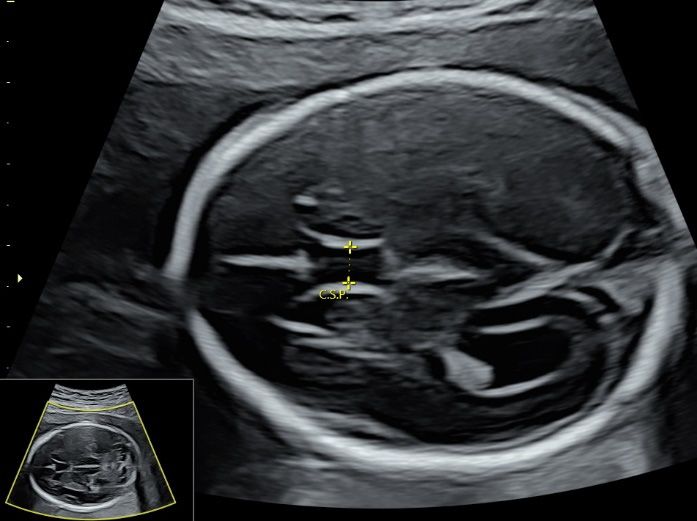
Diagnostics 2021, 11, 1217                                                                                                                    5 of 18
                             Diagnostics 2021, 11, x FOR PEER REVIEW                                                                                      5 of 1

                                       2.3. Early Pregnancy Scan
                                                           2.3. Early PregnancyisScan
                                            Transvaginal ultrasonography            essential in the assessment of pregnancy of unknown
                                       location, which can beTransvaginal
                                                                    due to earlyultrasonography       is essential or
                                                                                   pregnancy, miscarriage,         in the   assessment
                                                                                                                       ectopic            of pregnancy
                                                                                                                                 pregnancy.      It is     of un
                                                           known     location, which  can  be due   to early pregnancy,   miscarriage,
                                       important to avoid making a false-positive diagnosis of miscarriage by using transvaginal         or  ectopic  pregnancy
                                       sonography, carefulIt measurement
                                                              is important toof avoid
                                                                                  mean making    a false-positive
                                                                                          sac diameter             diagnosis
                                                                                                           and crown    rumpoflength,
                                                                                                                                  miscarriage    by using trans
                                                                                                                                          and using
                                                           vaginal sonography, careful measurement of mean sac diameter and crown rump length
                                       safe cut-off values of these measurements in defining miscarriage [19]. A recent study
                                                           and using safe cut-off values of these measurements in defining miscarriage [19]. A recen
                                       showed that amniotic sac sign (the presence of amniotic sac without a live embryo) is a
                                                           study showed that amniotic sac sign (the presence of amniotic sac without a live embryo
                                       reliable marker of ismiscarriage     [20]. While the presence of an extrauterine gestational sac
                                                              a reliable marker of miscarriage [20]. While the presence of an extrauterine gestationa
                                       with yolk sac and/or     embryo     with or without
                                                           sac with yolk sac and/or     embryocardiac    activity
                                                                                                   with or        is indicative
                                                                                                            without               of ectopic
                                                                                                                     cardiac activity           preg- of ectopi
                                                                                                                                        is indicative
                                       nancy, the presencepregnancy,
                                                             of an inhomogeneous        adnexal    mass   (‘blob’ sign)  or  extrauterine    sac-like
                                                                          the presence of an inhomogeneous adnexal mass (‘blob’ sign) or extrauterin
                                                           sac-like
                                       structure (‘bagel’ sign)       structure
                                                                  is very       (‘bagel’ofsign)
                                                                           suggestive            is very
                                                                                           a tubal        suggestive
                                                                                                     ectopic  pregnancyof a [21].
                                                                                                                            tubal In
                                                                                                                                   ectopic
                                                                                                                                      women  pregnancy
                                                                                                                                                 with [21]. I
                                                           womenultrasound
                                       prior Caesarean section,       with prior Caesarean
                                                                                   features ofsection,   ultrasound
                                                                                                  Caesarean           features of Caesarean
                                                                                                                scar pregnancy      includingscarlowpregnanc
                                       implantation of theincluding
                                                             gestationallow sac
                                                                             implantation
                                                                                within orofinthe   gestational
                                                                                                close  proximitysac within  or in closescar
                                                                                                                    to a Caesarean       proximity
                                                                                                                                              as wellto a Caesar
                                       as classical signs ofean scar as well
                                                             placenta         as classical
                                                                          accreta spectrum signs  of placenta
                                                                                               disorders       accreta
                                                                                                            should   bespectrum   disorders
                                                                                                                         looked out            should be looke
                                                                                                                                       for [22,23].
                                                              out for [22,23].
                                       2.4. First Trimester Scan
                                                              2.4. First Trimester Scan
                                             ISUOG and recently, AIUM published the practice guidelines on first-trimester fetal
                                       ultrasound scan [24,25].ISUOG   and recently,ultrasonography
                                                                 High-resolution     AIUM published the        practice
                                                                                                           allows   theguidelines  on first-trimester
                                                                                                                         early assessment     of      feta
                                                         ultrasound scan [24,25]. High-resolution ultrasonography allows the early assessment o
                                       fetal anatomy [11] (Figure 5a–d, Video S5a,b) and fetal malformations [12]. Fetal heart
                                                         fetal anatomy [11] (Figure 5a–d, Video S5a,b) and fetal malformations [12]. Fetal heart ca
                                       can be examined in the late first trimester [26], particularly with the use of color Doppler
                                                         be examined in the late first trimester [26], particularly with the use of color Dopple
                                       (Video S5c,d). ISUOG     recommends using high-frequency (6–12 MHz) transvaginal ul-
                                                         (Video S5c,d). ISUOG recommends using high-frequency (6–12 MHz) transvaginal ultra
                                       trasound to examine    fetal brain, especially
                                                         sound to examine    fetal brain,ifespecially
                                                                                            the focusif is
                                                                                                         theinfocus
                                                                                                                the posterior  fossa and
                                                                                                                    is in the posterior     the
                                                                                                                                        fossa and the ma
                                       maternal abdominal   wall  is thick [5].
                                                         ternal abdominal wall is thick [5].

                                                                   (a)                                                    (b)

                                                                   (c)                                                    (d)

                                       Figure 5. High-resolution ultrasonography of the fetus at 13 weeks’ gestation: (a) mid-sagittal view
                                       showing head, neck, and facial profile, (b) coronal view showing both eyes and ears, (c) the hand
                                       with five fingers, and (d) foot.
Applications of Advanced Ultrasound Technology in Obstetrics
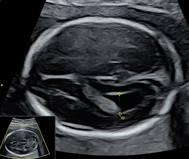
Diagnostics 2021, 11, 1217                                                                                           6 of 18

                             2.5. Doppler Ultrasound
                                  Doppler ultrasound is widely used in obstetrics. ISUOG has made recommendations
                             on how to perform Doppler ultrasonography of the fetoplacental circulation [27]. It is
                             a challenge to detect late-onset feral growth restriction (FGR). Although third-trimester–
                             cerebroplacental ratio (CPR = middle cerebral artery pulsatile index/umbilical artery
                             pulsatile index) is an independent predictor of stillbirth and perinatal mortality [28], CPR
                             with or without adjustment for estimated fetal weight centile showed a low prediction rate
                             for adverse perinatal outcome [29]. According to a meta-analysis, abnormal uterine artery
                             (UtA) Doppler in the third trimester is useful in predicting perinatal death in suspected
                             small-for-gestational age fetuses [30]. A recent prospective study suggested that cerebral–
                             placental–uterine ratio (CPUR = CPR divided by mean UtA pulsatile index) detected FGR
                             better than CPR or UtA Doppler alone [31].

                             2.6. Labour Ward Ultrasound
                                  The use of intrapartum ultrasonography is increasing. It can be performed by using a
                             portable machine equipped with a wide-sector and low-frequency (
Applications of Advanced Ultrasound Technology in Obstetrics
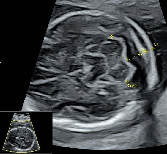
Diagnostics 2021, 11, x FOR PEER REVIEW                                                                                               7
Diagnostics 2021, 11, 1217                                                                                                               7 of 18

                                                         (a)                                                      (b)

                                                         (c)                                                      (d)
                      Figure 6. Ultrasonography        of a fetus atof
                                    Figure 6. Ultrasonography        13a weeks’
                                                                         fetus atgestation
                                                                                  13 weeks’ bygestation
                                                                                                a transabdominal     high-frequency
                                                                                                          by a transabdominal         linear transducer: (a
                                                                                                                                 high-frequency
                      transverse view    oftransducer:
                                    linear   fetal brain, (b) coronal view
                                                          (a) transverse     of face
                                                                          view        showing
                                                                                of fetal brain, both  orbits (OB),
                                                                                                (b) coronal   view (c)  coronal
                                                                                                                    of face     view of
                                                                                                                            showing      abdomen
                                                                                                                                      both orbits showing
                      both kidneys(OB),
                                      (Ki) (c)
                                           on coronal   view of abdomen showing both kidneys (Ki) on either side of the spine, and (d)showing
                                               either side of the spine, and (d) the three-vessel trachea transverse view with color Doppler   the  pulmo
                      nary artery (PA), aorta (Ao), superior vena cava (SVC), and trachea (T).
                                    three-vessel trachea transverse view with color Doppler showing pulmonary artery (PA), aorta (Ao),
                                    superior vena cava (SVC), and trachea (T).
                                                  4. Radiant Flow
                                   4. Radiant Flow Radiant flow shows the blood flow with a sense of depth by using a specific
                                                  rithm
                                        Radiant flow       to convert
                                                       shows             theflow
                                                                 the blood    index   of aerythrocyte
                                                                                   with     sense of depth density   in a acertain
                                                                                                               by using      specificarea  into a height
                                                                                                                                      algorithm
                                   to convert thewhich
                                                    indexisofthen    superimposed
                                                                erythrocyte    densityonin thea initial
                                                                                                 certaincoding    of color,
                                                                                                           area into          power
                                                                                                                       a height       Doppler,
                                                                                                                                   index  whichor high-d
                                                  tion flowon
                                   is then superimposed        [42].
                                                                   theOther  advantages
                                                                       initial  coding ofinclude        reducing
                                                                                              color, power         blood overflow
                                                                                                               Doppler,                and indicating th
                                                                                                                            or high-definition
                                   flow [42]. Other
                                                  sel advantages      include
                                                      with sharp edges.          reducing
                                                                             Special  displayblood     overflow
                                                                                                  produced     by and   indicating
                                                                                                                  similar             the vessel
                                                                                                                            technologies    include Micro
                                   with sharp edges.    Special
                                                  Imaging           display
                                                              (Philips),     produced
                                                                          MV-Flow,     andbyLumiFlow
                                                                                                similar technologies
                                                                                                            (Samsung).include MicroFlow
                                   Imaging (Philips), MV-Flow,
                                                        Radiant flow  andisLumiFlow     (Samsung).
                                                                             used to show        fast blood flow in the fetal heart and brachycep
                                        Radiant flow    is  used   to show    fast blood
                                                  arteries [42] (Videos S5d and S6a,b), as  flow    in well
                                                                                                       the fetal  heart and flow
                                                                                                            as slow-blood       brachycephalic
                                                                                                                                     in the neurovascula
                                   arteries [42] (Videos
                                                  culationS5d [43]and   S6a,b),
                                                                    (Video  S7). as well as slow-blood flow in the neurovascular
                                   circulation [43] (Video
                                                        The S7).
                                                               fetal umbilical–portal venous system is complex. High-definition flow im
                                        The fetal (HDFI)
                                                  umbilical–portal      venous
                                                             has been used      to system
                                                                                   assess theis complex.     High-definition
                                                                                                  normal anatomy                  flow imaging
                                                                                                                        of this system    or umbilical–p
                                   (HDFI) has been    used    to  assess  the  normal   anatomy        of this system    or umbilical–portal–
                                                  systemic venous shunts. Transverse and sagittal planes are used to examine the feta
                                   systemic venous     shunts. Transverse
                                                  bilical–portal     venous systemand (Video
                                                                                       sagittalS8a,b).
                                                                                                   planesInare   used case
                                                                                                              a recent  to examine      the authors
                                                                                                                               report, the   fetal used
                                   umbilical–portal   venous      system   (Video   S8a,b).    In  a recent  case  report,   the  authors
                                                  and radiant flow imaging to clearly delineate the aberrant course of the ductus ve        used
                                   HDFI and radiant      flowto
                                                  returning      imaging    to clearly
                                                                   the coronary    sinusdelineate
                                                                                           [44].        the aberrant course of the ductus
                                   venosus returning to the coronary sinus [44].
                                                 5. 3D/4D Ultrasound
                                   5. 3D/4D Ultrasound
                                                      Over the years, new 2D modes (such as high-density power imaging), new 3D
                                        Over the years, new 2D modes (such as high-density power imaging), new 3D volume
                                                 ume acquisition (such as Corpus callosum mode or matrix probe), and new analysis
                                   acquisition (such as Corpus callosum mode or matrix probe), and new analysis (such as
                                                 as semiautomated analysis) have been added in 3D/4D ultrasound examinations (
                                   semiautomated analysis) have been added in 3D/4D ultrasound examinations (Table 1).
                                                 1). The use of 3D multiplanar/multislice analysis facilitates the assessment of norma
Applications of Advanced Ultrasound Technology in Obstetrics
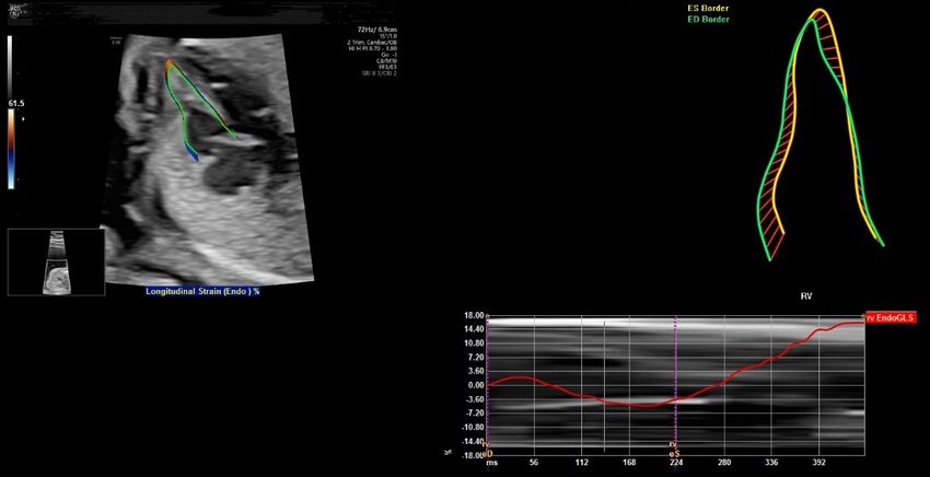
Diagnostics 2021, 11, 1217                                                                                            8 of 18

                             The use of 3D multiplanar/multislice analysis facilitates the assessment of normal and
                             abnormal structures in standard planes. This can also facilitate the detection of subtle fetal
                             defects [45]. The use of 3D rendered images can help counseling to the women when fetal
                             malformations are found or reassure the at-risk women when normal fetal anatomy is
                             found [45]. 3D/4D US is useful for the assessment of fetal brain, spine, face, heart, and
                             other structures [45,46].

                             Table 1. Commonly used scanning mode, volume acquisition, and analysis for three-/four-
                             dimensional (3D/4D) ultrasound examinations.

                                              Mode                   Volume Acquisition            3D/4D Analysis
                                          Gray scale                 3D: different modes            Multiplanar
                                          Color flow                          4D                     Multislice
                                        Power doppler                       STIC 1         Rendered view: different modes
                                  High-density power imaging            Matrix probe                 Cine loop
                                            B-flow                                            Semi-automatic analysis
                                                                                               Volume measurement
                                                                                            Power Doppler measurements
                             1   Spatiotemporal image correlation.

                                     Examples in the assessment of fetal abnormalities
                             a.       Cleft lip and palate: use gray-scale mode, after a 3D volume acquisition, perform
                                      multiplanar/multi-slice analysis and rendering techniques to assess the integrity
                                      of palate.
                             b.       Short-limbed and short-rib dysplasia: use gray-scale mode, after a 3D volume ac-
                                      quisition with skeletal mode, perform rendering techniques with skeletal mode to
                                      examine the long bones and ribs.
                             c.       Agenesis of ductus venosus: use high-density power imaging, after a 3D volume
                                      acquisition, perform multi-slice analysis to assess the precordial venous system.
                             d.       Cardiac outflow tract abnormalities: use color flow, after a STIC volume acquisition,
                                      perform multiplanar/multi-slice analysis in a cine-loop of cardiac cycle.
                             e.       Atrioventricular valve abnormalities: use matrix probe and gray-scale mode, real-
                                      time 4D cine-loop analysis to display the coronal view of atrioventricular valve.

                             5.1. 3D Neurosonography
                                  In targeted neurosonography, a systematic assessment of the fetal brain is required.
                             Although this assessment can be performed by a 2D ultrasound examination, a perfect
                             midsagittal view may not be achieved at all times, thus affecting a proper assessment.
                             ISUOG recommends using 3D ultrasound examination that can provide images of enhanced
                             resolution by displaying thicker ‘slices’ of the brain and thus increasing the signal-to-
                             background noise ratio on all three planes. In addition, multiplanar imaging correlation
                             allows the display of perfectly aligned views on the three orthogonal planes [5]. To avoid
                             shallowing by adjacent skull bones, it is important to acquire a 3D volume in a mid-
                             sagittal plane through the sagittal suture. If the focus is on the anterior complex, the
                             volume will be obtained from the anterior fontanelle or the anterior part of the sagittal
                             suture [5] (Figure 7a). If the focus is on the posterior fossa and cerebellar vermis, the volume
                             will be obtained from the posterior fontanelle or the posterior part of the sagittal suture
                             with the ultrasound beam being almost perpendicular to the tentorium [47] (Figure 7b). A
                             transvaginal and transabdominal approach is used when the fetal presentation is vertex and
                             breech, respectively. Then, the midlines structures including corpus callosum, brain stem,
                             and cerebellar vermis can be examined by multiplanar and multi-slice analysis [43,48,49].
                             An accurate measurement of corpus callosum and cerebellar vermis can be achieved.
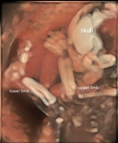
Applications of Advanced Ultrasound Technology in Obstetrics
Diagnostics
 Diagnostics   2021,
             2021, 11,11, x FOR
                       x FOR    PEER
                              PEER   REVIEW
                                   REVIEW                                                                                                      9 of 918of 18
Diagnostics 2021, 11, 1217                                                                                                                        9 of 18

                                    (a)(a)                                                                     (b) (b)
      Figure
     Figure
        Figure 7. Three-dimensional
              7.  Three-dimensional
                  7. Three-dimensional  ultrasonography
                                        ultrasonography
                                           ultrasonographyof
                                                           of fetal
                                                              fetal brain
                                                                    brain
                                                               of fetal    at
                                                                            at20
                                                                         brain atweeks’
                                                                               20 weeks’
                                                                                  20       gestation:
                                                                                      weeks’gestation: (a)
                                                                                                        (a)multiplanar
                                                                                                gestation:  multiplanar
                                                                                                           (a)         analysis
                                                                                                                         analysis
                                                                                                               multiplanar       after
                                                                                                                           analysis    a avolume
                                                                                                                                   after
                                                                                                                                      aftervolume
                                                                                                                                            a volume
      acquisition
     acquisition
        acquisition  with
                    with  corpus
                         corpus
                       with corpus callosum
                                  callosum
                                      callosummode
                                             modemodethrough
                                                    through
                                                        throughtheanterior
                                                             the    anterior
                                                                   the         partof
                                                                             part
                                                                         anterior    of the
                                                                                      the
                                                                                   part       sagittal
                                                                                         ofsagittal
                                                                                             the       suture
                                                                                                    suture
                                                                                                 sagittal      showing
                                                                                                            showing
                                                                                                           suture      corpus
                                                                                                                     corpus
                                                                                                                  showing       callosum
                                                                                                                             callosum
                                                                                                                            corpus          (CC),
                                                                                                                                         (CC),
                                                                                                                                     callosum  and
                                                                                                                                                (CC),
      and
        and(b)(b)
                multiplanar
                   multiplanaranalysis  after
                                  analysis    a volume
                                           after a volumeacquisition
                                                            acquisition through
                                                                           throughthe the
                                                                                       posterior   fontanelle
                                                                                            posterior          showing
                                                                                                       fontanelle       corpus
                                                                                                                  showing       callosum
                                                                                                                            corpus          (CC),(CC),
                                                                                                                                     callosum
     (b) multiplanar analysis after a volume acquisition through the posterior fontanelle showing corpus callosum (CC), cavum
      cavum septi pellucidi (C.S.P.), thalamus (TH), brainstem (BS), and cerebellar vermis (CV).
        cavum
     septi        septi(C.S.P.),
           pellucidi    pellucidi  (C.S.P.),(TH),
                                 thalamus    thalamus   (TH), brainstem
                                                   brainstem                (BS), and vermis
                                                               (BS), and cerebellar     cerebellar   vermis (CV).
                                                                                                 (CV).
                                           After a 3D volume acquisition of the fetal spine at mid-sagittal plane, a rendered view
                                              After
                                           After a 3Da 3D volume
                                                        volume      acquisition
                                                                 acquisition  of of
                                                                                 thethe fetal
                                                                                      fetal   spine
                                                                                            spine at at mid-sagittal
                                                                                                     mid-sagittal    plane,
                                                                                                                  plane,     a rendered
                                                                                                                          a rendered     view
                                                                                                                                       view
                                     of the fetal spine can be well displayed with various modes (Figure 8a,b). In addition, the
                                     of of
                                         the fetal
                                           the fetalspine
                                                      spinecan
                                                            canbebewell
                                                                     welldisplayed
                                                                          displayed with
                                                                                       with various modes (Figure
                                                                                                             (Figure 8a,b).
                                                                                                                       8a,b).InInaddition,
                                                                                                                                  addition,the
                                     coronal planes at the level of the vertebral bodies and/or posterior arches can be recon-
                                     thecoronal
                                          coronal    planes
                                                 planes   at at
                                                             thethe  level
                                                                  level of of
                                                                           thethe  vertebral
                                                                               vertebral       bodies
                                                                                            bodies      and/or
                                                                                                    and/or      posterior
                                                                                                            posterior       arches
                                                                                                                       arches       canrecon-
                                                                                                                                can be   be
                                     structed on multiplanar analysis [5].
                                     reconstructed     on multiplanar
                                        structed on multiplanar         analysis
                                                                     analysis [5].[5].

                                                    (a)                                                         (b)
                                                       (a)                                                            (b)
      Figure 8. Three-dimensional rendered views of fetal spine at 20 weeks’ gestation after a volume acquisition with skeletal
        Figure
      mode: (a)8. Three-dimensional
                usual mode, and (b)  rendered
                                    X-ray
                                Figure        views of fetal spine
                                        8. mode.
                                           Three-dimensional        at 20 weeks’
                                                               rendered          gestation
                                                                          views of         afterata 20
                                                                                   fetal spine      volume acquisition
                                                                                                       weeks’ gestation with
                                                                                                                        afterskeletal
                                                                                                                              a volume
        mode: (a) usual mode, and  (b) X-ray mode.
                                acquisition with skeletal mode: (a) usual mode, and (b) X-ray mode.
                                     5.2. Spatiotemporal Image Correlation
                                     5.2. Spatiotemporal
                                        5.2.              Image
                                             Spatiotemporal
                                           Spatiotemporal       Correlation
                                                            Image
                                                            Image Correlation
                                                                  Correlation (STIC) allows an automatic acquisition of a single
                                           Spatiotemporal
                                     3D volumeSpatiotemporal  Image
                                                    through slowImage  Correlation
                                                                    sweepCorrelation(STIC)
                                                                                       (STIC)
                                                                            and subsequent   allows
                                                                                                allowsanan
                                                                                                 analysisautomatic
                                                                                                            automatic
                                                                                                           in a loopedacquisition
                                                                                                                        acquisitionof of
                                                                                                                         cine sequencya single
                                                                                                                                         a of
                                                                                                                                           single
                                     3D   volume
                                        3D volume
                                     images         through   slow   sweep
                                                       through slow sweep and
                                               in the multiplanar/multi-sliceand  subsequent
                                                                                      subsequent
                                                                                 format           analysis  in  a
                                                                                                     analysisview.
                                                                                          and a rendered          looped
                                                                                                               in a This
                                                                                                                     loopedcine sequency
                                                                                                                          cancine        im-of of
                                                                                                                                   sequency
                                                                                                                              produce
                                     images
                                        images
                                     ages      in the multiplanar/multi-slice
                                                  in the multiplanar/multi-slice
                                            in a standardized                    format   and
                                                                                     format and
                                                                 plane while minimizing        a rendered   view.  This
                                                                                                    a rendereddependency
                                                                                               the operation            can  produce
                                                                                                                  view. This can       images
                                                                                                                                   produce
                                                                                                                               of the  ultra- im-
                                     in ages
                                         a standardized
                                     sound    examination. plane
                                               in a standardized  while
                                                             The recent   minimizing
                                                                    planeadvances        the operation
                                                                                     in gray
                                                                           while minimizing    scale and
                                                                                                   the    dependency
                                                                                                          color
                                                                                                        operationDoppler  of theprocessing
                                                                                                                           post
                                                                                                                    dependency    ultrasound
                                                                                                                                   of the ultra-
                                     examination.     The recentThe
                                        sound examination.       advances
                                                                      recent in gray scale
                                                                             advances    in and
                                                                                            graycolor
                                                                                                   scaleDoppler
                                                                                                         and colorpost processing
                                                                                                                     Doppler   postimproves
                                                                                                                                     processing
Applications of Advanced Ultrasound Technology in Obstetrics
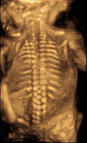
Diagnostics 2021, 11, x FOR PEER REVIEW                                                                                                10 of 18
Diagnostics 2021, 11, 1217                                                                                                        10 of 18

                                  improves
                                the  display the   display ofimages.
                                              of ultrasound     ultrasound
                                                                        Usingimages.     Using color
                                                                               color Doppler            Doppler
                                                                                                 with STIC    in thewith  STIC inmode
                                                                                                                     glass-body     the glass-
                                can show normal and abnormal anatomy of the fetal heart and major vessels [46] (Figure vessels
                                  body   mode    can  show   normal   and   abnormal    anatomy      of the fetal  heart  and   major   9,
                                  [46] (Figure
                                Video   S9). The9,matrix
                                                   Videoprobe
                                                           S9). The  matrix
                                                                 allows       probeacquisition
                                                                         the rapid   allows theof  rapid  acquisition
                                                                                                     an STIC   volume,of    an reducing
                                                                                                                         thus   STIC volume,
                                  thus
                                the     reducing
                                     motion        theand
                                              artifact  motion   artifactlive
                                                            facilitating   and4Dfacilitating  liveIn4D
                                                                                  display [46].         displaythe
                                                                                                      addition,   [46].
                                                                                                                     useInofaddition,  the use
                                                                                                                              the matrix
                                  of theallows
                                probe    matrixtheprobe   allows the
                                                     simultaneous      simultaneous
                                                                     examination        examination
                                                                                    of two  orthogonalofplanes
                                                                                                            two orthogonal
                                                                                                                   of the fetal planes
                                                                                                                                 heart inof the
                                the  ‘biplane
                                  fetal heart mode’.    Additional
                                               in the ‘biplane       use ofAdditional
                                                                  mode’.     image recognition     software
                                                                                         use of image        can help review
                                                                                                          recognition            cardiac
                                                                                                                          software   can help
                                structures   in the  standard  planes  [46].  The 3D   rendered   images    are useful  for
                                  review cardiac structures in the standard planes [46]. The 3D rendered images are useful   counseling
                                tofor
                                    parents.   In addition,
                                      counseling              STIC
                                                    to parents.      volume can
                                                                 In addition,   STICfacilitate
                                                                                       volumeinterdisciplinary       consultation and
                                                                                                 can facilitate interdisciplinary      consul-
                                teleconsultation    [42].
                                  tation and teleconsultation      [42].

                                 Figure9.9.Color
                                Figure      ColorDoppler
                                                  Doppler  with
                                                         with   spatiotemporal
                                                              spatiotemporal     image
                                                                             image      correlation
                                                                                    correlation       inglass-body
                                                                                                 in the  the glass-body
                                                                                                                   modemode    showing
                                                                                                                          showing
                                 multiplanar   view and a rendered  image of a normal fetal heart  at 20 weeks’  gestation.
                                multiplanar view and a rendered image of a normal fetal heart at 20 weeks’ gestation.

                                5.3.
                                 5.3.3D
                                      3DUltrasound
                                         UltrasoundExamination
                                                    Examinationof Face, Limbs,
                                                                  of Face,     andand
                                                                           Limbs,  Other Structures
                                                                                      Other  Structures
                                      While
                                       While2D 2Dultrasound
                                                   ultrasound   is aiskey tool
                                                                       a key   forfor
                                                                             tool  thethe
                                                                                       detection  of fetal
                                                                                          detection        anomalies,
                                                                                                      of fetal           therethere
                                                                                                                anomalies,      are some
                                                                                                                                      are some
                                anomalies    such  as facial  clefts,  micrognathia,   and club  foot  in  which   3D
                                 anomalies such as facial clefts, micrognathia, and club foot in which 3D ultrasound   ultrasound    may may
                                provide
                                 provideadditional
                                            additional information
                                                          information   or or
                                                                           help  counseling
                                                                              help           when
                                                                                    counseling   whensuchsuch
                                                                                                            anomaly    is suspected
                                                                                                                 anomaly               [9]. [9].
                                                                                                                             is suspected
                                Compared to 2D ultrasound alone, combined approach of 2D and 3D ultrasound with
                                 Compared to 2D ultrasound alone, combined approach of 2D and 3D ultrasound with
                                multiplanar/multi-slice analysis can improve the detection or exclusion of cleft palate
                                 multiplanar/multi-slice analysis can improve the detection or exclusion of cleft palate in
                                in fetuses with cleft lip [50] (Figure 10). Although 3D ultrasound is less sensitive for the
                                 fetuses with cleft lip [50] (Figure 10). Although 3D ultrasound is less sensitive for the de-
                                detection of isolated cleft palate, a recent study showed that an accurate evaluation of palate
                                 tection of
                                requires  3Disolated
                                              ultrasoundcleftexamination
                                                               palate, a recent
                                                                             withstudy
                                                                                  volume showed    that an
                                                                                           acquisition   in aaccurate   evaluation
                                                                                                              strictly axial          of palate
                                                                                                                              transverse
                                 requires   3D  ultrasound      examination     with  volume   acquisition    in  a strictly
                                view of the palate [16]. The use of 3D ultrasound multiplanar analysis and 3D rendering       axial transverse
                                 viewcan
                                view    of facilitate
                                           the palate   [16].
                                                      the       The of
                                                           display    use  of 3D ultrasound
                                                                         mid-sagittal plane ofmultiplanar
                                                                                               the fetal faceanalysis
                                                                                                                and thusand     3D rendering
                                                                                                                           improve     the
                                 view canoffacilitate
                                accuracy                the display
                                              measurements       of the of  mid-sagittal
                                                                         mandible         plane
                                                                                    and the      of theof
                                                                                            detection     fetal face and thus
                                                                                                            micrognathia     [51].improve the
                                 accuracy    of measurements
                                      Three-dimensional       (3D) of   the mandible
                                                                     rendering          and the
                                                                                technology   withdetection    of micrognathia
                                                                                                   skeletal mode     can display[51].
                                                                                                                                    skull,
                                vertebrae, ribs, long bones and fingers [52] (Figure 11a,b and Video S10a,b). Prenatal as-
                                sessment of the ribs and vertebral pattern can be performed by 3D ultrasound with skeletal
                                mode (Figure 8a,b), albeit it is not a routine practice. A review of 39 studies including
                                75,018 healthy subjects and 6130 subjects with structural or chromosomal anomalies or
                                adverse outcome showed an association between cervical ribs and other structural anoma-
                                lies including esophageal atresia and anorectal malformation [53]. Abnormalities such as
                                craniosynostosis [26,54], and extra ribs can be shown.
Diagnostics
Diagnostics   2021,
            2021, 11,11, x FOR PEER REVIEW
                      1217                                                                                                               11ofof1818
                                                                                                                                         11

                                                      Figure 10. Three-dimensional ultrasound assessment of the fetal face at 13 weeks’ gestation s
                                                      multiplanar views of lip and palate (reference dot).

                                                          Three-dimensional (3D) rendering technology with skeletal mode can display
                                                    vertebrae, ribs, long bones and fingers [52] (Figure 11a,b and Video S10a,b). Prena
                                                    sessment of the ribs and vertebral pattern can be performed by 3D ultrasound wit
                                                    etal mode (Figure 8a,b), albeit it is not a routine practice. A review of 39 studies inc
                                                    75,018 healthy subjects and 6,130 subjects with structural or chromosomal anoma
                                                    adverse outcome showed an association between cervical ribs and other structural
                                    Figure10.
                                   Figure  10.Three-dimensional
                                               Three-dimensional
                                                    alies includingultrasound  assessment
                                                                      esophageal
                                                                  ultrasound       atresiaofof
                                                                              assessment    andthefetal
                                                                                             the    fetalface
                                                                                                  anorectalfaceatat
                                                                                                                  1313weeks’
                                                                                                                       weeks’gestation
                                                                                                                 malformation gestation showing
                                                                                                                                  [53].showing
                                                                                                                                        Abnormalities s
                                    multiplanar views of lip and palate (reference dot).
                                   multiplanar viewscraniosynostosis    [26,54], and
                                                      of lip and palate (reference    extra ribs can be shown.
                                                                                   dot).

                                           Three-dimensional (3D) rendering technology with skeletal mode can display skull,
                                     vertebrae, ribs, long bones and fingers [52] (Figure 11a,b and Video S10a,b). Prenatal as-
                                     sessment of the ribs and vertebral pattern can be performed by 3D ultrasound with skel-
                                     etal mode (Figure 8a,b), albeit it is not a routine practice. A review of 39 studies including
                                     75,018 healthy subjects and 6,130 subjects with structural or chromosomal anomalies or
                                     adverse outcome showed an association between cervical ribs and other structural anom-
                                     alies including esophageal atresia and anorectal malformation [53]. Abnormalities such as
                                     craniosynostosis [26,54], and extra ribs can be shown.

                                                           (a)                                                       (b)
                        Figure 11. Three-dimensional  rendered images
                                   Figure 11. Three-dimensional         of theimages
                                                                   rendered    fetal skeleton
                                                                                       of the at 20–22
                                                                                              fetal    weeks’atgestation
                                                                                                    skeleton             showing:
                                                                                                                20–22 weeks’       (a) X-ray mode
                                                                                                                                gestation
                        the skull bone with frontal
                                   showing:         suture
                                            (a) X-ray mode(SS) andskull
                                                           of the   anterior
                                                                        bonefontanelle   (AF),
                                                                              with frontal     and(SS)
                                                                                            suture  (b) and
                                                                                                        HD anterior
                                                                                                            skeletal fontanelle
                                                                                                                     mode of the  skull,
                                                                                                                                (AF), andbones of t
                        upper and (b)
                                   lower limbs.
                                      HD skeletal mode of the skull, bones of the upper and lower limbs.

                                       It is difficult to It  is difficult
                                                           visualize       to visualize
                                                                       esophagus     on 2Desophagus
                                                                                              ultrasoundon      2D ultrasound
                                                                                                             examination.       Theexamination.
                                                                                                                                     use of 3D ul- The use
                                                     ultrasound with
                                 trasound with multiplanar                multiplanar
                                                                   analysis   and Crystal analysis   and Crystal
                                                                                              Vue rendering      may Vue
                                                                                                                       make rendering   may make the vis
                                                                                                                                the visualization
                                                     tion possible [55]. (3D)
                                 possible [55]. Three-dimensional           Three-dimensional
                                                                                  volumes are acquired(3D) volumes      are acquiredsection
                                                                                                               from a midsagittal        from aofmidsagit
                                 the thorax
                                        (a)    and   tion
                                                     upperof  the thorax
                                                             abdomen       and
                                                                          with  upper
                                                                               the   fetusabdomen
                                                                                             lying  in with
                                                                                                       supine
                                                                                                     (b)     the  fetus
                                                                                                                 position.lying   in supine  position.
                                       While 2D ultrasound Whileexamination
                                                                   2D ultrasound  with examination
                                                                                         gray scale and with  gray
                                                                                                            color    scale
                                                                                                                  flow       andstandard
                                                                                                                         is the    color flow
                                                                                                                                           for is
                                                                                                                                                thethe stand
       Figure 11. Three-dimensional   rendered   images
                                 antenatal diagnosis      of the fetal
                                                          of placenta
                                                     the antenatal     skeleton  at 20–22
                                                                         accreta spectrum
                                                                       diagnosis           weeks’
                                                                                    of placenta     gestation
                                                                                               disorders
                                                                                                  accreta[22],  showing:    (a)
                                                                                                                  3D ultrasound
                                                                                                             spectrum           X-ray mode
                                                                                                                           disorderswith     of
                                                                                                                                       [22],power
                                                                                                                                              3D ultrasoun
       the skull bone with frontalDoppler
                                   suture (SS)
                                             andand   anterior fontanelle
                                                  multiplanar     analysis(AF),   and an
                                                                            permits     (b) accurate
                                                                                            HD skeletal    mode of the
                                                                                                       assessment          skull,
                                                                                                                      of the      bones of the
                                                                                                                                placenta-bladder
       upper and lower limbs. interface, and the degree of bladder invasion by the placenta [56]. Three-dimensional (3D)
                                 rendered images can be used for patient counseling [56].
                                         It is difficult to visualize esophagus on 2D ultrasound examination. The use of 3D
                                   ultrasound
                                 5.4. 3D Printing with multiplanar analysis and Crystal Vue rendering may make the visualiza-
                                   tionWith
                                         possible   [55]. Three-dimensional
                                               advances     in 3D ultrasound, (3D)        volumesultrasound
                                                                                    the derived        are acquired    from
                                                                                                                    data    canabe midsagittal
                                                                                                                                     used for 3D sec-
                                   tion of  the thorax   and    upper   abdomen      with  the  fetus   lying  in supine
                                 printing of physical models of whole fetuses [57] and the fetal face [58]. A recent trial   position.
                                 showed  While
                                             that2Dtheultrasound    examination
                                                        use of 3D-printed       fetalwith    gray
                                                                                        facial     scale and
                                                                                                models          colorin
                                                                                                           resulted    flow    is theincreases
                                                                                                                           greater    standard in for
                                   the antenatal diagnosis of placenta accreta spectrum disorders [22], 3D ultrasound with
5.4. 3D Printing
                                     With advances in 3D ultrasound, the derived ultrasound data can be used for 3D
                              printing of physical models of whole fetuses [57] and the fetal face [58]. A recent tria
Diagnostics 2021, 11, 1217    showed that the use of 3D-printed fetal facial models resulted in greater increases                    12 of 18   in ma
                              ternal–fetal attachment in the third trimester than the use of ultrasonography only [58
                              Whether this can be translated into better pregnancy outcomes needs further studies. I
                             maternal–fetal attachment in the third trimester than the use of ultrasonography only [58].
                              addition,
                             Whether        a 3D-printed
                                          this                spinainto
                                               can be translated       bifida   model
                                                                           better         can beoutcomes
                                                                                    pregnancy      beneficial    for surgical
                                                                                                               needs            rehearsal
                                                                                                                      further studies.     In prior t
                              a  fetoscopic     repair  [59].
                             addition, a 3D-printed spina bifida model can be beneficial for surgical rehearsal prior to a
                                     Withrepair
                             fetoscopic      advances
                                                   [59]. in STIC, the derived data can be used for 3D printing of the fetal hear
                              which Withisadvances
                                            a fast-moving
                                                      in STIC,structure
                                                                 the derived [60].
                                                                                 dataIncan
                                                                                        a recent
                                                                                           be usedcase
                                                                                                     for 3Dreport,  theofauthors
                                                                                                              printing     the fetalfound
                                                                                                                                      heart, that th
                             which
                              3D model was useful in showing the complex anatomy of fetal transposition 3D
                                      is a fast-moving    structure   [60]. In a   recent case report,   the  authors  found  that  the   of great ar
                             model
                              terieswasanduseful    in showing
                                             in providing          the complex
                                                               prenatal     parentalanatomy    of fetal[61].
                                                                                        counseling       transposition of great arteries
                             and in providing prenatal parental counseling [61].
                                     Previously, after acquisition of a 3D/STIC volume dataset, a number of post-pro
                                    Previously, after acquisition of a 3D/STIC volume dataset, a number of post-processing
                              cessing steps are required to convert it from Cartesian.vol file through segmentation, re
                             steps are required to convert it from Cartesian.vol file through segmentation, refinement,
                              finement,
                             and             and optimization
                                   optimization     to a STL (Standard to a Triangle
                                                                              STL (Standard
                                                                                         Language)  Triangle     Language)
                                                                                                       file, the industry       file, the
                                                                                                                            standard     file industr
                             type for 3D Printing [60]. These steps take a long time, and whether the final produced STL the fina
                              standard      file type  for  3D   Printing    [60].   These   steps  take    a long  time,  and   whether
                              produced
                             file            STL fileforis3D
                                  is good enough            good    enough
                                                              printing    is notfor  3D printing
                                                                                  certain            is not certain
                                                                                           before processing.     With before    processing. Wit
                                                                                                                         recent advances
                              recent
                             in          advances
                                 ultrasound          in ultrasound
                                                technology,    a 3D/STIC  technology,      a 3D/STIC
                                                                              volume dataset      can bevolume
                                                                                                            directly dataset
                                                                                                                      exportedcanfrombethedirectly ex
                             ultrasound     machine    as an  STL   file that is  ready  for viewing    on  a  personal
                              ported from the ultrasound machine as an STL file that is ready for viewing on a persona   computer     using
                             common
                              computer    software
                                             using as   well as for
                                                     common           3D printing
                                                                  software     as well(Figure  12).
                                                                                          as for  3D printing (Figure 12).

                              Figure12.12.
                             Figure     AA   physical
                                           physical   model
                                                    model     of three-dimensional
                                                          of three-dimensional       printing
                                                                               printing         of the
                                                                                        of the fetal    fetal face.
                                                                                                     face.

                             6.6.FetalHQ
                                  FetalHQ
                                   FetalHQ, a novel heart and vascular analysis software, can allow assessment of the
                                    FetalHQ, a novel heart and vascular analysis software, can allow assessment of th
                             fetal heart shape, size, and contractibility by using speckle tracking to analyze the motion of
                              fetal heart shape, size, and contractibility by using speckle tracking to analyze the motio
                             multiple points of the fetal heart [62] (Figure 13). The global sphericity index (SI) is a simple
                              of multiple points
                             measurement              of the
                                              of cardiac     fetal heart
                                                          contractility, and[62]   (Figure
                                                                               it is        13).
                                                                                     equal to     The global mid-basal–apical
                                                                                               (end-diastolic  sphericity index (SI) is
                             length)/transverse length [63]. For 24-segment sphericity index, SI is computed for each mid-basal
                              simple   measurement       of cardiac   contractility,     and  it is equal  to (end-diastolic  of
                              apical
                             the      length)/transverse
                                 24 end-diastolic            length
                                                    transverse        [63]. which
                                                                segments,     For 24-segment      sphericity
                                                                                      are distributed from theindex,  SIthe
                                                                                                                base to  is computed
                                                                                                                            apex      fo
                             ofeach
                                eachofventricle, as well as the end-diastolic
                                        the 24 end-diastolic                     mid-basal–apical
                                                                 transverse segments,         whichlength   [62].
                                                                                                      are distributed   from the base t
                              the This
                                   apex24-segment     sphericity
                                          of each ventricle,   as index  is athe
                                                                   well as     comprehensive
                                                                                  end-diastolicmethod     to assess thelength
                                                                                                    mid-basal–apical    shape of
                                                                                                                               [62].
                             ventricular chambers [62]. The SI for each segment was independent of gestational age
                             and fetal biometry. The SI of the right ventricle was lower than that of the left ventricle
                             for segments 1–18. This index can be used when discordance between the size of the atrial
                             and/or ventricular cardiac chambers is found. Abnormal SI values are found in the fetuses
                             with cardiac abnormalities such as coarctation of aorta, pulmonary stenosis, and fetal
                             growth restriction [62]. Abnormal SI values are associated with increased risk of perinatal
                             complications and childhood and/or adult cardiovascular disease [64].
Diagnostics2021,
Diagnostics  2021,11,
                   11,1217
                       x FOR PEER REVIEW                                                                                                            13 of18
                                                                                                                                                    13 of 18

                                   Figure13.
                                  Figure  13.FetalHQ
                                              FetalHQassessment
                                                        assessment of
                                                                    ofthe
                                                                       thefetal
                                                                           fetalheart
                                                                                 heartshape,
                                                                                       shape,size,
                                                                                              size,and
                                                                                                    andcontractibility
                                                                                                         contractibilityby
                                                                                                                         byusing
                                                                                                                            usingspeckle
                                                                                                                                  speckle
                                   tracking to analyze the motion of multiple points of the fetal heart at 20 weeks’ gestation.
                                  tracking to analyze the motion of multiple points of the fetal heart at 20 weeks’ gestation.

                                          This 24-segment
                                         While                    sphericity
                                                 the initial results             index is aacomprehensive
                                                                         are promising,          recent review ofmethod
                                                                                                                    23 studies to showed
                                                                                                                                   assess the       shape of
                                                                                                                                                conflicting
                                   ventricular     chambers       [62].  The    SI  for  each    segment    was   independent
                                  results concerning the development of strain and strain rate during gestation [65]. Large         of   gestational      age
                                   and   fetal biometry.      The    SI of  the right   ventricle    was  lower   than   that
                                  longitudinal cohort studies with a standard protocol are needed to obtain reference values  of  the   left  ventricle    for
                                   segments
                                  for            1–18. This
                                        fetal cardiac            index caninbe
                                                          deformation               used when discordance
                                                                                 uncomplicated         pregnancies  between
                                                                                                                        [65]. Athe     size of
                                                                                                                                   recent          the atrial
                                                                                                                                               systematic
                                   and/or    ventricular     cardiac    chambers      is  found.   Abnormal     SI values
                                  review also showed heterogeneous results concerning gestational age and Doppler profiles.  are found       in  the  fetuses
                                   with    cardiac    abnormalities        such    as  coarctation     of  aorta,  pulmonary
                                  Large prospective longitudinal cohort studies are required to assess the clinical significance   stenosis,       and   fetal
                                   growth     restriction    [62].   Abnormal      SI values     are associated    with
                                  of deformation measurements of the fetal heart in growth restricted fetuses and normal  increased     risk   of  perinatal
                                   complications
                                  fetuses    [66].      and childhood and/or adult cardiovascular disease [64].
                                          While the initial results are promising, a recent review of 23 studies showed conflict-
                                  7.ing
                                      Artificial
                                         results Intelligence
                                                   concerning the development of strain and strain rate during gestation [65].
                                   Large    longitudinal
                                         Machine     learning,cohort    studies with
                                                                    in particular     deepa standard
                                                                                              learning,protocol     are neededimage
                                                                                                          allows ultrasound        to obtain       reference
                                                                                                                                              recognition
                                  and thus facilitates the automatic identification and measurement of fetal biometry system-
                                   values    for fetal   cardiac     deformation      in  uncomplicated       pregnancies      [65].  A   recent     [67]. It
                                  isatic reviewofalso
                                      a branch            showed
                                                      artificial       heterogeneous
                                                                   intelligence     (AI). In results  concerning
                                                                                                obstetric            gestationalthe
                                                                                                           ultrasonography,           age    and Doppler
                                                                                                                                         automation        of
                                   profiles. Largeof
                                  measurements           prospective
                                                           fetal biometry longitudinal       cohort useful
                                                                                is a potentially      studiestool
                                                                                                               are to
                                                                                                                    required
                                                                                                                       increasetothe assess     the clinical
                                                                                                                                         reliability     and
                                   significance ofof
                                  reproducibility        deformation
                                                          measurements     measurements
                                                                                as comparedoftothe       fetal measurements
                                                                                                     manual    heart in growth        restricted
                                                                                                                                  [68].               fetuses
                                                                                                                                           In addition,     it
                                   andreduce
                                  can    normal     fetuses [66].
                                                 scanning      time [68] and work-related fatigue and musculoskeletal disorders [69].
                                         With automatic image recognition technology applied on a frozen 2D ultrasound
                                   7. Artificial
                                  image,            Intelligence of fetal biometry including biparietal diameter (BPD), head
                                             auto measurement
                                  circumference
                                          Machine (HC),learning,abdominal      circumference
                                                                     in particular                  (AC),allows
                                                                                       deep learning,       and femur      length (FL)
                                                                                                                    ultrasound      image   becomes      fea-
                                                                                                                                               recognition
                                  sible.
                                   and thusA study     showed
                                                facilitates    the aautomatic
                                                                      success rate      of 91.43% and
                                                                                   identification     and 100%     for auto of
                                                                                                            measurement        measurement
                                                                                                                                  fetal biometry     of HC
                                                                                                                                                         [67].
                                  and
                                   It is BPD,   respectively
                                         a branch    of artificial [67].  Although(AI).
                                                                      intelligence       the Ininaccuracy    for the plane acceptance
                                                                                                  obstetric ultrasonography,                      check for
                                                                                                                                     the automation         of
                                  head    parameters      was     12.9%    [67],  such   inaccuracy     can   be corrected
                                   measurements of fetal biometry is a potentially useful tool to increase the reliability     by  fine-tuning        of the
                                                                                                                                                          and
                                  caliper    placement
                                   reproducibility       ofmanually.
                                                            measurements   In another      study, manual
                                                                                  as compared       to manualadjustment
                                                                                                                 measurementsof caliper[68].position    was
                                                                                                                                               In addition,
                                  not   required   in   about   two-thirds     of  cases    for HC   and  FL  measurements,
                                   it can reduce scanning time [68] and work-related fatigue and musculoskeletal disorders        but   it  was    required
                                  in  more than 80% for the measurement of AC [68]. Auto measuring AC is more difficult
                                   [69].
                                  than measuring
                                          With automatic HC because        of the low contrast
                                                                image recognition          technology between
                                                                                                          appliedtheonabdomen
                                                                                                                        a frozen 2D andultrasound
                                                                                                                                            surrounding   im-
                                  tissues and the large variability in abdominal shape and appearance [68]. The accuracy of
                                   age, auto measurement of fetal biometry including biparietal diameter (BPD), head cir-
                                  the auto measurement of HC, AC, and FL was high, and it compared well with previously
                                   cumference (HC), abdominal circumference (AC), and femur length (FL) becomes feasi-
                                  published manual-to-manual agreement, but the auto measurements had a tendency to
                                   ble. A study showed a success rate of 91.43% and 100% for auto measurement of HC and
                                  underestimate biometry, which requires further improvements in the algorithm [68].
                                   BPD, respectively [67]. Although the inaccuracy for the plane acceptance check for head
                                         With automatic image recognition technology applied on an acquired 3D ultrasound
                                   parameters was 12.9% [67], such inaccuracy can be corrected by fine-tuning of the caliper
                                  volume of the fetal head from the BPD plane, SonoCNS allows auto measurement of fetal
                                   placement manually. In another study, manual adjustment of caliper position was not re-
                                  biometry including BPD, HC, atrium of the posterior horn of the lateral ventricle (Vp),
                                   quired in about two-thirds of cases for HC and FL measurements, but it was required in
                                  transcerebellar diameter (TCD), and cisterna magnum (CM) [70] (Figure 14). A recent
                                   more than 80% for the measurement of AC [68]. Auto measuring AC is more difficult than
                                  study showed that this 3D automated technology reliably identified and measured BPD
                                   measuring HC because of the low contrast between the abdomen and surrounding tissues
                                   and the large variability in abdominal shape and appearance [68]. The accuracy of the auto
lished manual-to-manual agreement, but the auto measurements had a tendency to un-
                              derestimate biometry, which requires further improvements in the algorithm [68].
                                   With automatic image recognition technology applied on an acquired 3D ultrasound
                              volume of the fetal head from the BPD plane, SonoCNS allows auto measurement of fetal
Diagnostics 2021, 11, 1217    biometry including BPD, HC, atrium of the posterior horn of the lateral ventricle    14 (Vp),
                                                                                                                      of 18
                              transcerebellar diameter (TCD), and cisterna magnum (CM) [70] (Figure 14). A recent
                              study showed that this 3D automated technology reliably identified and measured BPD
                             and
                              andHCHCbut
                                      butwas
                                           wasless
                                                lessso
                                                     sofor
                                                        forTCD,
                                                            TCD,CM,
                                                                 CM,and
                                                                     andVp
                                                                         Vp[70].
                                                                            [70].Further
                                                                                  Furtheroptimization
                                                                                          optimizationofofthis
                                                                                                           thisautomated
                                                                                                                automated
                             technology
                              technologyisisrequired.
                                             required.

                              Figure14.
                             Figure   14. SonoCNS,
                                          SonoCNS, after
                                                    after volume
                                                          volume acquisition
                                                                   acquisition of
                                                                                of the
                                                                                    the fetal
                                                                                         fetalbrain
                                                                                               brainatatbiparietal
                                                                                                         biparietaldiameter
                                                                                                                    diameterplane
                                                                                                                             planeatat21
                              weeks gestation, showing auto measurement of biparietal diameter (BPD), head circumference
                             21 weeks gestation, showing auto measurement of biparietal diameter (BPD), head circumference
                              (HC), atrium of the posterior horn of the lateral ventricle (Vp), transcerebellar diameter (TCD), and
                             (HC), atrium of the posterior horn of the lateral ventricle (Vp), transcerebellar diameter (TCD), and
                              cisterna magnum (CM).
                             cisterna magnum (CM).
                                    Fetal Intelligent Navigation Echocardiography (FINE) applied on a STIC volume us-
                                   Fetal Intelligent Navigation Echocardiography (FINE) applied on a STIC volume
                              ing “intelligent navigation” technology allows the automatic display of nine standard fe-
                             using “intelligent navigation” technology allows the automatic display of nine standard
                              tal echocardiography views [71]. This can simplify fetal cardiac examinations, reduce op-
                             fetal echocardiography views [71]. This can simplify fetal cardiac examinations, reduce
                              erator dependency,
                             operator   dependency,and    andhelp
                                                                helpdetect
                                                                      detectcongenital
                                                                              congenitalheart heartdefects
                                                                                                       defects[71].[71].
                                    During     a real-time    2D  morphology        scan,
                                   During a real-time 2D morphology scan, identifying and   identifying       andinterpreting
                                                                                                                     interpretingfetalfetalstandard
                                                                                                                                              standard
                              scan   planes    are   highly    complex     tasks.    With     automatic        image
                             scan planes are highly complex tasks. With automatic image processing technology [46,70],    processing      technology
                              [46,70],
                             these      these
                                    tasks   cantasks    can beby
                                                 be assisted     assisted   by providing
                                                                    providing     feedback or   feedback
                                                                                                     guidance   or to
                                                                                                                    guidance      to an ultrasound
                                                                                                                        an ultrasound       operator
                              operator    on  whether     a  correct  standard     scan   plane    of   fetal  anatomy
                             on whether a correct standard scan plane of fetal anatomy is obtained, whether all parts        is  obtained,     whetherof
                              all partsare
                             anatomy      of anatomy
                                             checked, andare checked,     and whether
                                                               whether unusual               unusual
                                                                                      findings             findingsplane
                                                                                                  on a standard          on a standard       plane
                                                                                                                                are identified        are
                                                                                                                                                   [72].
                             The operator can use this technology as a second pass or confirmation to improve diagnosticto
                              identified   [72].  The   operator    can  use  this   technology       as   a  second      pass   or confirmation
                              improve[70].
                             accuracy     diagnostic
                                                This canaccuracy    [70].audit
                                                           also allow     This can
                                                                                andalso      allow
                                                                                        quality       audit and quality
                                                                                                  improvement           [73]. improvement [73].
                                    Based    on  deep   learning,   image   segmentation         is  an  image
                                   Based on deep learning, image segmentation is an image processing method        processing       methodthat thatcancan
                              automatically      recognize     the location    and     size  of  an   object     in
                             automatically recognize the location and size of an object in pixels. However, accurate pixels.   However,        accurate
                              segmentationofofmost
                             segmentation          mostanatomical
                                                            anatomicalstructures
                                                                          structuresininmedical
                                                                                              medicalultrasound
                                                                                                           ultrasoundisislimitedlimitedby  bythethelow
                                                                                                                                                     low
                              contrastbetween
                             contrast    betweenthe  thetarget
                                                          targetand
                                                                  andbackground
                                                                        background      ofof
                                                                                           thethe   images
                                                                                                images          [74].
                                                                                                             [74].   ToTo   improve
                                                                                                                         improve     thethe   segmen-
                                                                                                                                          segmenta-
                              tation
                             tion     performance
                                  performance       of of
                                                        thethe  thoracic
                                                             thoracic     wall
                                                                        wall  in in  fetal
                                                                                  fetal     ultrasound
                                                                                          ultrasound          videos,a anovel
                                                                                                           videos,          novelmodel-agnostic
                                                                                                                                    model-agnostic
                              methodusing
                             method      using    deep
                                               deep      learning
                                                      learning       techniques
                                                                 techniques          in processing
                                                                               in processing               time-series
                                                                                                   time-series              information
                                                                                                                     information               of ultra-
                                                                                                                                     of ultrasound
                              sound    videos    and   the  shape   of the  thoracic     wall   was    proposed
                             videos and the shape of the thoracic wall was proposed [75]. Accurate segmentation can    [75].  Accurate     segmenta-
                              tion ultrasonographers
                             assist can assist ultrasonographers           with the
                                                             with identifying     identifying       the thoracic
                                                                                        thoracic area                   area and its
                                                                                                           and its orientation,       andorientation,
                                                                                                                                            it has the
                              and it has
                             potential   tothe
                                            buildpotential
                                                   AI-based  to build  AI-based
                                                                 diagnostic   supportdiagnostic
                                                                                           modelssupport
                                                                                                       to assess  models     to assessview
                                                                                                                      four-chamber        four-cham-
                                                                                                                                                 [75].
                              ber There
                                   view [75].
                                           are emerging studies on the application of artificial intelligence in obstetric
                                    There
                             scan. It        are emerging
                                      is feasible   to use 3D   studies   on thetoapplication
                                                                  ultrasound         automatic measureof artificial      intelligence
                                                                                                                    thymic    volume in       obstetric
                                                                                                                                          [76].   Two-
                             dimensional      (2D) placental
                              scan. It is feasible    to use 3Dsonographic
                                                                   ultrasound to   images      can bemeasure
                                                                                        automatic         screened      for lacunae,
                                                                                                                      thymic    volumewhich         is a
                                                                                                                                           [76]. Two-
                             feature  of PAS   [77].  Preliminary    results  are  encouraging.         Further
                              dimensional (2D) placental sonographic images can be screened for lacunae, which is a improvement        of algorithm
                             and technology are required prior to using AI applications in clinical practice.

                             8. Conclusions
                                  The use of high-resolution ultrasonography can facilitate detailed diagnostic ultra-
                             sonography, in particular, fetal echocardiography and targeted neurosonography, in at-risk
                             pregnancies, as suggested by the recent guidelines. The use of radiant flow can improve
                             the display, especially in complex cardiac or vascular structures. The use of 3D/4D ul-
You can also read