A Tale of Two Viruses: The Distinct Spike Glycoproteins of Feline Coronaviruses - MDPI
←
→
Page content transcription
If your browser does not render page correctly, please read the page content below
viruses
Review
A Tale of Two Viruses: The Distinct Spike
Glycoproteins of Feline Coronaviruses
Javier A. Jaimes 1 , Jean K. Millet 2 , Alison E. Stout 1 , Nicole M. André 1              and
Gary R. Whittaker 1, *
 1    Department of Microbiology & Immunology, College of Veterinary Medicine, Cornell University,
      Ithaca, NY 14853, USA; jaj246@cornell.edu (J.A.J.); aek68@cornell.edu (A.E.S.); nma39@cornell.edu (N.M.A.)
 2    Virologie et Immunologie Moléculaires, INRAE, Université Paris-Saclay, 78352 Jouy-en-Josas, France;
      jean.millet@inra.fr
 *    Correspondence: grw7@cornell.edu

 Received: 13 November 2019; Accepted: 8 January 2020; Published: 10 January 2020

 Abstract: Feline coronavirus (FCoV) is a complex viral agent that causes a variety of clinical
 manifestations in cats, commonly known as feline infectious peritonitis (FIP). It is recognized that
 FCoV can occur in two different serotypes. However, differences in the S protein are much more
 than serological or antigenic variants, resulting in the effective presence of two distinct viruses. Here,
 we review the distinct differences in the S proteins of these viruses, which are likely to translate into
 distinct biological outcomes. We introduce a new concept related to the non-taxonomical classification
 and differentiation among FCoVs by analyzing and comparing the genetic, structural, and functional
 characteristics of FCoV and the FCoV S protein among the two serotypes and FCoV biotypes. Based
 on our analysis, we suggest that our understanding of FIP needs to consider whether the presence of
 these two distinct viruses has implications in clinical settings.

 Keywords:      feline coronavirus;           feline infectious peritonitis;     spike protein;      serotype;
 genetic characterization

1. Introduction
     The Coronaviridae family comprises a diverse group of viruses that affects birds and
mammals (including humans), resulting in a variety of disease manifestations spanning respiratory,
gastrointestinal, neurological and other tissue tropisms [1]. The diversity of viruses is encompassed by
four viral genera, namely the alpha-, beta-, gamma-, and delta-coronaviruses. Among the coronaviruses
(CoVs), severe acute respiratory syndrome coronavirus (SARS-CoV) and the Middle East respiratory
syndrome coronavirus (MERS-CoV), both betacoronaviruses, have received special attention as
emergent pathogens in humans, with the potential to create global epidemics [2–4]. However, CoVs
are also well known as important pathogens in both domesticated and wild birds and mammals [5].
Felids are no exception to this, and feline coronavirus (FCoV) is known to be the cause of disease
in both wild and domestic cats [6–8]. FCoV is grouped as a member of the Alphacoronavirus genus,
along with a range of other coronaviruses causing disease in dogs, pigs, and humans, as well as other
mammalian species.
     FCoV has been a focus of study for several decades due to its interesting behavior in the infected
animal and its ability to cause the systemic, often lethal illness, feline infectious peritonitis (FIP).
The virus is commonly proposed to exist as two forms or biotypes, either causing mainly sub-clinical
disease (where it is known as feline enteric coronavirus, or FECV) or an aggressive and severe form,
where it is known as feline infectious peritonitis virus (FIPV) [9]. While there is still no definitive
evidence to understand the transition between the two forms, several authors have reported that

Viruses 2020, 12, 83; doi:10.3390/v12010083                                        www.mdpi.com/journal/viruses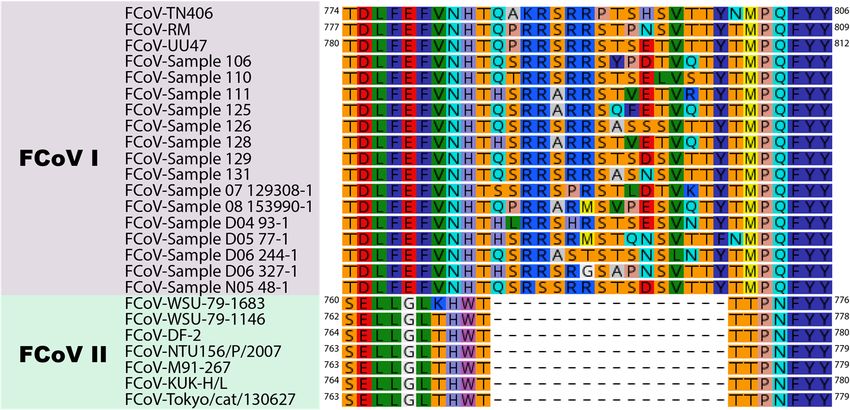
Viruses 2020, 12, 83                                                                                  2 of 14

mutations in the FCoV genome result in changes to the pathogenicity and tropism of the virus, with a
significant role for infected macrophages resulting in a severe-systemic and frequently lethal disease in
cats [10–14]. However, the dichotomous behavior (i.e., FECV-FIPV) has been questioned more recently
with evidence of “systemic” FCoVs that are not “FECVs” by definition, but that do not cause FIP
either (therefore cannot be considered as FIPVs), contributing to the diversity of FCoV and refuting the
discrete FECV-FIPV concept. [13]. While remaining a devastating disease in cat populations, recent
reports describing the role of nucleoside analogs and protease inhibitors, and their potential as a
treatment for FCoV infection have raised the possibility of preventing lethal FIP [15–18].
      Conventionally, FCoVs have been regarded to exist as two distinct serotypes, based on significant
antigenic differences between different viruses infecting cats. The designation of two different serotypes
initially based on antigenic differences found through characterization of spike-specific monoclonal
antibodies (MAbs) against FCoV and canine coronavirus (CCoV) strains [19,20]. From these studies,
FCoVs corresponding to each serotype have been described: feline (serotype I or type I) and canine
(serotype II or type II) [21,22]. While both serotypes have been categorized in FECV and FIPV forms,
both of which can cause FIP, serotype I viruses are much more prevalent in cat populations and so
are the leading cause of FIP [23–26]. Such viruses grow only poorly in cell culture and are hence
understudied compared to serotype II viruses, which have arisen by independent recombination events
with canine coronaviruses and replicate well in cell culture [27,28].
      According to established virus taxonomical criteria, all FCoVs are grouped as single species
(Alphacoronavirus 1), along with canine coronaviruses and transmissible gastroenteritis virus (TGEV)
of swine, a grouping based on sequences of whole genomes or the conserved ORF1ab gene. It is
clear, however, that this single species designation does not reflect the significant differences between
these viruses. In an attempt to provide a more refined and biologically-relevant grouping, we recently
provided evidence for such classification based on the viral spike protein, which is a major driver
of viral tropism and pathogenesis [29]. The study showed that the Alphacoronavirus 1 species can be
sub-grouped such that the different FCoVs are present in distinct clades, which classically would be
defined as monophyletic groups (as they share a common ancestor) but with members of each clade
sharing a set of distinctive characteristics [29]. In this designation, serotype I and serotype II FCoVs
have sufficiently distinguishing features to define them as separate biological entities, and so could be
considered to be distinct virus types.
      Here, we address new concepts related to the non-taxonomical classification and differentiation
among FCoVs. To do this, we analyzed and compared the genetic, structural and functional
characteristics of FCoV and the FCoV S protein among the two serotypes and the two biotypes
and conclude that serotype I and serotype II FCoVs are highly distinct viruses, which we will refer
to as type I and type II. We suggest that our understanding of FIP should consider the presence of
these two distinct viruses, in order to determine whether or not it has implications in clinical settings.
Such distinctions are suggested based on documented differences seen in cell culture between type
I and type II viruses, for example with regard to interferon responses and the activity of antiviral
drugs [30,31].

2. Feline Coronaviruses as Agents of Disease
      FCoV, like all coronaviruses, is an enveloped virus with a large (~30 kb) single-strand positive-sense
RNA genome (ssRNA+) [1,32]. The FCoV genome encodes 11 proteins with four structural proteins,
namely spike (S), envelope (E), matrix (M), and nucleocapsid (N), and five non-structural proteins,
namely the replicase 1a and 1b polyproteins (which are enzymatically cleaved to produce 16 functional
proteins involved in RNA synthesis), and the accessory proteins 3a, 3b, 3c, 7a, and 7b [6,33]. While
all viral proteins play a role during FCoV infection and replication, the structural proteins have the
important role of protecting the viral genome and facilitating the interactions of virions with susceptible
cells. The N protein binds to the viral genomic RNA to protect it, while the M and E proteins interact
with the cell membrane and play a role during the maturation and assembly of the virus [6]. The E
Viruses 2020, 12, 83                                                                                   3 of 14

protein has been also reported to play a major role in the regulation of pathogenicity of other CoVs [34].
The S protein is known to be viral regulator of the cell entry and the major antigenic element of the
virus [35–37]. Several studies have implicated this protein as the main driver of changes in viral
tropism and virulence [11–13,38]. In addition, the historical serotype classification of FCoV S, based on
antigenic differences, reaffirms the importance of this protein in FCoV pathogenesis [21,22].
     The extensive available literature has described that the initial FCoV infection is localized to the
enteric tract, with this infection linked to the “FECV” biotype and typically being subclinical or causing
only mild enteric disease [6]. However, in some cats, this initial infection can lead to extra-enteric
infections in other organs [6,8], and after initial infection, the virus is believed to mutate into the “FIPV”
biotype and produce a severe and often lethal disease called FIP, if left untreated [39,40]. Both biotypes
have been described for the two FCoV serotypes. The commonly accepted model is that, in 5–10% of
the cases, primary infection (by “FECV”) progresses to a systemic form that is characterized by an
acute accumulation of liquid in the peritoneal and thoracic cavities (known as the “wet” or “effusive”
form), hence the name feline infectious peritonitis [41,42]. In some cases, the host’s immune response
can alter the clinical manifestations of the FCoV infection, leading to a less aggressive, but equally
relevant FIP form, where the accumulation of fluid is not evident (the “dry” or “non-effusive” form)
and other major clinical signs arise (e.g., neurological signs and uveitis) [43,44]. This concept has been
recognized as the sole model of FCoV pathogenesis, and while it describes key elements in this disease,
it may fall short in describing the complexity and diversity of FCoV infection clinical outcomes.

3. Genetic Characterization of FCoV
      To gain a better understanding of the distinctive features of the two serotypes of FCoV, it is
important to consider the origins and evolutionary history of FCoV in the context of the other members
of the Alphacoronavirus 1 species (Figure 1). The evolution of FCoV and CCoV (like that of many RNA
viruses) is complex, but it is believed they originate from a common ancestor. During their evolutionary
process, it has been shown that several recombination events have led to the emergence of CCoVs and
FCoVs in their respective animal populations [28,29,45]. In the case of FCoV, recombination events
between FCoV and CCoV genomes resulted in the appearance of novel, chimeric FCoVs whose S
proteins have their origin in CCoV [28]. As a result of these recombination events, FCoV type I and II
differ both genetically and antigenically.
      Early FCoV studies have shown that CCoV antibodies are able to bind to type II FCoV, but
fail to bind type I FCoV [20,22]. This denotes major differences in antigenicity of the viruses, and
also reinforces findings that the biology of the two viral types (in particular with regard to receptor
usage and cell culture adaptation) differ greatly with type II FCoV being the most easily isolated and
grown in cell culture [46,47]. More recent work by Terada and colleagues analyzed the dynamics of
recombination events between FCoV and CCoV [48]. In their research, the authors compared the
sequences of type II FCoV and type II CCoV, to study the mechanisms of emergence of type II FCoVs,
as well as to determine potential recombination sites. Their results showed that, while the three studied
FCoV type II strains emerged from homologous recombination between type I FCoV and type II CCoV
viruses, they possessed different recombination sites, suggesting that the mechanisms of emergence
of these viruses are independent and unique to each strain, rather than uniform for all FCoV type II.
Interestingly, in all the strains, the S protein shared the same characteristics at the S1/S2 interface, which
we will discuss later. This contrasts with another study where the horizontal transmission of identical
FCoV type II viruses was reported [49]. In this report, an outbreak in an animal shelter was studied and
FCoV type II viruses were consistently detected in all the deceased animals, suggesting the effective
transmission of these viruses among the cats in this outbreak. Additionally, type I viruses were also
detected in other animals in the same facility, indicating the co-circulation of the two viruses and rising
questions about the possibility of FCoV type I and II coinfections and the possible consequences for
the host. While these two studies give different views of type II FCoV, it is important to note that the
typing method used by Wang et al. focused on the analysis of the 30 end of spike and the 3a and 3c
3. Genetic Characterization of FCoV
                To gain a better understanding of the distinctive features of the two serotypes of FCoV, it is
          important to consider the origins and evolutionary history of FCoV in the context of the other
          members of the Alphacoronavirus 1 species (Figure 1). The evolution of FCoV and CCoV (like that of
          many
Viruses 2020,     RNA viruses) is complex, but it is believed they originate from a common ancestor. During 4 of 14
              12, 83
          their evolutionary process, it has been shown that several recombination events have led to the
          emergence of CCoVs and FCoVs in their respective animal populations [28,29,45]. In the case of
genes rather
          FCoV,than     full genome
                   recombination      analysis
                                 events betweenasFCoV
                                                  described    by Terada
                                                        and CCoV    genomesand  collaborators,
                                                                            resulted              rendering
                                                                                     in the appearance       the side to
                                                                                                        of novel,
          chimeric
side analysis     of FCoVs   whose
                      these two    S proteins
                                 studies      have their origin in CCoV [28]. As a result of these recombination
                                          inconclusive.
          events, FCoV type I and II differ both genetically and antigenically.

     Figure 1. Diagram of the S gene flow within the Alphacoronavirus 1 species. In this schematic diagram
              Figure 1. Diagram of the S gene flow within the Alphacoronavirus 1 species. In this schematic diagram
     the evolution     of members
              the evolution            of the
                              of members        Alphacoronavirus
                                            of the Alphacoronavirus 11 species
                                                                       speciesisis summarized
                                                                                 summarized    usingusing   a S gene-centric
                                                                                                     a S gene-centric   view. view.
     It is thought    that clade
              It is thought         A (serotype
                             that clade             I) CCoV
                                          A (serotype   I) CCoVand
                                                                 and FCoV    sharea acommon
                                                                      FCoV share        common      ancestor.
                                                                                                ancestor.         A recombination
                                                                                                           A recombination
     event with
              event anwith
                       unknown
                           an unknowncoronavirus     allowed
                                           coronavirus   allowedthe
                                                                 the emergence
                                                                      emergence ofof    a clade
                                                                                     a clade     B (serotype
                                                                                             B (serotype         II) CCoV
                                                                                                          II) CCoV   with a Swith a S
              protein distinct
     protein distinct           from clade
                         from clade          A CCoV.
                                        A CCoV.        Multipleindependent
                                                    Multiple    independent recombination
                                                                                recombination  events led toled
                                                                                                   events     theto
                                                                                                                  emergence
                                                                                                                     the emergence
     of cladeofBclade   B (serotype
                   (serotype          II) FCoV
                                II) FCoV     andand   recombinant A/B
                                                   recombinant     A/B (serotype
                                                                         (serotype  I/II) CCoV.
                                                                                       I/II)     TGEV
                                                                                             CCoV.      is believed
                                                                                                     TGEV             to haveto have
                                                                                                               is believed
     emerged from clade B CCoV. Partial deletion of the N-terminal domain of the S protein of TGEV gave
     rise to PRCV.

      A recent study from our group suggested the inclusion of new parameters for the classification
within the Alphacoronavirus 1 species, which includes FCoV as one of its representative members [29].
In this study, we highlighted that the current taxonomic classification scheme (based on key domains
of the CoV replicase polyprotein (ORF1ab) and devised by the International Committee on Taxonomy
of Viruses (ICTV)), ignores genetic differences in the S gene that are key in differentiating FCoV types.
Analyses based on the S1 and S2 domains, and on the full-length S gene of Alphacoronavirus 1, found
that the viruses branched as two distinct clades where type I FCoV viruses grouped with type I CCoV
and recombinants (clade A—type I) and type II FCoV viruses grouped with CCoV II and TGEV viruses.
This S-centric phylogenetic grouping aligned well with the historical serotype classification of both
FCoV and CCoV, and when other alphacoronaviruses were taken into account, the grouping also
matched well with current taxonomical classification, suggesting that S gene-based phylogenetic
analyses could provide more accurate and meaningful sub-species classification.
      The results of the phylogenetic analysis offer new insights on FCoV classification, as well as
provide more evidence for the major differences between the two types. One of the main functional
differences of the FCoV S protein between the two types or clades is their proteolytic processing
requirements [12,50]. It has been described that the S protein of FCoV type I viruses possess two
distinct activation sites: the S1/S2 site which is located at the interface between the S1 and the S2
domains, and the S20 site which is located immediately upstream of the fusion peptide (FP) [29,51].
In contrast, the S protein of FCoV II viruses are believed to possess a single activation site at S20 , a
characteristic that is also shared with the majority of other alphacoronaviruses [2]. The additional
cleavage site of the FCoV type I spike protein, introduces additional steps in its activation pathway
that suggest differences in molecular mechanisms of its function. The S1/S2 site has been described
for the S proteins of other CoV mostly belonging to the Betacoronavirus and Gammacoronavirus species.
However, phylogenetic evidence did not show the homology of the S gene between FCoV type I and
that of betacoronaviruses [29].
Viruses 2020, 12, 83 5 of 14
Taking an S gene-centric view, the evolutionary histories of FCoV and other members of the
Alphacoronavirus 1 species depicted in Figure 1 highlights the many recombination events of the S gene
as well as the non-linear aspect of the evolution of members of Alphacoronavirus 1 species. The frequent
occurrence of recombination events and deletions is precisely why reconstructing virus phylogenies
is often difficult. Interestingly, in the case of Alphacoronavirus 1 there is a precedent for viruses that
depart from classical linear evolution, because of an important modification in the S gene. Indeed,
a deletion in the N-terminal part of the S protein of TGEV, which replicates in intestinal enterocytes and
is responsible for severe diarrhea in young pigs, gave rise to the emergence of a different virus named
porcine respiratory coronavirus (PRCV), which has an altered tissue tropism and infects respiratory
tract epithelial cells causing mild to subclinical infections [52].
From the diagram in Figure 1, it is clear to see why previous classification schemes based solely
on phylogenetic analyses that do not take into account the flow of S gene exchanges and modifications
(deletions) would fail to capture the complex relationships, histories and distinctive characteristics
of this group of coronaviruses. We believe that, much like the situation with TGEV and PRCV, and
despite the fact that type I (clade A) and type II (clade B) FCoV are grouped within the same species,
the distinct origins of their S genes and the significant functional differences between them is such that
they can be considered as two separate biological entities.
4. Growth Properties of FCoV in Cell Culture
FCoV type II viruses have uncertain and/or complex origins, but have been shown to be readily
isolated and grown in cell culture. This has facilitated their study and allowed the identification of
the type II cellular receptor, aminopeptidase N (APN), and the propagation of laboratory-adapted
strains, such as the well-studied FCoV-WSU-79-1683 (FECV II 1683) and FCoV-WSU-79-1146 (FIPV
II 1146) strains [28]. It is important to mention that while the FCoV-WSU-79-1683 strain has been
considered an FECV (hence the name FECV II 1683), there is strong evidence to consider that this strain
did not behave as an FECV when it was originally isolated [53–55]. In contrast, our knowledge of
the molecular mechanisms involved in type I virus infections is limited to a highly restricted number
of cell culture-adapted viruses such as FIPV-Black (TN406), which remains often difficult to grow in
culture [22]. As a result of this, the vast majority of in vitro, ex vivo, and in vivo FCoV studies have
been carried out using type II strains which are epidemiologically less common and do not represent
well the circulating population of type I viruses. This is a major limitation in our understanding of FIP.
While most of the available studies conducted with type II viruses are often accepted as comparable
with type I FCoV viruses, there is sufficient genetic, structural and functional evidence to support that
the S protein of type II FCoV (which regulates most of the viral entry processes) is significantly different
to its homologue in FCoV I strains, suggesting that the pathogenesis of the two types may also differ.
5. The Coronavirus S Protein
The coronavirus S protein is a large (approximately 200 kDa) class I viral fusion protein that is
distributed around the viral envelope and is considered as the main viral regulator of entry into host
cells [36,37,56]. As with other class I fusion proteins, S is mainly characterized by α-helix secondary
structure [37,57]. The functional S protein is composed of three individual monomers organized
in a trimeric form, and while there is no evidence regarding the number of FCoV S trimer units
necessary to induce fusion, studies conducted on the prototypical class I influenza hemagglutinin (HA)
have reported that at least three fusion protein trimers are necessary to induce viral-cell membrane
fusion [36,58]. S is considered to be the major regulator of entry into the host cell [35]. To accomplish
its function, the S protein undergoes a series of structural rearrangements that allow the exposure of an
important functional segment of the fusion protein, called the fusion peptide (FP). The S protein is
composed of three main domains: an ectodomain (which contains most of the functional elements to
bind to the host cell receptor and induce fusion), a transmembrane domain, and a small endodomain (aViruses 2020, 12, 83 6 of 14
short terminal tail or cytoplasmic domain) [56]. Within the ectodomain, two basic functional subunits
have been described
Viruses and
2019, 11, x FOR named
PEER S1 and S2 (Figure 2).
REVIEW 6 of 14
Figure 2. The 2.FCoV
Figure S gene
The FCoV andand
S gene protein
proteinfunctional components.
functional components. A schematic
A schematic of theof the FCoV
FCoV S gene (A)
S gene (A)
and theand
S protein (B left),
the S protein the the
(B left), S1 domain
S1 domainororRBD
RBD forms thehead
forms the head of the
of the protein,
protein, whilewhile
the S2 the S2 domain
domain
(fusion (fusion
domain)domain)
formsforms
thethe stalkof
stalk of the
the protein.
protein.Trimeric structure
Trimeric of the S of
structure protein
the Sof protein
HCoV-NL63 (PDB
of HCoV-NL63
# 5SZS) (B right). The structure of the transmembrane domain has not yet been solved. The two
(PDB # 5SZS) (B right). The structure of the transmembrane domain has not yet been solved. The
domains S1 (blue) and S2 (green), as well as the major functional components RBD (NTD and C-
two domains S1 (blue) and S2 (green), as well as the major functional components RBD (NTD and
domain), FP (bright blue), HR1, HR2 and the transmembrane domain (TM) are noted.
C-domain), FP (bright blue), HR1, HR2 and the transmembrane domain (TM) are noted.
The S1 subunit contains the receptor binding domain (RBD), the portion of the protein that
The S1 subunit
interacts with thecontains the receptor
cellular receptor [59]. Thebinding
S1 subunitdomain
forms the(RBD),
“head” the
of theportion of the2)protein
protein (Figure and that
interactscan
with the cellular
be further divided receptor
into two[59]. The S1
functional subunit
domains: aNforms the domain
terminal “head”(NTD)of theand
protein (Figure 2) and
a C-terminal
can be further divided into
domain (C-domain). Thetwo
NTDfunctional
of several CoVs domains:
have been aN terminal
described domain
to bind (NTD) and
to carbohydrate a C-terminal
receptors
like 9-O-acetylated neuraminic acid in BCoV and human coronavirus
domain (C-domain). The NTD of several CoVs have been described to bind to carbohydrate receptors HCoV-OC43, or α-2,3-linked
sialic acid and heparan sulfate (HS) in IBV [60–62]. However, in contrast to the carbohydrate-binding
like 9-O-acetylated neuraminic acid in BCoV and human coronavirus HCoV-OC43, or α-2,3-linked
examples, MHV NTD binds a cellular receptor, carcinoembryonic antigen-related cell adhesion
sialic acid and heparan sulfate (HS) in IBV [60–62]. However, in contrast to the carbohydrate-binding
molecule (CEACAM1) [63]. On the other hand, the C-domain is known to bind only protein receptors.
examples,Among MHV NTD
these binds
protein a cellular
cellular receptors, receptor, carcinoembryonic
angiotensin I converting enzyme antigen-related
2 (ACE2) hascell adhesion
been
molecule (CEACAM1) [63]. On the other hand, the C-domain is known
described as a receptor for HCoV-NL63 and SARS-CoV, dipeptidyl peptidase 4 (DPP4) for MERS- to bind only protein receptors.
CoV, and aminopeptidase N (APN) has been described as suitable receptors
Among these protein cellular receptors, angiotensin I converting enzyme 2 (ACE2) has been described for TGEV, PEDV and
HCoV-229E,
as a receptor as well as for type
for HCoV-NL63 andII SARS-CoV,
(clade B) FCoV [59,64,65]. peptidase 4 (DPP4) for MERS-CoV, and
dipeptidyl
The S2 domain includes the critical fusion peptide (FP), in addition to the two heptad repeats 1
aminopeptidase N (APN) has been described as suitable receptors for TGEV, PEDV and HCoV-229E,
and 2 (HR1 and HR2), the transmembrane domain and the terminal cytoplasmic tail (endodomain)
as well (Figure
as for type II (clade
2) [56,59]. The FPB) isFCoV
one of[59,64,65].
the most important components of the S2 domain. The FP is the
Theprimary regulator of virus-cell critical
S2 domain includes the membrane fusion
fusionpeptide (FP), inis addition
and its function controlled toby athe two
series ofheptad
molecularrepeats 1
and 2 (HR1
events, and HR2),structural
including the transmembrane
changes due todomainprotease and the terminal
activation, cytoplasmic
pH changes, tail (endodomain)
and ion binding [66,67].
(Figure While there are
2) [56,59]. Theseveral
FP is molecular
one of the and structural
most eventscomponents
important necessary for the FP to
of the S2bedomain.
exposed andThetoFP is the
fulfill its function, activation of the S protein by cellular proteases is perhaps the most critical of those
primary regulator of virus-cell membrane fusion and its function is controlled by a series of molecular
events. As mentioned previously, protease activation can occur at different sites in fusion proteins,
events, including structural changes due to protease activation, pH changes, and ion binding [66,67].
but CoV spikes are commonly activated at a site immediately upstream of the fusion peptide, which
While there areS2′.
is called several molecular
The other activation and structural
site, located atevents necessary
the interface forthe
between theS1FP tothe
and beS2exposed
domainsand andto fulfill
its function,
calledactivation of the
S1/S2, is found S protein by cellular
in Betacoronaviruses proteases is perhaps
and Gammacoronaviruses, theinmost
but also type I critical of those events.
Alphacoronavirus
1 FCoV (but
As mentioned not type II),
previously, suggesting
protease a key difference
activation can occur in the
at molecular
different mechanisms
sites in fusion usedproteins,
by the twobut CoV
types
spikes are of this virusactivated
commonly to gain entryat ainto
sitethe cell [35].
immediately upstream of the fusion peptide, which is called
0
S2 . The other activation site, located at the interface between the S1 and the S2 domains and called
6. Structural Differences between FCoV I and FCoV II S Protein
S1/S2, is found in Betacoronaviruses and Gammacoronaviruses, but also in type I Alphacoronavirus 1 FCoV
In the past few years, several cryo-electron microscopy structures of CoV S have been solved
(but not type II), suggesting a key difference in the molecular mechanisms used by the two types of
and reported, including the S proteins of SARS-CoV, MERS-CoV, MHV, HCoV-NL63, porcine
this virus to gain entry(PDCoV)
deltacoronavirus into theand cellIBV
[35].[68–73]. However, the structure of the FCoV S protein has not
6. Structural Differences between FCoV I and FCoV II S Protein
In the past few years, several cryo-electron microscopy structures of CoV S have been solved
and reported, including the S proteins of SARS-CoV, MERS-CoV, MHV, HCoV-NL63, porcineViruses 2020, 12, 83 7 of 14
deltacoronavirus (PDCoV) and IBV [68–73]. However, the structure of the FCoV S protein has
not been solved yet, hindering the study of this protein and its structural properties. To address that,
we initially used in silico analysis using structural models of three FCoV S, based on the structure of
the HCoV-NL63 S [35]. Major structural differences between the studied FCoV type I and type II S
proteins were predicted in the S1/S2 region, that include the specific S1/S2 cleavage site and a 10 amino
acid loop flanking this site in FCoV I S from the strain FCoV-TN406 (also known as FIPV I Black),
but missing in type II spikes from the strains FCoV-WSU-79-1683 and FCoV-WSU-79-1146. A similar
finding performed at the amino acid sequence level was described in another report, showing that the
S1/S2 cleavage site and the 10 amino acid insertion were also present in other FCoV I strains, as well as
in the type I CCoV strain CCoV 23/03 [29]. These findings suggest major differences in the activation
processes and the molecular pathways of the two FCoV serotypes, which can also affect the overall
pathogenesis of each type of the virus.
Here, we extend our structural studies and compare the S protein sequence of three prototypical
FCoV type I strains and three prototypical type II strains. Briefly, we compared the amino acid
sequences corresponding to the interface region between the S1 and the S2 domain of eighteen type I
and seven type II FCoV strains, through a pairwise alignment to determine differences between type I
and type II FCoVs at the S1/S2 cleavage site and surrounding sequences. Indeed, we observed that
the S1/S2 site and the 10 amino acid flanking sequence were only present in FCoV I strains (Figure 3).
This 16 amino acid loop was present in the FCoV type I analyzed sequences detected clinically by our
laboratory and also reported previously (Figure 3) [12]. In this analysis, we also included the following
type II FCoV sequences: M91-267, KUK-H/L and Tokyo/cat/130627, reported by Terada et al. (2014) [48].
Interestingly, the three reported type II FCoV S sequences also shared the same characteristics at the
S1/S2 interface, as they all lacked the S1/S2 cleavage site and the 10 amino acid flanking sequences
(Figure 3). It was previously reported that these three strains resulted from recombination events
between FCoV type I and CCoV type II strains, at different recombination sites, suggesting the FCoV
type II strains resulted from independent recombination events [48]. However, the fact that these
strains share the same characteristics at the S protein level highlights its importance in the FCoV S
biology and suggest a key role in the biology of this type of virus. We also performed an in silico
modeling of the S protein from six FCoV strains (three FCoV I and three FCoV II), following the
methods described in [35]. Briefly, FCoV S protein models were predicted based on the structure of
the HCoV-NL63 S protein (PDB ID: 5SZS). We first performed a pairwise alignment of amino acid
sequences of the HCoV-NL63 S and each of the to-be-modeled FCoV S. Then, we used the Modeller tool
(Modeller, v. 9.23, University of California) within the Chimera software package (v. 1.13.1, University
of California) to build a distance-comparison model of each FCoV S protein. This methodology was
previously described and used [35]. As observed previously, the S protein models showed differences
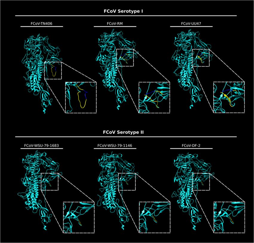
at the S1/S2 region between the two FCoV serotypes. In the type I S models, a loop was observed
corresponding to the S1/S2 cleavage site and the 10 amino acid insertion in the flanking regions of
this site (Figure 4). The loop is predicted to be exposed in the S monomers, but differences in the
folding and topology of the loop among the modeled S proteins suggest it can have some flexibility
in the structure (Figure 4, compare between FCoV I S models). In contrast to the S1/S2 region of
type I spike protein models, the serotype II S proteins lack the 16 amino acid insertion (including the
S1/S2 site) and the models predicted a similar conformation at this region among the three studied
proteins, suggesting that this site is not necessary for the FCoV II S function (Figure 4, compare the
FCoV II S models). The absence of the S1/S2 cleavage site in FCoV serotype II is a major structural
and functional difference when compared to serotype I FCoV S proteins, and its implications for the
S protein functionality are yet to be addressed in the literature. However, these differences suggest
that the mechanisms used by each serotype to induce fusion between the viral and the cell membrane
could be significantly different.Viruses 2020, 12, 83 8 of 14
Viruses 2019, 11, x FOR PEER REVIEW 8 of 14
Viruses 2019, 11, x FOR PEER REVIEW 8 of 14
FigureFigure 3. Alignment
3. Alignment of theof S1/S2
the S1/S2 region
region of FCoV
of FCoV I and
I and FCoVIIIIstrains.
FCoV strains. Sequences
Sequences corresponding
corresponding to to the
Figure 3. Alignment
the S1/S2 of theFCoV
S1/S2type
region of FCoV I and FCoV II strains.and
Sequences corresponding
threetoFCoV
S1/S2 region ofregion of three
three FCoV type I strainsI strains (FCoV-RM,
(FCoV-RM, FCoV-UU47
FCoV-UU47 FCoV-TN406)
and FCoV-TN406) andthree
and
the S1/S2 region of three FCoV type I strains (FCoV-RM, FCoV-UU47 and FCoV-TN406) and three
type IIFCoV type
strains II strains
(FCoV-DF-2, (FCoV-DF-2, FCoV-WSU-79-1683
FCoV-WSU-79-1683 and FCoV-WSU-79-1146)
and FCoV-WSU-79-1146) reported in
reported in GenBank,
GenBank, and
FCoV
and 15type
FCoVII strains
type I(FCoV-DF-2,
sequences from FCoV-WSU-79-1683
clinical samples and FCoV-WSU-79-1146)
reported in the Europeanreported in GenBank,
Nucleotide Archive
15 FCoV
and
type I sequences
15 aligned
FCoV type
from clinical
I sequences
samples reported
from clinical
in the European Nucleotide Archive were
were using the Geneious Prime samples reported in
2019 (v.2019.2.3) the European
software package Nucleotide Archive
(supplementary
aligned
wereusing the Geneious
aligned1).using Prime
the Geneious 2019 (v.2019.2.3)
Prime software
2019 (v.2019.2.3) package
software(supplementary information 1).
information A 16 amino acid insertion including the S1/S2 cleavage sitepackage
(6 amino(supplementary
acid) and a 10
A 16 amino acid
information insertion
1). A 16 including
amino acid the S1/S2
insertion cleavage
including site
the (6
S1/S2 amino acid)
cleavage and
site
amino acid flanking region was only observed in type I strains. FCoV type I sample are partial (6 a 10
amino amino
acid) acid
and aflanking
10
region was acid
amino onlyamino
sequences, observed
flanking in type
region
acid was Ionly
nomenclature strains. FCoV
observed
cannot in type
type II sample
be displayed. are partial
strains. FCoV type Isequences,
sample are amino
partial acid
nomenclature
sequences,cannot be displayed.
amino acid nomenclature cannot be displayed.
Figure 4. In silico modeling of the S protein from FCoV I and FCoV II strains. In silico FCoV S models
of three FCoV type I strains (FCoV-RM, FCoV-UU47 and FCoV-TN406) and three FCoV type II strains
FigureFigure
4. In4.silico
In silico modeling
modeling of of
thethe Sprotein
proteinfrom
S and from FCoV
FCoV IIand
andFCoV
FCoV II II
strains. In silico
strains. FCoV
In silico S models
FCoV S models
(FCoV-DF-2, FCoV-WSU-79-1683 FCoV-WSU-79-1146) were built using Chimera (UCSF Chimera
of
of three three FCoV type I strains (FCoV-RM, FCoV-UU47 and FCoV-TN406) and three FCoV type II strains
v. FCoV
1.13.1, type I strains
University of (FCoV-RM, FCoV-UU47
California). The and(dashed
S1/S2 region FCoV-TN406)
squares)and wasthree FCoV to
magnified type II strains
better
(FCoV-DF-2, FCoV-WSU-79-1683 and FCoV-WSU-79-1146) were built using Chimera (UCSF Chimera
(FCoV-DF-2,
visualizeFCoV-WSU-79-1683
the differences between andtheFCoV-WSU-79-1146)
two serotypes. The 16 were
aminobuilt using Chimera
acid insertion (UCSFtoChimera
corresponding the v.
v. 1.13.1, University of California). The S1/S2 region (dashed squares) was magnified to better
1.13.1,S1/S2 site (bright
University blue) and the
of California). 10 S1/S2
The amino region
acid flanking region
(dashed (yellow)
squares) waswere predictedtoasbetter
magnified a flexible
visualize
visualize the differences between the two serotypes. The 16 amino acid insertion corresponding to the
loop only between
the differences in the FCoV thetype
twoI Sserotypes.
models. The 16 amino acid insertion corresponding to the S1/S2 site
S1/S2 site (bright blue) and the 10 amino acid flanking region (yellow) were predicted as a flexible
(bright blue)
loop onlyand theFCoV
in the 10 amino
type I acid flanking region (yellow) were predicted as a flexible loop only in
S models.
the FCoV type I S models.Viruses 2020, 12, 83 9 of 14
7. Receptor Binding of FCoV S
Binding to a cellular receptor is the first step in the viral infection of a host cell, and in FCoV the S
protein is the component carrying out this function. The cellular receptor used by FCoV has been a
topic of discussion for several years. The primary receptor that has been described for FCoV is the
feline aminopeptidase N (fAPN) with the possibility of additional binding to lectin molecules such as
DC-SIGN, which would be considered a non-specific receptor, or attachment factor [74–76].
fAPN (also called CD13) is a membrane glycoprotein with metalloproteinase activity that is
expressed in a variety of tissues, including: granulocytes, monocytes, fibroblasts, endothelial cells,
synaptic membranes and several epithelial cells [64,77]. This receptor was first shown to be the key
FCoV receptor by Tresnan et al. [64] in 1996, where it was shown to be a common receptor for many
alphacoronaviruses, including the type I strain UCD-1. However, subsequent follow up studies have
shown that this receptor appears to be suitable for FCoV type II, but not for type I [74,78]. As such,
the primary receptor for serotype I viruses remains to be identified. While these major differences in
receptor usage are likely an underlying reason behind the known differences in virus isolation between
the type I and type II viruses in laboratory settings, other factors may also play a role, including
availability of proteases.
8. Activation and Fusion of FCoV S
Following receptor binding, the subsequent step in virus entry is the fusion of the virus envelope
with the host cell membrane. Fusion is mediated via the S2 domain and controlled by proteolytic
cleavage events at one or two positions. The cleavage of type II FCoV occurs at a single position
immediately adjacent to the viral fusion peptide (S20 ), whereas cleavage of type I viruses occurs at
both the S20 site and at the interface between the S1 and S2 domains (the S1/S2 site). In the case of type
I viruses, changes in viral tropism have been strongly linked to mutations in the S1/S2 site. However,
mutations are variable between different cats and tissues [10,12]. The overall model is that the S1/S2
region consists of an exposed loop, highly susceptible to proteolytic cleavage and consisting of a
recognition site for furin (a ubiquitous intracellular protease) and that mutations in the exposed loop
lead to an elimination of furin cleavage, possibly accompanied by a switch to a distinct protease in
distinct cell types and organs and transition to FIPV [12]. The finding of a distinct R793M mutation in
the spike S1/S2 site in certain neurological samples of cats with FIP would be in line with this model [10].
How mutations in the S20 site (shared between type I and type II viruses) link to pathogenesis is
currently less clear. Early studies on S20 site sequence changes and protease differences between the
serotype II laboratory strains FIPV II 1146 and FECV II 1683 do not appear to be widely reproduced in
clinical samples [79,80]. Studies on the S20 site of serotype I viruses also do not show a clear linkage
with disease outcome, although the site can often be mutated in viruses from FIP cases [80]. Overall
however, these significant differences in cleavage-activation underlies the different structure-function
relationships between type I and type II viruses and suggest a strong link to disease outcome.
For type I viruses, a mutation in a region within S2 (M1058A) also appears to correlate with
disease outcome [13]. Mutation at this amino acid was first described by Chang et al. in 2012, were
they described that this mutation allowed to distinguish FECV from FIP in most of their analyzed
cases [11]. The region of S containing M1058 was originally attributed to a fusion peptide region, but is
in fact down-stream of what is now considered the bona fide fusion peptide (based on the SARS-CoV
fusion peptide) [66]. However, M1058 is adjacent to the HR1 domain and so may modulate fusion in
some capacity. In a subsequent study, it was shown that the mutation M1058L was in fact indicative of
systemic spread of FCoV, finding that a methionine (M) was present in fecal samples from both FIP
and non-FIP cats, while leucine (L) was detected in tissues of both FIP and non-FIP cats, suggesting
that this mutation cannot predict the potential of FCoV to cause FIP [13]. This mutation has only been
studied in FCoVs corresponding to clinical cases that are most likely caused by type I viruses, so its
role in type II FCoV is still unknown.Viruses 2020, 12, 83 10 of 14
9. A Tale of Two Viruses
FCoV is a complex viral agent that causes a variety of clinical manifestations in cats. The virus
has been the focus of study for several decades, but the available literature still fails to completely
understand the nature and biology of this agent. Since the 1980s, it has been known that FCoV can
occur in two different types based on antigenic and genetic differences in the S protein [21,22]. Here,
we present evidence that the differences in the S protein are much more than serological or antigenic
variants, resulting in the effective presence of two distinct viruses.
We believe that the current paradigm for FCoV does not fully take into consideration the two
distinct viruses that circulate in cat populations. Our ability to molecularly diagnose FCoV infections in
cats is limited, and while efforts are being made to discriminate between the different pathogenic forms
of the virus (“FECV”, FIPV”, and “systemic”), information on the virus type (I or II) is not currently built
into current clinical or molecular testing regimes. We believe the distinct differences in the S protein
structure and function reviewed here are likely to translate into equally distinct biological and clinical
outcomes. Evidence for such biological differences is already apparent in laboratory settings, based on
the clear and well demonstrated differences in genetic background, structural features, receptor usage
and protease activation between the type I and type II S proteins. Whether these differences translate
into clinical settings remains opaque. However, the fact that molecular or antigenic differentiation
between the two types are not usually considered in routine diagnostic testing, does not allow to
understand the potential role of type II viruses in clinical settings. The realization that systemic
forms of FCoV may exist beyond the FECV-FIPV paradigm should be more fully considered. Without
consideration of the “type” of FCoV preset, our knowledge of FCoV transmission, pathogenesis, and
clinical impact leaves unanswered questions.
Funding: Work on FCoV in the author’s lab is supported by research grants from the Cornell Feline Health
Center and the Winn Feline Foundation. AES is supported by the NIH Comparative Medicine Training
Program T32OD011000.
Acknowledgments: We thank all members of the Whittaker lab for helpful comments.
Conflicts of Interest: The authors declare no conflict of interest.
References
1. Masters, P.S.; Perlman, S. Coronaviridae. In Fields Virology, 6th ed.; Knipe, D.M., Howley, P.M., Eds.; Lippincot
Williams & Wilkins: Philadelphia, PA, USA, 2013; Vol. 1, pp. 825–858.
2. Millet, J.K.; Whittaker, G.R. Host cell proteases: Critical determinants of coronavirus tropism and pathogenesis.
Virus Res. 2015, 202, 120–134. [CrossRef] [PubMed]
3. Song, Z.; Xu, Y.; Bao, L.; Zhang, L.; Yu, P.; Qu, Y.; Zhu, H.; Zhao, W.; Han, Y.; Qin, C. From SARS to MERS,
Thrusting Coronaviruses into the Spotlight. Viruses 2019, 11, 59. [CrossRef]
4. Zumla, A.; Alagaili, A.N.; Cotten, M.; Azhar, E.I. Infectious diseases epidemic threats and mass gatherings:
Refocusing global attention on the continuing spread of the Middle East Respiratory syndrome coronavirus
(MERS-CoV). BMC Med. 2016, 14, 132. [CrossRef] [PubMed]
5. Whittaker, G.R. Coronaviridae. In Fenner’s Veterinary Virology, 5th ed.; MacLachlan, N.J., Dubovi, E.J., Eds.;
Academic Press: London, UK, 2017; pp. 435–462.
6. Kipar, A.; Meli, M.L. Feline infectious peritonitis: Still an enigma? Vet. Pathol. 2014, 51, 505–526. [CrossRef]
[PubMed]
7. Wolfe, L.G.; Griesemer, R.A. Feline infectious peritonitis. Pathol. Vet. 1966, 3, 255–270. [CrossRef]
8. Pedersen, N.C. A review of feline infectious peritonitis virus infection: 1963–2008. J. Feline Med. Surg. 2009,
11, 225–258. [CrossRef]
9. Pedersen, N.C. An update on feline infectious peritonitis: Virology and immunopathogenesis. Vet. J. 2014,
201, 123–132. [CrossRef]
10. André, N.M.; Cossic, B.; Davies, E.; Miller, A.D.; Whittaker, G.R. Distinct mutation in the feline
coronavirus spike protein cleavage activation site in a cat with feline infectious peritonitis-associated
meningoencephalomyelitis. J. Feline Med. Surg. Open Rep. 2019, 5. [CrossRef]Viruses 2020, 12, 83 11 of 14
11. Chang, H.W.; Egberink, H.F.; Halpin, R.; Spiro, D.J.; Rottier, P.J. Spike protein fusion peptide and feline
coronavirus virulence. Emerg. Infect. Dis. 2012, 18, 1089–1095. [CrossRef]
12. Licitra, B.N.; Millet, J.K.; Regan, A.D.; Hamilton, B.S.; Rinaldi, V.D.; Duhamel, G.E.; Whittaker, G.R. Mutation
in spike protein cleavage site and pathogenesis of feline coronavirus. Emerg. Infect. Dis. 2013, 19, 1066–1073.
[CrossRef]
13. Porter, E.; Tasker, S.; Day, M.J.; Harley, R.; Kipar, A.; Siddell, S.G.; Helps, C.R. Amino acid changes in the
spike protein of feline coronavirus correlate with systemic spread of virus from the intestine and not with
feline infectious peritonitis. Vet. Res. 2014, 45, 49. [CrossRef] [PubMed]
14. Chang, H.W.; de Groot, R.J.; Egberink, H.F.; Rottier, P.J. Feline infectious peritonitis: Insights into feline
coronavirus pathobiogenesis and epidemiology based on genetic analysis of the viral 3c gene. J. Gen. Virol.
2010, 91, 415–420. [CrossRef] [PubMed]
15. Perera, K.D.; Rathnayake, A.D.; Liu, H.; Pedersen, N.C.; Groutas, W.C.; Chang, K.O.; Kim, Y. Characterization
of amino acid substitutions in feline coronavirus 3C-like protease from a cat with feline infectious peritonitis
treated with a protease inhibitor. Vet. Microbiol. 2019, 237, 108398. [CrossRef] [PubMed]
16. Murphy, B.G.; Perron, M.; Murakami, E.; Bauer, K.; Park, Y.; Eckstrand, C.; Liepnieks, M.; Pedersen, N.C. The
nucleoside analog GS-441524 strongly inhibits feline infectious peritonitis (FIP) virus in tissue culture and
experimental cat infection studies. Vet. Microbiol. 2018, 219, 226–233. [CrossRef] [PubMed]
17. Kim, Y.; Liu, H.; Galasiti Kankanamalage, A.C.; Weerasekara, S.; Hua, D.H.; Groutas, W.C.; Chang, K.-O.;
Pedersen, N.C. Reversal of the Progression of Fatal Coronavirus Infection in Cats by a Broad-Spectrum
Coronavirus Protease Inhibitor. PLoS Pathog. 2016, 12, e1005531. [CrossRef]
18. Pedersen, N.C.; Perron, M.; Bannasch, M.; Montgomery, E.; Murakami, E.; Liepnieks, M.; Liu, H. Efficacy
and safety of the nucleoside analog GS-441524 for treatment of cats with naturally occurring feline infectious
peritonitis. J. Feline Med. Surg. 2019, 21, 271–281. [CrossRef]
19. Pedersen, N.C.; Ward, J.; Mengeling, W.L. Antigenic relationship of the feline infectious peritonitis virus to
coronaviruses of other species. Arch. Virol. 1978, 58, 45–53. [CrossRef]
20. Corapi, W.V.; Olsen, C.W.; Scott, F.W. Monoclonal antibody analysis of neutralization and antibody-dependent
enhancement of feline infectious peritonitis virus. J. Virol. 1992, 66, 6695–6705. [CrossRef]
21. Hohdatsu, T.; Okada, S.; Koyama, H. Characterization of monoclonal antibodies against feline infectious
peritonitis virus type II and antigenic relationship between feline, porcine, and canine coronaviruses.
Arch. Virol. 1991, 117, 85–95. [CrossRef]
22. Pedersen, N.C.; Black, J.W.; Boyle, J.F.; Evermann, J.F.; McKeirnan, A.J.; Ott, R.L. Pathogenic differences
between various feline coronavirus isolates. Adv. Exp. Med. Biol. 1984, 173, 365–380.
23. Addie, D.D.; Schaap, I.A.; Nicolson, L.; Jarrett, O. Persistence and transmission of natural type I feline
coronavirus infection. J. Gen. Virol. 2003, 84, 2735–2744. [CrossRef] [PubMed]
24. Kummrow, M.; Meli, M.L.; Haessig, M.; Goenczi, E.; Poland, A.; Pedersen, N.C.; Hofmann-Lehmann, R.;
Lutz, H. Feline coronavirus serotypes 1 and 2: Seroprevalence and association with disease in Switzerland.
Clin. Diagn. Lab. Immunol. 2005, 12, 1209–1215. [CrossRef] [PubMed]
25. Li, C.; Liu, Q.; Kong, F.; Guo, D.; Zhai, J.; Su, M.; Sun, D. Circulation and genetic diversity of Feline
coronavirus type I and II from clinically healthy and FIP-suspected cats in China. Transbound Emerg. Dis.
2019, 66, 763–775. [CrossRef] [PubMed]
26. Benetka, V.; Kubber-Heiss, A.; Kolodziejek, J.; Nowotny, N.; Hofmann-Parisot, M.; Mostl, K. Prevalence
of feline coronavirus types I and II in cats with histopathologically verified feline infectious peritonitis.
Vet. Microbiol. 2004, 99, 31–42. [CrossRef]
27. Tekes, G.; Hofmann-Lehmann, R.; Bank-Wolf, B.; Maier, R.; Thiel, H.J.; Thiel, V. Chimeric feline coronaviruses
that encode type II spike protein on type I genetic background display accelerated viral growth and altered
receptor usage. J. Virol. 2010, 84, 1326–1333. [CrossRef]
28. Herrewegh, A.A.P.M.; Smeenk, I.; Horzinek, M.C.; Rottier, P.J.M.; de Groot, R.J. Feline Coronavirus Type II
Strains 79-1683 and 79-1146 Originate from a Double Recombination between Feline Coronavirus Type I and
Canine Coronavirus. J. Virol. 1998, 72, 4508–4514. [CrossRef]
29. Whittaker, G.R.; Andre, N.M.; Millet, J.K. Improving Virus Taxonomy by Recontextualizing Sequence-Based
Classification with Biologically Relevant Data: The Case of the Alphacoronavirus 1 Species. mSphere 2018, 3,
e00463-17. [CrossRef]Viruses 2020, 12, 83 12 of 14
30. Takano, T.; Endoh, M.; Fukatsu, H.; Sakurada, H.; Doki, T.; Hohdatsu, T. The cholesterol transport inhibitor
U18666A inhibits type I feline coronavirus infection. Antivir. Res. 2017, 145, 96–102. [CrossRef]
31. Doki, T.; Yabe, M.; Takano, T.; Hohdatsu, T. Differential induction of type I interferon by type I and type II
feline coronaviruses in vitro. Res. Vet. Sci. 2018, 120, 57–62. [CrossRef]
32. Olsen, C.W. A review of feline infectious peritonitis virus: Molecular biology, immunopathogenesis, clinical
aspects, and vaccination. Vet. Microbiol. 1993, 36, 1–37. [CrossRef]
33. Dye, C.; Siddell, S.G. Genomic RNA sequence of Feline coronavirus strain FIPV WSU-79/1146. J. Gen. Virol.
2005, 86, 2249–2253. [CrossRef] [PubMed]
34. Nieto-Torres, J.L.; DeDiego, M.L.; Verdia-Baguena, C.; Jimenez-Guardeno, J.M.; Regla-Nava, J.A.;
Fernandez-Delgado, R.; Castano-Rodriguez, C.; Alcaraz, A.; Torres, J.; Aguilella, V.M.; et al. Severe
acute respiratory syndrome coronavirus envelope protein ion channel activity promotes virus fitness and
pathogenesis. PLoS Pathog. 2014, 10, e1004077. [CrossRef] [PubMed]
35. Jaimes, J.A.; Whittaker, G.R. Feline coronavirus: Insights into viral pathogenesis based on the spike protein
structure and function. Virology 2018, 517, 108–121. [CrossRef] [PubMed]
36. Bosch, B.J.; van der Zee, R.; de Haan, C.A.M.; Rottier, P.J.M. The Coronavirus Spike Protein Is a Class I Virus
Fusion Protein: Structural and Functional Characterization of the Fusion Core Complex. J. Virol. 2003, 77,
8801–8811. [CrossRef]
37. White, J.M.; Delos, S.E.; Brecher, M.; Schornberg, K. Structures and mechanisms of viral membrane fusion
proteins: Multiple variations on a common theme. Crit. Rev. Biochem. Mol. Biol. 2008, 43, 189–219. [CrossRef]
38. Chang, H.W.; Egberink, H.F.; Rottier, P.J. Sequence analysis of feline coronaviruses and the circulating
virulent/avirulent theory. Emerg. Infect. Dis. 2011, 17, 744–746. [CrossRef]
39. Goodson, T.; Randell, S.; Moore, L. Feline infectious peritonitis. Compend. (YardleyPa) 2009, 31, E1–E8.
40. Pedersen, N.C.; Boyle, J.F.; Floyd, K.; Fudge, A.; Barker, J. An enteric coronavirus infection of cats and its
relationship to feline infectious peritonitis. Am. J. Vet. Res. 1981, 42, 368–377.
41. Addie, D.D.; Toth, S.; Murray, G.D.; Jarrett, O. Risk of feline infectious peritonitis in cats naturally infected
with feline coronavirus. Am. J. Vet. Res. 1995, 56, 429–434.
42. Tasker, S. Diagnosis of feline infectious peritonitis: Update on evidence supporting available tests. J. Feline
Med. Surg. 2018, 20, 228–243. [CrossRef]
43. Doherty, M.J. Ocular manifestations of feline infectious peritonitis. J. Am. Vet. Med. Assoc. 1971, 159, 417–424.
[PubMed]
44. Addie, D. Feline Coronavirus Infections. In Infectous Diseases of the Dog and Cat, 4th ed.; Greene, C.E., Ed.;
Elsevier: St. Louis, MO, USA, 2012; pp. 102–108.
45. Decaro, N.; Martella, V.; Elia, G.; Campolo, M.; Desario, C.; Cirone, F.; Tempesta, M.; Buonavoglia, C.
Molecular characterisation of the virulent canine coronavirus CB/05 strain. Virus Res. 2007, 125, 54–60.
[CrossRef] [PubMed]
46. O’Brien, A.; Mettelman, R.C.; Volk, A.; Andre, N.M.; Whittaker, G.R.; Baker, S.C. Characterizing replication
kinetics and plaque production of type I feline infectious peritonitis virus in three feline cell lines. Virology
2018, 525, 1–9. [CrossRef] [PubMed]
47. McKeirnan, A.J.; Evermann, J.F.; Davis, E.V.; Ott, R.L. Comparative properties of feline coronaviruses in vitro.
Rev. Can. De Rech. Vet. 1987, 51, 212–216.
48. Terada, Y.; Matsui, N.; Noguchi, K.; Kuwata, R.; Shimoda, H.; Soma, T.; Mochizuki, M.; Maeda, K. Emergence
of pathogenic coronaviruses in cats by homologous recombination between feline and canine coronaviruses.
PLoS ONE 2014, 9, e106534. [CrossRef]
49. Wang, Y.T.; Su, B.L.; Hsieh, L.E.; Chueh, L.L. An outbreak of feline infectious peritonitis in a Taiwanese shelter:
Epidemiologic and molecular evidence for horizontal transmission of a novel type II feline coronavirus.
Vet. Res. 2013, 44, 57. [CrossRef]
50. Jaimes, J.A.; Millet, J.K.; Goldstein, M.E.; Whittaker, G.R.; Straus, M.R. A Fluorogenic Peptide Cleavage
Assay to Screen for Proteolytic Activity: Applications for coronavirus spike protein activation. J. Vis. Exp.
2019. [CrossRef]
51. de Haan, C.A.; Haijema, B.J.; Schellen, P.; Wichgers Schreur, P.; te Lintelo, E.; Vennema, H.; Rottier, P.J.
Cleavage of group 1 coronavirus spike proteins: How furin cleavage is traded off against heparan sulfate
binding upon cell culture adaptation. J. Virol. 2008, 82, 6078–6083. [CrossRef]Viruses 2020, 12, 83 13 of 14
52. Rasschaert, D.; Duarte, M.; Laude, H. Porcine respiratory coronavirus differs from transmissible gastroenteritis
virus by a few genomic deletions. J. Gen. Virol. 1990, 71 (Pt. 11), 2599–2607. [CrossRef]
53. Pedersen, N.C.; Liu, H.; Dodd, K.A.; Pesavento, P.A. Significance of coronavirus mutants in feces and
diseased tissues of cats suffering from feline infectious peritonitis. Viruses 2009, 1, 166–184. [CrossRef]
54. Pedersen, N.C.; Liu, H.; Gandolfi, B.; Lyons, L.A. The influence of age and genetics on natural resistance
to experimentally induced feline infectious peritonitis. Vet. Immunol. Immunopathol. 2014, 162, 33–40.
[CrossRef] [PubMed]
55. McKeirnan, A.J.; Evermann, J.F.; Hargis, A.; Miller, L.M.; Ott, R.L. Isolation of feline coronaviruses from two
cats with diverse disease manifestations. Feline Pract. 1981, 11, 16–20.
56. Li, F. Structure, Function, and Evolution of Coronavirus Spike Proteins. Annu. Rev. Virol. 2016, 3, 237–261.
[CrossRef] [PubMed]
57. Harrison, S.C. Principles of virus structure. In Fields Virology, 6th ed.; Knipe, D.M., Howley, P.M., Eds.;
Lippincot Williams & Wilkins: Philadelphia, PA, USA, 2013; Vol. 1, pp. 52–86.
58. Floyd, D.L.; Ragains, J.R.; Skehel, J.J.; Harrison, S.C.; van Oijen, A.M. Single-particle kinetics of influenza
virus membrane fusion. Proc. Natl. Acad. Sci. USA 2008, 105, 15382–15387. [CrossRef] [PubMed]
59. Belouzard, S.; Millet, J.K.; Licitra, B.N.; Whittaker, G.R. Mechanisms of coronavirus cell entry mediated by
the viral spike protein. Viruses 2012, 4, 1011–1033. [CrossRef] [PubMed]
60. Madu, I.G.; Chu, V.C.; Lee, H.; Regan, A.D.; Bauman, B.E.; Whittaker, G.R. Heparan Sulfate Is a Selective
Attachment Factor for the Avian Coronavirus Infectious Bronchitis Virus Beaudette. Avian Dis. 2007, 51,
45–51. [CrossRef]
61. Winter, C.; Schwegmann-Wessels, C.; Cavanagh, D.; Neumann, U.; Herrler, G. Sialic acid is a receptor
determinant for infection of cells by avian Infectious bronchitis virus. J. Gen. Virol. 2006, 87, 1209–1216.
[CrossRef]
62. Li, W.; Hulswit, R.J.G.; Widjaja, I.; Raj, V.S.; McBride, R.; Peng, W.; Widagdo, W.; Tortorici, M.A.; van
Dieren, B.; Lang, Y.; et al. Identification of sialic acid-binding function for the Middle East respiratory
syndrome coronavirus spike glycoprotein. Proc. Natl. Acad. Sci. USA 2017, 114, E8508–E8517. [CrossRef]
63. Peng, G.; Sun, D.; Rajashankar, K.R.; Qian, Z.; Holmes, K.V.; Li, F. Crystal structure of mouse coronavirus
receptor-binding domain complexed with its murine receptor. Proc. Natl. Acad. Sci. USA 2011, 108,
10696–10701. [CrossRef]
64. Tresnan, D.B.; Levis, R.; Holmes, K.V. Feline aminopeptidase N serves as a receptor for feline, canine, porcine,
and human coronaviruses in serogroup I. J. Virol. 1996, 70, 8669–8674. [CrossRef]
65. Lu, G.; Hu, Y.; Wang, Q.; Qi, J.; Gao, F.; Li, Y.; Zhang, Y.; Zhang, W.; Yuan, Y.; Bao, J.; et al. Molecular basis of
binding between novel human coronavirus MERS-CoV and its receptor CD26. Nature 2013, 500, 227–231.
[CrossRef]
66. Lai, A.L.; Millet, J.K.; Daniel, S.; Freed, J.H.; Whittaker, G.R. The SARS-CoV Fusion Peptide Forms an
Extended Bipartite Fusion Platform that Perturbs Membrane Order in a Calcium-Dependent Manner. J. Mol.
Biol. 2017, 429, 3875–3892. [CrossRef] [PubMed]
67. White, J.M.; Whittaker, G.R. Fusion of Enveloped Viruses in Endosomes. Traffic 2016, 17, 593–614. [CrossRef]
[PubMed]
68. Song, W.; Gui, M.; Wang, X.; Xiang, Y. Cryo-EM structure of the SARS coronavirus spike glycoprotein in
complex with its host cell receptor ACE2. PLoS Pathog. 2018, 14, e1007236. [CrossRef] [PubMed]
69. Walls, A.C.; Xiong, X.; Park, Y.J.; Tortorici, M.A.; Snijder, J.; Quispe, J.; Cameroni, E.; Gopal, R.; Dai, M.;
Lanzavecchia, A.; et al. Unexpected Receptor Functional Mimicry Elucidates Activation of Coronavirus
Fusion. Cell 2019, 176, 1026–1039.e15. [CrossRef]
70. Walls, A.C.; Tortorici, M.A.; Bosch, B.J.; Frenz, B.; Rottier, P.J.; DiMaio, F.; Rey, F.A.; Veesler, D. Cryo-electron
microscopy structure of a coronavirus spike glycoprotein trimer. Nature 2016, 531, 114–117. [CrossRef]
71. Walls, A.C.; Tortorici, M.A.; Frenz, B.; Snijder, J.; Li, W.; Rey, F.A.; DiMaio, F.; Bosch, B.J.; Veesler, D. Glycan
shield and epitope masking of a coronavirus spike protein observed by cryo-electron microscopy. Nat. Struct.
Mol. Biol. 2016, 23, 899–905. [CrossRef]
72. Shang, J.; Zheng, Y.; Yang, Y.; Liu, C.; Geng, Q.; Luo, C.; Zhang, W.; Li, F. Cryo-EM structure of infectious
bronchitis coronavirus spike protein reveals structural and functional evolution of coronavirus spike proteins.
PLoS Pathog. 2018, 14, e1007009. [CrossRef]You can also read