ATM's Role in the Repair of DNA Double-Strand Breaks - MDPI
←
→
Page content transcription
If your browser does not render page correctly, please read the page content below
G C A T
T A C G
G C A T
               genes
Review
ATM’s Role in the Repair of DNA Double-Strand Breaks
Atsushi Shibata 1, *            and Penny A. Jeggo 2

                                          1   Signal Transduction Program, Gunma University Initiative for Advanced Research (GIAR),
                                              Gunma University, Gunma 371-8511, Japan
                                          2   Genome Damage and Stability Centre, School of Life Sciences, University of Sussex, Brighton BN1 9RQ, UK;
                                              p.a.jeggo@sussex.ac.uk
                                          *   Correspondence: shibata.at@gunma-u.ac.jp; Tel.: +81-27-220-7977; Fax: +81-27-220-7909

                                          Abstract: Ataxia telangiectasia mutated (ATM) is a central kinase that activates an extensive network
                                          of responses to cellular stress via a signaling role. ATM is activated by DNA double strand breaks
                                          (DSBs) and by oxidative stress, subsequently phosphorylating a plethora of target proteins. In the last
                                          several decades, newly developed molecular biological techniques have uncovered multiple roles
                                          of ATM in response to DNA damage—e.g., DSB repair, cell cycle checkpoint arrest, apoptosis, and
                                          transcription arrest. Combinational dysfunction of these stress responses impairs the accuracy of
                                          repair, consequently leading to dramatic sensitivity to ionizing radiation (IR) in ataxia telangiectasia
                                          (A-T) cells. In this review, we summarize the roles of ATM that focus on DSB repair.

                                          Keywords: DNA double-strand break; ionizing radiation; ATM; non-homologous end joining;
                                          homologous recombination


                                          1. Introduction
Citation: Shibata, A.; Jeggo, P.A.
ATM’s Role in the Repair of DNA
                                                Ataxia telangiectasia (A-T) was identified as a human disorder displaying radiosensi-
Double-Strand Breaks. Genes 2021, 12,
                                          tivity at both the cellular and clinical level in 1975 [1], and was amongst the first of the DNA
1370. https://doi.org/10.3390/            damage response disorders to be characterised. A-T has a broad clinical manifestation
genes12091370                             with individuals displaying progressive cerebellar degeneration, immunodeficiency and
                                          cancer predisposition. However, although dramatic sensitivity to ionising radiation (IR)
Academic Editors: Junya Kobayashi         and to radiomimetic drugs was evident in that first and additional early reports with cells
and Qiu-Mei Zhang-Akiyama                 derived from A-T individuals displaying marked sensitivity to cell killing and to chromo-
                                          some aberrations, subsequent studies failed to reveal any significant defect in the repair
Received: 30 June 2021                    of DNA double strand breaks (DSBs), the main lethal lesion induced by IR exposure [2].
Accepted: 30 August 2021                  The characterisation of a phenotype termed ‘radioresistant DNA synthesis’, which had
Published: 31 August 2021                 been amongst the earliest identified defects in A-T cells [3], raised the possibility that the
                                          inability to respond to DNA damage rather than a defect in the ability to repair the damage
Publisher’s Note: MDPI stays neutral      was at the root of the radiosensitivity. Fuel was added to the conundrum when A-T was
with regard to jurisdictional claims in   characterised as having a p53-dependent G1/S cell cycle checkpoint defect, resulting in its
published maps and institutional affil-   categorisation as the first human cell cycle checkpoint disorder [4], with both a G1/S and
iations.
                                          an intra-S phase checkpoint defect. A broad array of further phenotypes were observed
                                          in the ensuing years included defective meiotic recombination. Finally, in 1995 the causal
                                          genetic defect, the ataxia telangiectasia mutated (ATM) gene, was identified as a phos-
                                          phatidylinositol 3-kinase (PI3K) family member, which were known to play significant
Copyright: © 2021 by the authors.         roles in signal transduction [5]. Subsequently, ATM was shown to be a protein kinase rather
Licensee MDPI, Basel, Switzerland.        than a lipid kinase and classified as a PI3K-like kinase (PIKK). These important findings
This article is an open access article    helped to explain the broad phenotypes of A-T cells and the diverse clinical manifestation
distributed under the terms and           of the disorder. It is now appreciated that ATM, which is activated both by DNA DSBs
conditions of the Creative Commons        and by oxidative stress, has a vast array of substrates and, in response to DNA damage
Attribution (CC BY) license (https://
                                          or oxidative stress, initiates a plethora of responses. The co-ordination of these responses
creativecommons.org/licenses/by/
                                          optimises the repair of DSBs in the context of chromatin structure and the interface with
4.0/).

Genes 2021, 12, 1370. https://doi.org/10.3390/genes12091370                                                   https://www.mdpi.com/journal/genes
Genes 2021, 12, 1370                                                                                             2 of 13

                       other DNA metabolic processes, such as transcription. Failure to initiate this broad range
                       of responses impacts upon DSB repair in complex ways, which we detail here.
                             As techniques to monitor DNA DSB repair improved and primary rather than im-
                       mortalised cells were used, A-T was identified as having a subtle but significant defect in
                       the repair of a subset of radiation induced DSBs in non-cycling cells [6]. However, it is
                       also evident that additional consequences of the signalling defect in A-T cells can confer
                       deficiences in the repair of DSBs, with the defect often being manifest as a decrease in the
                       fidelity of repair rather than affecting the overall level of repair. In some cases, mis-repaired
                       DSBs may be tolerated and survival can ensue. However, frequently, mis-repaired DSBs,
                       such as chromosome rearrangements or large deletions, will be lethal. Thus, although the
                       outcome of misrepair versus no repair may be similar, simply assessing DSB repair levels
                       may not provide a good assessment of DSB repair. In this review, we will focus on the roles
                       that ATM plays to ensure efficient repair (of both the level and fidelity) of DSBs. We will
                       focus on the role of ATM in regulating four major processes, cell cycle checkpoint arrest,
                       the arrest of transcription in the vicinity of DSBs, the repair of a specific subset of DSBs,
                       and its influence on DSB repair pathway choice, largely involving its regulation of DSB
                       end-resection. However, at the core of most of these processes, is the role that ATM plays in
                       regulating chromatin structure at the DSB site. We will first discuss how ATM is activated
                       and how it influences chromatin structure. We will then consider how the four responses,
                       when perturbed, impede the level of DSB repair and its fidelity. Finally, we will evaluate
                       how these defects might contribute to the clinical manifestation of A-T.

                       2. ATM Activation
                             The primary activation mode of ATM kinase activity is its dissociation from a dimeric
                       form by autophosphorylation at Ser1981. In 2003, Kastan’s group elegantly demonstrated
                       that the ATM dimer is autophosphorylated in response to DNA damage, subsequently
                       the active form of an ATM monomer promotes the downstream phosphorylation and
                       interaction with ATM partners [7]. More recently, crystal structure analysis uncovered
                       the high-resolution structure of ATM dimers and monomers. Single-particle electron mi-
                       croscopy demonstrated that ATM active sites are buried, restricting access of the substrates
                       to these sites in the dimeric structure [8]. Furthermore, studies using electron cryomi-
                       croscopy (cryo-EM) suggest that the ATM dimeric structure has two distinct dynamic
                       forms, i.e., closed and open dimers (Figure 1) [9–11]. In the closed state, the PIKK reg-
                       ulatory domain blocks the peptide substrate–binding site. In contrast, the active site is
                       held in this closed conformation by interaction with a long helical hairpin in the TRD3
                       (tetratricopeptide repeats domain 3) domain of the symmetry-related molecule; this sug-
                       gests that the open conformation may be more active. Nevertheless, the kinase activity of
                       the monomer is ~10-fold higher than the open dimeric form [11]. Although the precise
                       in vivo regulation is unknown, it has been proposed that the open dimer is a transition
                       state between the closed dimer and monomer states. Alternatively, it could be a structure
                       to fine-tune ATM activity dependent on the biological situation.
                             Biologically, several different activation modes have been reported. A DSB is the pri-
                       mary form of triggering ATM activation by dimer dissociation. The MRE11/RAD50/NBS1
                       (MRN) complex acts as the DSB sensor, thereby recruiting ATM at DSB sites, particularly by
                       an interaction between ATM and Nijmegen breakage syndrome protein 1 (NBS1), resulting
                       in further accumulation of ATM signaling and positive feedback [12–14] (Figure 2). ATM
                       becomes hyperactive in DNA-dependent protein kinase catalytic subunit (DNA-PKcs) defi-
                       cient cells or cells treated with a DNA-PK inhibitor, suggesting that DNA-PKcs counteracts
                       the over-activation of ATM kinase activity under physiological conditions [15]. This mech-
                       anism is considered to maintain normal cell growth by preventing unnecessary apoptotic
                       pathway activation. A recent study using high-throughput chromosome conformation cap-
                       ture (Hi-C) and chromatin immunoprecipitation sequencing (ChIP-Seq) analyses showed
                       that the signal of ATM pS1981 (a marker of autophosphorylation and ATM activation)
                       shows a sharp peak at the DSB site compared with the γH2AX (a phosphorylation form
Genes 2021, 12, 1370 3 of 13
of H2AX) signal, a downstream substrate of ATM, suggesting that the active form of
ATM is predominantly located near DSBs [16]. The proposed model suggests that the
locally recruited ATM to DSB ends phosphorylates H2AX. Concomitantly, ATM activates a
cohesion-dependent loop extrusion that further promotes H2AX phosphorylation along
the chromatin until the loop extrusion is blocked at the topologically associating domain
x FOR PEER REVIEW boundary element [16,17] (Figure 2). Thus, ATM can reach and phosphorylate 3 of 13 multiple
targets along the chromatin even if localization of ATM near a DSB end is required to
sustain its activation (see also the discussion of ATM localization in Section 3).
1. ATM1.activation
FigureFigure after DSB
ATM activation formation.
after (A) ATM structure
DSB formation. (A) ATMisstructure
associatediswith the kinase
associated activity.
with The monomeric
the kinase
form has greater kinase activity than the dimeric form. (B) A two-dimensional ATM structure with some
activity. The monomeric form has greater kinase activity than the dimeric form. (B) A two-dimen- of ATM’s relevant
domains indicated.
sional ATM structure with some of ATM’s relevant domains indicated.
In addition, ATM can be activated under conditions of oxidative stress following
Biologically, several different activation modes have been reported. A DSB is the pri-
the formation of dimers via disulfide-crosslinks [18]. This mode of ATM activation is
mary form of triggering ATMseparable
genetically activation by the
from dimer dissociation.
process describedThe MRE11/RAD50/NBS1
above, requires distinct sites within
(MRN) complex acts as the DSB sensor, thereby recruiting ATM at
ATM and is MRN-independent. Whereas loss of DNA damage-induced DSB sites, particularly
ATM activation
by an interaction between ATM and Nijmegen breakage syndrome protein
confers loss of cell viability, failure to activate checkpoint arrest and 1 (NBS1), re-
end-resection, loss of
sulting in further oxidative
accumulation of ATM
activation signaling
has minimal and on
impact positive feedbackbut
these outcomes [12–14]
impedes (Figure
ATM-mediated
2). ATM becomes checkpoint
hyperactive response after oxidativeprotein
in DNA-dependent response, deficiency
kinase in mitochondrial
catalytic subunit (DNA- function and
PKcs) deficient cells or cells treated with a DNA-PK inhibitor, suggesting that DNA-PKcs alteration
autophagy [19]. ATM has also been reported to be activated in response to the
of chromatin structure
counteracts the over-activation of ATM without
kinase DNA damage
activity although
under whether this
physiological requires MRN and
conditions
the DNA damage or oxidative damage sites has not been established [7]. In response
[15]. This mechanism is considered to maintain normal cell growth by preventing unnec-
to DSBs, ATM is exported from the nucleus and can stimulate NF-κB–dependent signal
essary apoptotic pathway activation.
transduction A recent
[20]. Hence, ATM study usingmodes
has multiple high-throughput chromosome
for the activation of its kinase activity
conformation capture (Hi-C) and chromatin immunoprecipitation sequencing
in response to DSBs and cellular stress. These diverse activation modes (ChIP-Seq)
may be critical to
analyses showed select
that the
the signal of ATMbypS1981
best responses (a selective
targeting maker ofsubstrates
autophosphorylation and
from the multitudinous ATM
ATM activation) shows a sharp
substrates peak at
in response to the DSBtypes
distinct site of
compared
genotoxicwith the
stress. H2AX
Since here, (a
wephos-
will discuss the
phorylation form impact on DSB
of H2AX) repair,
signal, we will focus on
a downstream the canonical
substrate of ATM,activation of ATM
suggesting at DSBs.
that the
active form of ATM is predominantly located near DSBs [16]. The proposed model sug-
gests that the locally recruited ATM to DSB ends phosphorylates H2AX. Concomitantly,
ATM activates a cohesion-dependent loop extrusion that further promotes H2AX phos-
phorylation along the chromatin until the loop extrusion is blocked at the topologically
associating domain boundary element [16,17] (Figure 2). Thus, ATM can reach and phos-
phorylate multiple targets along the chromatin even if localization of ATM near a DSB
end is required to sustain its activation (see also the discussion of ATM localization in
Section 3).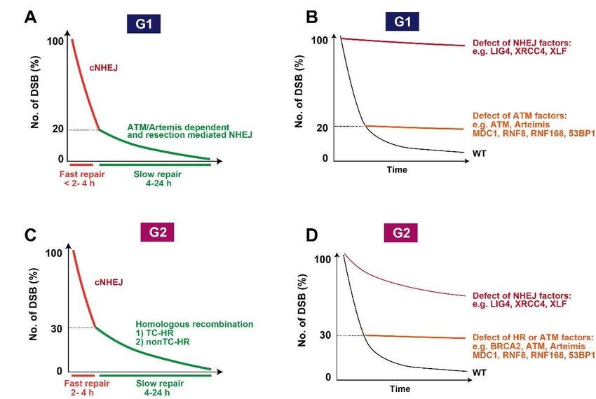
Genes 2021, 12, 1370
OR PEER REVIEW 4 of 13 4 of 13
Figure
Figure 2. ATM
2. ATM activation
activation at DSB
at DSB sites. ATMsites. ATM phosphorylates
phosphorylates H2AX at S139,H2AX
this, in at S139,
turn, this, in
promotes theturn, promotes
recruitment of MDC1,
the in
which, recruitment
its own turn,offacilitates
MDC1,the which, in its own turn, facilitates
RNF8/RNF168-dependent the RNF8/RNF168-dependent
ubiquitination of H2AX at K15. These signalsubiquiti-
recruit 53BP1
nation
to form nanoof H2AX
domains, atwhich
K15. These signals as
are visualized recruit 53BP1
nano foci. to form nanopost-translational
ATM-dependent domains, whichmodifications
are visualized are as
shown
nano
in the foci. panel.
bottom ATM-dependent
The interactingpost-translational modifications
proteins and downstream effectorsare
areshown
describedin the bottom
in more panel.
detail in theThe
text. CTCF:
interacting proteins
CCCTC-Binding Factor. and downstream effectors are described in more detail in the text. CTCF:
CCCTC-Binding Factor.
3. Role of ATM in DNA Damage Signalling
In addition, ATM can be activated under conditions of oxidative stress following the
ATM lies at the heart of a signal transduction process alerting cells to the presence of
formation of dimersDSBs. via disulfide-crosslinks
The most significant aspect[18].ofThis
this mode of the
is to alter ATM activation
chromatin in theisDSB
genet-
vicinity. For
ically separable from the
this process
process, described
multiple repairabove, requires distinct
mediators—mediator sitesdamage
of DNA withincheckpoint
ATM andprotein 1
is MRN-independent. Whereas
(MDC1), RINGloss
fingerof DNA damage-induced
ubiquitin E3 ligase (RNF8),ATM RING activation confers loss
finger 168 (RNF168), breast cancer
of cell viability, failure to activate checkpoint arrest and end-resection, loss of oxidative in an
susceptibility protein 1 (BRCA1), and p53 binding protein 1 (53BP1)—are recruited
ATM-dependent manner (see the details in our previous reviews [21–23]). As damage
activation has minimal impact on these outcomes but impedes ATM-mediated checkpoint
response mediators, MDC1 and 53BP1 contribute to the amplification of ATM activity
response after oxidative response, deficiency in mitochondrial function and autophagy
surrounding DSBs. MDC1 binds to γH2AX and the recruitment is identified as foci [24],
[19]. ATM has also been reported
suggesting that to
thebedistribution
activated in of response
MDC1 is also to the alteration
estimated of achromatin
to be million base pairs
structure without DNA arounddamage
DSBs. although
In addition,whether
MDC1 also thisinteracts
requireswithMRN and the
MRE11, whichDNA alsodam-
promotes the
tethering
age or oxidative damage of MRN,
sites has notand also
beenATM [25,26]. Furthermore,
established MDC1 interacts
[7]. In response to DSBs, with
ATMRNF8 isvia ATM-
dependent MDC1 phosphorylation [27–29]. This signal
exported from the nucleus and can stimulate NF-κB–dependent signal transduction [20]. cascade promotes the formation of
K63-linked ubiquitin chains in an RNF8/RNF168-dependent manner and subsequently
Hence, ATM has multiple modes for the activation of its kinase activity in response to
the recruitment of 53BP1 on chromatin. In addition to MDC1, 53BP1 interacts with the
DSBs and cellular stress. These diverse
MRN complex, activation
which promotes themodes mayofbe
activation ATMcritical
at DSBto sites
select theGiven
[30]. best that the
responses by targetingDNAselective substrates
damage response from the
mediators, multitudinous
MDC1, RNF8/RNF168, ATM andsubstrates
53BP1, playinmajorre- roles in
sponse to distinct types of genotoxic
ATM-dependent stress.
signal Since here,
expansion we deficient
and that will discuss the impact
cells have smalleron ATMDSB foci [31], it
is unclear how this previous model of
repair, we will focus on the canonical activation of ATM at DSBs. ATM recruitment and foci expansion is reconciled
with the recent findings discussed above, that activated ATM is only localised at the DSB
site. One possibility is that the initial activation of ATM may occur at close proximity
3. Role of ATM in DNA Damage Signalling
to DSB ends, and subsequently ATM can be recruited in a mediator-dependent manner
ATM lies at thealong
heartthe
ofchromatin,
a signal transduction process
with the signal alerting cells
being amplified to the
onto the presence
scaffold of Such a
proteins.
DSBs. The most significant aspect of this is to alter the chromatin in the DSB vicinity. For
this process, multiple repair mediators—mediator of DNA damage checkpoint protein 1
(MDC1), RING finger ubiquitin E3 ligase (RNF8), RING finger 168 (RNF168), breast can-
cer susceptibility protein 1 (BRCA1), and p53 binding protein 1 (53BP1)—are recruited in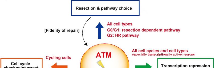
Genes 2021, 12, 1370 5 of 13
model is possible if the second phase of ATM binding is not detected by ChIP if it is not
tightly bound to chromatin. An alternative possibility is that the mediators promote ATM
turnover or tethering at the DSB site but not when distant from the DSB site.
4. Cell Cycle Checkpoint Arrest
An important role of ATM is the activation of cell cycle checkpoint arrest (G1/S,
intra-S and G2/M checkpoint arrest). In some cell types, apoptosis can also be activated
if an excessive amount of DNA damage is induced. Central to checkpoint activation
is the ATM-dependent phosphorylation of Chk2 (see Bartek & Lukas for a review) [32].
Although phosphorylated Chk2 spreads cell-wide [33], the initial phosphorylation event
arises at γH2AX foci and thus requires all the ATM-dependent signal transduction proteins
described above for optimal activation [34]. The processes of checkpoint arrest have been
well described previously and will not be detailed here [32]. The impact of checkpoint
arrest on DSB repair has also been discussed [35]. Importantly, the activation of checkpoint
arrest provides time to allow DSB repair to be completed prior to the onset of replication or
mitosis, and failure to arrest efficiently can dramatically impede the fidelity of DSB repair.
It is important to note, however, that this process is only significant for cycling cells and
will not impact upon G0 arrested cells, which actually represents the majority of cells in our
body. This demonstrates that, whilst cell cycle checkpoint arrest is an important process
enhancing survival post irradiation, it is certainly not the only ATM-regulated process
influencing radiosensitivity.
5. Impact of ATM Signaling on Transcription
In parallel to cell cycle arrest and DSB repair, the transcription machinery is also
arrested to prevent aberrant mRNA synthesis at damaged transcription regulatory se-
quences. ATM has a critical role in preventing transcription after DSB formation by RNA
polymerase II (RNAPII) that has responsibility for mRNA synthesis at gene loci, and RNA
polymerase I that synthesizes ribosomal RNA [36,37]. ATM-dependent transcriptional
silencing occurs via RNF8 and RNF168 dependent K63-linked ubiquitination. Histone
H2A ubiquitination which requires ATM activity also mediates RNAPII transcription si-
lencing. In addition, ATM phosphorylates BRG1-associated factor 180 (BAF180), a subunit
of polybromo-associated BAF complex (PBAF) that is a chromatin remodeler, to suppress
RNAPII elongation [38]. Because cohesin, which regulates chromatin looping, is also
required in this axis [39], the dynamic change of chromatin structure may affect the overall
transcription machinery. However, the silencing occurs in cis to DNA damage, i.e., possibly
the local relationship between damage and transcription repression is localised and pos-
sibly on the same chromosome. In G1 phase, failure to activate transcriptional arrest has
been shown to delay the rate of DSB repair without affecting the final level of repair [38].
However, loss of transcriptional arrest enhances chromosomal rearrangements suggesting
an impact on the fidelity of repair [39]. This process, however, affects a minor, although
very important, subset of DSBs.
6. Role of ATM in the Repair of a Subset of DSBs: Role of Chromatin Remodeling
Although initial studies suggested that the level of DSB repair was normal in A-T
deficient cells, accumulating evidence clearly shows that lack of ATM kinase activity causes
a defined DSB repair defect [6]. Nonetheless, the magnitude of the DSB repair defect is
not as big as expected considering that ATM-deficient cells exhibit strong radio-sensitivity
equivalent to cells defective in non-homologous end joining (NHEJ) [40]. In 2004, the
Jeggo and Lobrich groups applied γH2AX foci analysis to measure DSB repair capability by
enumerating γH2AX foci [6]. Since γH2AX foci analysis is a more sensitive assay compared
with the pre-existing physical techniques such as the neural comet assay or pulsed-field
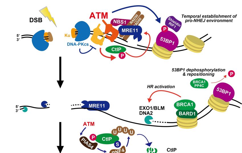
gel electrophoresis, the analysis uncovered the existence of an ATM-dependent DSB repair
fraction, which is approximately 15–20% of the total induced DSBs in irradiated G0/G1 cells
(Figure 3A,B). In addition, depletion of the other downstream factors such as 53BP1, MDC1,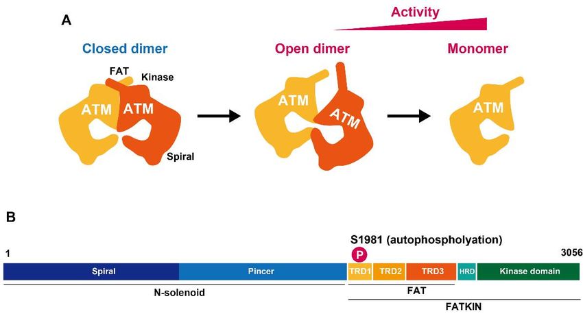
Genes 2021, 12, 1370                                                                                                               6 of 13

                                 and RNF8/RNF168 show a similar repair defect level, whereas a defect in core NHEJ factors
                                 such as X-ray cross complementing gene 4 (XRCC4), XRCC4-like-factor (XLF), or DNA
                                 ligase IV (LIG4) exhibit more substantial repair defects (Figure 3B) [6,31]. Interestingly,
                                 depletion of heterochromatin factors, KRAB-associated protein 1 (KAP-1), heterochromatin
                                 protein 1 (HP1), or histone deacetylase 1/2 (HDAC1/2), alleviates the repair defect in ATM-
                                 deficient cells [31,41]. ATM-dependent KAP-1 phosphorylation at Ser824 [42] promotes the
                                 dispersal of the nucleosome remodeler chromodomain helicase DNA-binding 3 (CHD3)
                                 from DSBs, which triggers chromatin relaxation at DSB sites [43] (see the details in our
                                 previous review [44]). In this axis, requirement for direct interaction between 53BP1 and
                                 γH2AX via the BRCA1 C-Terminal (BRCT) domain in 53BP1 was identified to amplify the
                                 ATM-pKAP1 signaling for ATM-dependent DSB repair [45]. In addition, suppressor of
                                 cancer cell invasion (SCAI), another 53BP1 interactor whose interaction is ATM kinase-
                                 dependent, facilitates this process [46]. Together, these studies suggest a role for ATM in
                                 coordinating chromatin remodeling during the repair process. Interestingly, cells defective
                                 in Artemis also show similar levels of defective DSB repair to ATM-deficient cells. Artemis-
R PEER REVIEW                    dependent DSB repair requires MRE11 exonuclease activity and CtBP-interacting7 of 13 protein
                                 (CtIP), suggesting that the subset of DSBs that undergo resection prior to the rejoining have
                                 a requirement for Artemis and ATM [47]. Importantly, ATM inhibition does not show an
                                 additive repair defect in Artemis cells, indicating that ATM and Artemis play a role in
             recombinase (RAD51) DSBrecruitment,      shows
                                       repair in the same     a defect
                                                            axis. Since itin  the repair
                                                                           is unlikely thatof ~30% directly
                                                                                            Artemis of DSBsparticipates
                                                                                                             in G2       in the
             phase. Similarly, ATM   inhibition    shows   the  same  level    of DSB  repair  defects in G2 phase
                                 chromatin remodeling event, it is still unknown how these two factors facilitate the repair
             [50] (Figure 3D).   process of DSBs in association with chromatin remodeling.

                 Figure
               Figure     DSB
                       3. 3.  repair
                             DSB     kinetics
                                 repair       after after
                                         kinetics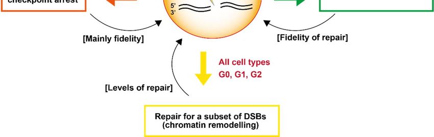
Genes 2021, 12, 1370                                                                                            7 of 13

                       7. Roles of ATM in DSB End Resection Influencing Pathway Choice
                             ATM plays a role in DSB end resection, which affects the fidelity of DSB repair. The
                       dysregulation of resection impairs the progression of homologous recombination (HR) in
                       S/G2 phase. The rejoining by these inefficiently resected DSBs by NHEJ likely leads to
                       deletion mutations. In S/G2 phase, the signaling machinery switches from ATM to ATR in
                       concert with the progression of resection because ATM is primarily activated at unresected
                       DSB ends. In contrast, ATR is activated on ssDNA-replication protein A (RPA) following
                       resection [48,49]. Therefore, the temporal switching of the two kinases occurs following the
                       progression of resection; however, ATM seems to be involved throughout the process of
                       HR, i.e., from initiation of resection to termination of resection (or restriction of excessive
                       resection). ATM is also required for the repair of 20–30% of DSBs after IR in S/G2, subtly
                       greater than the requirement for ATM in G1 phase (Figure 3C). Depletion of breast cancer
                       susceptibility gene 2 (BRCA2), which is an essential factor for HR by RAD51 recombinase
                       (RAD51) recruitment, shows a defect in the repair of ~30% of DSBs in G2 phase. Similarly,
                       ATM inhibition shows the same level of DSB repair defects in G2 phase [50] (Figure 3D).
                             In response to DSB induction in S/G2 phase, ATM is rapidly activated, similar to the
                       situation in G1 phase inducing the recruitment of DDR responders and promoting foci
                       formation such as γH2AX, MDC1, and 53BP1, followed by downstream signaling. 53BP1
                       hyper-phosphorylation, strictly dependent on ATM, occurs in S/G2 and G1 phases [51].
                       The hyper-phosphorylation of 53BP1 promotes the recruitment of replication timing reg-
                       ulatory factor 1 (RIF1), REV7, and Shieldin complexes that limit resection progression to
                       promote NHEJ by Polα primase recruitment, possibly to fill in the ssDNA gap [52,53].
                       Such a pro-NHEJ environment is turned into a pro-HR environment following 53BP1
                       dephosphorylation facilitated by BRCA1-protein phosphatase 4 catalytic subunit (PP4C) in
                       S/G2 phase [51] (Figure 4). In parallel, but possibly sequentially, ATM phosphorylates CtIP,
                       a central factor initiating resection, which activates MRE11 endonuclease activity [54], gen-
                       erating a nick (or nicks) to initiate bidirectional exonuclease dependent resection by MRE11
                       and Exonuclease 1 (EXO1)/BLM/DNA2 exonuclease activities [55–57] (Figure 4). Acti-
                       vated CtIP following its phosphorylation at Ser664/679/745 is required for resection and
                       HR [58,59]. This nicking commonly occurs to initiate resection at transcription-dependent
                       and independent DSBs [59,60]. However, it is unknown exactly how these phosphorylation
                       sites in CtIP affect the endonucleolytic activities although data suggest that phosphory-
                       lation promotes the recruitment or maintenance of CtIP at DSB sites [59,60]. In addition
                       to these sites, a total of eight SQ/TQ sites in CtIP are potentially ATM-dependent phos-
                       phorylation sites [58]. Particularly, disruption of the T859 site, with intact Ser664/679/745
                       residues impairs resection and HR. ATM-dependent CtIP phosphorylation occurs after
                       DSB formation. ATM-dependent phosphorylation event requires CDK-dependent phos-
                       phorylation of CtIP, suggesting that CtIP is effectively phosphorylated by ATM in S/G2
                       phase to promote resection after DNA damage [58,61]. The phosphorylation promotes
                       interaction between CtIP and NBS1, which helps (CtIP)-dependent endonucleolytic inci-
                       sion [62]. Following the progression of CtIP-dependent resection, 53BP1 dephosphorylation
                       releases RIF1, and its downstream factors from chromatin changes to promote a pro-HR
                       environment, i.e., HR-associated BRCA1 complex and exonucleases (EXO1/DNA2/BLM)
                       are recruited following 53BP1 repositioning, which generates a 53BP1-free chromatin area
                       in the immediate DSB vicinity [51,63,64] (Figure 4). The role of ATM-dependent chromatin
                       remodeling in HR is also proposed because the defective DSB repair in an ATM or 53BP1
                       deficient background is rescued by KAP-1 depletion in irradiated G2 cells [59,65]. However,
                       under ATM inhibition, because ATM is essential for the initiation of resection, NHEJ can
                       repair the DSBs, i.e., DSBs are repaired by NHEJ in ATM and KAP-1 double depleted
                       cells. However, again, the impact of pre-existing heterochromatin or active chromatin
                       remodeling is still under debate.
                             In parallel to the progression of resection, ATM also controls CtIP activity to restrict
                       excessive resection. A recent study showed that protein inhibitor of activated STAT4
                       (PIAS4) dependent SUMOylation of CtIP promotes RNF4 dependent CtIP ubiquitination,
Genes 2021, 12, 1370                                                                                                   8 of 13

                                which leads to its degradation [66] (Figure 4). ATM-dependent CtIP phosphorylation
                                precedes CtIP-SUMOylation and the lack of the phosphorylation impairs the downstream
                                CtIP degradation. Impaired CtIP ubiquitination results in excessive resection and defective
                                HR. Thus, CtIP degradation in the ATM-dependent signal cascade may serve to restrict
                                excessive resection during HR. In addition and interestingly, the inhibition of ATM after the
                                resection step slows down RAD51 removal from the chromatin, suggesting that ATM plays
                                a role in promoting RAD51 displacement [67]. Furthermore, ATM also phosphorylates
                                ubiquilin-4 (UBQLN4), a proteasome shuttling factor, which promotes the ubiquitylation of
                                MRE11 to fine-tune the magnitude of resection [68]. Thus, even if ATM is activated at the
                                beginning of the DSB repair process, the phosphorylation events comprehensively control
                                the overall HR process.
                                       The regulation of DNA-replication-fork-associated single-ended DSBs (seDSBs) in S
                                phase is different from two-ended DSBs because seDSBs must be directed toward HR be-
                                cause the lack of a DSB counterpart at seDSBs leads to chromosomal translocation if NHEJ
                                is used. Consistent with this notion, lack of ATM activity leads to toxic LIG4-mediated chro-
                                mosome fusions after DNA-replication-fork-associated seDSBs [69]. Appropriate removal
                                of NHEJ components (Ku/DNA-PKcs) is required to direct the repair pathway towards
                                HR at seDSB. The removal of the NHEJ component is achieved by MRE11/CtIP nuclease
                                activities [70]. At seDSB ends, ATM phosphorylates DNA-PKcs, and phosphorylation
                                at the ABCDE cluster of DNA-PKcs promotes the release of Ku from DSB ends in an
                                MRE11/CtIP-dependent manner [71]. Following the rapid removal of NHEJ components,
                                it is likely that a similar mechanism is used between seDSBs and two-ended DSBs.

                       Figure 4. Roles of ATM during DSB end resection in G2 phase. See the text for details.

                                     To summarise this section, ATM has a critical role in regulating resection at DSBs,
                                which arises via complex regulation of multiple substrates and critically depends on the
                                regulation of chromatin structure at the DSB site. Not surprisingly, this impact of ATM
                                affects DSBs in diverse ways. One affect is a failure to activate HR in G2 phase. However,
                                although less dramatic, resection also arises in G1 phase, impacting upon repair via NHEJ.
                                Here, two forms of NHEJ have been described, resection independent and dependent, and
                                the latter is ATM-dependent in G1 phase [47]. Failure to appropriately regulate resection
                                will predominantly affect the fidelity of DSB repair.
Genes 2021, 12, 1370 9 of 13
8. Summary
The details above reveal the significant functions that ATM has in determining the
response to DSBs and its influence on how they are repaired. Here, we describe how
ATM is essential for the repair of a small fraction of DSBs, its requirement for cell cycle
checkpoint arrest, transcriptional repression and resection, and hence an influence on
pathway choice, at DSBs (Figure 5). As briefly mentioned, ATM can also influence NF-κB
signaling following its export to the cytoplasm. These impacts can influence both the
level and fidelity of DSB repair. Indeed, one of the early hallmarks of ATM-deficient cells
was a profound increase in chromosomal aberrations following IR exposure. A major
question that arises is how these different consequences of ATM loss are manifest. For
example, which of these roles of ATM has the biggest influence on radiosensitivity or
impact clinically. Unfortunately, since all these processes interface and intertwine, it is
difficult to unravel their varying contributions. However, it is important to appreciate
that the precise tissue or cell type under analysis as well as cell cycle status is a major
determinant. Stem cells differ from differentiated cells, for example; lymphocytes differ
from fibroblasts. Cell cycle checkpoint arrest will not be influential in non-cycling cells.
Artemis deficiency, which is epistatic with ATM loss, confers significant radiosensitivity
in quiescent cells, demonstrating the significance of the modest DSB repair defect in A-T
Genes 2021, 12, x FOR PEER REVIEWcells [6]. Some neuronal cells are highly transcriptionally active, potentially rendering 10 of 13
transcription repression at DSBs of significance. Cytoplasmic signaling to NF-κB may be
more significant in cells that readily undergo apoptosis, such as progenitor cells. Clinically,
ATM
ataxiadeficiency causes
telangiectasia, ataxia telangiectasia,
a broad-based a broad-based
multi-system multi-system
disorder. Why disorder.
ATM deficiency Why
specifi-
ATM deficiency specifically results in marked loss of Purkinje cells and cerebellar function
cally results in marked loss of Purkinje cells and cerebellar function remains unclear. Why
remains unclear.
telangiectasia Why
arises telangiectasia
is not arises
well explained. is not
Since we well
now explained. Since we
have a reasonable now have a
understanding
reasonable understanding of the various steps regulated by ATM during DSB repair, a
of the various steps regulated by ATM during DSB repair, a current challenge is to under-
current challenge is to understand which processes are important in which situation.
stand which processes are important in which situation.
Figure 5.
Figure 5. Summary
Summary highlighting
highlighting the
the 44 pathways
pathways discussed
discussed in
in the text by
the text by which
which ATM
ATM influences
influences the
the repair
repair of
of DSBs.
DSBs. The
The
impact on the level or fidelity of DSB repair in each pathway is shown by brackets; the type of cell affected is highlighted
impact on the level or fidelity of DSB repair in each pathway is shown by brackets; the type of cell affected is highlighted in
in red. Additional impacts of ATM, such as its role in regulating apoptosis, may also influence survival levels in response
red. Additional impacts of ATM, such as its role in regulating apoptosis, may also influence survival levels in response to
to DSBs but here we focus on pathways that influence DSB repair.
DSBs but here we focus on pathways that influence DSB repair.
Funding: A.S. is supported by the Takeda Science Foundation, the SUNTORY Foundation for Life
Sciences, The Sumitomo Foundation, Program of the Network Type Joint Usage/Research Center
for Radiation Disaster Medical Science of Hiroshima University, Nagasaki University, and Fuku-
shima Medical University, and the Japan Society for the Promotion of Science (grant number
JP17H04713).
Institutional Review Board Statement: Not applicable.Genes 2021, 12, 1370 10 of 13
Funding: A.S. is supported by the Takeda Science Foundation, the SUNTORY Foundation for Life
Sciences, The Sumitomo Foundation, Program of the Network Type Joint Usage/Research Center for
Radiation Disaster Medical Science of Hiroshima University, Nagasaki University, and Fukushima
Medical University, and the Japan Society for the Promotion of Science (grant number JP17H04713).
Institutional Review Board Statement: Not applicable.
Informed Consent Statement: Not applicable.
Data Availability Statement: Not applicable.
Acknowledgments: The authors thank Akiko Shibata, Yoko Hayashi, Yasuyo Sekiguchi, Hiroko Iino,
and Naho Takashima for laboratory assistance. A.S. is a visiting associate professor of the Radiation
Biology Center, Graduate School of Biostudies, Kyoto University.
Conflicts of Interest: The authors declare that there are no conflicts of interest. The funders had no
role in the design of the study; in the collection, analyses, or interpretation of data; in the writing of
the manuscript, or in the decision to publish the results.
References
1. Taylor, A.M.; Harnden, D.G.; Arlett, C.F.; Harcourt, S.A.; Lehmann, A.R.; Stevens, S.; Bridges, B.A. Ataxia telangiectasia: A human
mutation with abnormal radiation sensitivity. Nature 1975, 258, 427–429. [CrossRef] [PubMed]
2. Lehman, A.R.; Stevens, S. The production and repair of double strand breaks in cells from normal humans and from patients
with ataxia telangiectasia. Biochim. Biophys. Acta 1977, 474, 49–60. [CrossRef]
3. Painter, R.B.; Young, B.R. Radiosensitivity in ataxia-telangiectasia: A new explanation. Proc. Natl. Acad. Sci. USA 1980, 77,
7315–7317. [CrossRef]
4. Kastan, M.B.; Zhan, Q.; el-Deiry, W.S.; Carrier, F.; Jacks, T.; Walsh, W.V.; Plunkett, B.S.; Vogelstein, B.; Fornace, A.J., Jr. A
mammalian cell cycle checkpoint pathway utilizing p53 and GADD45 is defective in ataxia-telangiectasia. Cell 1992, 71, 587–597.
[CrossRef]
5. Savitsky, K.; Bar-Shira, A.; Gilad, S.; Rotman, G.; Ziv, Y.; Vanagaite, L.; Tagle, D.A.; Smith, S.; Uziel, T.; Sfez, S.; et al. A single
ataxia telangiectasia gene with a product similar to PI-3 kinase. Science 1995, 268, 1749–1753. [CrossRef]
6. Riballo, E.; Kuhne, M.; Rief, N.; Doherty, A.; Smith, G.C.; Recio, M.J.; Reis, C.; Dahm, K.; Fricke, A.; Krempler, A.; et al. A pathway
of double-strand break rejoining dependent upon ATM, Artemis, and proteins locating to gamma-H2AX foci. Mol. Cell 2004, 16,
715–724. [CrossRef]
7. Bakkenist, C.J.; Kastan, M.B. DNA damage activates ATM through intermolecular autophosphorylation and dimer dissociation.
Nature 2003, 421, 499–506. [CrossRef]
8. Lau, W.C.; Li, Y.; Liu, Z.; Gao, Y.; Zhang, Q.; Huen, M.S. Structure of the human dimeric ATM kinase. Cell Cycle 2016, 15,
1117–1124. [CrossRef]
9. Baretic, D.; Pollard, H.K.; Fisher, D.I.; Johnson, C.M.; Santhanam, B.; Truman, C.M.; Kouba, T.; Fersht, A.R.; Phillips, C.; Williams,
R.L. Structures of closed and open conformations of dimeric human ATM. Sci. Adv. 2017, 3, e1700933. [CrossRef]
10. Yates, L.A.; Williams, R.M.; Hailemariam, S.; Ayala, R.; Burgers, P.; Zhang, X. Cryo-EM Structure of nucleotide-bound Tel1(ATM)
unravels the molecular basis of inhibition and structural rationale for disease-associated mutations. Structure 2020, 28, 96–104.E3.
[CrossRef]
11. Xiao, J.; Liu, M.; Qi, Y.; Chaban, Y.; Gao, C.; Pan, B.; Tian, Y.; Yu, Z.; Li, J.; Zhang, P.; et al. Structural insights into the activation of
ATM kinase. Cell Res. 2019, 29, 683–685. [CrossRef]
12. Lee, J.H.; Paull, T.T. ATM activation by DNA double-strand breaks through the Mre11-Rad50-Nbs1 complex. Science 2005, 308,
551–554. [CrossRef]
13. Uziel, T.; Lerenthal, Y.; Moyal, L.; Andegeko, Y.; Mittelman, L.; Shiloh, Y. Requirement of the MRN complex for ATM activation
by DNA damage. EMBO J. 2003, 22, 5612–5621. [CrossRef]
14. Falck, J.; Coates, J.; Jackson, S.P. Conserved modes of recruitment of ATM, ATR and DNA-PKcs to sites of DNA damage. Nature
2005, 434, 605–611. [CrossRef]
15. Zhou, Y.; Lee, J.H.; Jiang, W.; Crowe, J.L.; Zha, S.; Paull, T.T. Regulation of the DNA damage response by DNA-PKcs inhibitory
phosphorylation of ATM. Mol. Cell 2017, 65, 91–104. [CrossRef]
16. Arnould, C.; Rocher, V.; Finoux, A.L.; Clouaire, T.; Li, K.; Zhou, F.; Caron, P.; Mangeot, P.E.; Ricci, E.P.; Mourad, R.; et al. Loop
extrusion as a mechanism for formation of DNA damage repair foci. Nature 2021, 590, 660–665. [CrossRef]
17. Collins, P.L.; Purman, C.; Porter, S.I.; Nganga, V.; Saini, A.; Hayer, K.E.; Gurewitz, G.L.; Sleckman, B.P.; Bednarski, J.J.; Bassing,
C.H.; et al. DNA double-strand breaks induce H2Ax phosphorylation domains in a contact-dependent manner. Nat. Commun.
2020, 11, 3158. [CrossRef]Genes 2021, 12, 1370 11 of 13
18. Guo, Z.; Kozlov, S.; Lavin, M.F.; Person, M.D.; Paull, T.T. ATM activation by oxidative stress. Science 2010, 330, 517–521. [CrossRef]
19. Lee, J.H.; Mand, M.R.; Kao, C.H.; Zhou, Y.; Ryu, S.W.; Richards, A.L.; Coon, J.J.; Paull, T.T. ATM directs DNA damage responses
and proteostasis via genetically separable pathways. Sci. Signal. 2018, 11, eaan5598. [CrossRef]
20. Hadian, K.; Krappmann, D. Signals from the nucleus: Activation of NF-kappaB by cytosolic ATM in the DNA damage response.
Sci. Signal. 2011, 4, pe2. [CrossRef]
21. Shibata, A.; Jeggo, P.A. DNA double-strand break repair in a cellular context. Clin. Oncol. (R. Coll. Radiol.) 2014, 26, 243–249.
[CrossRef]
22. Shibata, A. Regulation of repair pathway choice at two-ended DNA double-strand breaks. Mutat. Res. 2017, 803–805, 51–55.
[CrossRef] [PubMed]
23. Shibata, A.; Jeggo, P. A historical reflection on our understanding of radiation-induced DNA double strand break repair in
somatic mammalian cells; interfacing the past with the present. Int. J. Radiat. Biol. 2019, 95, 945–956. [CrossRef]
24. Stucki, M.; Clapperton, J.A.; Mohammad, D.; Yaffe, M.B.; Smerdon, S.J.; Jackson, S.P. MDC1 directly binds phosphorylated
histone H2AX to regulate cellular responses to DNA double-strand breaks. Cell 2005, 123, 1213–1226. [CrossRef]
25. Melander, F.; Bekker-Jensen, S.; Falck, J.; Bartek, J.; Mailand, N.; Lukas, J. Phosphorylation of SDT repeats in the MDC1 N terminus
triggers retention of NBS1 at the DNA damage-modified chromatin. J. Cell Biol. 2008, 181, 213–226. [CrossRef] [PubMed]
26. Chapman, J.R.; Jackson, S.P. Phospho-dependent interactions between NBS1 and MDC1 mediate chromatin retention of the MRN
complex at sites of DNA damage. EMBO Rep. 2008, 9, 795–801. [CrossRef]
27. Mailand, N.; Bekker-Jensen, S.; Faustrup, H.; Melander, F.; Bartek, J.; Lukas, C.; Lukas, J. RNF8 ubiquitylates histones at DNA
double-strand breaks and promotes assembly of repair proteins. Cell 2007, 131, 887–900. [CrossRef]
28. Huen, M.S.; Grant, R.; Manke, I.; Minn, K.; Yu, X.; Yaffe, M.B.; Chen, J. RNF8 transduces the DNA-damage signal via histone
ubiquitylation and checkpoint protein assembly. Cell 2007, 131, 901–914. [CrossRef] [PubMed]
29. Kolas, N.K.; Chapman, J.R.; Nakada, S.; Ylanko, J.; Chahwan, R.; Sweeney, F.D.; Panier, S.; Mendez, M.; Wildenhain, J.; Thomson,
T.M.; et al. Orchestration of the DNA-damage response by the RNF8 ubiquitin ligase. Science 2007, 318, 1637–1640. [CrossRef]
30. Lee, J.H.; Goodarzi, A.A.; Jeggo, P.A.; Paull, T.T. 53BP1 promotes ATM activity through direct interactions with the MRN complex.
EMBO J. 2010, 29, 574–585. [CrossRef] [PubMed]
31. Noon, A.T.; Shibata, A.; Rief, N.; Lobrich, M.; Stewart, G.S.; Jeggo, P.A.; Goodarzi, A.A. 53BP1-dependent robust localized KAP-1
phosphorylation is essential for heterochromatic DNA double-strand break repair. Nat. Cell Biol. 2010, 12, 177–184. [CrossRef]
32. Bartek, J.; Lukas, J. DNA damage checkpoints: From initiation to recovery or adaptation. Curr. Opin. Cell Biol. 2007, 19, 238–245.
[CrossRef]
33. Lukas, C.; Falck, J.; Bartkova, J.; Bartek, J.; Lukas, J. Distinct spatiotemporal dynamics of mammalian checkpoint regulators
induced by DNA damage. Nat. Cell Biol. 2003, 5, 255–260. [CrossRef]
34. Fernandez-Capetillo, O.; Chen, H.T.; Celeste, A.; Ward, I.; Romanienko, P.J.; Morales, J.C.; Naka, K.; Xia, Z.; Camerini-Otero, R.D.;
Motoyama, N.; et al. DNA damage-induced G2-M checkpoint activation by histone H2AX and 53BP1. Nat. Cell Biol. 2002, 4,
993–997. [CrossRef]
35. Deckbar, D.; Jeggo, P.A.; Lobrich, M. Understanding the limitations of radiation-induced cell cycle checkpoints. Crit. Rev. Biochem.
Mol. Biol. 2011, 46, 271–283. [CrossRef]
36. Shanbhag, N.M.; Rafalska-Metcalf, I.U.; Balane-Bolivar, C.; Janicki, S.M.; Greenberg, R.A. ATM-dependent chromatin changes
silence transcription in cis to DNA double-strand breaks. Cell 2010, 141, 970–981. [CrossRef]
37. Kruhlak, M.; Crouch, E.E.; Orlov, M.; Montano, C.; Gorski, S.A.; Nussenzweig, A.; Misteli, T.; Phair, R.D.; Casellas, R. The ATM
repair pathway inhibits RNA polymerase I transcription in response to chromosome breaks. Nature 2007, 447, 730–734. [CrossRef]
38. Kakarougkas, A.; Ismail, A.; Chambers, A.L.; Riballo, E.; Herbert, A.D.; Kunzel, J.; Lobrich, M.; Jeggo, P.A.; Downs, J.A.
Requirement for PBAF in transcriptional repression and repair at DNA breaks in actively transcribed regions of chromatin. Mol.
Cell 2014, 55, 723–732. [CrossRef] [PubMed]
39. Meisenberg, C.; Pinder, S.I.; Hopkins, S.R.; Wooller, S.K.; Benstead-Hume, G.; Pearl, F.M.G.; Jeggo, P.A.; Downs, J.A. Repression
of transcription at DNA breaks requires cohesin throughout interphase and prevents genome instability. Mol. Cell 2019, 73,
212–223.E7. [CrossRef]
40. Kuhne, M.; Riballo, E.; Rief, N.; Rothkamm, K.; Jeggo, P.A.; Lobrich, M. A double-strand break repair defect in ATM-deficient
cells contributes to radiosensitivity. Cancer Res. 2004, 64, 500–508. [CrossRef] [PubMed]
41. Goodarzi, A.A.; Noon, A.T.; Deckbar, D.; Ziv, Y.; Shiloh, Y.; Lobrich, M.; Jeggo, P.A. ATM signaling facilitates repair of DNA
double-strand breaks associated with heterochromatin. Mol. Cell 2008, 31, 167–177. [CrossRef]
42. Ziv, Y.; Bielopolski, D.; Galanty, Y.; Lukas, C.; Taya, Y.; Schultz, D.C.; Lukas, J.; Bekker-Jensen, S.; Bartek, J.; Shiloh, Y. Chromatin
relaxation in response to DNA double-strand breaks is modulated by a novel ATM- and KAP-1 dependent pathway. Nat. Cell
Biol. 2006, 8, 870–876. [CrossRef] [PubMed]
43. Goodarzi, A.A.; Kurka, T.; Jeggo, P.A. KAP-1 phosphorylation regulates CHD3 nucleosome remodeling during the DNA
double-strand break response. Nat. Struct. Mol. Biol. 2011, 18, 831–839. [CrossRef] [PubMed]
44. Goodarzi, A.A.; Jeggo, P.A. The heterochromatic barrier to DNA double strand break repair: How to get the entry visa. Int. J. Mol.
Sci. 2012, 13, 11844–11860. [CrossRef]Genes 2021, 12, 1370 12 of 13
45. Baldock, R.A.; Day, M.; Wilkinson, O.J.; Cloney, R.; Jeggo, P.A.; Oliver, A.W.; Watts, F.Z.; Pearl, L.H. ATM Localization and
heterochromatin repair depend on direct interaction of the 53BP1-BRCT2 domain with gammaH2AX. Cell Rep. 2015, 13, 2081–2089.
[CrossRef]
46. Hansen, R.K.; Mund, A.; Poulsen, S.L.; Sandoval, M.; Klement, K.; Tsouroula, K.; Tollenaere, M.A.; Raschle, M.; Soria, R.;
Offermanns, S.; et al. SCAI promotes DNA double-strand break repair in distinct chromosomal contexts. Nat. Cell Biol. 2016, 18,
1357–1366. [CrossRef] [PubMed]
47. Biehs, R.; Steinlage, M.; Barton, O.; Juhasz, S.; Kunzel, J.; Spies, J.; Shibata, A.; Jeggo, P.A.; Lobrich, M. DNA double-strand break
resection occurs during non-homologous end joining in G1 but is distinct from resection during homologous recombination. Mol.
Cell 2017, 65, 671–684.E5. [CrossRef]
48. Jazayeri, A.; Falck, J.; Lukas, C.; Bartek, J.; Smith, G.C.; Lukas, J.; Jackson, S.P. ATM- and cell cycle-dependent regulation of ATR
in response to DNA double-strand breaks. Nat. Cell Biol. 2006, 8, 37–45. [CrossRef]
49. Shiotani, B.; Zou, L. Single-stranded DNA orchestrates an ATM-to-ATR switch at DNA breaks. Mol. Cell 2009, 33, 547–558.
[CrossRef] [PubMed]
50. Beucher, A.; Birraux, J.; Tchouandong, L.; Barton, O.; Shibata, A.; Conrad, S.; Goodarzi, A.A.; Krempler, A.; Jeggo, P.A.; Lobrich,
M. ATM and Artemis promote homologous recombination of radiation-induced DNA double-strand breaks in G2. EMBO J. 2009,
28, 3413–3427. [CrossRef]
51. Isono, M.; Niimi, A.; Oike, T.; Hagiwara, Y.; Sato, H.; Sekine, R.; Yoshida, Y.; Isobe, S.Y.; Obuse, C.; Nishi, R.; et al. BRCA1 directs
the repair pathway to homologous recombination by promoting 53BP1 dephosphorylation. Cell Rep. 2017, 18, 520–532. [CrossRef]
52. Setiaputra, D.; Durocher, D. Shieldin—The protector of DNA ends. EMBO Rep. 2019, 20, e47560. [CrossRef] [PubMed]
53. Mirman, Z.; de Lange, T. 53BP1: A DSB escort. Genes Dev. 2020, 34, 7–23. [CrossRef]
54. Sartori, A.A.; Lukas, C.; Coates, J.; Mistrik, M.; Fu, S.; Bartek, J.; Baer, R.; Lukas, J.; Jackson, S.P. Human CtIP promotes DNA end
resection. Nature 2007, 450, 509–514. [CrossRef]
55. Shibata, A.; Moiani, D.; Arvai, A.S.; Perry, J.; Harding, S.M.; Genois, M.M.; Maity, R.; van Rossum-Fikkert, S.; Kertokalio, A.;
Romoli, F.; et al. DNA double-strand break repair pathway choice is directed by distinct MRE11 nuclease activities. Mol. Cell
2014, 53, 7–18. [CrossRef]
56. Cannavo, E.; Cejka, P. Sae2 promotes dsDNA endonuclease activity within Mre11-Rad50-Xrs2 to resect DNA breaks. Nature 2014,
514, 122–125. [CrossRef] [PubMed]
57. Anand, R.; Ranjha, L.; Cannavo, E.; Cejka, P. Phosphorylated CtIP functions as a co-factor of the MRE11-RAD50-NBS1 endonucle-
ase in DNA end resection. Mol. Cell 2016, 64, 940–950. [CrossRef]
58. Wang, H.; Shi, L.Z.; Wong, C.C.; Han, X.; Hwang, P.Y.; Truong, L.N.; Zhu, Q.; Shao, Z.; Chen, D.J.; Berns, M.W.; et al. The
interaction of CtIP and Nbs1 connects CDK and ATM to regulate HR-mediated double-strand break repair. PLoS Genet. 2013, 9,
e1003277. [CrossRef]
59. Shibata, A.; Conrad, S.; Birraux, J.; Geuting, V.; Barton, O.; Ismail, A.; Kakarougkas, A.; Meek, K.; Taucher-Scholz, G.; Lobrich, M.;
et al. Factors determining DNA double-strand break repair pathway choice in G2 phase. EMBO J. 2011, 30, 1079–1092. [CrossRef]
60. Yasuhara, T.; Kato, R.; Hagiwara, Y.; Shiotani, B.; Yamauchi, M.; Nakada, S.; Shibata, A.; Miyagawa, K. Human Rad52 promotes
XPG-mediated R-loop processing to initiate transcription-associated homologous recombination repair. Cell 2018, 175, 558–570.E11.
[CrossRef]
61. Huertas, P.; Jackson, S.P. Human CtIP mediates cell cycle control of DNA end resection and double strand break repair. J. Biol.
Chem. 2009, 284, 9558–9565. [CrossRef]
62. Anand, R.; Jasrotia, A.; Bundschuh, D.; Howard, S.M.; Ranjha, L.; Stucki, M.; Cejka, P. NBS1 promotes the endonuclease activity
of the MRE11-RAD50 complex by sensing CtIP phosphorylation. EMBO J. 2019, 38, e101005. [CrossRef] [PubMed]
63. Chapman, J.R.; Sossick, A.J.; Boulton, S.J.; Jackson, S.P. BRCA1-associated exclusion of 53BP1 from DNA damage sites underlies
temporal control of DNA repair. J. Cell Sci. 2012, 125, 3529–3534. [CrossRef]
64. Kakarougkas, A.; Ismail, A.; Katsuki, Y.; Freire, R.; Shibata, A.; Jeggo, P.A. Co-operation of BRCA1 and POH1 relieves the barriers
posed by 53BP1 and RAP80 to resection. Nucleic Acids Res. 2013, 41, 10298–10311. [CrossRef]
65. Kakarougkas, A.; Ismail, A.; Klement, K.; Goodarzi, A.A.; Conrad, S.; Freire, R.; Shibata, A.; Lobrich, M.; Jeggo, P.A. Opposing
roles for 53BP1 during homologous recombination. Nucleic Acids Res. 2013, 41, 9719–9731. [CrossRef]
66. Han, J.; Wan, L.; Jiang, G.; Cao, L.; Xia, F.; Tian, T.; Zhu, X.; Wu, M.; Huen, M.S.Y.; Wang, Y.; et al. ATM controls the extent of DNA
end resection by eliciting sequential posttranslational modifications of CtIP. Proc. Natl. Acad. Sci. USA 2021, 118, e2022600118.
[CrossRef]
67. Bakr, A.; Oing, C.; Kocher, S.; Borgmann, K.; Dornreiter, I.; Petersen, C.; Dikomey, E.; Mansour, W.Y. Involvement of ATM in
homologous recombination after end resection and RAD51 nucleofilament formation. Nucleic Acids Res. 2015, 43, 3154–3166.
[CrossRef] [PubMed]
68. Jachimowicz, R.D.; Beleggia, F.; Isensee, J.; Velpula, B.B.; Goergens, J.; Bustos, M.A.; Doll, M.A.; Shenoy, A.; Checa-Rodriguez, C.;
Wiederstein, J.L.; et al. UBQLN4 represses homologous recombination and is overexpressed in aggressive tumors. Cell 2019, 176,
505–519.E22. [CrossRef]
69. Balmus, G.; Pilger, D.; Coates, J.; Demir, M.; Sczaniecka-Clift, M.; Barros, A.C.; Woods, M.; Fu, B.; Yang, F.; Chen, E.; et al. ATM
orchestrates the DNA-damage response to counter toxic non-homologous end-joining at broken replication forks. Nat. Commun.
2019, 10, 87. [CrossRef]Genes 2021, 12, 1370 13 of 13
70. Chanut, P.; Britton, S.; Coates, J.; Jackson, S.P.; Calsou, P. Coordinated nuclease activities counteract Ku at single-ended DNA
double-strand breaks. Nat. Commun. 2016, 7, 12889. [CrossRef]
71. Britton, S.; Chanut, P.; Delteil, C.; Barboule, N.; Frit, P.; Calsou, P. ATM antagonizes NHEJ proteins assembly and DNA-ends
synapsis at single-ended DNA double strand breaks. Nucleic Acids Res. 2020, 48, 9710–9723. [CrossRef] [PubMed]You can also read