Angiogenic Protein Cyr61 is Expressed by Podocytes in Anti- Thy-1 Glomerulonephritis
←
→
Page content transcription
If your browser does not render page correctly, please read the page content below
J Am Soc Nephrol 14: 1154–1163, 2003
Angiogenic Protein Cyr61 is Expressed by Podocytes in Anti-
Thy-1 Glomerulonephritis
KAZUTOMO SAWAI, KIYOSHI MORI, MASASHI MUKOYAMA,
AKIRA SUGAWARA, TAKAYOSHI SUGANAMI, MASAO KOSHIKAWA,
KENSEI YAHATA, HISASHI MAKINO, TETSUYA NAGAE, YURIKO FUJINAGA,
HIDEKI YOKOI, TETSURO YOSHIOKA, AKIHIRO YOSHIMOTO,
ISSEI TANAKA, and KAZUWA NAKAO
Department of Medicine and Clinical Science, Kyoto University Graduate School of Medicine, Kyoto, Japan.
Abstract. Dynamic recovery of glomerular structure occurs proximal straight tubules and afferent and efferent arterioles in
after severe glomerular damage in anti-Thy-1 glomerulone- normal rat kidneys and were intensely upregulated at podo-
phritis (Thy-1 GN), but its mechanism remains to be investi- cytes in Thy-1 GN. Platelet-derived growth factor–BB (PDGF-
gated. To identify candidate genes possibly involved in glo- BB) and transforming growth factor–1 (TGF-1), of which
merular reconstruction, screening was performed for genes that the gene expression in the glomeruli of Thy-1 GN was upregu-
are specifically expressed by podocytes and are upregulated in lated in similar time course as Cyr61, induced Cyr61 mRNA
glomeruli of Thy-1 GN. Among them, cysteine-rich protein 61 expression in cultured podocytes. Furthermore, supernatant of
(Cyr61 or CCN1), a soluble angiogenic protein belonging to Cyr61-overexpressing cells inhibited PDGF-induced mesan-
the CCN family, was identified. By Northern blot analysis, gial cell migration. In conclusion, it is shown that Cyr61 is
Cyr61 mRNA was markedly upregulated in glomeruli of Thy-1 strongly upregulated at podocytes in Thy-1 GN possibly by
GN from day 3 through day 7, when mesangial cell migration PDGF and TGF-. Cyr61 may be involved in glomerular
was most prominent. By in situ hybridization and immunohis- remodeling as a factor secreted from podocytes to inhibit
tochemistry, Cyr61 mRNA and protein were expressed by mesangial cell migration.
The glomeruli of the kidney seem to possess an ability to recovery by stimulating endothelial cell proliferation (6,7).
repair their own structure, because resolution of glomerular Other soluble factors may also be involved in this complex
sclerotic lesions occurs in diabetic patients after pancreas process.
transplantation (1). Rat anti-Thy-1 glomerulonephritis A characteristic feature of Thy-1 GN is that podocytes are
(Thy-1 GN) is one of the best-studied reversible models of kept almost intact throughout the course. Preceding minor
glomerulonephropathy (2–5). In this model, mesangiolysis podocyte injury to this model leads to irreversible mesangial
leads to severe destruction of glomerular structure, charac- alteration (8), demonstrating the importance of podocytes for
terized by so-called “ballooning lesions.” Glomerular recon- the glomerular recovery in this model. These findings suggest
struction begins with migration of mesangial cells from that podocytes contribute to maintenance and recovery of the
vascular poles and angiogenesis by immature endothelial glomerular structure by counteracting the hydrostatic force on
cells (2,3). During the course of glomerular remodeling, the glomerular filtration barrier mechanically. Furthermore, it
various soluble factors have been identified to be expressed is also possible that podocytes may regulate mesangial migra-
by mesangial cells, such as platelet-derived growth factor
tion, proliferation, or matrix accumulation by secreting unde-
(PDGF), basic fibroblast growth factor, and endothelin (5).
termined factors and that these factors might play a role in
However, most of them enhance mesangial cell proliferation
glomerular remodeling during Thy-1 GN. Therefore, to iden-
and potentially exacerbate glomerulonephritis. On the other
tify new candidate genes possibly involved in the reconstruc-
hand, vascular endothelial growth factor, secreted by mesan-
tion of damaged glomeruli, we screened for genes whose
gial cells, podocytes, and infiltrating leukocytes, enhances the
expressions are podocyte-specific and are upregulated in the
glomeruli of Thy-1 GN. Using the suppressive subtractive
Received October 11, 2002. Accepted January 23, 2003. hybridization method, we identified several such genes, one of
Correspondence to Dr. Kiyoshi Mori, Department of Medicine and Clinical which was cysteine-rich protein 61 (Cyr61 or CCN1).
Science, Kyoto University Graduate School of Medicine, 54 Shogoin Kawa-
Cyr61 is a secreted, heparin-binding extracellular matrix-
hara-cho, Sakyo-ku, Kyoto 606-8507, Japan. Phone: 81-75-751-4286; Fax:
81-75-771-9452; E-mail: keyem@kuhp.kyoto-u.ac.jp binding protein belonging to the CCN family, which also
1046-6673/1405-1154 includes connective tissue growth factor (CTGF or CCN2),
Journal of the American Society of Nephrology nephroblastoma overexpressed (Nov or CCN3), and Wnt-in-
Copyright © 2003 by the American Society of Nephrology duced secreted proteins-1, -2, and -3 (WISP-1, 2, 3 or CCN-4,
DOI: 10.1097/01.ASN.0000060576.61218.3D 5, 6, respectively) (9 –11).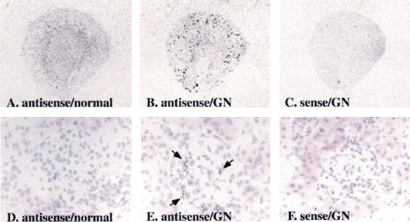
J Am Soc Nephrol 14: 1154–1163, 2003 Cyr61 Expression in Thy-1GN Podocytes 1155
Cyr61 stimulates migration and proliferation of vascular body against rat Thy-1 (CD90) antigen (CL005A; Cedarlane, Ontario,
endothelial cells and fibroblasts in culture and induces neovas- Canada) (17) was washed and concentrated in phosphate-buffered
cularization in rat corneas (12–14). To date, Cyr61 expression saline (PBS) using dialysis membrane (Slide-A-Lyser; Pierce, Rock-
or its role in the kidney remains unknown. ford, IL) for 16 h at 4°C. Glomerulonephritis was induced in Wistar
rats (150 to 200 g) by intravenous administration of 1.5 mg/kg
In this study, we revealed that Cyr61 is upregulated in the
anti-Thy-1 antibody diluted in 0.7 ml PBS from tail vein. The rats
glomeruli of Thy-1 GN. We investigated the sites of Cyr61 were killed after antibody administration for histologic examination of
expression in normal rat kidneys and in Thy-1 GN and its kidney tissues and for isolation of glomeruli to extract total RNA. For
regulation in cultured podocytes. Furthermore, we studied the light microscopic study, kidney tissues were fixed with Dubosq-Brazil
effect of Cyr61 on mesangial cell migration and proliferation. solution for 12 h at 4°C and embedded in paraffin.
Our data suggest that Cyr61 expressed in podocytes may be
involved in glomerular remodeling during Thy-1 GN. Library Screening for Genes Upregulated in Anti-Thy-1
Glomerulonephritis and Nucleotide Sequencing
Materials and Methods PSG were blotted identically on four different nylon membranes
Cell Culture (GeneScreen Plus) and hybridized with four different 32P-labeled
An immortalized mouse podocyte cell line, MPC5, was a kind gift cDNA pools derived from glomeruli of Thy-1 GN at days 0, 1, 3, and
from Dr. Peter Mundel, Albert Einstein College of Medicine, Bronx, 5 using PCR-Select Differential Screening Kit. In this screening,
New York (15). These cells proliferate when cultured with 10 U/ml mouse cDNAs blotted on membranes were hybridized with rat cDNA
murine interferon-␥ (INF-␥; Life Technologies, Gaithersburg, MD) at probes, because we expected that most, if not all, cDNAs have enough
33°C (permissive condition), whereas they halt growing and begin to sequence homologies for cross-hybridization. The nucleotide se-
differentiate to express podocyte-specific genes such as synaptopodin quences of cDNAs whose expression levels were increased in Thy-1
when cultured without INF-␥ at 37°C (nonpermissive condition). GN were determined by BigDye Terminator (Applied Biosystems,
Podocytes were cultured with RPMI 1640 medium (Nihonseiyaku, Foster City, CA) and ABI PRISM 310 Genetic Analyzer (Applied
Tokyo, Japan) supplemented with 10% fetal calf serum (FCS; Cansera Biosystems).
International, Ontario, Canada), 10 U/ml penicillin, and 10 g/ml
streptomycin (ICN Biomedicals, Costa Mesa, CA) on dishes coated Northern Blot Analyses
with 100 g/ml type I collagen (Cellgen IPC-03; KOKEN, Tokyo, Northern blot analysis was performed as described (18). In brief,
Japan). Before each experiment, cells were differentiated under non- total RNA (30 g in each lane) was electrophoresed on 1.0% agarose
permissive condition on type I collagen-coated dishes for 2 wk with- gels and transferred to nylon membranes (GeneScreen Plus). The
out passage and cultured with RPMI 1640 containing 0.5% bovine cDNA fragments of rat Cyr61 (see below), rat CTGF (nucleotides
serum albumin (BSA; Sigma, St. Louis, MO) for 24 h, until stimula- 1221 to 1803, GenBank accession number AF120275, generated by
tion with 5 ng/ml human transforming growth factor–1 (TGF-1) or reverse transcription [RT]-PCR) (19), rat PDGF-B (nucleotides 38 to
10 ng/ml human PDGF-BB (R&D Systems, Minneapolis, MN). Cells 666, GenBank accession number Z14117, generated by RT-PCR)
were used between passages 15 and 20 (15). (20), and human glyceraldehyde-3-phosphate dehydrogenase
Mesangial cells were established from glomeruli of 4 to 6-wk-old (GAPDH, Clontech) were used as probes. The membranes were
Sprague-Dawley rats as described previously (16). Mesangial cells hybridized with [32P]dCTP-labeled probes, and the blots were ex-
were cultured with Dulbecco Modified Eagle Medium (DMEM; In- posed to BAS-III imaging plate. The amount of RNA loaded in each
vitrogen, Carlsbad, CA) with 10% FCS and antibiotics and used lane was normalized for 28S ribosomal RNA or GAPDH.
between passages 7 and 10.
In Situ Hybridization Analyses
Generation of Podocyte-Specific cDNA Library Rat Cyr61 cDNA fragment was cloned by RT-PCR with sense
To generate a cDNA library of podocyte-specific genes (PSG), we primer 5'-tgcgcgccacaatgagctccagca-3' and antisense primer 5'-cccag-
applied the suppressive subtractive hybridization procedure between gagacctttagtccctgaa-3' (nucleotides 175 to 1337, GenBank accession
cDNA pools derived from differentiated mouse podocytes and from number AF218568) (21) using total RNA from Wistar rat kidneys.
C57BL/6 mouse whole kidney using PCR-Select cDNA Subtraction Sense and antisense [35S]CTP-labeled cRNAs were generated from
Kit (Clontech, Palo Alto, CA). Total RNA from each sample was the rat Cyr61 cDNA ligated in pGEM-T easy vector (Promega) using
extracted by TRIZOL Reagent (Invitrogen), and poly(A)⫹ mRNA T7 and SP6 RNA polymerases (Promega). In situ hybridization anal-
was isolated using PolyATtract mRNA Isolation System IV (Pro- ysis was performed as described previously (22). In brief, 10-m
mega, Madison, WI). To confirm podocyte-specific expression of cryosections of rat kidneys were mounted on poly-L-lysine-coated
each cDNA, we performed differential screening by reverse Northern slides, fixed with paraformaldehyde, acetylated, and hybridized with
blot analysis. cDNA fragments were blotted in quadruplicate on four the cRNA probes. Slides were washed, dehydrated, and apposed to
sets of nylon membranes (GeneScreen Plus; NEN Life Science Prod- Hyperfilm -max films (Amersham, Buckinghamshire, UK) for 10 d
ucts, Boston, MA) and hybridized with four different 32P-labaled or dipped into autoradiographic emulsion (NTB-2, Eastman Kodak,
cDNA pools using PCR-Select Differential Screening Kit (Clontech): Rochester, NY) and exposed for 6 wk and counterstained with hema-
podocyte (Pod) subtracted with whole kidney (WK) (Pod ⫺ WK); toxylin and eosin.
WK ⫺ Pod; Pod unsubtracted; and WK unsubtracted. The blots were
exposed to BAS-III imaging plate (Fuji, Tokyo, Japan). Immunohistochemical Analyses
Deparaffinized 3-m kidney sections were treated with microwave
Generation of Anti-Thy-1 Glomerulonephritis heating (5 min twice in 10 mM citrate buffer, pH 7.4). Endogenous
All animal experiments were conducted in accordance with our peroxidase was blocked by incubation with 3% hydrogen peroxide for
institutional guidelines for animal research. Mouse monoclonal anti- 15 min at room temperature. Goat anti-human Cyr61 antibody (sc-1156 Journal of the American Society of Nephrology J Am Soc Nephrol 14: 1154–1163, 2003
8561; Santa Cruz Biotechnology, Santa Cruz, CA) was diluted 1:300 with 0.5% Coomassie Brilliant Blue R 250 (Nacalai Tesque) in 50%
in PBS containing 1% BSA (1% BSA/PBS) and was incubated for 1 h methanol, 40% water, and 10% acetic acid. For each well, 5 high-
at room temperature. After washes with 1% BSA/PBS, the sections power field photographs (magnification, ⫻400) were taken to calcu-
were incubated with biotinylated secondary antibody (sc-2347, biotin- late the mean number of cells migrated per high-power field.
conjugated bovine anti-goat Ig; Santa Cruz Biotechnology) diluted
1:100 in 1% BSA/PBS for 30 min at room temperature. The sections
were further processed with avidin-biotin-peroxidase complex kit Cell Proliferation Assay
(Vector, Burlingame, CA) and 3,3'-diaminobenzidine tetrahydrochlo- Mesangial cells were plated on 24-well plates at 2 ⫻ 104 cells/well,
ride (Kanto Chemical, Tokyo, Japan), and counterstained with hema- grown for 24 h, and incubated with DMEM containing 0.2% FCS for
toxylin and coverslipped. Nonimmune goat serum was used as neg- the following 24 h. Cells were then treated with media containing
ative control. To further confirm the specificity of the signals, anti- supernatant of Cyr61-transfected or mock-transfected COS-7 cells
Cyr61 antibody was preincubated overnight at 4°C with blocking diluted 1:1 by fresh DMEM containing 10% FCS, with or without 10
peptide (sc-8561 P, Santa Cruz Biotechnology) at the five-times ng/ml PDGF-BB. [3H]-thymidine (3 Ci/ml, Amersham) was added
higher concentration (weight/volume) than the antibody concentration simultaneously with the above-described media. After 48 h of incu-
before incubation with the sections. bation, cells were washed with PBS and fixed with 10% TCA (Nacalai
Tesque). DNA was dissolved in 0.25 N NaOH, and incorporated
thymidine was counted in liquid scintillation counter (Aloka, Tokyo,
Overexpression of Cyr61 and Western Blot Analyses Japan) as described previously (25).
COS-7 cells were cultured with DMEM supplemented with 10%
FCS and antibiotics. Full-length mouse Cyr61 cDNA was generated
by RT-PCR using total RNA from C57BL/6 mouse kidneys and the Statistical Analyses
following primers: sense 5'-tgcgcgccacaatgagctccagca-3' and anti- Results are given as means ⫾ SEM. Mann-Whitney U test was
sense 5'-ttagtccctgaacttgtggatgtc-3' (nucleotides 179 to 1329, Gen- used to compare unpaired two-group means. The differences were
Bank accession number M32490) (23). The Cyr61 cDNA was first evaluated with a Stat View software package (Abacus Concepts Inc.,
TA-cloned into pGEM-T easy vector and transferred into EcoRI Berkeley, CA), and those with P ⬍ 0.05 were considered statistically
restriction site of expression vector pCXN2, a derivative of pCAGGS significant.
(24). COS-7 cells were transfected with Cyr61-pCXN2 or mock-
pCXN2 cDNA using Lipofectamine Plus Reagent (Invitrogen). Media
were changed 3 h after transfection, harvested 69 h later, and kept Results
⫺20°C until they were used for functional assay (see below). At 72 h Construction of Podocyte-Specific cDNA Library
after transfection, cells were lysed on ice in solution that contained 20 To construct a cDNA library of PSG, subtraction of cultured
mM Tris-HCl (pH 7.5), 12 mM-glycerophosphate, 0.1 M ethylene-
mouse Pod cDNA pool with mouse WK cDNA pool was
glycol-bis(-aminoethyl ether)-N,N'-tetraacetic acid, 1 mM pyrophos-
phate, 5 mM NaF, 5 mg/ml aprotinin, 2 mM dithiothreitol, 1 mM
performed (Pod ⫺ WK), and each of 1500 subtracted cDNA
phenylmethylsulfonyl fluoride, 1% Triton X-100 (Nacalai Tesque, clones was placed in grids to make four identical sets of PSG
Kyoto, Japan), and 1 mM sodium orthovanadate (Sigma) (25). The array. To confirm specific expression of PSG, we performed
lysate was centrifuged at 15,000 rpm for 20 min at 4°C, and the reverse Northern blot analyses using 32P-labeled Pod ⫺ WK,
supernatant was mixed with Laemmli sample buffer. Samples were WK ⫺ Pod, Pod, and WK cDNA probes, respectively. Repre-
separated by 12.5% SDS-PAGE in reducing conditions and electro- sentative results showed that thrombospondin-1 and cerulo-
phoretically transferred onto Immobilon polyvinylidine difluoride fil- plasmin are expressed intensely and specifically in cultured
ters (Millipore, Bedford, MA). The filters were incubated with anti- podocytes, whereas fibronectin is expressed abundantly in
Cyr61 antibody diltuted 1:1000 in Block Ace (Snow Brand Milk whole kidney (Figure 1). By this screening, 150 clones of
Products, Sapporo, Japan) for 2 h at room temperature and were highly podocyte-specific genes were isolated.
developed with horseradish peroxidase (HRP)– conjugated donkey
anti-goat IgG (sc-2020, Santa Cruz Biotechnology) and chemilumi-
nescence kit (ECL, Amersham). Histologic Examination of Anti-Thy-1
Glomerulonephritis
Cell Migration Assay We next examined the time course of histologic alteration
Migration of mesangial cells was analyzed by modified Boyden during Thy-1 GN with light microscopy (data not shown). In
chamber method using 96-well chemotaxis chambers (AB96, Neuro the present study, we used anti-Thy-1 antibody at the concen-
Probe, Gaithersburg, MD) (26). Polycarbonate filters (8-m pore size, tration sufficient to cause almost complete elimination of mes-
PFD8, Neuro Probe) coated with 20 ng/ml poly-L-lysine (Sigma) for angial cells at day 1. At day 3 of Thy-1 GN, most of mesangial
24 h at 25°C were placed in the middle of the chambers, and the cells were located at vascular poles, suggesting that mesangial
number of cells that moved from the upper chambers to the lower cells were migrating into glomerular tufts (2). They reached the
chambers was counted. Mesangial cells suspended in supernatant of
periphery of most glomerular tufts at day 7. Glomerular mi-
mock-transfected COS-7 cells (1 ⫻ 105 cells/100 l per well) were
placed in the top chambers. In the lower chambers, 30-l supernatant
croaneurysms appeared at day 3, progressing to ballooning
of Cyr61-transfected or mock-transfected COS-7 cells supplemented lesion at day 5, which indicated that severe destruction of the
with or without PDGF-BB was placed. The chambers were incubated capillary tufts occurred. At day 10, microaneurysms were
in humidified air with 5% CO2 for 3 h at 37°C. The cells remaining observed in only few glomeruli. At day 21, mesangial cell and
on the upper surfaces of the filters were scraped off, and the cells that matrix accumulation were still seen, but they subsided at day
had migrated to the lower surfaces were fixed in methanol and stained 28.J Am Soc Nephrol 14: 1154–1163, 2003 Cyr61 Expression in Thy-1GN Podocytes 1157 Figure 1. Screening for genes specifically expressed in cultured podocytes (Pod). Pod-derived cDNAs subtracted with whole kidney (WK)– derived cDNAs (Pod ⫺ WK) were blotted in quadruplicate and hybridized with four kinds of cDNAs obtained from Pod ⫺ WK, WK ⫺ Pod, Pod, and WK, respectively. Clones 1 and 2 were expressed more intensely in Pod than in WK, while expression of clone 3 was more abundant in WK. Identification of Cyr61 as PSG Upregulated in the Glomeruli of Anti-Thy-1 Glomerulonephritis To isolate PSG possibly involved in glomerular reconstruc- tion, we examined which PSG were upregulated in the glomer- uli of Thy-1 GN. We carried out second set of reverse Northern blot analyses by radiolabeling glomerular cDNA of Thy-1 GN at days 0, 1, 3, and 5. Of 150 PSG clones, we identified 40 to be upregulated, and we determined their nucleotide sequences. One of them was a cDNA fragment of Cyr61 (nucleotides 1290 to 1568, GenBank accession number M32490) (23), which is known as a soluble angiogenic protein associated with extra- cellular matrix (9,10). Cyr61 Expression in Normal Kidney and in Anti-Thy-1 Glomerulonephritis In Thy-1 GN, glomerular gene expression of Cyr61 was low at day 0, increased significantly from day 1 through day 7, reaching the peak at day 5 by 4.5-fold, and gradually decreased toward day 10 by Northern blot analysis (Figure 2). The gene expression of CTGF, another member of the CCN family, was similar to Cyr61, peaking at day 3. We also examined the gene expression of growth factors that have been implicated in Thy-1 GN (5) and found that gene expression of PDGF-B and TGF-1 in glomeruli was also induced maximally around days 3 to 5. Figure 2. Gene expression of Cyr61, connective tissue growth factor To examine the location of Cyr61 gene expression in normal (CTGF), platelet-derived growth factor-B (PDGF-B), and transform- kidney and in Thy-1 GN, in situ hybridization was performed ing growth factor–1 (TGF-1) in glomeruli of anti-Thy-1 glomeru- with 35S-labeled cRNA probe (Figure 3). At autoradiograph, lonephritis. Cyr61, CTGF, PDGF-B and TGF-1 expressions were all moderate hybridizing signals were observed in outer stripe of induced maximally around days 3 to 5 by Northern blot analysis. The outer medulla in normal kidney. At day 5 of Thy-1 GN, strong graphs on the bottom show relative gene expression level of each gene signals were seen in dot pattern in cortex. At photomicrograph, normalized for ethidium bromide staining intensity of 28S ribosomal these signals in Thy-1 GN were observed intensely in glomer- RNA. The level at day 0 was arbitrarily defined as 1. * P ⬍ 0.05 as uli, consistently with the findings by Northern blot analysis compared with day 0; n ⫽ 3. (Figure 2). Precise sites of Cyr61 expression were further studied by of outer medulla, which corresponded to brush border mem- immunohistochemistry with antibody against Cyr61 (Figure 4). brane-positive proximal straight tubules (S3 segment). At high- In normal kidney, Cyr61 protein was expressed in outer stripe power field, intense expression of Cyr61 protein was also
1158 Journal of the American Society of Nephrology J Am Soc Nephrol 14: 1154–1163, 2003
Figure 3. Localization of Cyr61 gene expression in normal kidney and in anti-Thy-1 glomerulonephritis by in situ hybridization. Antisense (A,
B, D, and E) or sense (C and F) Cyr61 cRNA probe was hybridized with sections from normal kidney (A and D) and Thy-1 GN (B, C, E, and
F), and autoradiograph (A through C, ⫻4) and photomicrograph with counterstaining by hematoxylin and eosin (D through F, ⫻400) were
carried out. In normal kidney, Cyr61 gene was expressed predominantly in outer stripe of outer medulla (A); in Thy-1 GN at day 5, it was
induced markedly in glomeruli (B and E, arrow). No specific signals were seen in sections hybridized with the sense probe (C and F).
observed in some of afferent and efferent arterioles in normal Inhibition of PDGF-Induced Mesangial Cell Migration
kidney, but the protein was not expressed in larger vessels. At by Supernatant of Cyr61-Overexpressing Cells
day 5 of Thy-1 GN, Cyr61 expression was also detected in To examine the functional role of Cyr61 upregulation in
glomeruli. The signals were observed on outer surfaces of podocytes during Thy-1GN, migration of mesangial cells in
glomerular basement membrane (GBM), which appeared to be supernatant of Cyr61-transfected or mock-transfected COS-7
podocytes. Anti-Cyr61 antibody specifically recognized Cyr61 cells was studied using modified Boyden chambers (Figure 7).
protein (40 kD) expressed in COS-7 cells by Western blot In the absence of PDGF-BB, supernatant of Cyr61-transfected
analysis. Specificity of the antibody binding in immunohisto- COS-7 cells had no significant effect on mesangial cell migra-
chemistry was further confirmed by disappearance of the sig- tion compared with that of mock-transfected COS-7 cells. With
nals when the antibody was preabsorbed with blocking peptide addition of 10 ng/ml PDGF-BB, cell migration in mock-trans-
(data not shown) or when nonimmune serum was used as fected supernatant was enhanced by 2.4-fold. On the other
primary antibody. The expression of Cyr61 mRNA and protein hand, conditioned media from Cyr61-overexpressing cells sig-
in glomeruli and podocytes increased in Thy-1 GN, whereas nificantly suppressed PDGF-induced mesangial cell migration
expression in tubules and arterioles did not seem to change in by 24%.
Thy-1 GN.
No Effects of Supernatant of Cyr61-Overexpressing
Cells on Mesangial Cell Proliferation
Cyr61 Induction by PDGF-BB and TGF-1 in We further examined whether culture media from Cyr61-
Cultured Podocytes transfected cells affect proliferation of mesangial cells (Figure
To explore the regulatory mechanism of Cyr61 expression in 8). PDGF-BB significantly increased mesangial cell numbers
podocytes, cultured podocytes were treated with PDGF-BB or by twofold after 48 h, but addition of supernatant of Cyr61-
TGF-1, because we found that PDGF-B, TGF-1, and Cyr61 transfceted cells had no significant effects on cell numbers both
genes were coordinately upregulated in Thy-1 GN. Treatment in the absence and presence of PDGF and on PDGF-induced
of podocytes with 10 ng/ml PDGF-BB upregulated Cyr61 gene DNA synthesis.
expression significantly at 10 min by 1.2-fold and at 24 h by
1.5-fold (Figure 5). Addition of 5 ng/ml TGF-1 to podocytes Discussion
induced Cyr61 expression significantly at 1 h by 2.2-fold and In the present study, we generated a podocyte-specific
24 h by 2.1-fold (Figure 6). We also observed that PDGF-BB cDNA library and investigated the expression levels of the
and TGF-1 caused upregulation of CTGF gene expression in cDNAs during Thy-1 GN, a reversible model of glomerular
podocytes significantly at 10 min to 24 h and at 1 to 24 h, disease, and identified that gene expression of soluble angio-
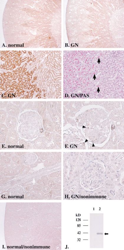
respectively. genic factor Cyr61 was highly induced in the glomeruli ofJ Am Soc Nephrol 14: 1154–1163, 2003 Cyr61 Expression in Thy-1GN Podocytes 1159 Figure 4. Immunohistochemical analyses of Cyr61 protein expression in normal kidney and in anti-Thy-1 glomerulonephritis. In normal kidney (A) and in Thy-1 GN at day 5 (B), Cyr61 protein was expressed in outer stripe of outer medulla. The analysis of high-power field at the border between outer and inner stripes (C and D) revealed that the signals were confined to proximal straight tubules (C) with brush border membranes, which were visualized as pink by periodic acid-Schiff (PAS) staining in the adjacent section (D, black arrow). Cyr61 protein was also expressed intensely in some, but not all, afferent and efferent arterioles in normal kidney (E). The protein was not expressed in larger vessels such as interlobular arteries (G, white arrow). In Thy-1 GN at day 5, Cyr61 expression was also seen in podocytes (F, arrowhead), which were present outside of glomerular basement membrane. Incubation of sections with nonimmune serum as primary antibody gave no signals in proximal straight tubules (I), arterioles or podocytes in Thy-1 GN (H). By Western blot analysis (J), anti-Cyr61 antibody specifically recognized a 40-kD protein in lysate of Cyr61-transfected COS-7 cells (lane 2, arrow), but not in that of mock-transfected cells (lane 1). Magnifications: ⫻40 in A, B, and I; ⫻200 in C, D, and G; ⫻400 in E, F, and H.
1160 Journal of the American Society of Nephrology J Am Soc Nephrol 14: 1154–1163, 2003
Figure 5. PDGF-BB-induced Cyr61 and CTGF gene expression in
cultured podocytes. PDGF-BB was added to podocytes and the time
course of Cyr61 and CTGF gene expression was examined by North- Figure 7. Effects of culture media of Cyr61-overexpressing cells on
ern blot analyses. The graphs on the bottom show relative gene PDGF-BB-induced mesangial cell migration. Mesangial cells plated
expression level of each gene normalized for GAPDH expression. The in modified Boyden chambers were incubated for 3 h with supernatant
level in untreated podocytes (control) was arbitrarily defined as 1. * P of Cyr61-transfected or mock-transfected COS-7 cells, which was
⬍ 0.05 as compared with control; n ⫽ 4. supplemented with or without PDGF-BB. In the absence of PDGF-
BB, supernatant of Cyr61-transfected COS-7 cells had no effects on
cell migration compared with that of mock-transfected COS-7 cells
(A). In culture media from mock-transfected cells, addition of 10
ng/ml PDGF-BB significantly increased mesangial cell migration (B
and D). Supernatant of Cyr61-overexpressing cells significantly sup-
pressed the chemotaxis in the presence of PDGF-BB (C and D). * P
⬍ 0.05; ns, not statistically significant; HPF, high-power field; n ⫽ 4
to 8. Magnification, ⫻400 in B through D.
gesting that Cyr61 may be involved in glomerular remodeling
in Thy-1 GN.
In glomeruli of Thy-1 GN, Cyr61 gene expression was
markedly upregulated from day 3 through day 7, when mes-
angial cell migration was most active, as shown by our histo-
logic analysis, which was consistent with previous reports
(2–5). Furthermore, neovascularization, characterized with im-
mature endothelial cell proliferation, is also reported to take
place in a period involving days 3 to 7 (2,17). Cyr61 exerts
various actions through interaction with cell surface integrin
Figure 6. TGF-1–induced Cyr61 and CTGF gene expression in
cultured podocytes. * P ⬍ 0.05 as compared with control; n ⫽ 3 to 5.
heterodimer complexes (13,27–29). Cyr61 induces endothelial
cell migration through integrin ␣ v3 and stimulates neovascu-
larization in rat cornea (12). In fibroblasts, Cyr61 induces cell
migration through ␣ v5 and proliferation through ␣ v3 (13).
Mesangial cells also express ␣ v 3 and ␣ v5 integrins (30);
Thy-1 GN. We revealed for the first time, at least to our therefore, an angiogenic factor Cyr61, which has high affinity
knowledge, that Cyr61 gene and protein expression was spe- to extracellular matrix (31), may be secreted by podocytes,
cifically induced in podocytes in Thy-1 GN and that physio- bound to GBM, and act upon endothelial and mesangial cells to
logic sites of Cyr61 expression were proximal straight tubules modulate migration and proliferation during remodeling of
and afferent and efferent arterioles. PDGF-B and TGF-1 were glomerular structure in Thy-1 GN.
upregulated in similar time course as Cyr61 in Thy-1 GN, and A regulatory role of podocytes on mesangial cells has been
they induced Cyr61 gene expression in cultured podocytes, implied by recent studies. One study showed that minor podo-
suggesting that they are inducers of podocyte Cyr61 expression cyte injury with puromycin preceding Thy-1 GN induction
in vivo. Furthermore, culture media of Cyr61-transfected cells results in irreversible mesangial lesions (8). Another report
inhibited mesangial cell migration induced by PDGF-BB, sug- demonstrated that mice deficient of podocyte-specific mole-J Am Soc Nephrol 14: 1154–1163, 2003 Cyr61 Expression in Thy-1GN Podocytes 1161
Previous reports showed that Cyr61 is expressed in endo-
thelial and smooth muscle cells in the developing mouse blood
vessels (34,35) but only weakly in vessels of normal adult mice
and humans (36). In the present study, Cyr61 was expressed in
normal afferent and efferent arterioles, in which Cyr61 expres-
sion might be upregulated by strong mechanical stretch due to
shear stress (37).
Cyr61 and CTGF share structural and functional similarity
and belong to the CCN family (9,10), but their tissue distribu-
tion (31) and intrarenal localization is clearly different. In
normal kidney, the main site of Cyr61 expression was proximal
straight tubules. On the contrary, we and others have shown
that neither CTGF gene (19,38) nor protein (31,39,40) are
Figure 8. Effects of culture media of Cyr61-overexpressing cells on
expressed in normal proximal tubules. In glomeruli of Thy-1
mesangial cell proliferation. Mesangial cells were incubated with
GN, Cyr61 protein expression was predominantly induced in
media containing supernatant of Cyr61-transfected or mock-trans-
fected COS-7 cells, together with or without 10 ng/ml PDGF-BB. The podocytes, whereas CTGF gene expression is broadly upregu-
cell numbers after 24 h and 48 h were not significantly different lated in podocytes, glomerular parietal epithelial cells, mesan-
between the treatments with Cyr61-conditioned and control media, gial cells, and periglomerular myofibroblasts (39). The spatial
both in the absence and presence of PDGF-BB (A). Supernatant of differences of Cyr61 and CTGF transcriptional regulation in
Cyr61-overexpressing cells also did not significantly affect PDGF- physiologic and pathophysiologic conditions suggest that
BB-induced mitogenesis as judged by [3H]-thymidine incorporation Cyr61 and CTGF play distinct roles in vivo. Furthermore,
after 48 h (B). ns, not statistically significant; n ⫽ 6 to 12. recent studies revealed functional difference between Cyr61
and CTGF; in fibroblasts CTGF upregulates collagen I and
cule CD2-associated protein exhibit mesangial cell prolifera- fibronectin expression (19,41), whereas Cyr61 does not up-
tion and mesangial matrix expansion (32). These findings seem regulate fibronectin expression but rather downregulates col-
to raise a possibility that factors secreted from podocytes may lagen I expression (42).
regulate mesangial cell activity. In the present study, we found In conclusion, we show that Cyr61, which exerts suppressive
that supernatant of cells transfected with Cyr61, a candidate for effects on mesangial cell migration, is strongly upregulated at
such podocyte-derived factors, inhibited PDGF-BB-induced podocytes in Thy-1 GN during a phase when mesangial cell
mesangial cell migration, implying that podocytes may sup- migration is active. These findings raise a possibility that
press migratory activity of mesangial cells when mesangial podocytes may participate in the reconstruction of glomeruli by
cells reach the periphery of glomerular tufts, that is GBM, and secreting factors that affect mesangial cell activity. Possible
may help the cessation of glomerulonephritis. Cyr61 enhances autocrine and paracrine role of Cyr61 not only in podocytes but
both migration and growth factor-induced proliferation in vas- also in arterioles and tubular cells also must be investigated in
cular endothelial cells and fibroblasts (12–14). However, here future studies.
we show that, in mesangial cells, Cyr61-conditioned media
inhibited PDGF-BB-induced migration but did not affect
PDGF-BB-induced proliferation. The discrepancy may be due Acknowledgments
to distinct responses in different cells or due to difference in The authors gratefully acknowledge Dr. Peter Mundel (Albert
Einstein College of Medicine) for providing mouse podocyte cell line
experimental settings: use of culture media of transfected cells
MPC5, Dr. Jun-ichi Miyazaki (Osaka University, Japan) for expres-
versus recombinant protein.
sion vector pCXN2, Dr. Akira Shimizu (Nippon Medical School,
Cyr61 protein has been reported to be synthesized by serum- Japan) for technical suggestion to generate Thy-1 GN, and Dr.
stimulated NIH 3T3 fibroblasts, but it is associated with ex- Takashi Kuwahara (Saiseikai Nakatsu Hospital, Japan) for technical
tracellular matrix and cannot be detected in the conditioned advice for histologic examination. We are also grateful to J. Naka-
media (31). When we overexpressed Cyr61 in COS-7 cells, mura and A. Wada for technical assistance, and A. Sonoda and S. Doi
Cyr61 protein was detected in the supernatant by Western blot for secretarial assistance. This work was supported in part by research
analysis (data not shown), but several possibilities can still be grants from the Japanese Ministry of Education, Science, Sports and
considered concerning the mechanism for migration inhibitory Culture, the Japanese Ministry of Health and Welfare, Smoking
effects of Cyr61. First, Cyr61 may bind directly to mesangial Research Foundation, and “Research for the Future” (RFTF) of Japan
cells and exert its action, presumably through cell surface Society for the promotion of Science.
integrin complexes (see above). Second, Cyr61 might interact
with PDGF in the media to interfere with its action, as is the References
case with CTGF and bone morphogenic protein 4 reported in 1. Fioretto P, Steffes MW, Sutherland DE, Goetz FC, Mauer M:
Xenopus (33). Third, Cyr61 may alter secretion of other soluble Reversal of lesions of diabetic nephropathy after pancreas trans-
factors such as extracellular matrix proteins (see below) from plantation. N Engl J Med 339: 69 –75, 1998
COS-7 cells, which have activity to modify mesangial cell 2. Hugo C, Shankland SJ, Bowen-Pope DF, Couser WG, Johnson
migration. RJ: Extraglomerular origin of the mesangial cell after injury. A1162 Journal of the American Society of Nephrology J Am Soc Nephrol 14: 1154–1163, 2003
new role of the juxtaglomerular apparatus. J Clin Invest 100: tubulointerstitial fibrosis. Am J Physiol Renal Physiol 282:
786 –794, 1997 F933–F942, 2002
3. Iruela-Arispe L, Gordon K, Hugo C, Duijvestijn AM, Claffey 20. Herren B, Weyer KA, Rouge M, Lotscher P, Pech M: Conser-
KP, Reilly M, Couser WG, Alpers CE, Johnson RJ: Participation vation in sequence and affinity of human and rodent PDGF
of glomerular endothelial cells in the capillary repair of glomer- ligands and receptors. Biochim Biophys Acta 1173: 294 –302,
ulonephritis. Am J Pathol 147: 1715–1727, 1995 1993
4. Morita T, Churg J: Mesangiolysis. Kidney Int 24:1–9, 1983 21. Albrecht C, von Der Kammer H, Mayhaus M, Klaudiny J,
5. Jefferson JA, Johnson RJ: Experimental mesangial proliferative Schweizer M, Nitsch RM: Muscarinic acetylcholine receptors
glomerulonephritis (the anti-Thy-1.1 model). J Nephrol 12: 297– induce the expression of the immediate early growth regulatory
307, 1999 gene CYR61. J Biol Chem 275: 28929 –28936, 2000
6. Ostendorf T, Kunter U, Eitner F, Loos A, Regele H, Kerjaschki 22. Mori K, Ogawa Y, Ebihara K, Tamura N, Tashiro K, Kuwahara
D, Henninger DD, Janjic N, Floege J: VEGF165 mediates glo- T, Mukoyama M, Sugawara A, Ozaki S, Tanaka I, Nakao K:
merular endothelial repair. J Clin Invest 104: 913–923, 1999 Isolation and characterization of CA XIV, a novel membrane-
7. Masuda Y, Shimizu A, Mori T, Ishiwata T, Kitamura H, Ohashi bound carbonic anhydrase from mouse kidney. J Biol Chem 274:
R, Ishizaki M, Asano G, Sugisaki Y, Yamanaka N: Vascular 15701–15705, 1999
endothelial growth factor enhances glomerular capillary repair 23. Latinkic BV, O’Brien TP, Lau LF: Promoter function and struc-
and accelerates resolution of experimentally induced glomerulo- ture of the growth factor-inducible immediate early gene cyr61.
nephritis. Am J Pathol 159: 599 – 608, 2001 Nucleic Acids Res 19: 3261–3267, 1991
8. Morioka Y, Koike H, Ikezumi Y, Ito Y, Oyanagi A, Gejyo F, 24. Niwa H, Yamamura K, Miyazaki J: Efficient selection for high-
Shimizu F, Kawachi H: Podocyte injuries exacerbate mesangial expression transfectants with a novel eukaryotic vector. Gene
proliferative glomerulonephritis. Kidney Int 60: 2192–2204, 2001 108: 193–199, 1991
9. Brigstock DR: The connective tissue growth factor/cysteine-rich 25. Suganami T, Mukoyama M, Sugawara A, Mori K, Nagae T,
61/nephroblastoma overexpressed (CCN) family. Endocr Rev 20: Kasahara M, Yahata K, Makino H, Fujinaga Y, Ogawa Y,
189 –206, 1999 Tanaka I, Nakao K: Overexpression of brain natriuretic peptide
10. Lau LF, Lam SC: The CCN family of angiogenic regulators: The in mice ameliorates immune-mediated renal injury. J Am Soc
integrin connection. Exp Cell Res 248:44 –57, 1999 Nephrol 12: 2652–2663, 2001
11. Proposal for a unified CCN nomenclature. Mol Pathol 54: 108, 26. Kohno M, Yasunari K, Minami M, Kano H, Maeda K, Mandal
2001 AK, Inoki K, Haneda M, Yoshikawa J: Regulation of rat mes-
12. Babic AM, Kireeva ML, Kolesnikova TV, Lau LF: CYR61, a angial cell migration by platelet-derived growth factor, angioten-
product of a growth factor-inducible immediate early gene, pro- sin II, and adrenomedullin. J Am Soc Nephrol 10: 2495–2502,
motes angiogenesis and tumor growth: Proc Natl Acad Sci USA 1999
95: 6355– 6360, 1998 27. Kireeva ML, Lam SC, Lau LF: Adhesion of human umbilical
13. Grzeszkiewicz TM, Kirschling DJ, Chen N, Lau LF: CYR61 vein endothelial cells to the immediate-early gene product Cyr61
stimulates human skin fibroblast migration through integrin is mediated through integrin ␣ v3. J Biol Chem 273: 3090 –3096,
␣ v5 and enhances mitogenesis through integrin ␣v3 indepen- 1998
dent of its carboxyl-terminal domain. J Biol Chem 276: 21943– 28. Jedsadayanmata A, Chen CC, Kireeva ML, Lau LF, Lam SC:
21950, 2001 Activation-dependent adhesion of human platelets to Cyr61 and
14. Kireeva ML, Mo FE, Yang GP, Lau LF: Cyr61, a product of a Fisp12/mouse connective tissue growth factor is mediated
growth factor-inducible immediate-early gene, promotes cell through integrin ␣IIb 3. J Biol Chem 274: 24321–24327, 1999
proliferation, migration, and adhesion. Mol Cell Biol 16: 1326 – 29. Chen N, Chen CC, Lau LF: Adhesion of human skin fibroblasts
1334, 1996 to Cyr61 is mediated through integrin ␣61 and cell surface
15. Mundel P, Reiser J, Zuniga Mejia Borja A, Pavenstadt H, Da- heparan sulfate proteoglycans. J Biol Chem 275: 24953–24961,
vidson GR, Kriz W, Zeller R: Rearrangements of the cytoskel- 2000
eton and cell contacts induce process formation during differen- 30. Hafdi Z, Lesavre P, Tharaux PL, Bessou G, Baruch D, Halbw-
tiation of conditionally immortalized mouse podocyte cell lines. achs-Mecarelli L: Role of ␣v integrins in mesangial cell adhesion
Exp Cell Res 236: 248 –258, 1997 to vitronectin and von Willebrand factor. Kidney Int 51: 1900 –
16. Ishibashi R, Tanaka I, Kotani M, Muro S, Goto M, Sugawara A, 1907, 1997
Mukoyama M, Sugimoto Y, Ichikawa A, Narumiya S, Nakao K: 31. Kireeva ML, Latinkic BV, Kolesnikova TV, Chen CC, Yang GP,
Roles of prostaglandin E receptors in mesangial cells under Abler AS, Lau LF: Cyr61 and Fisp12 are both ECM-associated
high-glucose conditions. Kidney Int 56: 589 – 600, 1999 signaling molecules: Activities, metabolism, and localization
17. Shimizu A, Masuda Y, Kitamura H, Ishizaki M, Sugisaki Y, during development. Exp Cell Res 233: 63–77, 1997
Yamanaka N: Recovery of damaged glomerular capillary net- 32. Shih NY, Li J, Karpitskii V, Nguyen A, Dustin ML, Kanagawa
work with endothelial cell apoptosis in experimental proliferative O, Miner JH, Shaw AS: Congenital nephrotic syndrome in mice
glomerulonephritis. Nephron 79: 206 –214, 1998 lacking CD2-associated protein. Science 286: 312–315, 1999
18. Makino H, Tanaka I, Mukoyama M, Sugawara A, Mori K, Muro 33. Abreu JG, Ketpura NI, Reversade B, De Robertis EM: Connec-
S, Suganami T, Yahata K, Ishibashi R, Ohuchida S, Maruyama tive-tissue growth factor (CTGF) modulates cell signalling by
T, Narumiya S, Nakao K: Prevention of diabetic nephropathy in BMP and TGF-. Nat Cell Biol 4: 599 – 604, 2002
rats by prostaglandin E receptor EP1-selective antagonist. J Am 34. Latinkic BV, Mo FE, Greenspan JA, Copeland NG, Gilbert DJ,
Soc Nephrol 13: 1757–1765, 2002 Jenkins NA, Ross SR, Lau LF: Promoter function of the angio-
19. Yokoi H, Mukoyama M, Sugawara A, Mori K, Nagae T, Makino genic inducer Cyr61gene in transgenic mice: Tissue specificity,
H, Suganami T, Yahata K, Fujinaga Y, Tanaka I, Nakao K: Role inducibility during wound healing, and role of the serum re-
of connective tissue growth factor in fibronectin expression and sponse element. Endocrinology 142: 2549 –2557, 2001J Am Soc Nephrol 14: 1154–1163, 2003 Cyr61 Expression in Thy-1GN Podocytes 1163
35. O’Brien TP, Lau LF: Expression of the growth factor-inducible imme- 39. Ito Y, Goldschmeding R, Bende R, Claessen N, Chand M, Kleij
diate early gene cyr61 correlates with chondrogenesis during mouse L, Rabelink T, Weening J, Aten J: Kinetics of connective tissue
embryonic development. Cell Growth Differ 3: 645–654, 1992 growth factor expression during experimental proliferative glo-
36. Hilfiker A, Hilfiker-Kleiner D, Fuchs M, Kaminski K, Lichtenberg merulonephritis. J Am Soc Nephrol 12: 472– 484, 2001
A, Rothkotter HJ, Schieffer B, Drexler H: Expression of CYR61, an 40. Wahab NA, Brinkman H, Mason RM: Uptake and intracellular
angiogenic immediate early gene, in arteriosclerosis and its regula- transport of the connective tissue growth factor: A potential
tion by angiotensin II. Circulation 106: 254 –260, 2002 mode of action. Biochem J 359: 89 –97, 2001
37. Tamura I, Rosenbloom J, Macarak E, Chaqour B: Regulation of 41. Frazier K, Williams S, Kothapalli D, Klapper H, Grotendorst
Cyr61 gene expression by mechanical stretch through multiple GR: Stimulation of fibroblast cell growth, matrix production, and
signaling pathways. Am J Physiol Cell Physiol 281: C1524 – granulation tissue formation by connective tissue growth factor.
1532, 2001 J Invest Dermatol 107: 404 – 411, 1996
38. Ito Y, Aten J, Bende RJ, Oemar BS, Rabelink TJ, Weening JJ, 42. Chen CC, Mo FE, Lau LF: The angiogenic factor Cyr61 activates
Goldschmeding R: Expression of connective tissue growth factor a genetic program for wound healing in human skin fibroblasts.
in human renal fibrosis. Kidney Int 53: 853– 861, 1998 J Biol Chem 276: 47329 – 47337, 2001You can also read


