Crosstalk Between Liver Macrophages and Surrounding Cells in Nonalcoholic Steatohepatitis
←
→
Page content transcription
If your browser does not render page correctly, please read the page content below
MINI REVIEW
                                                                                                                                             published: 24 June 2020
                                                                                                                                     doi: 10.3389/fimmu.2020.01169

                                            Crosstalk Between Liver
                                            Macrophages and Surrounding Cells
                                            in Nonalcoholic Steatohepatitis
                                            Haiou Li 1,2† , Yunjiao Zhou 1,2† , Haizhou Wang 1,2† , Meng Zhang 1,2 , Peishan Qiu 1,2 ,
                                            Mengna Zhang 1,2 , Ruike Zhang 1,2 , Qiu Zhao 1,2* and Jing Liu 1,2*
                                            1
                                              Department of Gastroenterology, Zhongnan Hospital of Wuhan University, Wuhan, China, 2 Hubei Clinical Center, Key Lab of
                                            Intestinal and Colorectal Diseases, Wuhan, China

                                            Nonalcoholic steatohepatitis (NASH), the advanced stage of nonalcoholic fatty liver
                                            disease (NAFLD), is emerging as a leading cause of progressive liver fibrosis and
                                            end-stage liver disease. Liver macrophages, mainly composed of Kupffer cells (KCs)
                                            and monocyte-derived macrophages (MoMFs), play a vital role in NASH progression
                        Edited by:          and regression. Recent advances suggest that cell–cell communication is a fundamental
                         Junji Xing,
Houston Methodist Research Institute,
                                            feature of hepatic microenvironment. The reprogramming of cell–cell signaling between
                      United States         macrophages and surrounding cells contributes to the pathogenesis of NASH. In this
                         Reviewed by:       review, we summarize the current knowledge of NASH regarding the composition of liver
                  Paramananda Saikia,
                                            macrophages and their communication with surrounding cells, which are composed of
       Cleveland Clinic, United States
                               Payel Sil,   hepatocytes, hepatic stellate cells (HSCs), liver sinusoidal endothelial cells (LSECs) and
    National Institute of Environmental     other immune cells. We also discuss the potential therapeutic strategies based on the
             Health Sciences (NIEHS),
                          United States
                                            level of macrophages.
                     *Correspondence:       Keywords: nonalcoholic steatohepatitis, cellular crosstalk, liver macrophages, liver cells, therapeutic strategies
                              Qiu Zhao
                   qiuzhao@whu.edu.cn
                               Jing Liu     INTRODUCTION
                 liujing_GI@whu.edu.cn

     † These   authors have contributed
                                            Nonalcoholic fatty liver disease (NAFLD), an increasingly common liver disease worldwide, ranges
                    equally to this work    from relatively benign NAFL to nonalcoholic steatohepatitis (NASH) (1, 2). NASH is strongly
                                            associated with progressive liver fibrosis and has further become a major cause of cirrhosis and liver
                   Specialty section:       cancer (3). Unlike isolated hepatic steatosis, NASH is characterized as the presence of inflammation,
         This article was submitted to      hepatocellular injury, and varying degrees of fibrosis (4). However, the underlying mechanisms
           Molecular Innate Immunity,       involved in pathogenesis of NASH are not fully understood. It was demonstrated that liver
               a section of the journal     macrophages orchestrate both the progression and restoration of NASH (5). Traditionally, liver
              Frontiers in Immunology
                                            macrophages mainly comprise liver-resident Kupffer cells (KCs) and circulating monocyte-derived
         Received: 24 February 2020         macrophages (MoMFs) (6). The activation of liver macrophages during NASH progression is a
            Accepted: 12 May 2020           dynamic procedure dependent on various stimuli such as cytokines, lipid metabolites, and other
           Published: 24 June 2020
                                            signal molecules (7, 8).
                             Citation:         Emerging evidence suggests that cellular networks rather than a single cell type modulate
      Li H, Zhou Y, Wang H, Zhang M,        NASH progression (9). In conjunction with surrounding cells, liver macrophages can trigger
Qiu P, Zhang M, Zhang R, Zhao Q and
                                            inflammation response, fibrogenesis, vascular remodeling, and so forth. In the development
 Liu J (2020) Crosstalk Between Liver
 Macrophages and Surrounding Cells
                                            of NASH, hepatocytes contribute to KC activation and MoMF recruitment via multiple signal
       in Nonalcoholic Steatohepatitis.     molecules such as damage-associated molecular patterns (DAMPs), extracellular vesicles (EVs),
             Front. Immunol. 11:1169.       and harmful lipids (5). In response to those signals, activated macrophages also signal back to
      doi: 10.3389/fimmu.2020.01169         modulate hepatocyte fate. Besides, those activated macrophages further mediate the activation

Frontiers in Immunology | www.frontiersin.org                                     1                                              June 2020 | Volume 11 | Article 1169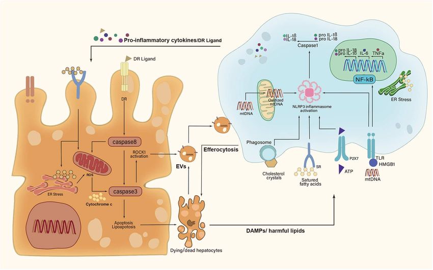
Li et al. Macrophage-Related Crosstalk in NASH
of hepatic stellate cells (HSCs) via producing cytokines and correlated with the degree of NASH-induced liver fibrosis (22).
chemokines, including transforming growth factor-β (TGFβ), More studies are needed to understand the ontology of hepatic
interleukin-1β (IL-1β), platelet—derived growth factor (PDGF) macrophage subpopulations in NASH.
receptor, and CC-chemokine ligand 2 (CCL2) (10). Moreover,
liver macrophages influence the biological functions of liver
sinusoidal endothelial cells (LSECs) and other immune cells INTERCELLULAR CROSSTALK OF LIVER
(11, 12). In turn, those surrounding cells can stimulate liver MACROPHAGES IN NONALCOHOLIC
macrophages during NASH progression (13, 14). Understanding STEATOHEPATITIS
the intercellular crosstalk between liver macrophages and their
surrounding cells is critical for developing novel therapeutic The growing consensus is that cell–cell communication within
interventions based on the level of macrophages. liver represents a key aspect that leads to the progression
In this review, we summarize the intercellular signaling toward NASH (9). The anatomical location of liver macrophages
between liver macrophages and surrounding cells involved allows them to interact with several liver resident cells and
in NASH development. The potential macrophage-targeted circulating immune cells (23). Histologically, the clusters of
therapeutic strategies for NASH are also discussed. KCs were characterized as microgranulomas, and those with
lipid droplets were characterized as lipogranulomas in human
NAFLD/NASH (24–26). A unique histological structure, where
THE COMPOSITION OF LIVER activated macrophages aggregated around hepatocytes with large
MACROPHAGES IN NONALCOHOLIC lipid droplets, was detected in the murine NASH models
STEATOHEPATITIS and patients with NASH, termed hepatic crown-like structures
(hCLS) (27). Conversely, activated KCs were not shown to
Liver macrophage populations comprise different subsets of cells. form hCLS in patients and mice with simple steatosis (28).
In particular, KCs and freshly recruited MoMFs are important This section focuses on liver macrophage-related crosstalk in
mediators of liver inflammation, fibrogenesis, and fibrinolysis NASH (Figure 1).
in the development of NASH (15, 16). In mice, circulating
monocytes were divided into two main subsets: lymphocyte
antigen 6C high (Ly-6Chi ) and Ly-6C low (Ly-6Clo ) expressing INTERACTION BETWEEN LIVER
monocytes. It was demonstrated that the hepatic infiltration MACROPHAGES AND HEPATOCYTES
of Ly-6Chi monocytes occurred early in murine NASH models
and patients with NASH (16, 17). Those monocytes gave rise Lipotoxicity is characterized as a key feature that differentiated
to phenotypically distinct populations of MoMFs upon external NASH from isolated steatosis (29, 30). Various lipotoxic
stimulus. Briefly, KCs and MoMFs could be differentiated compounds (e.g., free cholesterol, ceramides, and saturated fatty
toward either a classic proinflammatory phenotype (M1 acids) induce metabolic stress, oxidative stress, and endoplasmic
macrophages) or an alternative anti-inflammatory phenotype reticulum-related stress in hepatocytes, resulting in hepatocyte
(M2 macrophages) in vitro (18). The M1 macrophages produced injury and death (31). Hepatocyte stress and death cause the
proinflammatory cytokines such as tumor necrosis factor α release of their cellular contents into extracellular space, which
(TNFα), IL-1β, CCL2, and CCL5. In contrast, M2 macrophages contributes to macrophage activation (29).
secreted a distinct set of mediators including IL-13, IL-10, IL-4,
and TGFβ (19). It was noted that KCs and MoMFs in NASH liver Kupffer Cells-Hepatocytes
exhibited a notable shift toward a proinflammatory phenotype The DAMPs, such as cytosolic proteins, purine nucleotides,
on the basis of their gene expression signatures at the single-cell and mitochondrial compounds, primarily acted on pattern
level (20). recognition receptors (PRRs) to promote inflammatory
In a recent single-cell RNA sequencing (scRNA-seq) study, responses of KCs (32). High mobility group box-1 (HMGB1)
two distinct subpopulations of liver macrophages are exhibited was a widely studied DAMP that induced cytokine release
in western diet (WD)-induced NASH models in mice, including of macrophages (33). It bound toll-like receptor 4 (TLR4) to
MoMFs with high lysozyme 2 (Lyz2) expression and KCs induce nuclear factor (NF)-κB translocation and TNFα release
with high C-type lectin domain family 4 member F (Clec4f) in KCs (34). Besides, the mitochondrial DNA (mtDNA) released
expression (21). Besides, those MoMFs segregated into three from damaged hepatocytes activated TLR9 on KCs to promote
subtypes owing to their striking heterogeneity (21). Furthermore, inflammatory response (35). Recently, it was reported that the
a NASH-specific macrophage population, marked by high mtDNA was recognized by the stimulator of IFN genes (STING)
expression of triggering receptors expressed on myeloid cells in KCs to induce TNFα and IL-6 production under lipid
2 (Trem2), was observed in NASH livers of both mice and overload (36). Adenosine triphosphate (ATP) was also released
humans, termed NASH-associated macrophages (NAMs) (20). into extracellular space from injured hepatocytes. Being sensed
Consistently, another scRNA-seq study identified a pathogenic by P2X purinoceptor 7 (P2X7) receptor on KCs, ATP could
subpopulation of TREM2+ CD9+ macrophages in the fibrotic medicate the induction of NLR family pyrin domain-containing
niche of human liver with NASH, named scar-associated 3 (NLRP3) inflammasome and the consequent production of
macrophages (SAMacs). The expansion of SAMacs was positively proinflammatory cytokines (37, 38).
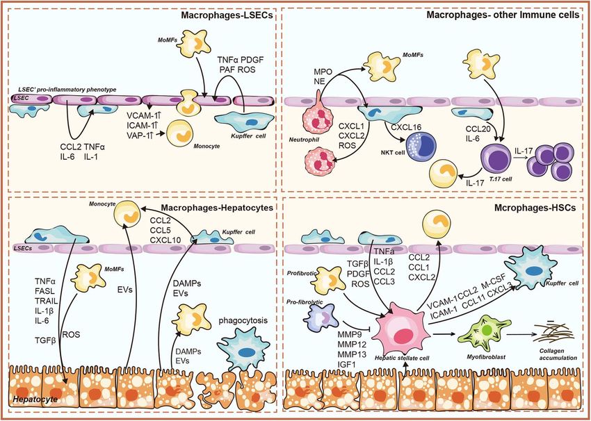
Frontiers in Immunology | www.frontiersin.org 2 June 2020 | Volume 11 | Article 1169Li et al. Macrophage-Related Crosstalk in NASH FIGURE 1 | Overview of liver macrophage-related intercellular signaling in nonalcoholic steatohepatitis (NASH). The illustration consists of four groups, as follows: liver macrophages–hepatocytes; liver macrophages–hepatic stellate cells (HSCs); liver macrophages–liver sinusoidal endothelial cells (LSECs); liver macrophages–immune cells. DAMPs, damage-associated molecular patterns; EVs, extracellular vesicles; TNFα, tumor necrosis factor α; TRAIL, TNF-related apoptosis-inducing ligand; FasL, Fas ligand; ROS, reactive oxygen species; CCL, chemokine (C-C) motif ligand; CXCL, chemokine (C-X-C motif) ligand; IL, interleukin; MMP, matrix metalloproteinase; IGF1, insulin-like growth factor 1; TGFβ, transforming growth factor-β; M-CSF, macrophage colony-stimulating factor; PDGF, platelet-derived growth factor; PAF, platelet-activating factor; ICAM-1, intercellular adhesion molecule-1; VCAM-1, vascular cell adhesion molecule-1; VAP-1, vascular adhesion protein-1; MPO, myeloperoxidase; NO, nitric oxide; IFNγ, interferon γ. Recent studies implicated lipotoxic hepatocyte-derived EVs cytokines (e.g., IL-6, TNFα, and IL-1β) (47). TNFα allowed for (LPC-EVs) in mediating cell–cell communication by transferring the activation of caspase-8 in hepatocytes by binding to TNF various cargos (39). Apoptotic bodies formed by apoptotic receptor 1 (TNFR1), which not only triggered apoptotic caspase hepatocytes fall in the category of EVs. Engulfment of apoptotic cascade directly but also induced mitochondrial dysfunction to bodies by KCs promoted the production of TNFα, TNF-related amplify the signals indirectly (8, 48). In addition, KC-derived apoptosis-inducing ligand (TRAIL), and Fas ligand (FasL) (40). IL-1β signaling was associated with de novo lipogenesis in These death receptor (DR) ligands further induced hepatocyte hepatocytes and promoted hepatic lipid deposition (49–51). IL-6 apoptosis in a feed-forward loop (Figure 2). Moreover, KCs were contributed to insulin resistance in hepatocytes by disrupting key shown to aggregate around dead hepatocytes to form hCLSs. steps in the insulin signal transduction (52). Additionally, KCs Specifically, the cholesterol crystals within remnant lipid droplets were shown to remove apoptotic hepatocytes via efferocytosis (9). of dead hepatocytes were processed by KCs, which then activated The efferocytic clearance of dead hepatocytes prevents the release the NLRP3 inflammasome in KCs, causing proinflammatory of DAMPs and subsequent DAMP-mediated inflammation. cytokines production (41). In this line, NLRP3 inflammasome Efferocytosis could be triggered by a series of “eat-me” signals blockade improved cholesterol crystal-derived inflammation and from apoptotic hepatocytes (53). The well-studied “eat-me” fibrosis in experimental NASH (42). signal was the presence of phosphatidylserine (PtdSer) on the In response to those signals sent by hepatocytes, KCs also outer leaflet of the cell membrane during apoptosis (54). KCs signaled back to the hepatocytes and regulated their fate (43–46) were thought to be the most important hepatic efferocytes (Figure 2). Firstly, activated KCs exerted actions via producing with the expression of several different PtdSer receptors, such Frontiers in Immunology | www.frontiersin.org 3 June 2020 | Volume 11 | Article 1169
Li et al.                                                                                                                   Macrophage-Related Crosstalk in NASH

  FIGURE 2 | A feed-forward regulatory loop between lipotoxic hepatocytes and Kupffer cells. Upon metabolic stress, dying and dead hepatocytes release
  damage-associated molecular patterns (DAMPs), extracellular vesicles (EVs), and harmful lipids to activate Kupffer cells (KCs). In turn, activated KCs secrete
  proinflammatory cytokines and death receptor (DR) ligands to aggravate hepatocyte damage. However, KCs can remove apoptotic hepatocytes via efferocytosis.
  IL-1β, interleukin-1β; IL-6, interleukin-6; IL-18, interleukin-18; TNFα, tumor necrosis factor α; HMGB1, high mobility group box-1; ATP, adenosine triphosphate;
  mtDNA, mitochondrial DNA; ROCK1, rho-associated, coiled-coil containing protein kinase 1; NF-κB, nuclear factor-κB; NLRP3, NLR family pyrin domain-containing 3;
  ER, endoplasmic reticulum; ROS, reactive oxygen species; P2X7, P2X purinoceptor 7; TLR, toll-like receptor; SR, scavenger receptor.

as T cell immunoglobulin, mucin domain-containing molecule                           (59, 60). Besides, integrin β (ITGβ) enriched LPC-EVs mediated
3 (Tim3), Tim4, macrophage c-mer tyrosine kinase (MerTK),                            monocyte adhesion to LSECs, an essential step for hepatic
stabilin-1, and stabilin-2 (53). Strikingly, both Tim3 and Tim4                      recruitment of MoMFs in murine NASH (61).
were overexpressed in all detected liver macrophage subsets in
methionine- and choline-deficient diet (MCD)-induced NASH
mice (55, 56). Their absence led to increased production of
reactive oxygen species (ROS), IL-1β, and IL-18 in macrophages,                      INTERACTION BETWEEN LIVER
concomitant with the aggravation of steatosis and liver fibrosis                     MACROPHAGES AND HEPATIC
(55, 56). Further studies are urgently needed to explore that how                    STELLATE CELLS
those PtdSer receptors participate in efferocytosis mechanisms of
macrophages during NASH development.                                                 Studies revealed that macrophages were key regulators in
                                                                                     the pathogenesis of NASH-driven fibrosis (9, 62). Similarly,
Monocyte-Derived                                                                     therapeutic inhibition of macrophage infiltration accelerated
Macrophages-Hepatocytes                                                              liver fibrosis regression in murine NASH (16, 63, 64). Besides,
NASH-induced hepatocyte damage recruited MoMFs indirectly                            macrophages aggregated to form hCLS where they could interact
by stimulating KCs to release proinflammatory chemokines                             with HSCs (27). The hCLS was located close to fibrogenic lesions
including CCL2, CCL5, and CXCL10 (57). Lipotoxic hepatocytes                         and the number of hCLS significantly linked to the extent of
also release EVs to induce the hepatic recruitment of MoMFs.                         liver fibrosis (27, 41). In turn, activated HSCs were shown to
TRAIL-enriched LPC-EVs induced the expression of IL-1β                               regulate macrophage accumulation and proliferation through
and IL-6 via NF-κB activation in mouse bone marrow-                                  paracrine effects (65). Moreover, a scRNA-seq analysis showed
derived macrophages (58). Ceramide and chemokine (C-X-C                              that activated HSCs were implicated in modulating the functions
motif) ligand 10 (CXCL10) within EVs contributed to MoMF                             of macrophages via a series of stellakines (e.g., CCL2, CCL11, and
recruitment to the liver via activating macrophage chemotaxis                        CXCL2) in murine NASH models (20).

Frontiers in Immunology | www.frontiersin.org                                    4                                             June 2020 | Volume 11 | Article 1169
Li et al. Macrophage-Related Crosstalk in NASH
Kupffer Cells–Hepatic Stellate Cells Kupffer Cells–Liver Sinusoidal
On the molecular level, KCs regulated HSC activation by Endothelial Cells
producing cytokines and chemokines such as TGFβ, PDGF, At the early stage of NAFLD, LSECs exhibited an anti-
TNFα, and IL-1β (10). KC-derived TGFβ promoted HSC inflammatory property by inhibiting KC activation and
differentiation into a profibrogenic phenotype, concomitant with monocyte migration (81, 82). At the stage of NASH, LSEC
increased collagen and α-smooth muscle actin expression (66). capillarization happened, and capillarized LSECs were necessary
Recently, Cai et al. proved that the MerTK signaling in KCs for activation of KCs (83). LSECs acquired a proinflammatory
promoted HSC activation and liver fibrosis in NASH mice via phenotype to produce proinflammatory mediators, leading to
TGFβ1 production (67). Moreover, TGFβ induced oxidative KC activation (14). Activated KCs were shown to be involved
DNA damage in HSCs through downregulation of cytoglobin in angiogenesis through the secretion of ROS and cytokines
(68). Besides, in murine NASH models, the enhancement of including TNFα, PDGF, and platelet-activating factor (PAF) (84).
TNFα signaling following KC activation facilitated HSC survival
via activating the NF-κB pathway in HSCs (69, 70). Activated KCs Monocyte-Derived Macrophages–Liver
caused the HSC migration and recruitment through the secretion Sinusoidal Endothelial Cells
of CCL2 and CCL5 (71, 72). On the other hand, the HSC-derived In NASH, the proinflammatory phenotype of LSECs increased
chemokines that included CCL2 and macrophage colony- proinflammatory chemokine CCL2 to facilitate hepatic
stimulating factor (M-CSF) further activated KCs, amplifying the recruitment of monocytes (14). Moreover, in mice models
inflammatory response (73). In response to lipopolysaccharide of NASH, the overexpression of adhesion molecules ICAM-1,
(LPS), HSCs secreted intercellular adhesion molecule-1 (ICAM- VCAM-1, and vascular adhesion protein-1 (VAP-1) in LSECs
1), vascular cell adhesion molecule-1 (VCAM-1), and E-selectin were critical for the adhesion and transmigration of monocytes
to induce KC migration (74). The underlying mechanisms to amplify local inflammatory response (14, 85, 86). Little is
governing this process have not been fully elucidated. known about the pathophysiological roles of MoMFs toward
LSECs in NASH.
Monocyte-Derived Macrophages–Hepatic
Stellate Cells INTERACTION BETWEEN LIVER
Infiltrating MoMFs are divided into two major subsets: Ly- MACROPHAGES AND OTHER
6Chi macrophages and Ly-6Clo macrophages. Similar to KCs, IMMUNE CELLS
proinflammatory Ly-6Chi macrophages activated HSCs by
secreting TGFβ, IL-1β, PDGF, and CCL2, enhancing the fibrotic
Kupffer Cells–Other Immune Cells
The interactions of immune cells in homeostasis and disease
response. Recently, Ramachandran P et al. demonstrated that
have been reviewed in detail elsewhere (87). Firstly, KCs
the TREM2+ CD9+ SAMacs, differentiating from circulating
contribute to the hepatic infiltration of neutrophils in NASH.
monocytes, performed a profibrogenic characteristic with
The inflammatory activation of KCs resulted in the production of
multiple profibrogenic genes expression (22). Of note, during the
chemokines (e.g., CXCL1, CXCL2, and CXCL8) and ROS, which
regression stage, the pro-restorative Ly-6Clo macrophages
stimulated neutrophil recruitment to expanded inflammation
promoted HSC apoptosis and accelerated extracellular
(44, 72). Hepatic neutrophil content and neutrophil elastase
matrix degradation by increasing the expression of matrix
(NE) activity were significantly increased in high-fat diet (HFD)-
metalloproteinase 9 (MMP9), MMP12, MMP13, and insulin-
fed mice. NE treatment caused the proinflammatory markers
like growth factor 1 (IGF1) (75, 76). This pro-restorative
of macrophages to largely increase (88). Neutrophil-derived
subpopulation also expressed chemokine (C-X3-C motif)
myeloperoxidase (MPO) was also associated with the formation
receptor 1 (CX3CR1), and its ligand CX3C ligand 1 (CX3CL1)
of hCLS in NASH (89). Besides, activated KCs promote natural
was mainly expressed by HSCs (77). The CX3CL1–CX3CR1
killer T (NKT) cell over-activation and subsequent deficiency
interaction negatively regulated the inflammatory properties in
in the pathogenesis of NAFLD (90). KC-derived IL-12 was
macrophages (78).
associated with the reduced numbers of hepatic NKT cells in
hepatosteatosis (91). Conversely, Syn et al. described that NKT
cells were associated with NASH-related fibrosis (92). CXCL16
INTERACTION BETWEEN LIVER secreted by KCs triggered the hepatic accumulation of CXCR6+
MACROPHAGES AND LIVER SINUSOIDAL NKT cells, thereby accentuating liver inflammation and fibrosis
ENDOTHELIAL CELLS in murine liver (93).
LSECs constituted a unique vascular bed with fenestrae in liver Monocyte-Derived Macrophages–Other
and interacted directly with the immune cells and antigens in Immune Cells
the blood flow (79). Monocyte’s adhesion to LSECs is a crucial A proinflammatory phenotype of macrophages showed a close
step for inflammation response in NASH, which verified the relationship with diverse T-cell subsets by secreting IL-6, TNFα,
“gatekeeper” role of LSECs in the progression from simple IL-1β, IL-12, and IL-23 in the pathogenesis of NAFLD (12).
steatosis to NASH (80). Although these cytokines are well-established drivers of T-cell
Frontiers in Immunology | www.frontiersin.org 5 June 2020 | Volume 11 | Article 1169Li et al. Macrophage-Related Crosstalk in NASH
differentiation, their roles in controlling T-cell differentiation in Notably, non-coding RNAs (ncRNAs) offered new possibilities
NASH are not fully understood (87). T helper type 17 (Th17) in developing therapeutic strategies for NASH on the basis of the
cells and their production of IL-17 facilitated the transition level of macrophages (104, 105). For instance, in murine fibrotic
from simple steatosis to steatohepatitis in NAFLD (94). They NASH models, treatment with miR-223-3p mimic ameliorated
favored the further activation of monocytes, leading to the activation of HSCs and fibrosis development through its NLRP3-
release of proinflammatory cytokines that, in turn, amplified liver targeted effect in KCs (106). In addition, miR-146b acted as
inflammation (95). a promising approach to attenuate HFD-induced NASH in
mice by directly targeting the IL-1 receptor-associated kinase
MACROPHAGE-TARGETED THERAPEUTIC 1 and TNFR-associated factor 6 in macrophages, resulting in
suppression of TNFα and IL-6 (107). A cell-specific delivery
INTERVENTIONS IN NONALCOHOLIC
system with efficiency and safety is essential for the clinical
STEATOHEPATITIS application of those miRNAs.
Currently, there are still no Food and Drug Administration
(FDA)-approved effective drugs for NASH despite its high CONCLUSION AND FUTURE
prevalence. Owing to their critical roles in NASH, liver PERSPECTIVES
macrophages are emphasized as attractive targets for NASH
treatment. Specifically, there are some options that exert potential Multiple studies have shown that liver macrophages play a central
therapeutic effects by regulating cell–cell communication role in the progression and regression of NASH. They sense
in NASH. various external signals and act as key mediators of hepatic
Because the recruited MoMFs widely interact with resident inflammation. Importantly, owing to their strategic location, liver
cells, interfering with recruiting signals would disrupt macrophages can interact with different cells such as hepatocytes,
intercellular communication at the level of macrophages. HSCs, and LSECs. However, there are several issues that need
Cenicriviroc (CVC), a dual CCR2/5 antagonist, efficiently to be addressed. Firstly, most of the observed interactive effects
reduced the hepatic recruitment of MoMFs that ameliorated are in specific cytokine-dependent manner. The core intracellular
hepatic inflammation and fibrosis in NASH mice models (64). pathways of macrophages in mediating intercellular signaling in
This drug was evaluated in a phase II clinical trial in NASH NASH are still unclear, which points out a future research goal.
patients and was found to be effective in reducing fibrosis after Secondly, owing to their striking heterogeneity, more studies are
CVC administration (96). The RNA-aptamer molecule mNOX- needed to reveal the complex cell–cell communication network
E36 also relieved steatohepatitis and accelerated regression of based on the large spectrum of macrophage phenotypes. In
liver fibrosis in experimental mouse models via antagonizing addition, most findings from murine models are insufficient to
CCL2 (63). Maraviroc, a CCL5 inhibitor, ameliorated hepatic reflect the complex cellular networks during NASH progression
steatosis in HFD-induced NAFLD in mice (97). Moreover, in humans. Further exploration of the macrophage function in
monocyte’s adhesion to LSECs is an essential step for hepatic human NASH liver is warranted.
recruitment of MoMFs. The VAP-1 inhibitor, also called amine Moreover, liver macrophages are identified as attractive
oxidase copper containing three (AOC3) inhibitor, decreased targets for NASH treatment. As described in this review, some
inflammatory cell recruitment and reduced fibrosis (85). This signaling pathways that mediated cellular crosstalk are potentially
drug was tested in a phase II trial in patients with NASH, but it druggable. Besides, the rapid advancement in nanomedicine
was discontinued owing to the risk of drug interactions in NASH allows for targeted delivery of drugs to macrophages, such
patients (98). as miRNA mimic. Taken together, deciphering macrophage
Another potential NASH treatment is to regulate KC function and their role in intercellular signaling network will
activation. Because hepatocyte-derived DAMPs trigger the sterile facilitate the design of novel targeted therapies to treat NASH.
inflammatory response of KCs by acting on PRRs, targeting
released DAMPs or PRRs can inhibit KC activation, thus AUTHOR CONTRIBUTIONS
ameliorating liver inflammation (99). HMGB1 neutralizing
antibodies and PRR antagonists (e.g., TLR2, TLR3, and TLR4 HL and YZ searched the literature and wrote the manuscript. HW
antagonists) were shown to attenuate liver inflammation prepared the figures. MengZ, PQ, MengnaZ, and RZ carefully
in murine models (100, 101). Targeting macrophage-derived checked the manuscript and helped to improve paragraphs.
profibrogenic molecules may be promising to improve NASH QZ and JL designed and revised the manuscript. All authors
fibrosis. Galectin-3 is a profibrogenic protein that is highly contributed to the article and approved the submitted version.
expressed in macrophages surrounding lipotoxic hepatocytes.
Treatment with galectin-3 inhibitor (GR-MD-02) markedly ACKNOWLEDGMENTS
improved fibrosis in a murine model of NASH (102). A phase
IIb trial showed that GR-MD-02 reduced the hepatic-portal vein This work was supported by a research grant from the National
pressure gradient in patients with NASH cirrhosis (103). Natural Science Foundation of China (JL, grant no. 81472735);
Another potential option is regulating intracellular pathways Wuhan University (JL, 2042019kf0206); and National Basic
in macrophages, which has been reviewed elsewhere (5). Research Program of China (973 program, 2015CB932600).
Frontiers in Immunology | www.frontiersin.org 6 June 2020 | Volume 11 | Article 1169Li et al. Macrophage-Related Crosstalk in NASH
REFERENCES inflammatory phenotype during obesity-related steatohepatitis. Gut. (2020)
69:551–63. doi: 10.1136/gutjnl-2019-318382
1. Di Sessa A, Cirillo G, Guarino S, Marzuillo P, Miraglia Del Giudice 22. Ramachandran P, Dobie R, Wilson-Kanamori JR, Dora EF, Henderson BEP,
E. Pediatric non-alcoholic fatty liver disease: current perspectives on Luu N T, et al. Resolving the fibrotic niche of human liver cirrhosis at
diagnosis and management. Pediatric Health Med Ther. (2019) 10:89–97. single-cell level. Nature. (2019) 575:512–8. doi: 10.1038/s41586-019-1631-3
doi: 10.2147/PHMT.S188989 23. Krenkel O, Tacke F. Liver macrophages in tissue homeostasis and disease.
2. Diehl AM, Day C. Cause, pathogenesis, and treatment of Nat Rev Immunol. (2017) 17:306–21. doi: 10.1038/nri.2017.11
nonalcoholic steatohepatitis. N Engl J Med. (2017) 377:2063–72. 24. Rensen SS, Slaats Y, Nijhuis J, Jans A, Bieghs V, Driessen A, et al.
doi: 10.1056/NEJMra1503519 Increased hepatic myeloperoxidase activity in obese subjects with
3. Doycheva I, Issa D, Watt KD, Lopez R, Rifai G, Alkhouri N. Nonalcoholic nonalcoholic steatohepatitis. Am J Pathol. (2009) 175:1473–82.
steatohepatitis is the most rapidly increasing indication for liver doi: 10.2353/ajpath.2009.080999
transplantation in young adults in the united states. J Clin Gastroenterol. 25. Brunt EM. Pathology of nonalcoholic fatty liver disease. Nat Rev
(2018) 52:339–46. doi: 10.1097/MCG.0000000000000925 Gastroenterol Hepatol. (2010) 7:195–203. doi: 10.1038/nrgastro.2010.21
4. Sheka AC, Adeyi O, Thompson J, Hameed B, Crawford PA, Ikramuddin 26. Tiniakos DG, Vos MB, Brunt EM. Nonalcoholic fatty liver disease:
S. Nonalcoholic steatohepatitis: a Review. JAMA. (2020) 323:1175–83. pathology and pathogenesis. Annu Rev Pathol. (2010) 5:145–71.
doi: 10.1001/jama.2020.2298 doi: 10.1146/annurev-pathol-121808-102132
5. Kazankov K, Jorgensen SMD, Thomsen KL, Moller HJ, Vilstrup H, George 27. Itoh M, Kato H, Suganami T, Konuma K, Marumoto Y, Terai S,
J, et al. The role of macrophages in nonalcoholic fatty liver disease and et al. Hepatic crown-like structure: a unique histological feature in non-
nonalcoholic steatohepatitis. Nat Rev Gastroenterol Hepatol. (2019) 16:145– alcoholic steatohepatitis in mice and humans. PLoS ONE. (2013) 8:e82163.
59. doi: 10.1038/s41575-018-0082-x doi: 10.1371/journal.pone.0082163
6. Tacke F, Zimmermann HW. Macrophage heterogeneity in liver injury and 28. Ioannou GN, Haigh WG, Thorning D, Savard C. Hepatic cholesterol crystals
fibrosis. J Hepatol. (2014) 60:1090–6. doi: 10.1016/j.jhep.2013.12.025 and crown-like structures distinguish nASH from simple steatosis. J Lipid
7. Hundertmark J, Krenkel O, Tacke F. Adapted immune responses of Res. (2013) 54:1326–34. doi: 10.1194/jlr.M034876
myeloid-Derived cells in fatty liver disease. Front Immunol. (2018) 9:2418. 29. Caligiuri A, Gentilini A, Marra F. Molecular pathogenesis of nASH. Int J Mol
doi: 10.3389/fimmu.2018.02418 Sci. (2016) 17:1575. doi: 10.3390/ijms17091575
8. Hirsova P, Gores GJ. Death receptor-Mediated cell death and 30. Pan X, Wang P, Luo J, Wang Z, Song Y, Ye J, et al. Adipogenic changes
proinflammatory signaling in nonalcoholic steatohepatitis. Cell Mol of hepatocytes in a high-fat diet-induced fatty liver mice model and
Gastroenterol Hepatol. (2015) 1:17–27. doi: 10.1016/j.jcmgh.2014.11.005 non-alcoholic fatty liver disease patients. Endocrine. (2015) 48:834–47.
9. Schwabe RF, Tabas I, Pajvani UB. Mechanisms of fibrosis doi: 10.1007/s12020-014-0384-x
development in nASH. Gastroenterology. (2020) 158:1913–28. 31. Marra F, Svegliati-Baroni G. Lipotoxicity and the gut-liver axis in nASH
doi: 10.1053/j.gastro.2019.11.311 pathogenesis. J Hepatol. (2018) 68:280–95. doi: 10.1016/j.jhep.2017.11.014
10. Tsuchida T, Friedman SL. Mechanisms of hepatic stellate cell activation. Nat 32. Mihm S. Danger-Associated molecular patterns (DAMPs): molecular
Rev Gastroenterol Hepatol. (2017) 14:397–411. doi: 10.1038/nrgastro.2017.38 triggers for sterile inflammation in the liver. Int J Mol Sci. (2018) 19:3104.
11. Ramirez-Pedraza M, Fernandez M. Interplay between macrophages and doi: 10.3390/ijms19103104
angiogenesis: a Double-Edged sword in liver disease. Front Immunol. (2019) 33. Yang H, Wang H, Chavan SS, Andersson U. High mobility group box protein
10:2882. doi: 10.3389/fimmu.2019.02882 1 (HMGB1): the prototypical endogenous danger molecule. Mol Med. (2015)
12. Van Herck MA, Weyler J, Kwanten WJ, Dirinck EL, De Winter BY, Francque 21 Suppl 1:S6–S12. doi: 10.2119/molmed.2015.00087
S M, et al. The differential roles of t Cells in non-alcoholic fatty liver 34. Li L, Chen L, Hu L, Liu Y, Sun H Y, Tang J, et al. Nuclear factor high-
disease and obesity. Front Immunol. (2019) 10:82. doi: 10.3389/fimmu.2019. mobility group box1 mediating the activation of toll-like receptor 4 signaling
00082 in hepatocytes in the early stage of nonalcoholic fatty liver disease in mice.
13. Cai J, Zhang XJ, Li H. The role of innate immune cells in nonalcoholic Hepatology. (2011) 54:1620–30. doi: 10.1002/hep.24552
steatohepatitis. Hepatology. (2019) 70:1026–37. doi: 10.1002/hep.30506 35. Garcia-Martinez I, Santoro N, Chen Y, Hoque R, Ouyang X, Caprio S,
14. Hammoutene A, Rautou PE. Role of liver sinusoidal endothelial cells et al. Hepatocyte mitochondrial dNA drives nonalcoholic steatohepatitis by
in non-alcoholic fatty liver disease. J Hepatol. (2019) 70:1278–91. activation of tLR9. J Clin Invest. (2016) 126:859–64. doi: 10.1172/JCI83885
doi: 10.1016/j.jhep.2019.02.012 36. Yu Y, Liu Y, An W, Song J, Zhang Y, Zhao X. STING-mediated inflammation
15. Reid DT, Reyes JL, McDonald BA, Vo T, Reimer RA, Eksteen B. Kupffer in kupffer cells contributes to progression of nonalcoholic steatohepatitis. J
cells undergo fundamental changes during the development of experimental Clin Invest. (2019) 129:546–55. doi: 10.1172/JCI121842
nASH and are critical in initiating liver damage and inflammation. PLoS 37. Elliott MR, Chekeni FB, Trampont PC, Lazarowski ER, Kadl A, Walk SF, et al.
ONE. (2016) 11:e0159524. doi: 10.1371/journal.pone.0159524 Nucleotides released by apoptotic cells act as a find-me signal to promote
16. Miura K, Yang L, van Rooijen N, Ohnishi H, Seki E. Hepatic phagocytic clearance. Nature. (2009) 461:282–6. doi: 10.1038/nature08296
recruitment of macrophages promotes nonalcoholic steatohepatitis through 38. Ishimaru M, Yusuke N, Tsukimoto M, Harada H, Takenouchi T, Kitani H,
cCR2. Am J Physiol Gastrointest Liver Physiol. (2012) 302:G1310–21. et al. Purinergic signaling via p2Y receptors up-mediates iL-6 production
doi: 10.1152/ajpgi.00365.2011 by liver macrophages/Kupffer cells. J Toxicol Sci. (2014) 39:413–23.
17. Gadd VL, Skoien R, Powell EE, Fagan KJ, Winterford C, Horsfall L, et al. The doi: 10.2131/jts.39.413
portal inflammatory infiltrate and ductular reaction in human nonalcoholic 39. Hirsova P, Ibrahim SH, Verma VK, Morton LA, Shah VH, LaRusso NF, et al.
fatty liver disease. Hepatology. (2014) 59:1393–405. doi: 10.1002/hep.26937 Extracellular vesicles in liver pathobiology: small particles with big impact.
18. Zhou D, Yang K, Chen L, Wang Y, Zhang W, Xu Z, et al. Macrophage Hepatology. (2016) 64:2219–33. doi: 10.1002/hep.28814
polarization and function: new prospects for fibrotic disease. Immunol Cell 40. Canbay A, Feldstein AE, Higuchi H, Werneburg N, Grambihler A,
Biol. (2017) 95:864–9. doi: 10.1038/icb.2017.64 Bronk SF, et al. Kupffer cell engulfment of apoptotic bodies stimulates
19. Arrese M, Cabrera D, Kalergis AM, Feldstein AE. Innate immunity death ligand and cytokine expression. Hepatology. (2003) 38:1188–98.
and inflammation in nAFLD/NASH. Dig Dis Sci. (2016) 61:1294–303. doi: 10.1053/jhep.2003.50472
doi: 10.1007/s10620-016-4049-x 41. Ioannou GN, Subramanian S, Chait A, Haigh WG, Yeh MM, Farrell GC,
20. Xiong X, Kuang H, Ansari S, Liu T, Gong J, Wang S, et al. et al. Cholesterol crystallization within hepatocyte lipid droplets and its role
Landscape of intercellular crosstalk in healthy and nASH liver revealed in murine nASH. J Lipid Res. (2017) 58:1067–79. doi: 10.1194/jlr.M072454
by single-Cell secretome gene analysis. Mol Cell. (2019) 75:644–60 e5. 42. Mridha AR, Wree A, Robertson AAB, Yeh MM, Johnson CD, Van Rooyen
doi: 10.1016/j.molcel.2019.07.028 DM, et al. NLRP3 inflammasome blockade reduces liver inflammation
21. Krenkel O, Hundertmark J, Abdallah AT, Kohlhepp M, Puengel T, Roth T, and fibrosis in experimental nASH in mice. J Hepatol. (2017) 66:1037–46.
et al. Myeloid cells in liver and bone marrow acquire a functionally distinct doi: 10.1016/j.jhep.2017.01.022
Frontiers in Immunology | www.frontiersin.org 7 June 2020 | Volume 11 | Article 1169Li et al. Macrophage-Related Crosstalk in NASH
43. Krenkel O, Tacke F. Macrophages in nonalcoholic fatty liver 62. Pellicoro A, Ramachandran P, Iredale JP, Fallowfield JA. Liver fibrosis and
disease: a Role model of pathogenic immunometabolism. repair: immune regulation of wound healing in a solid organ. Nat Rev
Semin Liver Dis. (2017) 37:189–97. doi: 10.1055/s-0037-16 Immunol. (2014) 14:181–94. doi: 10.1038/nri3623
04480 63. Baeck C, Wei X, Bartneck M, Fech V, Heymann F, Gassler N, et al.
44. Schuster S, Cabrera D, Arrese M, Feldstein AE. Triggering and resolution Pharmacological inhibition of the chemokine c-C motif chemokine ligand
of inflammation in nASH. Nat Rev Gastroenterol Hepatol. (2018) 15:349–64. 2 (monocyte chemoattractant protein 1) accelerates liver fibrosis regression
doi: 10.1038/s41575-018-0009-6 by suppressing ly-6C(+) macrophage infiltration in mice. Hepatology. (2014)
45. Huang W, Metlakunta A, Dedousis N, Zhang P, Sipula I, Dube JJ, 59:1060–72. doi: 10.1002/hep.26783
et al. Depletion of liver kupffer cells prevents the development of diet- 64. Krenkel O, Puengel T, Govaere O, Abdallah AT, Mossanen JC, Kohlhepp
induced hepatic steatosis and insulin resistance. Diabetes. (2010) 59:347–57. M, et al. Therapeutic inhibition of inflammatory monocyte recruitment
doi: 10.2337/db09-0016 reduces steatohepatitis and liver fibrosis. Hepatology. (2018) 67:1270–83.
46. Baeck C, Wehr A, Karlmark KR, Heymann F, Vucur M, Gassler N, et al. doi: 10.1002/hep.29544
Pharmacological inhibition of the chemokine cCL2 (MCP-1) diminishes liver 65. Cai X, Wang J, Wang J, Zhou Q, Yang B, He Q, et al. Intercellular crosstalk
macrophage infiltration and steatohepatitis in chronic hepatic injury. Gut. of hepatic stellate cells in liver fibrosis: new insights into therapy. Pharmacol
(2012) 61:416–26. doi: 10.1136/gutjnl-2011-300304 Res. (2020) 155:104720. doi: 10.1016/j.phrs.2020.104720
47. Oates JR, McKell MC, Moreno-Fernandez ME, Damen M, Deepe GS, 66. Kiagiadaki F, Kampa M, Voumvouraki A, Castanas E, Kouroumalis E, Notas
Jr. et al. Macrophage function in the pathogenesis of non-alcoholic G. Activin-A causes hepatic stellate cell activation via the induction of
fatty liver disease: the mac attack. Front Immunol. (2019) 10:2893. tNFalpha and tGFbeta in kupffer cells. Biochim Biophys Acta Mol Basis Dis.
doi: 10.3389/fimmu.2019.02893 (2018) 1864:891–9. doi: 10.1016/j.bbadis.2017.12.031
48. Liedtke C, Trautwein C. The role of tNF and fas dependent signaling in 67. Cai B, Dongiovanni P, Corey KE, Wang X, Shmarakov IO, Zheng Z, et al.
animal models of inflammatory liver injury and liver cancer. Eur J Cell Biol. Macrophage merTK promotes liver fibrosis in nonalcoholic steatohepatitis.
(2012) 91:582–9. doi: 10.1016/j.ejcb.2011.10.001 Cell Metab. (2020) 31:406–21 e7. doi: 10.1016/j.cmet.2019.11.013
49. Negrin KA, Roth Flach RJ, DiStefano MT, Matevossian A, Friedline RH, Jung 68. Okina Y, Sato-Matsubara M, Matsubara T, Daikoku A, Longato L, Rombouts
D, et al. IL-1 signaling in obesity-induced hepatic lipogenesis and steatosis. K, et al. TGF-beta-driven reduction of cytoglobin leads to oxidative dNA
PLoS ONE. (2014) 9:e107265. doi: 10.1371/journal.pone.0107265 damage in stellate cells during non-alcoholic steatohepatitis. J Hepatol.
50. Almog T, Kandel Kfir M, Levkovich H, Shlomai G, Barshack I, Stienstra R, (2020) doi: 10.1016/j.jhep.2020.03.051. [Epub ahead of print].
et al. Interleukin-1alpha deficiency reduces adiposity, glucose intolerance 69. Tomita K, Tamiya G, Ando S, Ohsumi K, Chiyo T, Mizutani A, et al. Tumour
and hepatic de-novo lipogenesis in diet-induced obese mice. BMJ Open necrosis factor alpha signalling through activation of kupffer cells plays an
Diabetes Res Care. (2019) 7:e000650. doi: 10.1136/bmjdrc-2019-000650 essential role in liver fibrosis of non-alcoholic steatohepatitis in mice. Gut.
51. Stienstra R, Saudale F, Duval C, Keshtkar S, Groener JE, van Rooijen N, (2006) 55:415–24. doi: 10.1136/gut.2005.071118
et al. Kupffer cells promote hepatic steatosis via interleukin-1beta-dependent 70. Pradere JP, Kluwe J, De Minicis S, Jiao JJ, Gwak GY, Dapito DH, et al. Hepatic
suppression of peroxisome proliferator-activated receptor alpha activity. macrophages but not dendritic cells contribute to liver fibrosis by promoting
Hepatology. (2010) 51:511–22. doi: 10.1002/hep.23337 the survival of activated hepatic stellate cells in mice. Hepatology. (2013)
52. Senn JJ, Klover PJ, Nowak IA, Mooney RA. Interleukin-6 induces 58:1461–73. doi: 10.1002/hep.26429
cellular insulin resistance in hepatocytes. Diabetes. (2002) 51:3391–9. 71. Seki E, De Minicis S, Gwak GY, Kluwe J, Inokuchi S, Bursill CA, et al. CCR1
doi: 10.2337/diabetes.51.12.3391 and cCR5 promote hepatic fibrosis in mice. J Clin Invest. (2009) 119:1858–70.
53. Horst AK, Tiegs G, Diehl L. Contribution of macrophage efferocytosis doi: 10.1172/jci37444
to liver homeostasis and disease. Front Immunol. (2019) 10:2670. 72. Marra F, Tacke F. Roles for chemokines in liver disease. Gastroenterology.
doi: 10.3389/fimmu.2019.02670 (2014) 147:577–94 e1. doi: 10.1053/j.gastro.2014.06.043
54. Morioka S, Maueroder C, Ravichandran KS. Living on the edge: efferocytosis 73. Friedman SL. Hepatic stellate cells: protean, multifunctional,
at the interface of homeostasis and pathology. Immunity. (2019) 50:1149–62. and enigmatic cells of the liver. Physiol Rev. (2008) 88:125–72.
doi: 10.1016/j.immuni.2019.04.018 doi: 10.1152/physrev.00013.2007
55. Du X, Wu Z, Xu Y, Liu Y, Liu W, Wang T, et al. Increased tim- 74. Koyama Y, Brenner DA. Liver inflammation and fibrosis. J Clin Invest. (2017)
3 expression alleviates liver injury by regulating macrophage activation 127:55–64. doi: 10.1172/JCI88881
in mCD-induced nASH mice. Cell Mol Immunol. (2019) 16:878–86. 75. Ramachandran P, Pellicoro A, Vernon MA, Boulter L, Aucott RL, Ali
doi: 10.1038/s41423-018-0032-0 A, et al. Differential ly-6C expression identifies the recruited macrophage
56. Liu W, Bai F, Wang H, Liang Y, Du X, Liu C, et al. Tim-4 inhibits phenotype, which orchestrates the regression of murine liver fibrosis.
nLRP3 inflammasome via the lKB1/AMPKalpha pathway in macrophages. Proc Natl Acad Sci U S A. (2012) 109:E3186–95. doi: 10.1073/pnas.11199
J Immunol. (2019) 203:990–1000. doi: 10.4049/jimmunol.1900117 64109
57. Lanthier N. Targeting kupffer cells in non-alcoholic fatty liver disease/non- 76. Campana L, Iredale JP. Regression of liver fibrosis. Semin Liver Dis. (2017)
alcoholic steatohepatitis: why and how? World J Hepatol. (2015) 7:2184–8. 37:1–10. doi: 10.1055/s-0036-1597816
doi: 10.4254/wjh.v7.i19.2184 77. Seki E, Schwabe RF. Hepatic inflammation and fibrosis: functional links and
58. Hirsova P, Ibrahim SH, Krishnan A, Verma VK, Bronk SF, Werneburg key pathways. Hepatology. (2015) 61:1066–79. doi: 10.1002/hep.27332
NW, et al. Lipid-Induced signaling causes release of inflammatory 78. Karlmark KR, Zimmermann HW, Roderburg C, Gassler N, Wasmuth HE,
extracellular vesicles from hepatocytes. Gastroenterology. (2016) 150:956–67. Luedde T, et al. The fractalkine receptor cX(3)CR1 protects against liver
doi: 10.1053/j.gastro.2015.12.037 fibrosis by controlling differentiation and survival of infiltrating hepatic
59. Kakazu E, Mauer AS, Yin M, Malhi H. Hepatocytes release ceramide- monocytes. Hepatology. (2010) 52:1769–82. doi: 10.1002/hep.23894
enriched pro-inflammatory extracellular vesicles in an iRE1alpha-dependent 79. Sorensen KK, Simon-Santamaria J, McCuskey RS, Smedsrod B.
manner. J Lipid Res. (2016) 57:233–45. doi: 10.1194/jlr.M063412 Liver sinusoidal endothelial cells. Compr Physiol. (2015) 5:1751–74.
60. Ibrahim SH, Hirsova P, Tomita K, Bronk SF, Werneburg NW, Harrison doi: 10.1002/cphy.c140078
SA, et al. Mixed lineage kinase 3 mediates release of c-X-C motif ligand 80. Knolle PA, Wohlleber D. Immunological functions of liver
10-bearing chemotactic extracellular vesicles from lipotoxic hepatocytes. sinusoidal endothelial cells. Cell Mol Immunol. (2016) 13:347–53.
Hepatology. (2016) 63:731–44. doi: 10.1002/hep.28252 doi: 10.1038/cmi.2016.5
61. Guo Q, Furuta K, Lucien F, Gutierrez Sanchez LH, Hirsova P, Krishnan 81. McMahan RH, Porsche CE, Edwards MG, Rosen HR. Free fatty acids
A, et al. Integrin beta1-enriched extracellular vesicles mediate monocyte differentially downregulate chemokines in liver sinusoidal endothelial
adhesion and promote liver inflammation in murine nASH. J Hepatol. (2019) cells: insights into non-Alcoholic fatty liver disease. PLoS ONE. (2016)
71:1193–205. doi: 10.1016/j.jhep.2019.07.019 11:e0159217. doi: 10.1371/journal.pone.0159217
Frontiers in Immunology | www.frontiersin.org 8 June 2020 | Volume 11 | Article 1169Li et al. Macrophage-Related Crosstalk in NASH
82. Tateya S, Rizzo NO, Handa P, Cheng AM, Morgan-Stevenson V, Daum G, of nonalcoholic steatohepatitis with fibrosis. Hepatology. (2018) 67:1754–67.
et al. Endothelial nO/cGMP/VASP signaling attenuates kupffer cell activation doi: 10.1002/hep.29477
and hepatic insulin resistance induced by high-fat feeding. Diabetes. (2011) 97. Perez-Martinez L, Perez-Matute P, Aguilera-Lizarraga J, Rubio-Mediavilla
60:2792–801. doi: 10.2337/db11-0255 S, Narro J, Recio E, et al. Maraviroc, a cCR5 antagonist, ameliorates
83. Miyao M, Kotani H, Ishida T, Kawai C, Manabe S, Abiru H, et al. Pivotal role the development of hepatic steatosis in a mouse model of non-alcoholic
of liver sinusoidal endothelial cells in nAFLD/NASH progression. Lab Invest. fatty liver disease (NAFLD). J Antimicrob Chemother. (2014) 69:1903–10.
(2015) 95:1130–44. doi: 10.1038/labinvest.2015.95 doi: 10.1093/jac/dku071
84. Coulon S, Heindryckx F, Geerts A, Van Steenkiste C, Colle I, Van Vlierberghe 98. Sumida Y, Yoneda M, Ogawa Y, Yoneda M, Okanoue T, Nakajima A. Current
H. Angiogenesis in chronic liver disease and its complications. Liver Int. and new pharmacotherapy options for non-alcoholic steatohepatitis. Expert
(2011) 31:146–62. doi: 10.1111/j.1478-3231.2010.02369.x Opin Pharmacother. (2020) 2020:1–15. doi: 10.1080/14656566.2020.17
85. Weston CJ, Shepherd EL, Claridge LC, Rantakari P, Curbishley SM, 44564
Tomlinson JW, et al. Vascular adhesion protein-1 promotes liver 99. van der Heide D, Weiskirchen R, Bansal R. Therapeutic targeting of hepatic
inflammation and drives hepatic fibrosis. J Clin Invest. (2015) 125:501–20. macrophages for the treatment of liver diseases. Front Immunol. (2019)
doi: 10.1172/JCI73722 10:2852. doi: 10.3389/fimmu.2019.02852
86. Miyachi Y, Tsuchiya K, Komiya C, Shiba K, Shimazu N, Yamaguchi S, et al. 100. Li X, Wang LK, Wang LW, Han XQ, Yang F, Gong ZJ. Blockade of high-
Roles for cell-Cell adhesion and contact in obesity-Induced hepatic myeloid mobility group box-1 ameliorates acute on chronic liver failure in rats.
cell accumulation and glucose intolerance. Cell Rep. (2017) 18:2766–79. Inflamm Res. (2013) 62:703–9. doi: 10.1007/s00011-013-0624-1
doi: 10.1016/j.celrep.2017.02.039 101. Brenner C, Galluzzi L, Kepp O, Kroemer G. Decoding cell
87. Heymann F, Tacke F. Immunology in the liver–from homeostasis death signals in liver inflammation. J Hepatol. (2013) 59:583–94.
to disease. Nat Rev Gastroenterol Hepatol. (2016) 13:88–110. doi: 10.1016/j.jhep.2013.03.033
doi: 10.1038/nrgastro.2015.200 102. Traber PG, Zomer E. Therapy of experimental nASH and
88. Talukdar S, Oh DY, Bandyopadhyay G, Li D, Xu J, McNelis J, et al. fibrosis with galectin inhibitors. PLoS ONE. (2013) 8:e83481.
Neutrophils mediate insulin resistance in mice fed a high-fat diet through doi: 10.1371/journal.pone.0083481
secreted elastase. Nat Med. (2012) 18:1407–12. doi: 10.1038/nm.2885 103. Chalasani N, Abdelmalek MF, Garcia-Tsao G, Vuppalanchi R, Alkhouri N,
89. Rensen SS, Bieghs V, Xanthoulea S, Arfianti E, Bakker JA, Shiri-Sverdlov Rinella M, et al. Effects of belapectin, an inhibitor of galectin-3, in patients
R, et al. Neutrophil-derived myeloperoxidase aggravates non-alcoholic with nonalcoholic steatohepatitis with cirrhosis and portal hypertension.
steatohepatitis in low-density lipoprotein receptor-deficient mice. PLoS Gastroenterology. (2019) 12:534–45. doi: 10.1053/j.gastro.2019.11.296
ONE. (2012) 7:e52411. doi: 10.1371/journal.pone.0052411 104. Sulaiman SA, Muhsin NIA, Jamal R. Regulatory non-coding rNAs
90. Tang T, Sui Y, Lian M, Li Z, Hua J. Pro-inflammatory activated kupffer cells network in non-alcoholic fatty liver disease. Front Physiol. (2019) 10:279.
by lipids induce hepatic nKT cells deficiency through activation-induced cell doi: 10.3389/fphys.2019.00279
death. PLoS ONE. (2013) 8:e81949. doi: 10.1371/journal.pone.0081949 105. Su Q, Kumar V, Sud N, Mahato RI. MicroRNAs in the pathogenesis and
91. Kremer M, Thomas E, Milton R J, Perry AW, van Rooijen N, Wheeler MD, treatment of progressive liver injury in nAFLD and liver fibrosis. Adv Drug
et al. Kupffer cell and interleukin-12-dependent loss of natural killer t cells in Deliv Rev. (2018) 129:54–63. doi: 10.1016/j.addr.2018.01.009
hepatosteatosis. Hepatology. (2010) 51:130–41. doi: 10.1002/hep.23292 106. Jimenez Calvente C, Del Pilar H, Tameda M, Johnson CD, Feldstein
92. Syn WK, Oo YH, Pereira TA, Karaca GF, Jung Y, Omenetti A, et al. AE. MicroRNA 223 3p negatively regulates the nLRP3 inflammasome
Accumulation of natural killer t cells in progressive nonalcoholic fatty liver in acute and chronic liver injury. Mol Ther. (2020) 28:653–63.
disease. Hepatology. (2010) 51:1998–2007. doi: 10.1002/hep.23599 doi: 10.1016/j.ymthe.2019.09.013
93. Wehr A, Baeck C, Heymann F, Niemietz PM, Hammerich L, Martin C, 107. Jiang W, Liu J, Dai Y, Zhou N, Ji C, Li X. MiR-146b attenuates high-fat
et al. Chemokine receptor cXCR6-dependent hepatic nK t Cell accumulation diet-induced non-alcoholic steatohepatitis in mice. J Gastroenterol Hepatol.
promotes inflammation and liver fibrosis. J Immunol. (2013) 190:5226–36. (2015) 30:933–43. doi: 10.1111/jgh.12878
doi: 10.4049/jimmunol.1202909
94. Rau M, Schilling AK, Meertens J, Hering I, Weiss J, Jurowich C, et al. Conflict of Interest: The authors declare that the research was conducted in the
Progression from nonalcoholic fatty liver to nonalcoholic steatohepatitis is absence of any commercial or financial relationships that could be construed as a
marked by a higher frequency of th17 cells in the liver and an increased potential conflict of interest.
th17/Resting regulatory t Cell ratio in peripheral blood and in the liver. J
Immunol. (2016) 196:97–105. doi: 10.4049/jimmunol.1501175 Copyright © 2020 Li, Zhou, Wang, Zhang, Qiu, Zhang, Zhang, Zhao and Liu.
95. Tang Y, Bian Z, Zhao L, Liu Y, Liang S, Wang Q, et al. Interleukin- This is an open-access article distributed under the terms of the Creative Commons
17 exacerbates hepatic steatosis and inflammation in non- Attribution License (CC BY). The use, distribution or reproduction in other forums
alcoholic fatty liver disease. Clin Exp Immunol. (2011) 166:281–90. is permitted, provided the original author(s) and the copyright owner(s) are credited
doi: 10.1111/j.1365-2249.2011.04471.x and that the original publication in this journal is cited, in accordance with accepted
96. Friedman SL, Ratziu V, Harrison SA, Abdelmalek MF, Aithal GP, Caballeria academic practice. No use, distribution or reproduction is permitted which does not
J, et al. A randomized, placebo-controlled trial of cenicriviroc for treatment comply with these terms.
Frontiers in Immunology | www.frontiersin.org 9 June 2020 | Volume 11 | Article 1169You can also read