Mitophagy in Pancreatic Cancer - Frontiers
←
→
Page content transcription
If your browser does not render page correctly, please read the page content below
MINI REVIEW
                                                                                                                                           published: 26 February 2021
                                                                                                                                        doi: 10.3389/fonc.2021.616079

                                              Mitophagy in Pancreatic Cancer
                                              Yangchun Xie 1*, Jiao Liu 2, Rui Kang 3 and Daolin Tang 3*
                                              1 Department of Oncology, The Second Xiangya Hospital, Central South University, Changsha, China, 2 The Third Affiliated

                                              Hospital, Guangzhou Medical University, Guangzhou, China, 3 Department of Surgery, UT Southwestern Medical Center,
                                              Dallas, TX, United States

                                              Pancreatic ductal adenocarcinoma (PDAC), one of the most aggressive solid
                                              malignancies, is characterized by the presence of oncogenic KRAS mutations, poor
                                              response to current therapies, prone to metastasis, and a low 5-year overall survival rate.
                                              Macroautophagy (herein referred to as autophagy) is a lysosome-dependent degradation
                                              system that forms a series of dynamic membrane structures to engulf, degrade, and
                                              recycle various cargoes, such as unused proteins, damaged organelles, and invading
                                              pathogens. Autophagy is usually upregulated in established cancers, but it plays a dual
                                              role in the regulation of the initiation and progression of PDAC. As a type of selective
                                              autophagy, mitophagy is a mitochondrial quality control mechanism that uses ubiquitin-
                                              dependent (e.g., the PINK1-PRKN pathway) and -independent (e.g., BNIP3L/NIX,
                          Edited by:
                                              FUNDC1, and BNIP3) pathways to regulate mitochondrial turnover and participate in
                    Eugenia Morselli,         the modulation of metabolism and cell death. Genetically engineered mouse models
    Pontificia Universidad Católica de        indicate that the loss of PINK1 or PRKN promotes, whereas the depletion of BNIP3L
                          Chile, Chile
                                              inhibits oncogenic KRAS-driven pancreatic tumorigenesis. Mitophagy also play a dual role
                     Reviewed by:
                     Maria Markaki,           in the regulation of the anticancer activity of certain cytotoxic agents (e.g., rocaglamide A,
       Foundation for Research and            dichloroacetate, fisetin, and P. suffruticosa extracts) in PDAC cells or xenograft models. In
 Technology Hellas (FORTH), Greece
                 Patricia V. Burgos,
                                              this min-review, we summarize the latest advances in understanding the complex role of
     San Sebastián University, Chile         mitophagy in the occurrence and treatment of PDAC.
                 *Correspondence:
                                              Keywords: mitophagy, autophagy, PDAC - pancreatic ductal adenocarcinoma, tumorigenesis, therapy
                       Yangchun Xie
          xieyangchun88@csu.edu.cn
                        Daolin Tang
     daolin.tang@utsouthwestern.edu
                                              INTRODUCTION
                  Specialty section:          More than 90% of pancreatic cancers are ductal adenocarcinoma (PDAC), which is highly
        This article was submitted to         malignant, difficult to diagnose early, and has a very poor prognosis. It is estimated that by 2030,
     Molecular and Cellular Oncology,
                                              pancreatic cancer will become the second largest tumor-related death in humans (1). Although
               a section of the journal
                Frontiers in Oncology
                                              there have been a variety of “precision” targeted therapies for certain solid cancers (such as lung and
                                              breast cancer), the clinical treatment of PDAC is still in the “non-precision” era. The effective rate of
         Received: 11 October 2020
                                              the widely used gemcitabine regimen is only 30%, while FOLFIRINOX (a regimen consisting of 5-
         Accepted: 22 January 2021
        Published: 26 February 2021
                                              fluorouracil, leucovorin, irinotecan, and oxaliplatin) has serious adverse reactions, and the targeted
                                              drug erlotinib (an oral tyrosine kinase inhibitor of epidermal growth factor receptor [EGFR]) as well
                               Citation:
Xie Y, Liu J, Kang R and Tang D (2021)
                                              as cutting-edge immune checkpoint inhibitors have limited efficacy in patients with PDAC (2). How
      Mitophagy in Pancreatic Cancer.         to achieve “precise” diagnosis and treatment of PDAC is a challenging issue in clinical practice. This
               Front. Oncol. 11:616079.       clinical goal may require in-depth basic research to understand the complex pathological
       doi: 10.3389/fonc.2021.616079          mechanisms of PDAC initiation and development.

Frontiers in Oncology | www.frontiersin.org                                          1                                     February 2021 | Volume 11 | Article 616079
Xie et al. Mitophagy in Pancreatic Cancer
Cells produce a large amount of waste every day, which needs (14, 15), and mitophagy (16) can selectively degrade invading
to be removed by an integrated degradation system to maintain pathogens, aggregated circadian protein aryl hydrocarbon
normal cell functions. In addition to the ubiquitin-proteasome receptor nuclear translocator like (ARNTL), and damaged
system (UPS), autophagy is a lysosomal-dependent pathway that mitochondria, respectively (Figure 1A). This selective autophagy
can remove various endogenous cellular materials (such as mainly depends on the molecular bridge-like autophagy receptor
proteins and organelles) and exogenous invading pathogens. (also called adaptor protein), which not only specifically binds
Autophagy dysfunction (including defects or over-activation) to the substrate, but also binds to members of the ATG8/LC3
may cause abnormal cell components and functions, leading to family (MAP1LC3A, MAP1LC3B, MAP1LC3C, GABARAP,
various pathological conditions and diseases (3). Therefore, it is GABARAPL1/GEC1, GABARAPL2/GATE-16, and
important to understand the types, functions, and regulation of GABARAPL3) through different structure domains (12). The
autophagy under different conditions (4). The focus on number of genes in the ATG8/LC3 family may be caused by
autophagy provides a promising alternative to the development gene duplication and loss events during evolution. LC3-II is a
of new treatment options for human diseases. In this min-review, standard marker for autophagosomes, which is produced by
we describe the types of autophagy and the mechanism of conjugating cytoplasmic LC3-I with phosphatidylethanolamine
mitophagy, and then analyze the effects of mitophagy on on the surface of newborn autophagosomes (17). It is worth
PDAC, including tumorigenesis and tumor treatment. noting that certain autophagy receptors (such as sequestosome 1
[SQSTM1/p62]) act on both selective and non-selective autophagy
during stress (18). In addition, the protein level of SQSTM1 is also
regulated by the crosstalk between the UPS and autophagy
TYPE OF AUTOPHAGY pathways (19). Collectively, these kinetics indicate that a
According to the different ways of transporting cellular components complex feedback network is involved in metabolism and signal
to lysosomes, autophagy is divided into the following categories transduction to control substrate degradation (20).
(Figure 1A) (4). 1) Macroautophagy. The process of
macroautophagy is a dynamic membrane reforming process
involving the formation and maturation of three special TYPE OF MITOPHAGY
structures: phagophore (also known as separated membrane
produced by endoplasmic reticulum, mitochondria, or other Mitochondria are organelles composed of two membranes (inner
subcellular membrane organelles), autophagosome (a double- membrane and outer membrane) found in most cells. Normal
membrane organelle phagocytosing degradable materials), and mitochondria act as a “power factory” whose main function is to
autolysosome (a hybrid organelle formed by the fusion of perform aerobic respiration to produce adenosine triphosphate
autophagosomes and lysosomes) where sequestered material is (ATP). In addition, the interaction between mitochondrial and
degraded by lysosomal hydrolases. 2) Microautophagy: lysosome non-mitochondrial metabolic pathways is important for generating
membrane directly envelops longevity protein and then degrades secondary signals (such as reactive oxygen species [ROS] and
in lysosome; 3) Chaperone-mediated autophagy (5): proteins calcium) and biological macromolecules (such as proteins,
containing KFERQ-like motifs bind to molecular chaperones carbohydrates, lipids, and nucleic acids). Therefore, maintaining
(such as heat shock protein family A (Hsp70) member 8 healthy mitochondria, including quantity and quality, is essential
[HSPA8/HSC70]), and then are transported to the lysosome for cell homeostasis. Conversely, damage to the mitophagy pathway
cavity by lysosomal associated membrane protein 2 (LAMP2/ can cause various pathological conditions (such as inflammation)
LAMP2A) to be digested by lysosomal enzymes. It is worth noting and diseases (such as neurodegenerative diseases and cancer) (21–
that the multimerization of LAMP2 is required to transport the 23). As an important component of the mitochondrial quality
substrate into the lysosomal cavity (6, 7). Among them, control mechanism, mitophagy can be activated through either
macroautophagy (hereinafter referred to as autophagy) is the ubiquitin (Ub)-dependent or independent pathway (Figure 1A),
most common and well-studied form of autophagy in which is regulated by various proteins, including mitochondrial
mammalian cells. The so-called autophagy-related (ATG) genes inner or outer membrane proteins (Figure 1B).
or proteins play a key role in the regulation of autophagy Mitophagy that rely on Ub can be further divided into
membrane dynamics through protein-protein interaction, and classical and non-classical pathways (24). The classical pathway
post-translational modifications (especially phosphorylation) is mediated by the PTEN induced kinase 1 (PINK1, a serine–
further regulate autophagic process by affecting ATG function (8). threonine protein kinase) and parkin RBR E3 ubiquitin protein
According to the selectivity of the substrate to be degraded, ligase (PRKN/PARK2) (25). Mutations in PINK1 and PRKN are
autophagy is further divided into selective autophagy and one of the important causes of Parkinson’s disease, a progressive
non-selective autophagy to control cell fate (9, 10) (Figure 1A). neurodegenerative disease with motor and non-motor symptoms.
Non-selective autophagy refers to non-specific degradation The impaired PINK1-PRKN-dependent mitophagy pathway also
processes, such as starvation-induced autophagic degradation. In promotes various types of tumor formation, including PDAC
addition to the core autophagy mechanism, selective autophagy also (discussed later). Mechanistically, oxidative damage to the
requires specific autophagy receptors to selectively degrade specific mitochondria causes the accumulation of PINK1 on the
cargo (9, 11, 12). For example, xenophagy (13), clockophagy mitochondrial outer membrane and the recruitment of PRKN
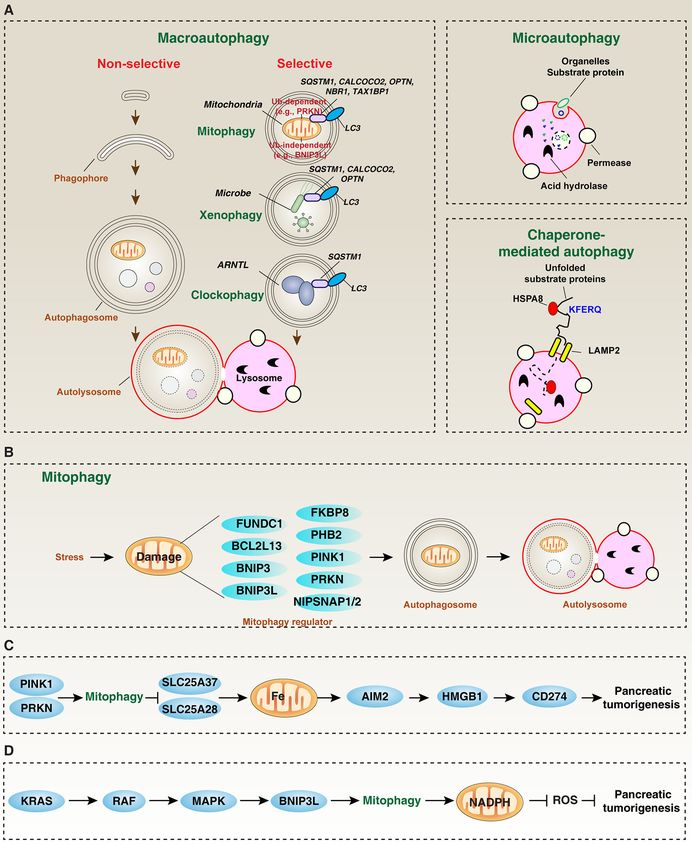
Frontiers in Oncology | www.frontiersin.org 2 February 2021 | Volume 11 | Article 616079Xie et al. Mitophagy in Pancreatic Cancer FIGURE 1 | The role of mitophagy in pancreatic tumorigenesis. (A) In mammalian cells, there are three main types of autophagy: microautophagy, macroautophagy, and chaperone-mediated autophagy. Macroautophagy can be further divided into selective and non-selective forms. (B) Core mitophagy regulators mediate mitochondrial clearance. (C, D) PINK1/PRKN and BNIP3L-dependent mitophagy play different roles in inhibiting or promoting pancreatic tumorigenesis, respectively. from the cytoplasm to the mitochondria, leading to subsequent (OPTN) (26), neighbor of BRCA1 gene 1 (NBR1) (27), calcium assembly of phosphorylated Ub chains on mitochondrial outer binding and coiled-coil domain 2 (CALCOCO2/NDP52) (28), membrane proteins (25). In addition to the earliest reported and tax1 binding protein 1 (TAX1BP1) (28), also help to SQSTM1 (25), other autophagy receptors, such as optineurin recognize and degrade damaged mitochondria after activating Frontiers in Oncology | www.frontiersin.org 3 February 2021 | Volume 11 | Article 616079
Xie et al.                                                                                                                       Mitophagy in Pancreatic Cancer

the PINK1-PRKN pathway (Figure 1A). Moreover, PINK1-                             be morphologically classified into four types: conventional, tubulo-
PRKN-mediated mitophagy can be reversed by deubiquitinating                      papillary, squamous, and “composite”, which exhibit different
enzymes, such as ubiquitin-specific peptidase 8 (USP8), USP15,                    molecular and genetic characteristics (55). Generally, autophagy
USP30, and USP35 (29). Non-classical Ub-dependent mitophagy                      inhibits the growth of PDAC in the early stage by limiting DNA
is mediated by non-PRKN E3 ubiquitin ligases (such as                            damage or inflammation, and upregulated autophagy in the later
mitochondrial E3 ubiquitin protein ligase 1 [MUL1] (30), siah                    stage can promote PDAC survival by limiting cell death or anti-
E3 ubiquitin protein ligase 1 [SIAH1] (31), SMAD specific E3                      tumor immunity (56–60). Since covering all the effects of autophagy
ubiquitin protein ligase 1 [SMURF1] (32), and autocrine motility                 in PDAC is outside the scope of this min-review, we will only
factor receptor [AMFR/GP78]) (33). The impact of crosstalk                       discuss the modulation and function of mitophagy in PDAC as
between Ub-dependent classical and non-classical mitophagy                       described below.
pathways on tumors is still poorly understood.
    Ub-independent mitophagy is mediated by receptors, rather                    Pancreatic Tumorigenesis
than E3 ligases. Recently, depending on the stimulus and cell                    Evidence is accumulating that both intrinsic genetic factor and
type, the list of mitophagy receptors is increasing (34). In addition to         extrinsic environmental factor are important for tumorigenesis.
the early reports of BCL2 interacting protein 3 like (BNIP3L/NIX)                For pancreatic cancer, the oncogenic KRAS mutation is a key
acting as a mitophagy receptor in red cells (35), other mitophagy                driving force for the formation of precursor lesions and
receptors, including FUN14 domain containing 1 (FUNDC1) (36),                    subsequent development of PDAC with stromal response (61).
BCL2 interacting protein 3 (BNIP3) (37), nipsnap homolog 1                       KRAS activation is related to changes in mitochondrial
(NIPSNAP1) (38), nipsnap homolog 2 (NIPSNAP2) (38),                              morphology (e.g., increased mitochondrial fragmentation) and
prohibitin 2 (PHB2) (39), BCL2 like 13 (BCL2L13) (40) and                        function (for example, reduction of mitochondrial respiratory
FKBP prolyl isomerase 8 (FKBP8) (41), have also been identified                   complex I activity, enhancement of glycolytic activity, promotion
in cancer and non-cancer cells (Figure 1B). These unique receptors               of ROS production and induction of mitophagy) in various
are responsible for binding to different mitochondrial membrane                  cancers (including PDAC) (62–66). Moreover, the conditional
components in response to various stresses (such as hypoxia and                  expression of endogenous KrasG12D in the pancreas of mice
oxidative damage). In addition to protein, non-protein                           (Pdx1-Cre;Kras G12D; called KC mice) can mimic most of
mitochondrial components, such as cardiolipin and ceramide                       pathological development of human PDAC (67). This
(42), also mediate mitophagy in some case, indicating that there                 spontaneous transgenic PDAC mouse model is widely used to
are complex mitophagy sub-routes to regulate mitochondrial                       further consume or overexpress additional genes to evaluate the
turnover and function.                                                           function of target genes in pancreatic tumorigenesis (Table 1).
                                                                                 For example, based on KC mice, further depletion of the tumor
                                                                                 suppressor high mobility group box 1 (HMGB1, Pdx1-Cre;
MITOPHAGY IN PANCREATIC CANCER                                                   KrasG12D;Hmgb1-/-) (69) or overexpression of tumor protein
                                                                                 p53 (TP53) mutation (Pdx1-Cre;KrasG12D;Tp53R172H, termed
Compared to normal cells, pancreatic cancer cells generally                      KPC mice) (70) can significantly promote the development of
exhibit highly fragmented mitochondria, which is associated                      KRAS-driven PDAC. HMGB1 is a positive regulator of autophagy
with increased mitochondrial fission and numbers as well as                       and mitophagy, coupled with TP53 signaling in a variety of
enhanced mitochondrial oxidative phosphorylation or glycolysis                   tumors (71–74). Cytoplasmic HMGB1 is a BECN1-binding
(43–45). Therefore, understanding the mechanism of mitochondrial                 protein that contributes to the formation of autophagosomes
biogenesis and turnover in different stages of pancreatic cancer,                (72). Nuclear HMGB1 promotes the expression of heat shock
including initiation, progression, and metastasis, is essential for the          protein b-1 (HSPB1) and subsequent HSPB1-mediated
next generation of cancer treatments. Indeed, increased autophagy                cytoskeletal integrity, which is required for the membrane
or mitophagy levels are observed in various types of pancreatic                  dynamics of mitophagy (71). In addition, mitochondrial
cancer (46–48). However, autophagy plays a dual role in various                  HMGB1 repairs mitochondrial genomic DNA damage, which
cancer (including PDAC), depending on many factors, such as                      also plays a potential role in suppressing tumorigenesis (75).
tumor stage, tumor microenvironment, gene mutation status                        However, depletion of mitophagy regulators, including PINK1
involving oncogenes and tumor suppressor genes, and metabolic                    [Pdx1-Cre;KrasG12D;Pink1-/-] (22), PRKN [Pdx1-Cre;KrasG12D;
reprogramming (49–54). PDAC is a heterogeneous disease and can                   Prkn-/-] (22), or BNIP3L/NIX [Pdx1-Cre;KrasG12D;Bnip3l-/- or

TABLE 1 | Mitophagy regulators in PDAC.

Mitophagy regulator        Expression in human PDAC         Function                                         Mechanism                                    Refs

BNIP3L                     Upregulation                 Tumor   promoter         Increases glucose metabolism and antioxidant capacity                     (68)
PINK1                      Upregulation                 Tumor   suppressor       Inhibits inflammation and mitochondrial iron-related antitumor immunity    (22)
PRKN                       Downregulation               Tumor   suppressor       Inhibits inflammation and mitochondrial iron-related antitumor immunity    (22)
HMGB1                      Upregulation                 Tumor   suppressor       Inhibits genomic instability and mitochondrial dysfunction                (69)
TP53                       Upregulation                 Tumor   suppressor       Inhibits genomic instability and mitochondrial dysfunction                (70)

Frontiers in Oncology | www.frontiersin.org                                  4                                      February 2021 | Volume 11 | Article 616079
Xie et al. Mitophagy in Pancreatic Cancer
Pdx1-Cre;KrasG12D;Tp53R172H; Bnip3l-/- (68), in KC mice exhibits to be seen. In addition, various types of regulated cell death are
different phenotype in pancreatic tumorigenesis. These transgenic closely related to autophagy (85–87), which may accelerate the
animal studies show that Ub-dependent and independent complexity of the immune characteristics of the tumor
mitophagy pathways play different roles in PDAC. microenvironment, thereby affecting anti-tumor immunity.
Dysregulated autophagy promotes or inhibits the growth of There is emerging evidence that impaired mitophagy is related to
pancreatic cancer by interfering with different metabolic pathways or epithelial-mesenchymal transition and pancreatic cancer stem cells
tumor signals, such as carbohydrate metabolism, fatty acid b- (pCSCs), which are pluripotent, self-renewable, and capable of
oxidation, and amino acid transport (48). For example, the forming tumors (88). In particular, the interferon signaling
reduced glycolysis gene PKM2 promotes survival by maintaining pathway-mediated the upregulation of Ub-like modifier interferon-
autophagy induced by low glucose in PDAC cells (76). Autophagy- stimulated gene 15 (ISG15) and its modification ISGylation maintain
mediated lipid degradation and subsequent fatty acid b-oxidation mitophagy and metabolic plasticity of pCSCs (89), suggesting a
may provide additional resources for ATP production during PDAC potential link between interferon, mitophagy, and metabolism in
growth (77, 78). Autophagy-mediated degradation of cellular pCSCs. PDAC patients with high ISG15 levels showed increased
material provides reusable amino acids for PDAC cell proliferation expression of genes related to the CSC pathway, including epithelial-
during glutamine deprivation (79). In addition, the PINK1-PRKN mesenchymal transition and oxidative phosphorylation (89). In
pathway can degrade mitochondrial iron importers (such as solute contrast, the inhibition of ISG15/ISGylation impairs PRKN-
carrier family 25 member 37 [SLC25A37] and solute carrier family dependent mitophagy, causing pCSCs to fail to eliminate
25 member 28 [SLC25A28]) through SQSTM1-mediated mitophagy dysfunctional and unhealthy mitochondria (89). Overall, these
to inhibit carcinogenic KRAS-driven pancreatic tumorigenesis in findings support a role of ISG15 in pCSCs by regulating
mice, thereby inhibiting mitochondrial iron-mediated absent in mitochondrial dynamics and energy metabolism. The role of
melanoma 2 (AIM2)-dependent inflammasome activation and the ISG15 in pancreatic tumorigenesis needs to be further studied
subsequent activation of damage associated molecular pattern using transgenic mice.
(DAMP, such as HMGB1)-dependent immune checkpoint
expression (e.g., CD274/PD-L1) (Figure 1C) (22). These findings Pancreatic Cancer Therapy
establish a role of PINK1/PRKN-mediated mitophagy to inhibit The purpose of tumor treatment is to induce death in tumor cells
pancreatic tumorigenesis by limiting chronic inflammation-related without damaging normal cells. Cell death can be divided into
immunosuppression in the hypoxic tumor microenvironment (80). accidental or regulated cell death (90). Regulated cell death further
Of note, high expression of PRKN mRNA was found to be associated includes apoptotic and non-apoptotic forms. In addition to the
with improved survival of pancreatic cancer patients, whereas extensively studied apoptosis (91, 92), the induction of non-
mRNA expression of PINK1 did not influence patient survival apoptotic regulated cell death [such as necroptosis (93, 94),
(22), indicating that PINK1 is a contributor of PDAC, but it is not alkaliptosis (95, 96), and ferroptosis (97–101)] in preclinical
a potential biomarker. In addition, both PINK1 and PRKN may have PDAC models has shown promising results in inhibiting tumor
mitophagy-independent functions in controlling the quality of growth. As a metabolic center, mitochondria play a complex role in
mitochondria during pancreatic tumorigenesis (22). regulating apoptosis and non-apoptotic cell death by cooperating
In contrast, in a precursor lesion called pancreatic intraepithelial with other subcellular organelles (102). Accordingly, mitophagy-
neoplasia (PanIN), oncogenic KRAS-mediated BNIP3L expression mediated mitochondrial degradation and turnover is reasonable to
may activate mitophagy in a rapidly accelerated fibrosarcoma affect the anti-cancer activity of cytotoxic agents in PDAC cells. The
(RAF)-mitogen-activated protein kinase (MAPK)-dependent best cell models for studying mitochondrial biology and mitophagy
manner to limit the flux of glucose to mitochondria and enhance of pancreatic cancer are various human PDAC cell lines with KRAS
reduced nicotinamide adenine dinucleotide phosphate (NADPH)- mutations. For example, rocaglamide A, a natural product from the
dependent redox capacity, thereby promoting pancreatic plant Aglaia elliptifolia, has the ability to induce PINK1/PRKN-
tumorigenesis (Figure 1D) (68). In KC and KPC pancreatic mediated mitophagy as a negative feedback mechanism to limit
cancer models, the depletion of additional BNIP3L will increase rocaglamide A-induced apoptosis in various human PDAC cell
the content of mitochondria in PanIN, thereby increasing the lines with KRAS mutations (103). In contrast, the inhibition of
production of mitochondrial ROS to limit the development of mitophagy by Mdivi-1 enhances the anti-cancer activity of
PanIN to PDAC (68). These observations indicate that BNIP3L- rocaglamide A in PDAC cells (103). Overexpression of serine/
mediated mitophagy have different roles in promoting pancreatic threonine kinase 25 (STK25, also known as MST1) in various
tumorigenesis by enhancing the antioxidant capacity of cancer cells PDAC cells induces apoptosis by inhibiting mitophagy mediated by
for cell proliferation and metastasis. However, oxidative stress and mitofusin 2 (MFN2) (104). In contrast, leflunomide, an FDA-
redox regulation are double-edged swords in tumorigenesis (81). approved arthritis drug, can inhibit the growth of PDAC tumors
Certain types of oxidative cell death, such as necroptosis (a caspase- by inducing MFN2 expression and subsequent mitophagy (44). In
independent regulated necrosis) and ferroptosis (an iron- addition, in vitro and xenograft models, the combination of cyst(e)
dependent regulated necrosis), can promote KRAS-driven PDAC inase (an engineered human enzyme) and anuranofin (a
by activating inflammation-related immune suppression (82–84). thioredoxin reductase inhibitor) can inhibit mitophagy, thereby
Whether PINK1, PRKN2, and BNIP3L have non-mitochondrial increase ROS production and apoptosis in the human PDAC cells
functions in the modulation of the oncogene KRAS signal remains (105). In other cases, dichloroacetate (an inhibitor of pyruvate
Frontiers in Oncology | www.frontiersin.org 5 February 2021 | Volume 11 | Article 616079Xie et al. Mitophagy in Pancreatic Cancer
dehydrogenase kinase) (106), fisetin (a bioactive flavonoid molecule tumor-promoting and anti-tumor effect. In addition, the degree
found in fruits and vegetables) (107), and P. suffruticosa extracts of substrate degradation (such as complete or partial degradation)
(108) may play a context-related role in the induction of mitophagy also affects the function of autophagy in tumors. Similarly,
and tumor suppression in PDAC cells. These findings further mitochondrial coupling with mitochondrial biogenesis also plays
indicate that the complex relationship between mitophagy and a dual role in cancer. In this min-review, we discussed the context-
mitochondrial dynamics can affect the effects of chemotherapy and dependent role of mitophagy in pancreatic cancer. Although this
targeted therapy. information enhances our understanding of the role of
In addition to PDAC cells, pCSCs is another cell model for mitochondrial homeostasis in pancreatic cancer, there are still
studying mitochondrial dysfunction. pCSCs not only promote some key questions about the process and function of mitophagy
the growth and metastasis of pancreatic tumors, but also mediate in PDAC. How does the multi-step mitophagy actually proceed at
chemoresistance. Metformin is a biguanide anti-diabetic drug that different stages of PDAC? What are the key molecules or signals
activates AMP-activated protein kinase (AMPK) to trigger that distinguish the functions of mitophagy in promoting or
autophagy (109). Retrospective studies have shown that inhibiting pancreatic tumorigenesis? Do tumor cells and non-
compared with patients receiving insulin or sulfonylureas, many tumor cells (such as immune cells or stromal cells) in the
diabetic patients with solid tumors (including pancreatic cancer) pancreatic tumor microenvironment have different mitophagy
treated with metformin have a survival benefit (109). The loss of activities? In the pancreatic tumor microenvironment, what is
ISG15 in pCSCs by CRISPR-Cas9 technology results in sensitivity the synergy or competition between mitophagy and other types of
to metformin therapy in xenograft models (89). These findings selective autophagy? How to develop specific mitophagy targeted
further indicate a potential role of ISG15 in regulating the anti- drugs to kill pancreatic tumors? Are there specific markers to
cancer activity of metformin in pCSCs. Further investigations are assess the level of mitophagy in PDAC patients?
still needed to determine whether ISG15 directly regulates AMPK
activation in pCSCs.
AUTHOR CONTRIBUTIONS
CONCLUSION AND PERSPECTIVES YX and DT conceived of the topic for this review. All authors
contributed to the article and approved the submitted version.
In the past decade, basic and clinical research on autophagy has
involved various diseases, including pancreatic cancer (48, 59,
110–112). With the deepening of research, the functions of FUNDING
autophagy in tumor biology show diversity and complexity.
One of the important reasons is that autophagy can have YX was supported by the National Natural Science Foundation
different degradation substrates, and these substrates can play a of China (No. 81802476).
REFERENCES macroautophagy. Autophagy (2015) 11:28–45. doi: 10.4161/
15548627.2014.984267
1. Rahib L, Smith BD, Aizenberg R, Rosenzweig AB, Fleshman JM, Matrisian 9. Liu J, Kuang F, Kroemer G, Klionsky DJ, Kang R, Tang D. Autophagy-
LM. Projecting cancer incidence and deaths to 2030: the unexpected burden Dependent Ferroptosis: Machinery and Regulation. Cell Chem Biol (2020)
of thyroid, liver, and pancreas cancers in the United States. Cancer Res 27:420–35. doi: 10.1016/j.chembiol.2020.02.005
(2014) 74:2913–21. doi: 10.1158/0008-5472.CAN-14-0155 10. Green DR, Levine B. To be or not to be? How selective autophagy and cell
2. Amanam I, Chung V. Targeted Therapies for Pancreatic Cancer. Cancers death govern cell fate. Cell (2014) 157:65–75. doi: 10.1016/j.cell.2014.02.049
(Basel) (2018) 2:36. doi: 10.3390/cancers10020036 11. Vernon PJ, Tang D. Eat-me: autophagy, phagocytosis, and reactive oxygen
3. Levine B, Kroemer G. Biological Functions of Autophagy Genes: A Disease species signaling. Antioxid Redox Signal (2013) 18:677–91. doi: 10.1089/
Perspective. Cell (2019) 176:11–42. doi: 10.1016/j.cell.2018.09.048 ars.2012.4810
4. Dikic I, Elazar Z. Mechanism and medical implications of mammalian 12. Gatica D, Lahiri V, Klionsky DJ. Cargo recognition and degradation by
autophagy. Nat Rev Mol Cell Biol (2018) 19:349–64. doi: 10.1038/s41580- selective autophagy. Nat Cell Biol (2018) 20:233–42. doi: 10.1038/s41556-
018-0003-4 018-0037-z
5. Bandyopadhyay U, Kaushik S, Varticovski L, Cuervo AM. The chaperone- 13. Sharma V, Verma S, Seranova E, Sarkar S, Kumar D. Selective Autophagy
mediated autophagy receptor organizes in dynamic protein complexes at the and Xenophagy in Infection and Disease. Front Cell Dev Biol (2018) 6:147.
lysosomal membrane. Mol Cell Biol (2008) 28:5747–63. doi: 10.1128/ doi: 10.3389/fcell.2018.00147
MCB.02070-07 14. Yang M, Chen P, Liu J, Zhu S, Kroemer G, Klionsky DJ, et al. Clockophagy is
6. Chiang HL, Terlecky SR, Plant CP, Dice JF. A role for a 70-kilodalton heat a novel selective autophagy process favoring ferroptosis. Sci Adv (2019) 5:
shock protein in lysosomal degradation of intracellular proteins. Science eaaw2238. doi: 10.1126/sciadv.aaw2238
(1989) 246:382–5. doi: 10.1126/science.2799391 15. Liu J, Yang M, Kang R, Klionsky DJ, Tang D. Autophagic degradation of the
7. Cuervo AM, Dice JF. A receptor for the selective uptake and degradation of circadian clock regulator promotes ferroptosis. Autophagy (2019) 15:2033–5.
proteins by lysosomes. Science (1996) 273:501–3. doi: 10.1126/ doi: 10.1080/15548627.2019.1659623
science.273.5274.501 16. Palikaras K, Lionaki E, Tavernarakis N. Mechanisms of mitophagy in
8. Xie Y, Kang R, Sun X, Zhong M, Huang J, Klionsky DJ, et al. cellular homeostasis, physiology and pathology. Nat Cell Biol (2018)
Posttranslational modification of autophagy-related proteins in 20:1013–22. doi: 10.1038/s41556-018-0176-2
Frontiers in Oncology | www.frontiersin.org 6 February 2021 | Volume 11 | Article 616079Xie et al. Mitophagy in Pancreatic Cancer
17. Kabeya Y, Mizushima N, Ueno T, Yamamoto A, Kirisako T, Noda T, et al. Mi toph agy. D ev C e l l (2 01 9) 49 :50 9– 52 5 e12 . doi : 1 0.1 01 6/
LC3, a mammalian homologue of yeast Apg8p, is localized in j.devcel.2019.03.013
autophagosome membranes after processing. EMBO J (2000) 19:5720–8. 39. Wei Y, Chiang WC, Sumpter RJr., Mishra P, Levine B. Prohibitin 2 Is an
doi: 10.1093/emboj/19.21.5720 Inner Mitochondrial Membrane Mitophagy Receptor. Cell (2017) 168:224–
18. Katsuragi Y, Ichimura Y, Komatsu M. p62/SQSTM1 functions as a signaling 238 e10. doi: 10.1016/j.cell.2016.11.042
hub and an autophagy adaptor. FEBS J (2015) 282:4672–8. doi: 10.1111/ 40. Murakawa T, Yamaguchi O, Hashimoto A, Hikoso S, Takeda T, Oka T, et al.
febs.13540 Bcl-2-like protein 13 is a mammalian Atg32 homologue that mediates
19. Liu WJ, Ye L, Huang WF, Guo LJ, Xu ZG, Wu HL, et al. p62 links the mitophagy and mitochondrial fragmentation. Nat Commun (2015) 6:7527.
autophagy pathway and the ubiqutin-proteasome system upon doi: 10.1038/ncomms8527
ubiquitinated protein degradation. Cell Mol Biol Lett (2016) 21:29. doi: 41. Bhujabal Z, Birgisdottir AB, Sjottem E, Brenne HB, Overvatn A, Habisov S,
10.1186/s11658-016-0031-z et al. FKBP8 recruits LC3A to mediate Parkin-independent mitophagy.
20. Xie Y, Li J, Kang R, Tang D. Interplay Between Lipid Metabolism and EMBO Rep (2017) 18:947–61. doi: 10.15252/embr.201643147
Autophagy. Front Cell Dev Biol (2020) 8:431. doi: 10.3389/fcell.2020.00431 42. Chu CT, Ji J, Dagda RK, Jiang JF, Tyurina YY, Kapralov AA, et al.
21. Kang R, Zeng L, Xie Y, Yan Z, Zhou B, Cao L, et al. A novel PINK1- and Cardiolipin externalization to the outer mitochondrial membrane acts as
PARK2-dependent protective neuroimmune pathway in lethal sepsis. an elimination signal for mitophagy in neuronal cells. Nat Cell Biol (2013)
Autophagy (2016) 12:2374–85. doi: 10.1080/15548627.2016.1239678 15:1197–205. doi: 10.1038/ncb2837
22. Li C, Zhang Y, Cheng X, Yuan H, Zhu S, Liu J, et al. PINK1 and PARK2 43. Anderson GR, Wardell SE, Cakir M, Yip C, Ahn YR, Ali M, et al.
Suppress Pancreatic Tumorigenesis through Control of Mitochondrial Iron- Dysregulation of mitochondrial dynamics proteins are a targetable feature
Mediated Immunometabolism. Dev Cell (2018) 46:441–455 e8. doi: 10.1016/ of human tumors. Nat Commun (2018) 9:1677. doi: 10.1038/s41467-018-
j.devcel.2018.07.012 04033-x
23. Sliter DA, Martinez J, Hao L, Chen X, Sun N, Fischer TD, et al. Parkin and 44. Yu M, Nguyen ND, Huang Y, Lin D, Fujimoto TN, Molkentine JM, et al.
PINK1 mitigate STING-induced inflammation. Nature (2018) 561:258–62. Mitochondrial fusion exploits a therapeutic vulnerability of pancreatic
doi: 10.1038/s41586-018-0448-9 cancer. JCI Insight (2019) 4:e126915. doi: 10.1172/jci.insight.126915
24. Harper JW, Ordureau A, Heo JM. Building and decoding ubiquitin chains 45. Dai S, Peng Y, Zhu Y, Xu D, Zhu F, Xu W, et al. Glycolysis promotes the
for mitophagy. Nat Rev Mol Cell Biol (2018) 19:93–108. doi: 10.1038/ progression of pancreatic cancer and reduces cancer cell sensitivity to
nrm.2017.129 gemcitabine. BioMed Pharmacother (2020) 121:109521. doi: 10.1016/
25. Geisler S, Holmstrom KM, Skujat D, Fiesel FC, Rothfuss OC, Kahle PJ, et al. j.biopha.2019.109521
PINK1/Parkin-mediated mitophagy is dependent on VDAC1 and p62/ 46. Daskalakis K, Alexandraki KI, Kloukina I, Kassi E, Felekouras E, Xingi E,
SQSTM1. Nat Cell Biol (2010) 12:119–31. doi: 10.1038/ncb2012 et al. Increased autophagy/mitophagy levels in primary tumours of patients
26. Wong YC, Holzbaur EL. Optineurin is an autophagy receptor for damaged with pancreatic neuroendocrine neoplasms. Endocrine (2020) 68:438–47.
mitochondria in parkin-mediated mitophagy that is disrupted by an ALS- doi: 10.1007/s12020-020-02228-1
linked mutation. Proc Natl Acad Sci U S A (2014) 111:E4439–48. doi: 47. Ko YH, Cho YS, Won HS, Jeon EK, An HJ, Hong SU, et al. Prognostic
10.1073/pnas.1405752111 significance of autophagy-related protein expression in resected pancreatic
27. Gao F, Chen D, Si J, Hu Q, Qin Z, Fang M, et al. The mitochondrial protein ductal adenocarcinoma. Pancreas (2013) 42:829–35. doi: 10.1097/
BNIP3L is the substrate of PARK2 and mediates mitophagy in PINK1/PARK2 MPA.0b013e318279d0dc
pathway. Hum Mol Genet (2015) 24:2528–38. doi: 10.1093/hmg/ddv017 48. Li J, Chen X, Kang R, Zeh H, Klionsky DJ, Tang D. Regulation and function
28. Lazarou M, Sliter DA, Kane LA, Sarraf SA, Wang C, Burman JL, et al. The of autophagy in pancreatic cancer. Autophagy (2020), 1–22. doi: 10.1080/
ubiquitin kinase PINK1 recruits autophagy receptors to induce mitophagy. 15548627.2020.1847462
Nature (2015) 524:309–14. doi: 10.1038/nature14893 49. Levy JMM, Towers CG, Thorburn A. Targeting autophagy in cancer. Nat
29. Wang Y, Serricchio M, Jauregui M, Shanbhag R, Stoltz T, Di Paolo CT, et al. Rev Cancer (2017) 17:528–42. doi: 10.1038/nrc.2017.53
Deubiquitinating enzymes regulate PARK2-mediated mitophagy. 50. Monkkonen T, Debnath J. Inflammatory signaling cascades and autophagy
Autophagy (2015) 11:595–606. doi: 10.1080/15548627.2015.1034408 in cancer. Autophagy (2018) 14:190–8. doi: 10.1080/15548627.2017.1345412
30. Yun J, Puri R, Yang H, Lizzio MA, Wu C, Sheng ZH, et al. MUL1 acts in parallel 51. New M, Tooze S. The Role of Autophagy in Pancreatic Cancer-Recent
to the PINK1/parkin pathway in regulating mitofusin and compensates for loss Advances. Biol (Basel) (2019) 9:7. doi: 10.3390/biology9010007
of PINK1/parkin. Elife (2014) 3:e01958. doi: 10.7554/eLife.01958 52. Piffoux M, Eriau E, Cassier PA. Autophagy as a therapeutic target in
31. Szargel R, Shani V, Abd Elghani F, Mekies LN, Liani E, Rott R, et al. The pancreatic cancer. Br J Cancer (2021) 1244:333–44. doi: 10.1038/s41416-
PINK1, synphilin-1 and SIAH-1 complex constitutes a novel mitophagy 020-01039-5
pathway. Hum Mol Genet (2016) 25:3476–90. doi: 10.1093/hmg/ddw189 53. Gorgulu K, Diakopoulos KN, Kaya-Aksoy E, Ciecielski KJ, Ai J, Lesina M,
32. Orvedahl A, Sumpter RJr., Xiao G, Ng A, Zou Z, Tang Y, et al. Image-based et al. The Role of Autophagy in Pancreatic Cancer: From Bench to the Dark
genome-wide siRNA screen identifies selective autophagy factors. Nature Bedside. Cells (2020) 9:1063. doi: 10.3390/cells9041063
(2011) 480:113–7. doi: 10.1038/nature10546 54. Kang R, Tang D. Autophagy in pancreatic cancer pathogenesis and
33. Fu M, St-Pierre P, Shankar J, Wang PT, Joshi B, Nabi IR. Regulation of treatment. Am J Cancer Res (2012) 2:383–96.
mitophagy by the Gp78 E3 ubiquitin ligase. Mol Biol Cell (2013) 24:1153–62. 55. S NK, Wilson GW, Grant RC, Seto M, O’Kane G, Vajpeyi R, et al.
doi: 10.1091/mbc.e12-08-0607 Morphological classification of pancreatic ductal adenocarcinoma that
34. Xie Y, Liu J, Kang R, Tang D. Mitophagy Receptors in Tumor Biology. Front predicts molecular subtypes and correlates with clinical outcome. Gut
Cell Dev Biol (2020) 8:594203. doi: 10.3389/fcell.2020.594203 (2020) 69:317–28. doi: 10.1136/gutjnl-2019-318217
35. Sandoval H, Thiagarajan P, Dasgupta SK, Schumacher A, Prchal JT, Chen 56. Yang S, Imamura Y, Jenkins RW, Canadas I, Kitajima S, Aref A, et al.
M, et al. Essential role for Nix in autophagic maturation of erythroid cells. Autophagy Inhibition Dysregulates TBK1 Signaling and Promotes
Nature (2008) 454:232–5. doi: 10.1038/nature07006 Pancreatic Inflammation. Cancer Immunol Res (2016) 4:520–30. doi:
36. Liu L, Feng D, Chen G, Chen M, Zheng Q, Song P, et al. Mitochondrial 10.1158/2326-6066.CIR-15-0235
outer-membrane protein FUNDC1 mediates hypoxia-induced mitophagy in 57. Loncle C, Molejon MI, Lac S, Tellechea JI, Lomberk G, Gramatica L, et al.
mammalian cells. Nat Cell Biol (2012) 14:177–85. doi: 10.1038/ncb2422 The pancreatitis-associated protein VMP1, a key regulator of inducible
37. O’Sullivan TE, Johnson LR, Kang HH, Sun JC. BNIP3- and BNIP3L- autophagy, promotes Kras(G12D)-mediated pancreatic cancer initiation.
Mediated Mitophagy Promotes the Generation of Natural Killer Cell Cell Death Dis (2016) 7:e2295. doi: 10.1038/cddis.2016.202
Memory. Immunity (2015) 43:331–42. doi: 10.1016/j.immuni.2015.07.012 58. Rosenfeldt MT, O’Prey J, Morton JP, Nixon C, MacKay G, Mrowinska A,
38. Princely Abudu Y, Pankiv S, Mathai BJ, Hakon Lystad A, Bindesboll C, et al. p53 status determines the role of autophagy in pancreatic tumour
Brenne HB, et al. NIPSNAP1 and NIPSNAP2 Act as “Eat Me” Signals for development. Nature (2013) 504:296–300. doi: 10.1038/nature12865
Frontiers in Oncology | www.frontiersin.org 7 February 2021 | Volume 11 | Article 616079Xie et al. Mitophagy in Pancreatic Cancer
59. Yamamoto K, Venida A, Yano J, Biancur DE, Kakiuchi M, Gupta S, et al. Adenocarcinoma. Cancers (Basel) (2020) 12:2477. doi: 10.3390/
Autophagy promotes immune evasion of pancreatic cancer by degrading cancers12092477
MHC-I. Nature (2020) 581:100–5. doi: 10.1038/s41586-020-2229-5 78. Maan M, Peters JM, Dutta M, Patterson AD. Lipid metabolism and
60. Kang R, Loux T, Tang D, Schapiro NE, Vernon P, Livesey KM, et al. 3rd, The lipophagy in cancer. Biochem Biophys Res Commun (2018) 504:582–9. doi:
expression of the receptor for advanced glycation endproducts (RAGE) is 10.1016/j.bbrc.2018.02.097
permissive for early pancreatic neoplasia. Proc Natl Acad Sci U.S.A. (2012) 79. Seo JW, Choi J, Lee SY, Sung S, Yoo HJ, Kang MJ, et al. Autophagy is
109:7031–6. doi: 10.1073/pnas.1113865109 required for PDAC glutamine metabolism. Sci Rep (2016) 6:37594. doi:
61. Waters AM, Der CJ. KRAS: The Critical Driver and Therapeutic Target for 10.1038/srep37594
Pancreatic Cancer. Cold Spring Harb Perspect Med (2018) 8:a031435. doi: 80. Kang R, Xie Y, Zeh HJ, Klionsky DJ, Tang D. Mitochondrial quality control
10.1101/cshperspect.a031435 mediated by PINK1 and PRKN: links to iron metabolism and tumor
62. Lin HH, Chung Y, Cheng CT, Ouyang C, Fu Y, Kuo CY, et al. Autophagic immunity. Autophagy (2019) 15:172–3. doi: 10.1080/15548627.2018.
reliance promotes metabolic reprogramming in oncogenic KRAS-driven 1526611
tumorigenesis. Autopha gy (2018) 14:1481–98. doi: 10.1080/ 81. Reuter S, Gupta SC, Chaturvedi MM, Aggarwal BB. Oxidative stress,
15548627.2018.1450708 inflammation, and cancer: how are they linked? Free Radic Biol Med
63. Nagdas S, Kashatus JA, Nascimento A, Hussain SS, Trainor RE, Pollock SR, (2010) 49:1603–16. doi: 10.1016/j.freeradbiomed.2010.09.006
et al. Drp1 Promotes KRas-Driven Metabolic Changes to Drive Pancreatic 82. Seifert L, Werba G, Tiwari S, Giao Ly NN, Alothman S, Alqunaibit D, et al.
Tumor Growth. Cell Rep (2019) 28:1845–1859 e5. doi: 10.1016/ The necrosome promotes pancreatic oncogenesis via CXCL1 and Mincle-
j.celrep.2019.07.031 induced immune suppression. Nature (2016) 532:245–9. doi: 10.1038/
64. Palorini R, De Rasmo D, Gaviraghi M, Sala Danna L, Signorile A, Cirulli C, nature17403
et al. Oncogenic K-ras expression is associated with derangement of the 83. Dai E, Han L, Liu J, Xie Y, Zeh HJ, Kang R, et al. Ferroptotic damage
cAMP/PKA pathway and forskolin-reversible alterations of mitochondrial promotes pancreatic tumorigenesis through a TMEM173/STING-
dynamics and respiration. Oncogene (2013) 32:352–62. doi: 10.1038/ dependent DNA sensor pathway. Nat Commun (2020) 11:6339. doi:
onc.2012.50 10.1038/s41467-020-20154-8
65. Meng N, Glorieux C, Zhang Y, Liang L, Zeng P, Lu W, et al. Oncogenic K-ras 84. Dai E, Han L, Liu J, Xie Y, Kroemer G, Klionsky DJ, et al. Autophagy-
Induces Mitochondrial OPA3 Expression to Promote Energy Metabolism in dependent ferroptosis drives tumor-associated macrophage polarization via
Pancreatic Cancer Cells. Cancers (Basel) (2020) 12:65. doi: 10.3390/ release and uptake of oncogenic KRAS protein. Autophagy (2020) 16:2069–
cancers12010065 83. doi: 10.1080/15548627.2020.1714209
66. Guo JY, Karsli-Uzunbas G, Mathew R, Aisner SC, Kamphorst JJ, Strohecker 85. Tang D, Kang R, Berghe TV, Vandenabeele P, Kroemer G. The molecular
AM, et al. Autophagy suppresses progression of K-ras-induced lung tumors machinery of regulated cell death. Cell Res (2019) 29:347–64. doi: 10.1038/
to oncocytomas and maintains lipid homeostasis. Genes Dev (2013) s41422-019-0164-5
27:1447–61. doi: 10.1101/gad.219642.113 86. Bialik S, Dasari SK, Kimchi A. Autophagy-dependent cell death - where, how
67. Hingorani SR, Petricoin EF, Maitra A, Rajapakse V, King C, Jacobetz MA, and why a cell eats itself to death. J Cell Sci (2018) 131:jcs215152. doi:
et al. Preinvasive and invasive ductal pancreatic cancer and its early 10.1242/jcs.215152
detection in the mouse. Cancer Cell (2003) 4:437–50. doi: 10.1016/S1535- 87. Zhou B, Liu J, Kang R, Klionsky DJ, Kroemer G, Tang D. Ferroptosis is a
6108(03)00309-X type of autophagy-dependent cell death. Semin Cancer Biol (2020) 66:89–
68. Humpton TJ, Alagesan B, DeNicola GM, Lu D, Yordanov GN, Leonhardt 100. doi: 10.1016/j.semcancer.2019.03.002
CS, et al. Oncogenic KRAS Induces NIX-Mediated Mitophagy to Promote 88. Guerra F, Guaragnella N, Arbini AA, Bucci C, Giannattasio S, Moro L.
Pancreatic Cancer. Cancer Discovery (2019) 9:1268–87. doi: 10.1158/2159- Mitochondrial Dysfunction: A Novel Potential Driver of Epithelial-to-
8290.CD-18-1409 Mesenchymal Transition in Cancer. Front Oncol (2017) 7:295. doi:
69. Kang R, Xie Y, Zhang Q, Hou W, Jiang Q, Zhu S, et al. Intracellular HMGB1 10.3389/fonc.2017.00295
as a novel tumor suppressor of pancreatic cancer. Cell Res (2017) 27:916–32. 89. Alcala S, Sancho P, Martinelli P, Navarro D, Pedrero C, Martin-Hijano L,
doi: 10.1038/cr.2017.51 et al. ISG15 and ISGylation is required for pancreatic cancer stem cell
70. Hingorani SR, Wang L, Multani AS, Combs C, Deramaudt TB, Hruban RH, mitophagy and metabolic plasticity. Nat Commun (2020) 11:2682. doi:
et al. Trp53R172H and KrasG12D cooperate to promote chromosomal 10.1038/s41467-020-16395-2
instability and widely metastatic pancreatic ductal adenocarcinoma in 90. Galluzzi L, Vitale I, Aaronson SA, Abrams JM, Adam D, Agostinis P, et al.
mice. Cancer Cell (2005) 7:469–83. doi: 10.1016/j.ccr.2005.04.023 Molecular mechanisms of cell death: recommendations of the Nomenclature
71. Tang D, Kang R, Livesey KM, Kroemer G, Billiar TR, Van Houten B, et al. Committee on Cell Death 2018. Cell Death Differ (2018) 25:486–541. doi:
High-mobility group box 1 is essential for mitochondrial quality control. Cell 10.1038/s41418-018-0102-y
Metab (2011) 13:701–11. doi: 10.1016/j.cmet.2011.04.008 91. Huang J, Chen P, Liu K, Liu J, Zhou B, Wu R, et al. CDK1/2/5 inhibition
72. Tang D, Kang R, Livesey KM, Cheh CW, Farkas A, Loughran P, et al. overcomes IFNG-mediated adaptive immune resistance in pancreatic
Endogenous HMGB1 regulates autophagy. J Cell Biol (2010) 190:881–92. cancer. Gut (2020). doi: 10.1136/gutjnl-2019-320441
doi: 10.1083/jcb.200911078 92. Arlt A, Muerkoster SS, Schafer H. Targeting apoptosis pathways in
73. Livesey KM, Kang R, Vernon P, Buchser W, Loughran P, Watkins SC, et al. pancreatic cancer. Cancer Lett (2013) 332:346–58. doi: 10.1016/
p53/HMGB1 complexes regulate autophagy and apoptosis. Cancer Res j.canlet.2010.10.015
(2012) 72:1996–2005. doi: 10.1158/0008-5472.CAN-11-2291 93. Xie Y, Zhu S, Zhong M, Yang M, Sun X, Liu J, et al. Inhibition of Aurora
74. Chang HW, Kim MR, Lee HJ, Lee HM, Kim GC, Lee YS, et al. p53/BNIP3- Kinase A Induces Necroptosis in Pancreatic Carcinoma. Gastroenterology
dependent mitophagy limits glycolytic shift in radioresistant cancer. (2017) 153:1429–1443 e5. doi: 10.1053/j.gastro.2017.07.036
Oncogene (2019) 38:3729–42. doi: 10.1038/s41388-019-0697-6 94. Huang C, Lan W, Fraunhoffer N, Meilerman A, Iovanna J, Santofimia-
75. Ito H, Fujita K, Tagawa K, Chen X, Homma H, Sasabe T, et al. HMGB1 Castano P. Dissecting the Anticancer Mechanism of Trifluoperazine on
facilitates repair of mitochondrial DNA damage and extends the lifespan of Pancreatic Ductal Adenocarcinoma. Cancers (Basel) (2019) 11:1869. doi:
mutant ataxin-1 knock-in mice. EMBO Mol Med (2015) 7:78–101. doi: 10.3390/cancers11121869
10.15252/emmm.201404392 95. Song X, Zhu S, Xie Y, Liu J, Sun L, Zeng D, et al. JTC801 Induces pH-
76. Li X, Deng S, Liu M, Jin Y, Zhu S, Deng S, et al. The responsively decreased dependent Death Specifically in Cancer Cells and Slows Growth of Tumors
PKM2 facilitates the survival of pancreatic cancer cells in hypoglucose. Cell in Mice. Gastroenterology (2018) 154:1480–93. doi: 10.1053/
Death Dis (2018) 9:133. doi: 10.1038/s41419-017-0158-5 j.gastro.2017.12.004
77. Lee JS, Oh SJ, Choi HJ, Kang JH, Lee SH, Ha JS, et al. ATP Production Relies 96. Liu J, Kuang F, Kang R, Tang D. Alkaliptosis: a new weapon for cancer
on Fatty Acid Oxidation Rather than Glycolysis in Pancreatic Ductal therapy. Cancer Gene Ther (2020) 27:267–9. doi: 10.1038/s41417-019-0134-6
Frontiers in Oncology | www.frontiersin.org 8 February 2021 | Volume 11 | Article 616079Xie et al. Mitophagy in Pancreatic Cancer
97. Li C, Zhang Y, Liu J, Kang R, Klionsky DJ, Tang D. Mitochondrial DNA stress-dependent pathways. Cell Death Dis (2019) 10:142. doi: 10.1038/
stress triggers autophagy-dependent ferroptotic death. Autophagy (2020), 1– s41419-019-1366-y
13. doi: 10.1080/15548627.2020.1739447 108. Liu YH, Weng YP, Tsai HY, Chen CJ, Lee DY, Hsieh CL, et al. Aqueous
98. Zhu S, Zhang Q, Sun X, Zeh HJ3rd3rd, Lotze MT, Kang R, et al. HSPA5 extracts of Paeonia suffruticosa modulates mitochondrial proteostasis by
Regulates Ferroptotic Cell Death in Cancer Cells. Cancer Res (2017) reactive oxygen species-induced endoplasmic reticulum stress in pancreatic
77:2064–77. doi: 10.1158/0008-5472.CAN-16-1979 cancer cells. Phytomedicine (2018) 46:184–92. doi: 10.1016/
99. Xie Y, Kuang F, Liu J, Tang D, Kang R. DUSP1 Blocks Autophagy- j.phymed.2018.03.037
Dependent Ferroptosis in Pancreatic Cancer. J Pancreatol (2020) 3:154– 109. Saraei P, Asadi I, Kakar MA, Moradi-Kor N. The beneficial effects of
60. doi: 10.1097/JP9.0000000000000054 metformin on cancer prevention and therapy: a comprehensive review of
100. Badgley MA, Kremer DM, Maurer HC, DelGiorno KE, Lee HJ, Purohit V, recent advances. Cancer Manag Res (2019) 11:3295–313. doi: 10.2147/
et al. Cysteine depletion induces pancreatic tumor ferroptosis in mice. CMAR.S200059
Science (2020) 368:85–9. doi: 10.1126/science.aaw9872 110. Liang C, Xu J, Meng Q, Zhang B, Liu J, Hua J, et al. TGFB1-induced
101. Hou W, Xie Y, Song X, Sun X, Lotze MT, Zeh HJ3rd3rd, et al. Autophagy autophagy affects the pattern of pancreatic cancer progression in distinct
promotes ferroptosis by degradation of ferritin. Autophagy (2016) 12:1425–8. ways depending on SMAD4 status. Autophagy (2020) 16:486–500. doi:
doi: 10.1080/15548627.2016.1187366 10.1080/15548627.2019.1628540
102. Bock FJ, Tait SWG. Mitochondria as multifaceted regulators of cell death. 111. Yang A, Herter-Sprie G, Zhang H, Lin EY, Biancur D, Wang X, et al.
Nat Rev Mol Cell Biol (2020) 21:85–100. doi: 10.1038/s41580-019-0173-8 Autophagy Sustains Pancreatic Cancer Growth through Both Cell-
103. Zhao C, He R, Shen M, Zhu F, Wang M, Liu Y, et al. PINK1/Parkin- Autonomous and Nonautonomous Mechanisms. Cancer Discovery (2018)
Mediated Mitophagy Regulation by Reactive Oxygen Species Alleviates 8:276–87. doi: 10.1158/2159-8290.CD-17-0952
Rocaglamide A-Induced Apoptosis in Pancreatic Cancer Cells. Front 112. Bryant KL, Stalnecker CA, Zeitouni D, Klomp JE, Peng S, Tikunov AP, et al.
Pharmacol (2019) 10:968. doi: 10.3389/fphar.2019.00968 Combination of ERK and autophagy inhibition as a treatment approach for
104. Hu Y, Wang B, Wang L, Wang Z, Jian Z, Deng L. Mammalian STE20like kinase 1 pancreatic cancer. Nat Med (2019) 25:628–40. doi: 10.1038/s41591-019-
regulates pancreatic cancer cell survival and migration through Mfn2mediated 0368-8
mitophagy. Mol Med Rep (2020) 22:398–404. doi: 10.3892/mmr.2020.11098
105. Kshattry S, Saha A, Gries P, Tiziani S, Stone E, Georgiou G, et al. Enzyme- Conflict of Interest: The authors declare that the research was conducted in the
mediated depletion of l-cyst(e)ine synergizes with thioredoxin reductase absence of any commercial or financial relationships that could be construed as a
inhibition for suppression of pancreatic tumor growth. NPJ Precis Oncol potential conflict of interest.
(2019) 3:16. doi: 10.1038/s41698-019-0088-z
106. Tataranni T, Agriesti F, Pacelli C, Ruggieri V, Laurenzana I, Mazzoccoli C, Copyright © 2021 Xie, Liu, Kang and Tang. This is an open-access article distributed
et al. Dichloroacetate Affects Mitochondrial Function and Stemness- under the terms of the Creative Commons Attribution License (CC BY). The use,
Associated Properties in Pancreatic Cancer Cell Lines. Cells (2019) 8:478. distribution or reproduction in other forums is permitted, provided the original
doi: 10.3390/cells8050478 author(s) and the copyright owner(s) are credited and that the original publication in
107. Jia S, Xu X, Zhou S, Chen Y, Ding G, Cao L. Fisetin induces autophagy in this journal is cited, in accordance with accepted academic practice. No use,
pancreatic cancer cells via endoplasmic reticulum stress- and mitochondrial distribution or reproduction is permitted which does not comply with these terms.
Frontiers in Oncology | www.frontiersin.org 9 February 2021 | Volume 11 | Article 616079You can also read


