Phase Ib study of patients with metastatic castrate-resistant prostate cancer treated with different sequencing regimens of atezolizumab and ...
←
→
Page content transcription
If your browser does not render page correctly, please read the page content below
Open access Original research
Phase Ib study of patients with
J Immunother Cancer: first published as 10.1136/jitc-2021-002931 on 10 August 2021. Downloaded from http://jitc.bmj.com/ on October 23, 2021 by guest. Protected by copyright.
metastatic castrate-resistant prostate
cancer treated with different sequencing
regimens of atezolizumab
and sipuleucel-T
Tanya Dorff ,1 Yosuke Hirasawa,2 Jared Acoba,3 Ian Pagano,3 David Tamura,3
Sumanta Pal ,1 Minlu Zhang,4 Rebecca Waitz,4 Abhilash Dhal,4
Winston Haynes,4 John Shon,4 Mark Scholz,5 Hideki Furuya ,2 Owen T M Chan,3
Jeffrey Huang,3 Charles Rosser 2
To cite: Dorff T, Hirasawa Y, ABSTRACT Trial registration number NCT03024216.
Acoba J, et al. Phase Ib study Background Combining an immune checkpoint inhibitor
of patients with metastatic with a tumor vaccine may modulate the immune system
castrate-resistant prostate
to leverage complementary mechanisms of action that
cancer treated with different INTRODUCTION
sequencing regimens of lead to sustained T-cell activation and a potent prolonged
Prostate cancer is the most common non-
atezolizumab and sipuleucel-T. immunotherapeutic response in metastatic castration
resistant prostate cancer (mCRPC). cutaneous malignancy in North American
Journal for ImmunoTherapy
of Cancer 2021;9:e002931. Methods Subjects with asymptomatic or minimally men accounting for 29% of new cancer cases.
doi:10.1136/jitc-2021-002931 symptomatic mCRPC were randomly assigned in More importantly, metastatic prostate cancer
a 1:1 ratio to receive either atezolizumab followed is the third-leading cancer-related cause of
►► Additional supplemental by sipuleucel-T (Arm 1) or sipuleucel-T followed by death in the USA.1 Up to 80% of patients
material is published online only. atezolizumab (Arm 2). The primary endpoint was safety, with metastatic prostate cancer demonstrate
To view, please visit the journal while secondary endpoints included preliminary clinical objective and symptomatic responses with
online (http://dx.d oi.org/10. activity such as objective tumor response and systemic androgen deprivation.2 However, castration-
1136/j itc-2021-0 02931).
immune responses that could identify key molecular resistant prostate cancer (CRPC) inevitably
and immunological changes associated with sequential develops,3–5 and at this stage, median overall
TD and YH are joint first authors. administration of atezolizumab and sipuleucel-T. survival (OS) is approximately 3.5 years.6
Results A total of 37 subjects were enrolled. The median
Accepted 11 July 2021 Thus, additional treatment strategies are
age was 75.0 years, median prostate specific antigen
needed.
(PSA) was 21.9 ng/mL, and subjects had a median number
of three prior treatments. Most subjects (83.8%) had at
In the past decade, six drugs with different
least one treatment-related adverse event. There were mechanisms of action have been shown to
no grade 4 or 5 toxicities attributed to either study drug. prolong OS in patients with CRPC. These
Immune-related adverse events and infusion reactions include the tubulin targeting chemotherapy
occurred in 13.5% of subjects, and all of which were grade cabazitaxel,7 the immunotherapy sipuleu-
1 or 2. Of 23 subjects with Response Evaluation Criteria cel-T,8 the androgen biosynthesis inhib-
in Solid Tumors measurable disease, only one subject itor abiraterone,9 the second generation
in Arm 2 had a partial response (PR) and four subjects androgen receptor antagonists enzalutamide,
overall had stable disease (SD) at 6 months reflecting an apalutamide and darolutamide,10–12 the alpha-
objective response rate of 4.3% and a disease control rate emitting radiopharmaceutical radium-223,13
© Author(s) (or their of 21.7%. T-cell receptor diversity was higher in subjects and the PARP inhibitors olaparib and ruca-
employer(s)) 2021. Re-use with a response, including SD. Immune response to three
parib in cancers expressing BRCA1, BRCA2
permitted under CC BY-NC. No novel putative antigens (SIK3, KDM1A/LSD1, and PIK3R6)
commercial re-use. See rights or ATM.14 15 However, these treatments only
appeared to increase with treatment.
and permissions. Published by Conclusions Overall, regardless of the order in which they
extend survival by a few months, and patients
BMJ.
were administered, the combination of atezolizumab with with metastatic CRPC (mCRPC) still have a
For numbered affiliations see sipuleucel-T appears to be safe and well tolerated with a poor prognosis and are in need of treatments
end of article. that provide a durable benefit.
comparable safety profile to each agent administered as
Correspondence to monotherapy. Correlative immune studies may suggest the It is known that sipuleucel- T prolongs
Dr Charles Rosser; combination to be beneficial; however, further studies are survival in patients with mCRPC previ-
charles.rosser@c shs.org needed. ously treated with chemotherapy. The first
Dorff T, et al. J Immunother Cancer 2021;9:e002931. doi:10.1136/jitc-2021-002931 1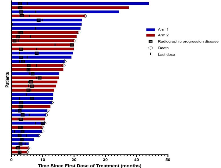
Open access
J Immunother Cancer: first published as 10.1136/jitc-2021-002931 on 10 August 2021. Downloaded from http://jitc.bmj.com/ on October 23, 2021 by guest. Protected by copyright.
randomized, placebo-controlled, phase III trial for sipu- with asymptomatic or minimally symptomatic mCRPC.
leucel-T (D9901) enrolled 127 men with asymptomatic Immune activation was evaluated using general immune
mCRPC randomly assigned at a 2:1 ratio of treatment function assays (T-cell receptor (TCR) clonality and cyto-
versus control. Median OS was improved by 4 months kine levels) and also sipuleucel- T product parameters
compared with placebo (25.9 vs 21.4 months; p=0.01).16 including CD54 upregulation, cumulative final product
Subsequent real-world studies, IMPACT study, and the CD54+ cell numbers, and cumulative total nucleated cell
PROCEED registry have confirmed and extended the counts, since these have been associated with OS.30
finding that sipuleucel-T is beneficial in chemotherapy
naïve mCRPC.8 17–19
Immune checkpoint inhibitors have demonstrated METHODS
some activity in prostate cancer, but not enough as a Study design and subjects
monotherapy to warrant approval for the treatment of This was an open-labeled randomized, dual-arm, phase
mCRPC. Ipilimumab, an immune checkpoint inhibitor Ib study assessing the safety and preliminary efficacy of
targeting CTLA-4, demonstrated activity when adminis- sequential administration of atezolizumab and sipuleu-
tered in combination with radiotherapy in patients with cel-T in subjects with asymptomatic or minimally symp-
mCRPC who had disease progression after docetaxel.20 tomatic mCRPC. Subjects were randomly assigned in a 1:1
However, in phase III trials, ipilimumab failed to prolong ratio to receive either atezolizumab followed by sipuleu-
OS in an unselected patient population. Furthermore, cel-T (Arm 1) or sipuleucel-T followed by atezolizumab
a phase II study investigating pembrolizumab mono- (Arm 2). The primary endpoint was safety and tolerability
therapy, an immune checkpoint inhibitor targeting the of the combination treatment. Preliminary efficacy was
programmed death-1 (PD-1) protein, demonstrated anti- a secondary objective. Correlative studies investigating
tumor activity in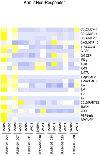
Open access
J Immunother Cancer: first published as 10.1136/jitc-2021-002931 on 10 August 2021. Downloaded from http://jitc.bmj.com/ on October 23, 2021 by guest. Protected by copyright.
by intravenous infusion on weeks 1 and 4 followed by had undergone at least one leukapheresis procedure
sipuleucel-T administered intravenously on weeks 6, 8, or atezolizumab infusion were included in the safety
and 10. Subjects in Arm 2 received induction treatment population. Adverse events and laboratory values were
as follows: sipuleucel-T administered intravenously on graded using the National Cancer Institute Common
weeks 1, 3, and 5 followed by atezolizumab 1200 mg intra- Terminology Criteria for Adverse Events, V.4.0. Immune-
venously on weeks 7 and 10. Subjects with an objective related AEs (irAEs) were defined as per previous report.33
response, defined as either a complete response (CR) or Multiple occurrences of specific events were counted
partial response (PR), or stable disease (SD) at the end once per patient; the event with the greatest severity was
of week 12 could receive atezolizumab 1200 mg intrave- summarized. Additional anticancer interventions and
nously every 3 weeks until disease progression or a loss of causes of death were collected for all subjects. The cut-off
clinical benefit occurred (maintenance phase). Subjects date for data presented here was October 15, 2020.
were categorized into one of three Halabi risk groups
(Low, Group 1; Intermediate, Group 2; High, Group 3) Immunohistochemical staining
based on the following variables: Eastern Cooperative A diagnostic antihuman PD- L1 monoclonal antibody
Oncology Group (ECOG) performance status (0, 1, or 2), (Clone 22C3, Agilent Technologies/Dako, Carpinteria,
baseline PSA, lactate dehydrogenase, alkaline phospha- California, USA) was used to detect PD-L1 expression
tase, albumin, hemoglobin, presence of bone metastases, on formalin- fixed, paraffin-
embedded tumor tissue by
and presence of lymph node metastasis.31 Once catego- immunohistochemistry (IHC), as previously described.34
rized into one of the three risk groups, subjects were strat- PD-L1 positivity was defined as a combined positive score
ified and randomized using a block design. Subjects who (CPS) of >10. The CPS was calculated by: (1) For an area
discontinued or withdrew from the induction phase of of 200 tumor cells, count the number of PD-L1 positive
the study were replaced. cells (tumor cells, lymphocytes, and macrophages) and
Samples for hematology, serum chemistries, coagu- divide by 200 and then multiply by 100 to generate a score
lation, and urinalysis were obtained prior to each infu- for this 200 tumor cell area. (2) Repeat this for a total of
sion. Furthermore, these blood-based analyses along with four 200 tumor cell areas. (3) Calculate the average of the
serum PSA were evaluated at week 12 and subsequently scores from these four areas to calculate the final CPS. A
every 12 weeks thereafter until disease progression. board certified pathologist (OTMC) reviewed all slides.
Sipuleucel-
T infusions were prepared from periph-
eral blood mononuclear cells (PBMCs) as previously Correlative immune testing
reported.8 The dose level of atezolizumab in this study Correlative testing was performed to assess immune
was 1200 mg (equivalent to an average body weight−based response associated with study drug administration.
dose of 15 mg/kg) administered by intravenous infusion Whole blood samples were collected at baseline and
over 30 (±10) min every 3 weeks (±2 days). The initial weeks 12, 16, 20, 32, 45, and 58 (Arm 1) or weeks 7, 11,
dose of atezolizumab was delivered over 60 (±15) min. 15, 27, 40, and 53 (Arm 2). Immune assays, as detailed
below, were performed at Dendreon (Seal Beach, Cali-
Response fornia, USA).
Tumor assessments were conducted at baseline, every
12 weeks thereafter, and at the end of treatment by CT T-cell responses
and bone scanning. Efficacy outcome measures included Antigen-specific memory T-cell responses were evaluated
radiographic progression-free survival (rPFS), objective by an interferon (IFN)-γ enzyme- linked immunospot
response rate (ORR), and disease control rate (DCR) by (ELISpot) assay. PVDF ELISpot plates (Millipore, Burl-
Prostate Cancer Working Group 3- modified Response ington, Massachusetts, USA) were coated with an anti-
Evaluation Criteria in Solid Tumors (RECIST) V.1.1,32 IFN-γ antibody (clone D1K, MabTech, Cincinnati, Ohio,
and OS. PSA levels were not used to determine disease USA) overnight, then plates were blocked and rinsed
progression or to trigger radiographic evaluations. At the with phosphate-buffered saline (PBS)/Tween. Cryopre-
conclusion of the study, a blinded, independent radio- served PBMCs were defrosted and rested overnight in
logical review was used to confirm the time to objective media then aliquoted at 3×10⁵ PBMC/well in a volume of
disease progression. 200 µL/well with media alone or with media containing
antigen (PA2024: PAP- GMCSF fusion protein, PAP, or
Adverse events CEFT peptide pool (control)) in triplicate. Plates were
All treatment- emergent adverse events were reported incubated for 40–48 hours then washed and incubated
until the time of objective disease progression. There- with Streptavidin conjugated anti-IFN-γ antibody (clone
after, only events that were determined by the investiga- B6-1, MabTech). After incubation, plates were rinsed with
tors to be at least possibly related to sipuleucel-T and/or PBS/Tween and incubated with biotin conjugated with
atezolizumab were reported. Monitoring for treatment- alkaline peroxidase for another hour. Afterwards, the
emergent adverse events (AEs) and survival occurred at plates were rinsed with PBS/Tween and incubated with
2 and 6 months after disease progression and at inter- BCIP (5-bromo-4-chloro-3-indolyl phosphate) to visualize
vals of 6 months or less thereafter. All subjects who IFN-γ secreting cells. ELISpot data are depicted with the
Dorff T, et al. J Immunother Cancer 2021;9:e002931. doi:10.1136/jitc-2021-002931 3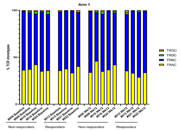
Open access
J Immunother Cancer: first published as 10.1136/jitc-2021-002931 on 10 August 2021. Downloaded from http://jitc.bmj.com/ on October 23, 2021 by guest. Protected by copyright.
median of triplicates minus background (PBMCs incu- propagated cells, followed by two rounds of PCR; the first
bated with media). amplifying the peptide-encoding DNA, and the second
adding barcodes with well-specific indices. Samples were
T-cell proliferation normalized to 4 nM, pooled, and sequenced on the Illu-
Sipuleucel-T infusions are associated with a T cell prolif- mina NextSeq500 (Illumina, San Diego, California, USA).
erative response to PA2024 and prostatic acid phospha-
tase (PAP). Thus, antigen-specific T-cell proliferation to Protein-based immunome-wide association study (PIWAS)
PA2024, PAP, and phytohaemagglutinin (PHA, control) Using the prostate cancer samples as cases and samples
were tested via a tritiated thymidine incorporation assay from 2514 healthy individuals as controls, we ran a
deploying 96- well plates. Cryopreserved PBMCs were PIWAS analysis against a modified human proteome.36
defrosted and rested overnight in media then plated at PIWAS was parameterized to have a window size of 5, the
1×10⁵ PBMC/well in a total volume of 200 µL/well with number of SD approach, and the maximum peak signal.
either media alone or with media containing antigen A PIWAS value was calculated per sample per antigen and
(PA2024: PAP-GMCSF fusion protein, PAP, or PHA) in the outlier sum false discovery rate as defined previously
triplicate. Plates were incubated for a total of 5 days, then was used to prioritize antigens.36 The reference human
the wells were pulsed with 0.5 µCi of 3H-thymidine over- proteome was downloaded from Uniprot on March 20,
night. The amount of 3H-thymidine incorporated into 2020. A modified human proteome was then assembled
the cell was quantified by a γ-radiation counter with the by masking regions on the reference human proteome
degree of proliferation (ie, stimulation index (SI)) being that may reflect atezolizumab binding signals due to high
defined as the amount of 3H-thymidine incorporation sequence similarity.
divided by 3H-thymidine incorporation with media alone.
Serial sample PIWAS analysis
Humoral response For a subject with multiple samples at different time
Antibody responses against PAP and PA2024 were assessed points, serial sample PIWAS analysis was applied to iden-
by ELISA. First, 96-well plates were coated overnight with tify antigens with significant signal increase. A PIWAS
either PAP, PA2024, or Tetanus (control). Subsequently, value at each time point was calculated per subject. A
plates were blocked with PBS/casein and rinsed with Z-score was then calculated based on the difference
PBS/Tween. Serially diluted serum was then aliquoted between two PIWAS values when compared with a refer-
in duplicate to each set of plates and incubated at room ence null distribution indicating no biological variability.
temperature for 2 hours followed by rinsing the plates Using these Z-scores, antigens with significantly increased
with PBS/Tween and incubated with a mixture of anti-IgG PIWAS values were identified with Bonferroni adjusted
and anti- IgM antibodies (Jackson Immunoresearch, p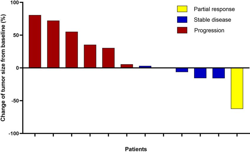
Open access
J Immunother Cancer: first published as 10.1136/jitc-2021-002931 on 10 August 2021. Downloaded from http://jitc.bmj.com/ on October 23, 2021 by guest. Protected by copyright.
TNF-α, VEGF, FGF-basic, IL1α/IL-1F1) using a custom- 75.0 years (range, 53–86 years), 73.0% of subjects had an
ized Luminex assay (Cat # FCSTM18-22 R&D Systems, ECOG performance status 0, 18.9% had received prior
Minneapolis, MN). Measurements were performed using docetaxel-based chemotherapy, and 62.2% had received
a Luminex 200 instrument (Luminex, Austin, Texas, two or more previous antiandrogen therapies. Median
USA) and were analyzed using a standard curve for each PSA level for the total population was 21.9 ng/mL at study
molecule (xPONENT software, Luminex). entry
Peripheral blood mononuclear cell analysis Safety
PBMCs were stained and analyzed by flow cytometry as
At least one treatment- related AE was reported in 31
previously described.38 The following antibodies were
subjects (83.8%), including 7 (18.9%) with at least
used for cell surface staining: FITC- conjugated anti-
one grade 3 treatment-related AE. The most common
human CD45 (Biolegend, Catalog #368508), PerCP-
treatment- related AEs were diarrhea, nausea, fatigue,
Cyanine5.5- conjugated antihuman CD3 (Tonbo
and hypertension in Arm 1 and fatigue, pain, joint pain,
Biosciences, Catalog #65–0037), PE- conjugated anti-
platelet decrease, constipation, and anemia in Arm 2
human CD4 (Tonbo, Catalog #50–0048), APC-conjugated
antihuman CD8 (Tonbo, Catalog # 20–0087), Bril- (table 2A). irAEs, which were based on a list of common
liant Violet 421- conjugated antihuman CD279 (PD-1, terms, occurred in 5 (13.5%) subjects with similar inci-
Biolegend, Catalog #329920). Ghost Dye UV 450 (Tonbo) dences between the two arms and all of which were grade
was used to assess live versus dead status of cells. Samples 1 or 2 (table 2B). There were no grade 4 or five toxici-
were acquired on a LSRII analyzer (BD Biosciences) and ties attributed to either study drug and no irAE required
analyzed with FlowJo software (Treestar). For all FACS, systemic steroid therapy.
experiments debris and dead cells were excluded from
the analyzed gates. Antitumor activity
In total, 23 subjects were evaluable for response (table 3).
Statistical analysis No patient in either arm had a CR and only one patient
This study was designed to obtain preliminary safety and in Arm 2 had a PR. Three subjects (30%) in Arm 1 and
clinical activity information in subjects with asymptom- one patient (7.7%) in Arm 2 had SD. Thus, for the
atic or mildly symptomatic mCRPC. Analyses were based overall population the ORR was 4.3% and the DCR was
on all subjects who received any amount of study treat- 21.7%. For the individual arms, the DCR was 30% for
ment (safety evaluable population). The ORR and DCR Arm 1 and 15.4% for Arm 2. These subjects are subse-
with corresponding 95% CIs were calculated using the quently noted as responders. Among the 12 subjects
Clopper-Pearson method. The rPFS and OS were assessed who had at least one measurable target lesion by CT at
using the Kaplan-Meier method, with 95% CIs for median baseline, 4 (33.3%) had a decrease from baseline in the
rPFS and OS estimated using the Brookmeyer-Crowley sum of target lesions, including 1 (8.3%) with a PR and
method. While this study was not powered to detect a
2 (16.7%) subjects with SD (figure 1). Among the five
survival difference between the two treatment arms,
subjects with radiographic response, one remained on
there was a protocol-specified requirement to follow each
treatment at data cut-off, while the remaining three had
patient for survival (and treatment-related AEs) for up to
experienced subsequent disease progression (figure 2A).
12 months after randomization.
Overall, the median rPFS was 3.0 months (95% CI 2.8 to
5.6 months). Median rPFS was 3.3 months (95% CI 2.7
to 7.8 months) for Arm 1 and 2.9 months (95% CI 2.6
RESULTS
to 5.5 months) for Arm 2 (figure 2B). Median OS was
Subjects
not reached (NR; 95% CI 11.1 to NR) in Arm 1 and 21.4
From January 15, 2017 through October 30, 2019, 37
months (95% CI 16.6 to NR) in Arm 2 (figure 2C). The
subjects were enrolled in this study (n=19 Arm 1, n=18 Arm
2). All 37 subjects were included in the safety population estimated 12-month survival rates were 70.6% in Arm 1
and thus analyzed for both safety and efficacy (online and 83.3% in Arm 2. For both arms, median OS was 23.6
supplemental figure 1). Notably, three subjects did not months (95% CI 16.6 to NR) and the estimated 12-month
complete induction treatment. These three subjects were survival rate was 77.2%. A total of 15 subjects (40.5%) out
replaced (two in Arm 1 and one in Arm 2); otherwise, of 37 had some decrease in PSA level, including 1 patient
all subjects received three doses of sipuleucel- T and (5.3%) in Arm 1 and 3 subjects (16.7%) in Arm 2 who
two doses of atezolizumab. The most common reason had a >50% decrease (table 3 and online supplemental
for treatment discontinuation was disease progression. figure 2). PD-L1 expression in patient samples is shown
Median duration of treatment was 4.9 months (range, in online supplemental figure 3). Only one subject, who
0–9.9 months) for Arm 1 and 5.1 months (range, 1.4–13.7 was classified as a non-responder in Arm 2, had CPS >10.
months) for Arm 2. Baseline characteristics were gener- Furthermore, non-responders in both arms had numer-
ally as expected for a sipuleucel-T eligible mCRPC popu- ically higher levels of PD-L1 expression compared with
lation (table 1). Median age for the total population was responders.
Dorff T, et al. J Immunother Cancer 2021;9:e002931. doi:10.1136/jitc-2021-002931 5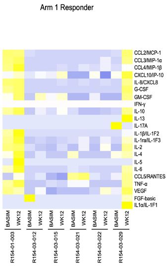
Open access
J Immunother Cancer: first published as 10.1136/jitc-2021-002931 on 10 August 2021. Downloaded from http://jitc.bmj.com/ on October 23, 2021 by guest. Protected by copyright.
Table 1 Patient characteristics
Total (n=37) Arm 1 (n=20) Arm 2 (n=17)
Parameters No. % No. % No. % p-value
Age, years 0.67
Median 75 75 74
Range 53–86 53–86 55–84
Race: white 4 10.80 2 10.00 2 11.80 0.94
Disease location 0.55
Bone only 19 51.40 9 45 10 58.80
Soft tissue only 5 13.50 2 10 3 17.60
Bone and soft tissue 11 29.70 7 35 4 23.50
Visceral 2 5.40 2 10 0
Number of bone mets 0.51
0 7 18.90 3 15 4 23.50
1-10 17 45.90 11 55 6 35.30
>10 13 35.10 6 30 7 41.20
ECOG performance status 0.57
0 27 73 14 70 13 76.50
1 9 24.3 6 30 3 17.60
2 1 2.70 0 1 5.90
Median PSA, ng/mL 21.9 20.2 26.3 0.71
Range 0.33–636.8 2.1–636.8 0.33–529
Median alkaline phosphatase, 76 68.5 92 0.28
U/L 41–741 41–321 46–741
Range
Median hemoglobin, g/dL 13 12.9 13 0.98
Range 9.8–15.3 10.2–15.3 9.8–14.9
Median LDH, U/L 181 180 181 0.32
Range 114–677 114–677 130–229
Gleason Score 1
≤7 17 45.90 9 45 8 47.10
≥8 15 40.50 8 40 7 41.20
Unknown 5 13.50 3 15 2 11.80
Patients with prior 7 18.90 3 15 4 23.50 0.68
chemotherapy for mCRPC
Patients receiving docetaxel- 11 29.70 6 30 5 29.40 1
based chemotherapy
subsequent to study treatment
Patients with two or more 23 62.20 14 70 9 52.90 0.33
previous anti-androgen
therapies
Immune activity TRBC population tended to increase after the treatment
First, we performed TCR repertoire analysis using whole in Arm 1, but not in Arm 2. In addition, we found that
blood samples from 16 subjects (n=9 Arm 1 and n=7 Arm TCR diversity was relatively higher in the one responder
2) to assess the functional phenotype associated with each in Arm 2 compared with non-responders in both arms
treatment. TCR clonotype analysis revealed that the main (figure 3B).
population consists of (T Cell Receptor Alpha Constant) Next, using a panel of T- cell related cytokines, we
TRAC and (T Cell Receptor Beta Constant) TRBC compared cytokine expression in serum pretreatment
subtypes (figure 3A, online supplemental figure 4). Inter- and post-treatment using specimens from 22 subjects (11
estingly, the TRAC population tended to decrease and in each arm). Overall, the heatmap shows that cytokine
6 Dorff T, et al. J Immunother Cancer 2021;9:e002931. doi:10.1136/jitc-2021-002931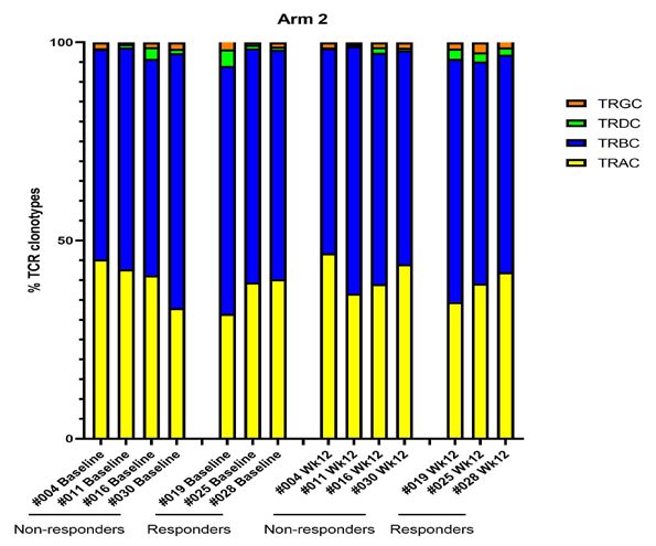
Open access
J Immunother Cancer: first published as 10.1136/jitc-2021-002931 on 10 August 2021. Downloaded from http://jitc.bmj.com/ on October 23, 2021 by guest. Protected by copyright.
Table 2 (A) Summary of Treatment related AEs occurring in ≥ 5% of Patients in Either Treatment, (B) Immune-related adverse
events (irAEs)
(A)
Arm 1 Arm 1 Arm 2 Arm 2
Total % Grade1-2 % Grade3-4 % Grade1-2 % Grade3-4 %
Any AEs 31 83.8 13 68.4 3 15.8 11 61.1 4 22.2
Fatigue 9 24.3 3 15.8 0 0 6 33.3 0 0
Diarrhea 6 16.2 4 21.1 0 0 2 11.1 0 0
Hypertension 5 13.5 3 15.8 0 0 1 5.6 1 5.6
Joint Pain 5 13.5 2 10.5 0 0 3 16.7 0 0
Nausea 5 13.5 4 21.1 0 0 1 5.6 0 0
Platelet count 5 13.5 2 10.5 0 0 3 16.7 0 0
decreased
Pain 5 13.5 0 0 0 0 4 22.2 1 5.6
Anemia 4 10.8 1 5.3 0 0 2 11.1 1 5.6
Chills 4 10.8 2 10.5 0 0 2 11.1 0 0
Constipation 4 10.8 1 5.3 0 0 3 16.7 0 0
Fever 3 8.1 1 5.3 0 0 2 11.1 0 0
Hypoglycemia 3 8.1 1 5.3 0 0 2 11.1 0 0
Increased Alkaline 3 8.1 2 10.5 0 0 1 5.6 0 0
Phosphatase
Shortness of breath 3 8.1 1 5.3 0 0 2 11.1 0 0
Chest pain 2 5.4 0 0 0 0 2 11.1 0 0
Dizziness 2 5.4 1 5.3 0 0 1 5.6 0 0
Flu like symptoms 2 5.4 0 0 0 0 2 11.1 0 0
Headache 2 5.4 0 0 0 0 2 11.1 0 0
Hyperglycemia 2 5.4 1 5.3 0 0 1 5.6 0 0
Hypermagnesemia 2 5.4 1 5.3 0 0 1 5.6 0 0
Hypernatremia 2 5.4 1 5.3 0 0 1 5.6 0 0
Hypotension 2 5.4 0 0 1 5.3 0 0 1 5.6
Infusion related 2 5.4 1 5.3 0 0 1 5.6 0 0
reaction
(B)
All % Arm 1 % Arm 1 % Arm 2 % Arm 2 %
Grade1-2 Grade3-4 Grade1-2 Grade3-4
Any 5 13.5 2 10.5 0 0 3 16.7 0 0
Dermatologic 3 8.1 2 10.5 0 0 1 5.6 0 0
Endocrine 1 2.7 0 0 0 0 1 5.6 0 0
Hepatitis 1 2.7 0 0 0 0 1 5.6 0 0
Infusion related 2 5.4 1 5.3 0 0 1 5.6 0 0
reaction
levels in the post- treatment responder is higher than significantly increased compared with baseline at each
those in baseline (figure 3C and online supplemental postbaseline visit (Arm 1: baseline vs week 12; p=0.022 and
figure 5) compared with non-responder changes from Arm 2: baseline vs week 11; p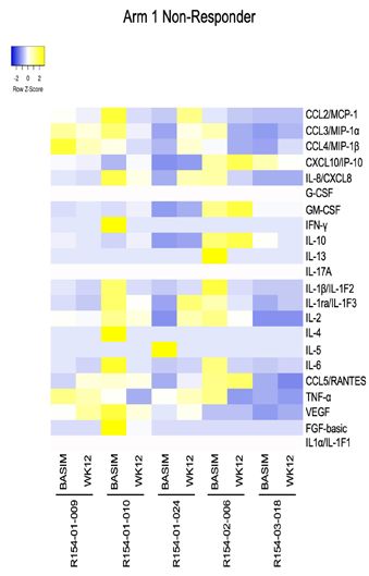
Open access
J Immunother Cancer: first published as 10.1136/jitc-2021-002931 on 10 August 2021. Downloaded from http://jitc.bmj.com/ on October 23, 2021 by guest. Protected by copyright.
Table 3 Summary of responses
Variable All Arm-1 Arm-2 p-value
Response assessed per RECIST criteria 23 10 13 0.38
No. of patients who can be assessed by RECIST
PR 1 (4.3%) 0 1 (7.7%)
SD 4 (17.4%) 3 (30%) 1 (7.7%)
Non-CR/Non-PD 5 (21.7%) 1 (10%) 4 (30.8%)
PD 13 (56.5%) 6 (60%) 7 (53.8%)
ORR, No (%) 1 (4.3%) 0 1 (7.7%) 1
DCR,* No (%) 5 (21.7%) 3 (30%) 2 (15.4%) 0.62
Response assessed per PCWG3 27 16 11 1
No. of patients who can be assessed by RECIST
PR 0 0 0
SD 10 (37%) 6 (37.5%) 4 (36.4%)
PD 17 (63%) 10 (62.5%) 7 (63.6%)
ORR, No (%) 0 0 0 1
DCR, No (%) 10 (37%) 6 (37.5%) 4 (36.4%) 1
rPFS 37 20 17 0.27
No. of patients who can be assessed 3.0 months 3.3 months 2.9 months
median (95% CI) (2.8 to 5.6) (2.6 to 7.8) (2.6 to 5.6)
PSA response † in patients with baseline 30 20 17
PSA measurement
No. of patients who can be assessed
PSA decline 50% † 4 (10.8%) 1 (5%) 3 (17.6%) 0.32
*Define as the percentage of patients with confirmed complete or partial response or stable disease. Patients who died without evidence of
disease progression before death were considered to have stable disease.
†Define as the percentage of patients with a reduction in PSA level from baseline by 50% or greater as confirmed on an additional PSA
evaluation performed > 3 weeks later.
CR, complete response; DCR, disease control rate; NE, not evaluable; ORR, overall response rate; PCWG3, Prostate Cancer Working Group
3; PD, progressive disease; PR, partial response; PSA, prostate specific antigen; rPFS, radiographic progression free survival.
significantly higher compared with baseline in both arms
(Arm 1; p=0.034; figure 3D, baseline vs week 12; p=0.0097)
(Arm 2; p=0.019; figure 3D, baseline vs week 11; p=0.011,
baseline vs week 11; p=0.049, and baseline vs week 15;
p=0.014). All subjects developed PA2024-specific T-cell
responses after sipuleucel-T treatment. PA2024 antibody
titers after sipuleucel-T treatment in Arm 1 and Arm 2
were 16.8 times (p=0.00066) and 12.8 times (p=0.00025)
higher, respectively, on average compared with base-
line, and similar between arms, remaining significantly
elevated through week 32 (figure 3 and online supple-
mental table 1).
Exploratory analysis of PA2024 T- cell stimulation
index was performed in subjects for whom cells could be
Figure 1 Waterfall plot of antitumor activity. Percentage
processed within 24 hours of collection, thus precluding
change from baseline in the sum of longest diameter of the need to freeze the cells before analysis. The median
target lesions as assessed by RECIST V.1.1 by central ratio of the T-cell stimulation index at 12 weeks in Arm 1
review. Thirteen subjects assigned to Arm 1 and 12 subjects and 11 weeks in Arm 2 was 32.9 times (p=0.00033) and
assigned to Arm 2 were not evaluable for change from 24.0 times (p=0.0014) higher, respectively, on average
baseline in tumor size because they did not have one or more compared with baseline (preinfusion). Increased IgG
evaluable postbaseline imaging assessments or did not have levels to secondary antigens such as PSA, PAPi, and PAPm
any target lesions. RECIST, Response Evaluation Criteria in were observed in both arms at all time points through
Solid Tumors.
8 Dorff T, et al. J Immunother Cancer 2021;9:e002931. doi:10.1136/jitc-2021-002931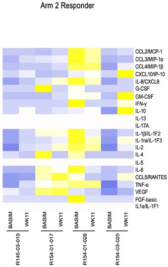
Open access
J Immunother Cancer: first published as 10.1136/jitc-2021-002931 on 10 August 2021. Downloaded from http://jitc.bmj.com/ on October 23, 2021 by guest. Protected by copyright.
Figure 2
A
B C
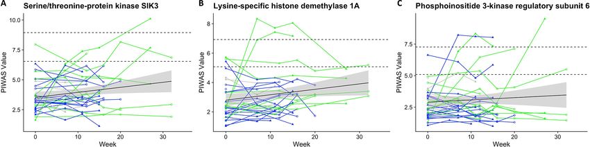
Figure 2 Antitumor activity. (A) Swimmer plot showing subjects with confirmed response as assessed by PCWG3-modified
RECIST. (B) Kaplan-Meier plot of rPFS by treatment arm. (C) Kaplan-Meier plot of OS by treatment arm. OS, overall survival;
PCWG3, Prostate Cancer Working Group 3; RECIST, Response Evaluation Criteria in Solid Tumors; rPFS, radiographic
progression-free survival.
week 32 (online supplemental table 1); however, there observed either at baseline or after therapy between
was no significant difference between responders and responders and non- responders (online supplemental
non-responders in either arm. figure B,D). PIWAS identified 92 outlier antigens that
In addition, to assess the T- cell population, PBMCs were significantly different in responders versus. non-
were collected and analyzed by flow cytometry for CD45+ responders at baseline (online supplemental table
(general leucocyte marker) and CD4+ and CD8+ T cells 2).39–45 Serial PIWAS analysis also identified 40 antigens
expression. The percentage of total T cells (Arm 1 29.4% that increased between baseline and week 7 from Arm
vs Arm 2 30.3%, p=0.86), CD4+ T cells (Arm 1 13.2% vs 2 (online supplemental table 3) and 18 antigens that
Arm 2 12.5%, p=0.85), and CD8+ T cells (Arm 1 14.4% vs increased in responders versus non-responders in either
Arm 2 14.8%, p=0.92) were similar in both arms (repre- Arm 1 or Arm 2 (online supplemental table 4). Arms were
sentative results are shown in figure 3E). Interestingly, combined for this analysis given the limited numbers of
the percentage of total T cells was significantly higher in responders. The trajectories of response to three putative
responders versus non-responders in Arm 1 (p=0.044) antigens, SIK3, KDM1A/LSD1, and PIK3R6, that were
but not in Arm 2 (p=0.51). However, CD4+ or CD8+ shared in at least two subjects in response to sipuleucel-T
T cells were numerically but not statistically higher in at (week 7, Arm 2) and also in response to combined
responders compared with non-responders in both arms therapy (week 12, either Arm 1 and Arm 2) are shown in
(CD4+ T cell; p=0.72 for Arm 1 and p=0.92 for Arm 2, figure 4.
CD8+ T cell; p=0.15 for Arm 1 and p=0.46 for Arm 2).
The SERA assay was performed on samples from 37
subjects to assess autoantigen signal in subjects pretreat- DISCUSSION
ment and post-treatment in both arms. SERA uses a large This is the first report of the safety, clinical activity, and
random bacterial peptide display library with the PIWAS immune activity associated with combination immuno-
method to assess outlier antigens in samples. At baseline, therapy using sipuleucel-T and atezolizumab in subjects
a sum- of-
IWAS calculation identifies a non- significant with mCRPC. Sipuleucel-T and atezolizumab each target
difference in the baseline number of outlier antigens in the immune system by two distinct mechanisms and
subjects with prostate cancer relative to a large cohort each, independently, has modest activity in mCRPC. The
of healthy controls that becomes significant over time rationale for this trial is predicated on the hope that by
(online supplemental figure A,C). No differences were combining the different mechanisms of action of these
Dorff T, et al. J Immunother Cancer 2021;9:e002931. doi:10.1136/jitc-2021-002931 9Open access
J Immunother Cancer: first published as 10.1136/jitc-2021-002931 on 10 August 2021. Downloaded from http://jitc.bmj.com/ on October 23, 2021 by guest. Protected by copyright.
Figure 3
A
B p=0.12 p=0.31 p=0.52 p=0.45
C
D PA2024 PA2024
PA2024 PA2024
PA2024 PA2024 PA2024
PA2024
PA2024
PA2024
PA2024 PA2024
E Arm 1 Non-responder Arm 2 Non-responder
Arm 2 Responder
Arm 1 Responder
Figure 3 On study immunological landscape. (A) TCR clonotype analysis. (B) TCR DI. (C) Heatmap illustrating differences in
22 serum cytokines. The scale represents SD from the mean after a Z-transformation of signal values of a cytokine across all
samples. Yellow represents a higher level of cytokine expression and blue a lower level, relative to the mean across all samples
for each cytokine. (D) ELISpot analysis of PA2024. (E) Flow cytometry analysis of T cell population. Each central bar in the box
indicates median, and the two whisker boundaries indicate the 5th and 95th percentiles. DI, diversity index; ELISpot, enzyme-
linked immunospot; TCR, T-cell receptor.
10 Dorff T, et al. J Immunother Cancer 2021;9:e002931. doi:10.1136/jitc-2021-002931Open access
J Immunother Cancer: first published as 10.1136/jitc-2021-002931 on 10 August 2021. Downloaded from http://jitc.bmj.com/ on October 23, 2021 by guest. Protected by copyright.
Figure 4 Candidate shared antigens among 6-month responders. (A) SIK3, (B) KDM1A/LSD1, and (C) PIK3R6 are candidate
shared antigens with significantly increased signals from baseline to week 12 in at least two 6-month responders. These
antigens also have increased signals from baseline to week 7 in Arm 2 subjects treated with Sipuleucel-T prior to Atezolizumab
treatment. Horizontal dashed lines indicate 95th and 99th percentile of control PIWAS values. Green dots: responders, n=10;
Blue dots: non-responder, n=19; gray dots: missing response information, n=5. Circle: a sample is from Arm 1; triangle: a
sample is from Arm 2. Black line is trend line by linear regression using PIWAS values of all samples, and gray area indicates the
95% CI of the trend line. PIWAS, protein-based immunome-wide association study.
two agents would lead to increased anticancer activity TCR diversity. Interestingly, the patient with the highest
without increased toxicity. Overall, the combination of TCR diversity in Arm 2 (patient #028) had radiological
sipuleucel-T and atezolizumab was well-tolerated in this SD at 15 months and a dramatic reduction in PSA levels
study, regardless of which was administered first. There (96.8% reduction). Note that serum PSA at diagnosis was
were no grade 4 or 5 AEs observed in this study. The most 236 ng/dL, serum PSA at study entry was 12.3 ng/dL, and
reported AE, fatigue, was observed in 24.3% of subjects bony metastatic disease was noted in spine and pelvic
overall, which was similar to a previous study using atezoli- girdle.
zumab as monotherapy for advanced solid tumors.46 Furthermore, we observed a difference in autoan-
There were no unexpected AEs and limited irAEs, and the tigen signal at baseline between subjects with cancer and
safety profile of the present study was consistent with what healthy controls that increased over time. The difference
has been published with each agent as monotherapy.8 16 is most notable in non-responders and is likely due to
During a time when there are many other treatment an increase in tumor burden and tumor antigens as has
options, a median OS of 23.6 months was noted, which is been previously described for known tumor antigens.50 A
similar to the 25.8 months previously reported by Kantoff limited number of autoantigens that are shared between
et al for sipuleucel-T monotherapy.8 Of note, in the study at least two subjects increased in response to sipuleu-
by Kantoff et al, nearly three-quarters of the subjects had cel-T, and a limited number of these are also increased
a Gleason score of 7 or less, while in our study approxi- at 6 months in responders versus non-responders. Inter-
mately only 50% had Gleason score of 7 or less. Although estingly, all three antigens are members of cell-signaling
the observed ORR was a modest 4.3%, it is similar to that kinase pathways associated with tumorigenesis,51–53 and it
seen with immune checkpoint inhibitor monotherapy could be postulated that an increase in humoral response
in mCRPC.19 Notably, the responses that did occur were against these antigens might lead to decreased oncogenic
rather durable, and eight subjects (21.6%) had SD lasting signaling. Exploration of the significance and prevalence
more than 6 months. These data add to a growing body of humoral signaling against these antigens should thus
of evidence suggesting that despite its more immunosup- be considered with future studies.
pressive microenvironment, certain patients with mCRPC In summary, this study suggests that the combination of
may benefit from immunotherapy. Although the combi- sipuleucel-T and atezolizumab is safe and well tolerated
nation studied did not demonstrate clear superiority over with a comparable safety profile to each agent adminis-
monotherapy, immune activation appeared to be greater tered as monotherapy. Furthermore, correlative immune
in Arm 1. studies suggest that the combination may be beneficial;
Identification of predictive biomarkers for cancer nevertheless, objective responses were rare. Better predic-
immunotherapy has been a significant challenge. Our tive biomarkers to select patients who may respond and
exploratory biomarker research assessed changes in additional combination strategies are needed for immu-
cytokines, neoantigens, TCRs, and PBMCs. Exploratory notherapy to make a greater impact on mCRPC.
biomarker analysis hinted at a relationship between
response and a higher TCR diversity, and an increased Author affiliations
1
putative immune response to three antigens, SIK3, Department of Medical Oncology and Therapeutics Research, City of Hope National
KDM1A/LSD1, and PIK3R6. Furthermore, outcomes Medical Center, Duarte, California, USA
2
Samuel Oschin Comprehensive Cancer Institute, Cedars-Sinai Medical Center, Los
were not dependent on PD- L1 status (online supple-
Angeles, California, USA
mental figure 5). TCR diversity has been associated with 3
University of Hawaii Cancer Center, Honolulu, Hawaii, USA
clinical benefit in immune therapy for cancer.47–49 In the 4
Serimmune, Goleta, California, USA
present study, we can also confirm the trend of increased 5
Prostate Oncology Specialists, Marina del Rey, California, USA
Dorff T, et al. J Immunother Cancer 2021;9:e002931. doi:10.1136/jitc-2021-002931 11Open access
J Immunother Cancer: first published as 10.1136/jitc-2021-002931 on 10 August 2021. Downloaded from http://jitc.bmj.com/ on October 23, 2021 by guest. Protected by copyright.
Acknowledgements This work was funded by Dendreon Corp and Genentech Inc. 11 Smith MR, Saad F, Chowdhury S, et al. Apalutamide treatment
We thank the CSMC Flow cytometry core (A Lopez and Dr J Suda) for their support. and metastasis-free survival in prostate cancer. N Engl J Med
2018;378:1408–18.
Contributors Conception and design: CR. Provision of study materials or patients: 12 Fizazi K, Shore N, Tammela TL, et al. Darolutamide in
TD, SP, DT, JA, MS. Collection and assembly of data: CR, TD, SP, DT, JA, OTMC, MS. nonmetastatic, castration-resistant prostate cancer. N Engl J Med
Data analysis and interpretation: CR, YH, HF, JH, OTMC, IP, MZ, RW, AD, WH, JS. 2019;380:1235–46.
Manuscript writing: CR, TD, JH. Final approval of manuscript: CR, TD, SP, DT, JA, MS, 13 Parker C, Nilsson S, Heinrich D, et al. Alpha emitter radium-223
YH, HF, JH, OTMC, IP, MZ, RW, AD, WH, JS. and survival in metastatic prostate cancer. N Engl J Med
2013;369:213–23.
Funding This study was funded by the Dendreon Corporation and Genentech. 14 de Bono J, Mateo J, Fizazi K, et al. Olaparib for metastatic
Competing interests MZ, RW, AD, WH and JS are employees of Serimmune. CR castration-resistant prostate cancer. N Engl J Med
2020;382:2091–102.
received funding from Dendreon for the conduct of this study. 15 Abida W, Patnaik A, Campbell D, et al. Rucaparib in Men With
Patient consent for publication Not required. Metastatic Castration-Resistant Prostate Cancer Harboring a BRCA1
or BRCA2 Gene Alteration. JCO 2020;38:3763–72.
Ethics approval This study was approved by Western IRB (#20152124). 16 Small EJ, Schellhammer PF, Higano CS, et al. Placebo-controlled
Provenance and peer review Not commissioned; externally peer reviewed. phase III trial of immunologic therapy with sipuleucel-T (APC8015) in
patients with metastatic, asymptomatic hormone refractory prostate
Data availability statement Data are available on reasonable request. Additional cancer. J Clin Oncol 2006;24:3089–94.
data are available on reasonable request. 17 Higano CS, Armstrong AJ, Sartor AO, et al. Real-World outcomes
of sipuleucel-T treatment in proceed, a prospective registry of
Supplemental material This content has been supplied by the author(s). It has men with metastatic castration-resistant prostate cancer. Cancer
not been vetted by BMJ Publishing Group Limited (BMJ) and may not have been 2019;125:4172–80.
peer-reviewed. Any opinions or recommendations discussed are solely those 18 Sartor O, Armstrong AJ, Ahaghotu C, et al. Survival of African-
of the author(s) and are not endorsed by BMJ. BMJ disclaims all liability and American and Caucasian men after sipuleucel-T immunotherapy:
responsibility arising from any reliance placed on the content. Where the content outcomes from the proceed registry. Prostate Cancer Prostatic Dis
includes any translated material, BMJ does not warrant the accuracy and reliability 2020;23:517–26.
of the translations (including but not limited to local regulations, clinical guidelines, 19 McKay RR, Hafron JM, Ferro C, et al. A retrospective observational
analysis of overall survival with Sipuleucel-T in Medicare beneficiaries
terminology, drug names and drug dosages), and is not responsible for any error
treated for advanced prostate cancer. Adv Ther 2020;37:4910–29.
and/or omissions arising from translation and adaptation or otherwise. 20 Fizazi K, Drake CG, Beer TM, et al. Final analysis of the ipilimumab
Open access This is an open access article distributed in accordance with the versus placebo following radiotherapy phase III trial in Postdocetaxel
Creative Commons Attribution Non Commercial (CC BY-NC 4.0) license, which metastatic castration-resistant prostate cancer identifies an excess
of long-term survivors. Eur Urol 2020;78:822–30.
permits others to distribute, remix, adapt, build upon this work non-commercially,
21 Antonarakis ES, Piulats JM, Gross-Goupil M, et al. Pembrolizumab
and license their derivative works on different terms, provided the original work is for treatment-refractory metastatic castration-resistant prostate
properly cited, appropriate credit is given, any changes made indicated, and the use cancer: multicohort, open-label phase II KEYNOTE-199 study. J Clin
is non-commercial. See http://c reativecommons.org/licenses/by-nc/4.0 /. Oncol 2020;38:395–405.
22 Sweeney CJ, Gillessen S, Rathkopf D. IMbassador250: a phase III
ORCID iDs trial comparing atezolizumab with enzalutamide vs enzalutamide
Tanya Dorff http://orcid.org/0000-0001-5990-298X alone in patients with metastatic castration-resistant prostate cancer
Sumanta Pal http://orcid.org/0000-0002-1 712-0848 (mCRPC). Cancer Res 2020;80:CT014.
Hideki Furuya http://orcid.org/0000-0002-9536-8662 23 Kaur HB, Lu J, Guedes LB, et al. TP53 missense mutation is
Charles Rosser http://orcid.org/0 000-0002-6052-4223 associated with increased tumor-infiltrating T cells in primary
prostate cancer. Hum Pathol 2019;87:95–102.
24 Vidotto T, Saggioro FP, Jamaspishvili T, et al. PTEN-deficient
prostate cancer is associated with an immunosuppressive tumor
microenvironment mediated by increased expression of IDO1 and
REFERENCES infiltrating Foxp3+ T regulatory cells. Prostate 2019;79:969–79.
1 Siegel RL, Miller KD, Jemal A. Cancer statistics, 2020. CA A Cancer 25 Rizvi NA, Hellmann MD, Snyder A, et al. Cancer immunology.
J Clin 2020;70:7–30. mutational landscape determines sensitivity to PD-1 blockade in
2 Sun S, Sprenger CCT, Vessella RL, et al. Castration resistance non-small cell lung cancer. Science 2015;348:124–8.
in human prostate cancer is conferred by a frequently occurring 26 Snyder A, Makarov V, Merghoub T, et al. Genetic basis for clinical
androgen receptor splice variant. J Clin Invest 2010;120:2715–30. response to CTLA-4 blockade in melanoma. N Engl J Med
3 Gandaglia G, Karakiewicz PI, Briganti A, et al. Impact of the site of 2014;371:2189–99.
metastases on survival in patients with metastatic prostate cancer. 27 Taube JM, Klein A, Brahmer JR, et al. Association of PD-1, PD-1
Eur Urol 2015;68:325–34. ligands, and other features of the tumor immune microenvironment
4 Attar RM, Takimoto CH, Gottardis MM. Castration-resistant prostate with response to anti-PD-1 therapy. Clin Cancer Res
cancer: locking up the molecular escape routes. Clin Cancer Res 2014;20:5064–74.
2009;15:3251–5. 28 Lawrence MS, Stojanov P, Mermel CH, et al. Discovery and
5 Watson PA, Chen YF, Balbas MD, et al. Constitutively active saturation analysis of cancer genes across 21 tumour types. Nature
androgen receptor splice variants expressed in castration-resistant 2014;505:495–501.
prostate cancer require full-length androgen receptor. Proc Natl Acad 29 Fong L, Carroll P, Weinberg V, et al. Activated lymphocyte recruitment
Sci U S A 2010;107:16759–65. into the tumor microenvironment following preoperative sipuleucel-T
6 Nuhn P, De Bono JS, Fizazi K, et al. Update on systemic prostate for localized prostate cancer. J Natl Cancer Inst 2014;106:dju268.
cancer therapies: management of metastatic castration-resistant 30 Sheikh NA, Petrylak D, Kantoff PW, et al. Sipuleucel-T immune
prostate cancer in the era of precision oncology. Eur Urol parameters correlate with survival: an analysis of the randomized
2019;75:88–99. phase 3 clinical trials in men with castration-resistant prostate
7 de Bono JS, Oudard S, Ozguroglu M, et al. Prednisone plus cancer. Cancer Immunol Immunother 2013;62:137–47.
cabazitaxel or mitoxantrone for metastatic castration-resistant 31 Halabi S, Lin C-Y, Kelly WK, et al. Updated prognostic model for
prostate cancer progressing after docetaxel treatment: a randomised predicting overall survival in first-line chemotherapy for patients
open-label trial. Lancet 2010;376:1147–54. with metastatic castration-resistant prostate cancer. J Clin Oncol
8 Kantoff PW, Higano CS, Shore ND, et al. Sipuleucel-T 2014;32:671–7.
immunotherapy for castration-resistant prostate cancer. N Engl J 32 Scher HI, Morris MJ, Stadler WM, et al. Trial design and objectives
Med 2010;363:411–22. for castration-resistant prostate cancer: updated recommendations
9 Ryan CJ, Smith MR, de Bono JS, et al. Abiraterone in metastatic from the prostate cancer clinical trials Working group 3. J Clin Oncol
prostate cancer without previous chemotherapy. N Engl J Med 2016;34:1402–18.
2013b;368:138–48. 33 Brahmer JR, Lacchetti C, Schneider BJ. Thompson, and in
10 Scher HI, Fizazi K, Saad F, et al. Increased survival with collaboration with the National comprehensive cancer network.
enzalutamide in prostate cancer after chemotherapy. N Engl J Med management of immune-related adverse events in patients treated
2012;367:1187–97. with immune checkpoint inhibitor therapy: American Society
12 Dorff T, et al. J Immunother Cancer 2021;9:e002931. doi:10.1136/jitc-2021-002931Open access
J Immunother Cancer: first published as 10.1136/jitc-2021-002931 on 10 August 2021. Downloaded from http://jitc.bmj.com/ on October 23, 2021 by guest. Protected by copyright.
of clinical oncology clinical practice guideline. J Clin Oncol 44 Zhang Q, Bastard P, Liu Z, et al. Inborn errors of type I IFN
2018;36:1714–68. immunity in patients with life-threatening COVID-19. Science
34 Vennapusa B, Baker B, Kowanetz M, et al. Development of a 2020;370:eabd4570.
PD-L1 complementary diagnostic immunohistochemistry assay 45 Tate JG, Bamford S, Jubb HC, et al. Cosmic: the Catalogue of
(SP142) for Atezolizumab. Appl Immunohistochem Mol Morphol somatic mutations in cancer. Nucleic Acids Res 2019;47:D941–7.
2019;27:92–100. 46 Jung KH, LoRusso P, Burris H, et al. Phase I study of the indoleamine
35 Kamath K, Reifert J, Johnston T, et al. Antibody epitope repertoire 2,3-dioxygenase 1 (IDO1) inhibitor Navoximod (GDC-0919)
analysis enables rapid antigen discovery and multiplex serology. Sci administered with PD-L1 inhibitor (Atezolizumab) in advanced solid
Rep 2020;10:1–9. tumors. Clin Cancer Res 2019;25:3220–8.
36 Haynes WAet al. "Protein-based Immunome Wide Association 47 Postow MA, Manuel M, Wong P, et al. Peripheral T cell receptor
Studies (PIWAS) for the discovery of significant disease-associated diversity is associated with clinical outcomes following ipilimumab
antigens.". bioRxiv 2020. treatment in metastatic melanoma. J Immunother Cancer 2015;3:23.
37 Yu X, Almeida JR, Darko S, et al. Human syndromes of 48 GuhaThakurta D, Sheikh NA, Fan L-Q, et al. Humoral immune
immunodeficiency and dysregulation are characterized by distinct response against nontargeted tumor antigens after treatment with
defects in T-cell receptor repertoire development. J Allergy Clin Sipuleucel-T and its association with improved clinical outcome. Clin
Immunol 2014;133:1109–15. Cancer Res 2015;21:3619–30.
38 Bankoti R, Ogawa C, Nguyen T, et al. Differential regulation 49 Cha E, Klinger M, Hou Y, et al. Improved survival with T cell
of effector and regulatory T cell function by Blimp1. Sci Rep clonotype stability after anti-CTLA-4 treatment in cancer patients. Sci
2017;7:12078. Transl Med 2014;6:238ra70.
39 Almeida LG, Sakabe NJ, deOliveira AR, et al. CTdatabase: a 50 Paulson KG, Carter JJ, Johnson LG, et al. Antibodies to merkel cell
knowledge-base of high-throughput and curated data on cancer- polyomavirus T antigen oncoproteins reflect tumor burden in merkel
testis antigens. Nucleic Acids Res 2009;37:D816–9. cell carcinoma patients. Cancer Res 2010;70:8388–97.
40 Uhlén M, Fagerberg L, Hallstrom BM, et al. Tissue-based map of the 51 Sun Z, Jiang Q, Li J, et al. The potent roles of salt-inducible kinases
human proteome. Science 2015;347:1260419. (SIKs) in metabolic homeostasis and tumorigenesis. Signal Transduct
41 Bausch-Fluck D, Goldmann U, Müller S, et al. The in silico human Target Ther 2020;5:150.
surfaceome. Proc Natl Acad Sci U S A 2018;115:E10988–97. 52 Wang Z, Gao S, Han D, et al. Lsd1 activates PI3K/Akt signaling
42 Lucas C, Wong P, Klein J, et al. Longitudinal analyses reveal through regulating p85 expression in prostate cancer cells. Front
immunological misfiring in severe COVID-19. Nature 2020;584:463–9. Oncol 2019;9:721.
43 Ku C-L, Chi C-Y, von Bernuth H, et al. Autoantibodies against 53 Edlind MP, Hsieh AC. Pi3K-Akt-mTOR signaling in prostate cancer
cytokines: phenocopies of primary immunodeficiencies? Hum Genet progression and androgen deprivation therapy resistance. Asian J
2020;139:1–12. Androl 2014;16:378–86.
Dorff T, et al. J Immunother Cancer 2021;9:e002931. doi:10.1136/jitc-2021-002931 13You can also read