Comprehensive comparative genomics reveals over 50 phyla of free living and pathogenic bacteria are associated with diverse members of the ...
←
→
Page content transcription
If your browser does not render page correctly, please read the page content below
www.nature.com/scientificreports
OPEN Comprehensive comparative
genomics reveals over 50 phyla
of free‑living and pathogenic
bacteria are associated with diverse
members of the amoebozoa
Yonas I. Tekle*, Janae M. Lyttle, Maya G. Blasingame & Fang Wang
The Amoebozoa, a group containing predominantly amoeboid unicellular protists has been shown
to play an important ecological role in controlling environmental bacteria. Amoebozoans not only
graze bacteria but also serve as a safe niche for bacterial replication and harbor endosymbiotic
bacteria including dangerous human pathogens. Despite their importance, only a few lineages
of Amoebozoa have been studied in this regard. In this research, we conducted a comprehensive
genomic and transcriptomic study with expansive taxon sampling by including representatives from
the three known clades of the Amoebozoa. We used culture independent whole culture and single
cell genomics/transcriptomics to investigate the association of bacteria with diverse amoebozoans.
Relative to current published evidence, we recovered the largest number of bacterial phyla (64)
and human pathogen genera (51) associated with the Amoebozoa. Using single cell genomics/
transcriptomics we were able to determine up to 24 potential endosymbiotic bacterial phyla, some
potentially endosymbionts. This includes the majority of multi-drug resistant pathogens designated
as major public health threats. Our study demonstrates amoebozoans are associated with many
more phylogenetically diverse bacterial phyla than previously recognized. It also shows that all
amoebozoans are capable of harboring far more dangerous human pathogens than presently
documented, making them of primal public health concern.
The study of microbial interactions is a complex and fascinating field of r esearch1–3. Microorganisms occupy
diverse ecological niches and are usually found in large communities that result in inherent interactions. Coevolu-
tionary processes have been shaping these interactions, which gave rise to various types of adaptation, specializa-
tion and establishment of temporary and stable (obligate) associations2,4–6. Understanding microbial interactions
have profound evolutionary implications; among other notable insights, it has contributed to our understanding
of the origin of eukaryotic c ells7, ecosystem health and f unction8 as well as disease and pathogen e volution9–11.
While the biodiversity of microbes is generally poorly understood, many examples of well-established associa-
tions are known among various m icrobes12. Among these, the interactions of bacteria with protists (single-cell
eukaryotes) have been a subject of immense scientific interest and substantial investigations9–11,13,14. Protists
comprise some of the most important primary grazers of environmental bacteria. They play an integral role in
major biogeochemical and ecological processes of microbial food webs, substantially contributing to nutrient
recycling and energy transfer to higher trophic levels both in aquatic and terrestrial e cosystems2,15. Furthermore,
many animal and human pathogenic bacteria are directly or indirectly associated with protists. Several studies
have shown that many bacteria, including some that are well-known multi-drug resistant (e.g. Legionella), are
capable of evading digestion by p rotists3,16–18. These bacteria use protist hosts as safe haven to reproduce and as
intermediate agents to infect their final hosts. Many examples of this type of relationship are known in ciliates13,
flagellates19 and amoeboids3,9,15. In this study, we will focus on the association of bacteria with the predominantly
amoeboid supergroup, Amoebozoa.
The association of bacteria with Amoebozoa has been mostly studied from two representatives, Acanthamoeba
and Dictyostelium20–24. These two amoebozoans are extensively studied as models in many important cellular
Department of Biology, Spelman College, 350 Spelman Lane Southwest, Atlanta, GA 30314, USA. *email: ytekle@
spelman.edu
Scientific Reports | (2021) 11:8043 | https://doi.org/10.1038/s41598-021-87192-0 1
Vol.:(0123456789)www.nature.com/scientificreports/
processes and pathogenesis10,21,25–28. A substantial work on Vermamoeba vermiformis and sporadic reports on
other amoebae (e.g. Vannella, Arcella) on association with bacteria are also a vailable18,21,29–33. These studies dem-
onstrated that amoebozoans are both grazers and hosts of some bacterial epibionts (attached to the surface of
the amoebozoan) and endosymbionts (within the cytoplasm of the amoebozoan), the latter including dangerous
human pathogens. Amoebozoans have been implicated as training ground for emerging pathogens and vehicles
for their d ispersal4,21. These studies also gave insights on mechanism of pathogen evasion and host d efense17,21,27,34.
Despite these major advances in the field, the number of amoebozoans examined for association with bacteria
remain limited; and the studied lineages are not representative of the extremely diverse groups currently recog-
nized within the supergroup. Amoebozoa encompasses members characterized by diverse morphology, ecology,
behavior and life cycle35–38. The limited taxa used to study association with bacteria, undoubtedly has missed
the vast diversity of bacteria that could potentially be associated with the Amoebozoa. Consequently, this under
sampling hampers our knowledge of the positive contributions, and impact, that amoebozoans might have on
the environment; and their role in major public health concerns.
Over ten amoeba-associated bacterial pathogens (in humans and other eukaryotes) belonging to the com-
monly discovered bacterial phyla (Proteobacteria, Bacteroidetes, Chlamydiae, Firmicutes and Actinobacteria) have
been reported in the A moebozoa4,9,18,21,25,30. Additionally, many non-pathogenic members of the above five phyla
and some less known bacteria phyla (e.g. Candidatus Dependentiae), and unclassified or novel bacterial lineages,
have been reported to form temporary or stable endosymbiotic associations with some amoebozoans6,39,40. These
reports are mostly based on culture-dependent studies, which focus on the microbiome of bacteria that can be
isolated and cultured independently or grown concurrently with the amoebozoan host. Culture-dependent stud-
ies fail to capture those bacteria that are unculturable under conventional laboratory conditions and with estab-
lished culture media. Studies that used a culture-independent approach also suffer from limited taxon sampling,
or they are limited to specialized or specific e nvironments30,41. In order to capture the complete microbiome of the
Amoebozoa-associated bacteria, we used a culture-independent, comprehensive genomic approach and surveyed
49 samples (38 species) covering most known lineages of Amoebozoa. The sampled organisms belong to the
three major clades of Amoebozoa (Discosea, Evosea and Tubulinea), consisting of lineages of different morphol-
ogy, ecology and b ehavior35. We used large molecular data, including genome data derived from whole culture
and single cells maintained in our laboratory and transcriptome data obtained from prior published research.
We assessed the impact of sampling and culturing conditions on the types and number of bacteria discovered.
Our study reveals representatives from 64 bacterial phyla, including over 50 known human pathogenic species,
potentially associated with the various members of the Amoebozoa. Our study reports the largest number of
associated bacteria, including new phyla and pathogen genera, not reported in previous studies. Our findings
reinforce previous reports that showed Amoebozoa as a major grazer of environmental bacteria, and host of many
bacterial endosymbionts, some that pose a threat to public health. This study also lays foundation for further
investigations on mechanisms of predator–prey relationships, evasion of host defense (immunity) and forms and
types of associations of newly discovered epi- and endosymbionts, including internalized pathogenic bacteria.
Results
Overall composition of amoebozoa associated bacteria. Taxonomic assignment of the various
genetic datasets analyzed, combining genome data generated in this study with transcriptomes from previous
studies, yielded a large number of amoebozoan-associated representative bacteria phyla with overall similar
taxonomic compositions across the three clades of Amoebozoa and methods used (Tables S1-S3). Both Kraken
2 and Centrifuge analyses recovered a similar number of bacteria phyla across samples analyzed (Tables S1-S3).
Overall Centrifuge yielded more bacterial phyla than Kraken 2 except for few samples (Fig. S1, Tables S1-S4).
This is expected since the data used in Centrifuge included non-ribosomal genomic and transcriptomic data.
The classification and number of bacterial phyla covered in Kraken 2 and Centrifuge databases are slightly dif-
ferent (Fig. 3, Table S1). The results are reported from both analyses side by side in our supplementary Table S1
using common taxonomic naming. Since the results from these methods are similar and 16S ribosomal gene is
commonly used for species identification in bacteria, we will report results mostly based on Kraken 2 analysis. A
combined total of 61 bacterial phyla (57 in Kraken 2 analysis) were discovered from all of the datasets examined
(Figs. 1, 2, 3, Tables S1-S3). Since the majority of bacterial phyla, 56, were found in the whole culture RNA-Seq
dataset, we will focus our comparison among the clades of Amoebozoa based on this dataset mostly (Fig. 2). One
additional phylum, MAT-CR-M4-B07, besides others was found in the whole culture genome dataset (Table S3).
Discosea, with the highest number of taxa analyzed in whole culture RNA-seq dataset, had 52 (60 in Centrifuge)
associated bacterial phyla, while Evosea and Tubulinea had 44 (53 in Centrifuge) and 39 (49 in Centrifuge)
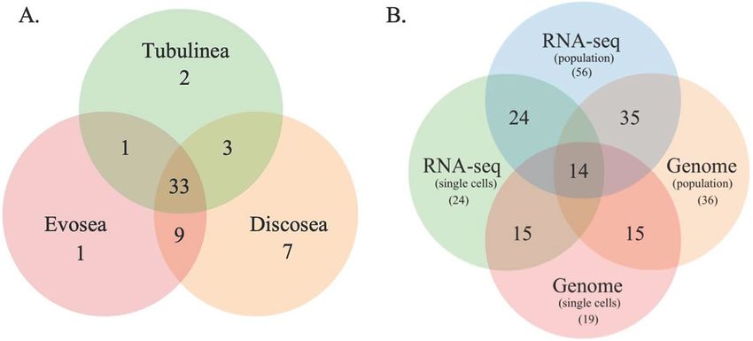
phyla, respectively (Fig. 1, Table S1). Among these discovered phyla, 33 phyla are shared among the three clades
(Fig. 2A). While the bacterial taxon sampling for Tubulinea in the transcriptome data is smaller than Evosea and
Discosea, the latter two clades shared more bacterial phyla between them (i.e. 9), when compared to the phyla
that they each mutually shared with Tubulinea (i.e. 1 and 3, respectively) as shown in Fig. 2A. We also found
some bacterial phyla specifically associated with each clade; namely, 7 in Discosea, 2 in Tubulinea and 1 in Evo-
sea (Fig. 2A, Table S1). However, in future research, the specific representative bacterial phyla recovered in each
clade might change with more taxon sampling, and in relation to the nature of the acquired data. For instance,
two samples from the same species, C. minus, in the whole culture genome data showed variation in the number
of bacterial phyla recovered and shared (Table S3). This indicates that a thorough and even sampling is required
to make such comparisons. Overall, phyla recovered were proportional to data size and taxon sampling (Fig. 1,
Tables S1-S3).
The total number of genera and their representation differed by bacterial phyla in our datasets. The most
abundant bacterial phylum recovered in all datasets and amoebozoan clades is Proteobacteria (Tables S1-S3).
Scientific Reports | (2021) 11:8043 | https://doi.org/10.1038/s41598-021-87192-0 2
Vol:.(1234567890)www.nature.com/scientificreports/
Key
Discosea
Evosea
30 Tubulinea
No. of genera
20
10
0
Modulibacteria
Epsilonbacteraeota
Kiritimatiellaeota
Deinococcus-Thermus
Entotheonellaeota
Elusimicrobia
Actinobacteria
BRC1
Gemmatimonadetes
Calditrichaeota
FCPU426
Dependentiae
Halanaerobiaeota
Hydrogenedentes
Chloroflexi
Firmicutes
MAT-CR-M4-B07
Acidobacteria
Cyanobacteria
Dadabacteria
Omnitrophicaeota
Deferribacteres
Acetothermia
Fusobacteria
Margulisbacteria
Marinimicrobia
Aerophobetes
Latescibacteria
LCP-89
Chlamydiae
WS1
Zixibacteria
Schekmanbacteria
Aquificae
Armatimonadetes
Atribacteria
Cloacimonetes
Lentisphaerae
Planctomycetes
BHI80-139
FBP
TA06
Caldiserica
Rokubacteria
Proteobacteria
Nitrospinae
Nitrospirae
PAUC34f
WS2
Patescibacteria
Thermotogae
Verrucomicrobia
Bacteroidetes
Fibrobacteres
Spirochaetes
Synergistetes
Tenericutes
Phylum (Bacterium)
Figure 1. Distribution of sequences identified (number of genera) representing the 57 Bacterial phyla
discovered in the three major clades of Amoebozoa across all datasets analyzed using Kraken 2.
Figure 2. Venn diagram showing bacterial phyla shared among the three major clades of Amoebozoa of the
whole culture RNA-Seq data (A) and among the four types of datasets analyzed (B) analyzed using Kraken 2.
Class Gammaproteobacteria, a subdivision of Proteobacteria, was represented by a higher number of genera and
total number of sequences that were representative for its genera (Tables 1, S1-S4). Other bacterial phyla that
were represented by over 1000 sequences for the genera recovered, in Kraken 2 analysis, include Bacteroidetes
and Firmicutes (Table S1). Generally, a higher number of sequences representing a given phylum were observed
in the whole culture genome data (Fig. 3, Table S3).
Comparison of data types and potential endosymbiont bacterial phyla. The four data types,
genome (whole-culture and single cells) and RNA-Seq (whole-culture and single cells), analyzed yielded bacte-
rial phyla that are commonly shared among samples and amoebozoan clades analyzed (Figs. 2, S1, Tables S1-S3).
We observed some variations in taxonomic breadth and the total number of genera/species recovered depending
on data type and taxon sampling size (Figs. 2, S1, Tables S1-S3). As mentioned above all except one bacterial
phylum reported here were present in whole culture RNA-Seq datasets (Table S1). While the large number of
bacterial phyla in the whole culture RNA-Seq dataset can be partly attributed to the size of taxon sampling used
for this dataset, these results clearly indicate that RNA-Seq is a good data source for this type of study. The whole
culture genome data is represented by two independent samples from a single species, C. minus (Table S3). A
total of 36 bacterial phyla were recovered from these two samples, 35 of these are shared with the whole culture
RNA-Seq dataset (Fig. 2B). The single cell genome data yielded the lowest number,19 bacterial phyla (Table S3),
Scientific Reports | (2021) 11:8043 | https://doi.org/10.1038/s41598-021-87192-0 3
Vol.:(0123456789)www.nature.com/scientificreports/
B. cepacia comp
Meth
p
grou
6 more
y
5 more
7 more
libiu
ens
e
0.7% Cupriavidus
5 mor
ae
ore
s
mp
na
resc
aligenace
20 m
mo
Va
etro
12
o
rio
ore
pho
P. flu
lex 1%
mo
vo
leiph
5m
otro
rax
re
Ac
0.9% Alc
re
ilum
pa
n
0.6%
id
mo
Ste
ov
s
rad
ale
or
15
ax
ox
ia
ter
4%
%
er
sp
us
ac
ct
0.7
.K
ba
ob
1%
KS
eo
ter
10
ot
deria
En
2 m Burkhol Pse
pr
udo
ylibiu
lta
4% mon
Meth Burkholderiaceae
De
1%
x X as
Rh x
o ra sedis Pse antho
r a e ud.. 10
8%
od
ob d
. . ov
ar. ae Burkho
l. .. .acemona djl-
i ae da sp.
0.
V s X
Rh
ac
ter Ac x c e e ri ale P s e
an
th cea m
od ac d a hold udo o e riu
fie .ra ad mon mon cte
3m
os
pir ea si i.. on Burk ada ad ba
e co
unc
ore illa as Ac am teria de les ales My
ce
ae
1%
n cl m o bac G l t
las
Ca
ulo 0.9 u C o
e ta pro te am
m
a/
ep 11%
a
sifie
bac % B pro sil
2m ter teo o
My
uncla ore 0.8 bac n s
dM
My
cob
% ter ub
h.. les
ssifie
d Bre ia di
cob
yco
s R illa
vis
.es
Cory
e
vund
acte
cea
imon as io
...le ospir
acte
bac
as n s
imon
Eryth 1%
rium
n
tera
robact
eba
ter
riac
eracea
od
e 0.7%
ium
Actin
und
bac
5 more Rh
cteri
eae
Actin
a
Cau
lo
teri
Brev
ales
obact
Cau
Sphingopyxis 10 more
bac
o
1%
Sphingomonadales
bacte
Sphingomonadaceae
teo
eria
Sp...um
Pro
ria
Bacteria
e
unclassified Sphingobium 1% 0.9% Mycolicibacterium
ycetacea
p
grou
0.7% Nocardiaceae
4 more
teria
Sp...as
Alp
1% Strep
Streptom
tom yces
hap
abac
0.7% 0.7%
igenens Microco PVC group 0.2%
sanxan
rote
monas 0.7% ccales
as taxi
Terr
Sphingo gomon
Phy pho..
0.8%
oba
Sphin si s
Hy
n Spirochaetia 0.1%
shuie
...e .ac
cte
n g 14 m
nas h
e FCB group ore
ae ea t...iu
gomo
ria
s
Sphin te Fusobacteriales 0.08%
Me e
re
9m
o
0.7
% icu
t...
Rh rm
m Fi
ea
obiu izo illi
M
Rh Thermotogae 0.06%
hiz % hlorobi group c s
e
e
bia
sor 0.8 izo
les Bacteroidetes/C Ba l al
e
ae
Me m 1% Ag bi cil ce
ore iu Rh ac
Ba lla
m
rob s Acidobacteriia 0.06%
6m rum
ro i.. e ae ci illu
mic b ba .o ib
a
ac
ph
o
lor
u ct up Bra Bacteroidetes en ib
Hy h y
9%
er dyrh en Aquificae 0.06%
Me
t
0.
iu Bra izob Pa
m
d.. iace Pa
e m .biu ae 14
m or riu unc m % Nitrospiraceae 0.03%
3 t e las
ac
8%
s...i Bosea Pa
lob zob
1%
0.
e en
hy mor ium
1%
ib Synergistaceae 0.01%
et
ex
unclass...d ac
um
M 3 Bosea
pl
d illu
m
bi
tris
i e
%
s
sif
co
izo
gr Deferribacteraceae 0.01%
0.9
lus
s
ns
am
Rh
cla
1% Flavob
pa
ie
un in
2%
ac
is
01
as
Thermodesulfobacteriaceae 0.01%
ef
A0
m
on
2%
tu
viae
ore
05 3%
om
E-L
m
ore
iu
4m
Elusimicrobia 0.005%
ud
avilo
642
CG
er
acteria
se
3m
ct
18 m
ba
op
p. C
MC 26
Bosea sp. RAC
7 mo
ea v
ro
Candidatus Babela massiliensis 0.002%
od
5 mor
Ag
5 more
ms
Rh
ore
les
Bos
re
sp. PA
obiu
Dictyoglomus thermophilum 0.002%
e
hiz
dyr
Desulfurispirillum indicum 0.002%
Bosea
Bra
Figure 3. Krona plot of bacterial sequences identified in the whole culture genome data (10× genomics) of
Cochliopodium minus analyzed using Centrifuge.
after the single cell RNA-Seq data (24 phyla) (Table S2). Using the four datasets we were able to identify 14 poten-
tial endosymbionts/epibionts by taking a subset of the bacterial phyla discovered in each dataset (Fig. 2B). Use
of single cells datasets, both genome and RNA-Seq, primarily aimed at reducing bacteria contamination from
external environment, enabled us to deduce these 14 putative endosymbionts/epibionts. A total of 24 potential
endosymbionts/epibionts phyla can be recognized if we considered taxa shared among three datasets i.e. all the
phyla discovered in single cell RNA-Seq dataset (Fig. 2B). Among these seven putative endosymbiont phyla (5
shared in all and 2 shared among 3 datasets, see also Tables S1-S3) included members (human pathogen genera/
species) previously reported to associate with or found in the cytoplasm of amoebozoan h osts3,9,23,25,34,42. Centri-
fuge analysis yielded the same or less number of bacterial phyla from the genome data compared to the Kraken
2 analysis (see Tables S1-S3, Fig. S1). The numbers of bacterial phyla recovered in the RNA-Seq datasets were
comparable between the two approaches, with Centrifuge recovering more phyla than Kraken 2 (Tables S1, S2).
In order to assess the impact of culturing techniques and types of bacteria that may be associated due to
difference in the environment of isolation and types of food sources used between labs, we compared RNA-Seq
data of three taxa sequenced in two different labs. Our comparison showed a similar total number of bacterial
phyla recovery but with some differences in the number of overlapping phyla (Table S1). The variation of non-
overlapping phyla in these three pairs of species ranged from 5 to 7. This observed difference using the RNA-Seq
data is smaller compared to the variation observed in the number of non-overlapping phyla found in the genome
data samples (Table S3). The whole culture genome data used two samples from the same species that were cul-
tured under the same conditions. These two samples had 9 non-overlapping bacterial phyla, which indicate that
other technical factors, such as sample quality (e.g. starting RNA/DNA material) and sequencing (e.g. depth/
coverage), might affect the recovery rate of overlapping bacterial community in samples of the same species.
Human pathogenic bacterial phyla and genera associated with amoebozoa. Our survey of lit-
erature and databases (e.g., https://globalrph.com/bacteria/) for bacterial pathogens yielded over 50 known and
potential human pathogen genera along their representative species (Table S4). We used this list to investigate
the presence of pathogenic species in our datasets (Tables S4). We used both Kraken 2 and Centrifuge to ana-
lyze our samples for pathogens, but Kraken 2 taxonomic classifications is only to a genus level and will not be
discussed further. Of the human pathogenic genera surveyed, over 50 genera spanning eight different bacterial
phyla were recovered (Fig. S2, Table 1, Table S4). The number of pathogens recovered in the three clades was
the same (51 pathogenic genera) in all clades despite taxon sampling differences in the whole culture RNA-Seq
dataset (Table S4). These genera are represented by 84 well-known and described species in Discosea and Evo-
sea, and 85 species in Tubulinea (Table S4). One extra species, Streptococcus viridans, was found in one species
Scientific Reports | (2021) 11:8043 | https://doi.org/10.1038/s41598-021-87192-0 4
Vol:.(1234567890)www.nature.com/scientificreports/
Phylum Genus Species
Corynebacterium C. diphtheriae (18), C. jeikeium (20), C. urealyticum (11)
Gardnerella G. vaginalis (26)
Actinobacteria
Mycobacterium M. leprae (11), M. tuberculosis (16)
Nocardia N.brasiliensis (23)
Bacteroides Bacteroides fragilis (30)
Bacteroidetes Capnocytophaga C. canimorsus (26), C. ochracea (17)
Porphyromonas P. gingivalis (17)
Chlamydiae Chlamydia C. pneumoniae (11), C. trachomatis (12), C. psittaci (7)
Bacillus B. anthracis (30), B. cereus (43), B. subtilis (43)
Clostridium C. botulinum (43), C. perfringens (38), C. tetani (30)
Enterococcus E. faecalis (30), E. faecium (31)
Firmicutes
Listeria L. monocytogenes (35)
Staphylococcus S. aureus (39), S. epidermis (36), S. hemolyticus (29), S. saprophyticus (26)
Streptococcus S. pneumoniae (33), S. agalactiae (30), S. pyogenes (30), S. viridans (2)
Fusobacteria Fusobacterium Fusobacterium nucleatum (45)
Proteobacteria
Anaplasma A. phagocytophilum (9)
Bartonella B. bacilliformis (13), B. henselae (15), B. quintana (12)
Alpha- Brucella B. suis (15)
Ehrlichia E. canis (21), E. chaffensis (20)
Rickettsia R. rickettsii (4)
Achromobacter A. xylosoxidans (36)
Bordetella B. pertussis (22)
Beta-
Burkholderia B. cepacia (35), B. pseudomallei (39)
Neisseria N. gonorrhoeae (14), N. meningitidis (35)
Campylobacter C. fetus (28), Campylobacter jejuni (36)
Epsilon-
Helicobacter H. pylori (42)
Acinetobacter A. baumannii (40)
Aeromonoas A. hydrophila (40), A. veronii (21), A. schubertii (20)
Citrobacter C. koseri (16), C. freundii (34)
Coxiella C. burnetii (30)
Enterobacter E. cloacae (35)
Escherichia E. coli (44)
Francisella F. tularensis (32)
Haemophilus H. influenzae (36)
Klebsiella K. pneumoniae (41)
Legionella L. pneumophila (38)
Gamma- Moraxella M. catarrhalis (23)
Morganella M. morganii (16)
Pasteurella P. multocida (24)
Proteus P. mirabilis (24), P. vulgaris (19)
Providencia P. stuartii (23)
Pseudomonas P. aeruginosa (40)
Salmonella S. enterica (39)
Serratia S. marcescens (40)
Shigella S. dysenteriae (16), S. flexneri (34), S. sonnei (31)
Vibrio V. cholerae (30), V. parahaemolyticus (38), V. vulnificus (33)
Yersinia Y. pestis (28)
Borrelia B. recurrentis (9)
Spirochaetaes
Leptospira L. borgpetersenii (31), L. interrogans (34), L. santarosai (20)
Treponema T. pallidum (15)
Tenericutes
Mycoplasma M. pneumoniae (14)
Table 1. List of potential human pathogens discovered in all analyzed samples. Numbers of amoebae samples
possessing listed pathogens are shown in parenthesis. Species level identification of pathogens was based
on Centrifuge analysis; the true nature of association and identification should be confirmed further by
experimental evidence.
Scientific Reports | (2021) 11:8043 | https://doi.org/10.1038/s41598-021-87192-0 5
Vol.:(0123456789)www.nature.com/scientificreports/
of Tubulinea (Cryptodifflugia operculata, Table S4). The detection of pathogens across clades and samples was
uniform in the RNA-Seq dataset (Table S4). The minimum number of pathogen genera in this dataset was 23,
which was recorded in two members of Discosea (Parvamoeba monoura (YT) and Stenamoeba limacina). Inter-
estingly, the number of pathogens recovered from another, Parvamoeba monoura, sequenced in a different lab
(Kang et al.35) had much higher associated pathogens (38 genera) than the one sequenced in our lab. Similar
number of pathogenic genera (26–49) were recovered from whole culture genome and single cell transcriptome
datasets (Table S4). The single cells genome data yielded the least pathogenic genera (0–8), which is similar to
results found in Kraken 2 analysis (data not shown).
The top three phyla with the highest number of pathogenic bacterial genera (4 or more genera and associated
species per phylum) recovered include Proteobacteria, Actinobacteria and Firmicutes (Table 1). Among the classes
of Proteobacteria, Gammaproteobacteria had the largest number (21 pathogen genera, 29 species) compared to
any group analyzed (Table 1). Majority of the detected pathogen genera are found in several amoebae belonging
to the three major clades of Amoebozoa (Table 1, S4, Fig. S2).
Discussion
Large amoebozoa associated bacterial phyla recovered. Our study using whole culture and single
cell genomics and transcriptomics recovered the largest number of bacterial phyla that are potentially associated
with the supergroup Amoebozoa to date. The majority of the known bacterial phyla (~ 50) recovered in our anal-
ysis of the amoebozoans are reported here for the first time (Fig. 1, Tables S1-S3). We also found well known and
common amoebozoan-associated bacterial phyla (5–10) reported in previous studies3,4,6,9,16,25,29–31,41. The large
and taxonomically diverse discovery of amoebozoans associated bacterial phyla in this study could be attributed
to the comprehensive taxon sampling and molecular genetic approach employed. We analyzed amoebozoans
characterized by diverse ecology, behavior and evolutionary history that represented the three major clades of
the Amoebozoa. We used monoclonal cultures of amoebozoans isolated directly from nature or acquired from
culture collection agents35,36,38. Research methods using monoclonal cultures typically include addition of food
bacteria (e.g., E. coli or Klebsiella); but once the culture starts to advance, it is common to see more bacterial
communities, besides food bacteria, growing among the amoebozoan cells. Amoebozoans are known to carry
undigested food bacteria vertically through g enerations43. These food bacteria are used presumably as seeds to
be conserved for potential replenishment within new environments encountered by the amoebozoan, and then
harvested as food; this behavior led some to metaphorically call amoebozoans, ‘farmers’21,43–45. Therefore, the
bacteria found in monoclonal samples analyzed likely reflect a bacterial community that might be expected
to occur naturally in nature; although we cannot rule out that some are acquired from contamination during
laboratory culture as for example from contact with instruments used in culturing or from air-borne bacteria
introduced from the laboratory environment. The taxonomic composition of bacteria found in amoebozoans
grown in different labs, or obtained from different culture collection agents or nature, in the RNA-Seq data were
similar (Table S1). The consistent recovery of similar bacterial phyla across different amoebozoan samples and
taxonomic groups, that we have found in our analyses for this research study, also indicates that all bacterial line-
ages discovered in our analysis are potentially associated with the Amoebozoa, and may mitigate against possible
contamination from sources largely derived from the laboratory environment. While the confirmation and type
of association of the newly discovered bacteria awaits further investigation, our study reinforces amoebozoans
as key players in controlling environmental bacteria through grazing. Our study also suggests that Amoebozoa
potentially can harbor more taxonomically diverse bacteria, with 64% of the 89 bacterial phyla in SILVA data-
base recovered, than previously reported.
The large taxonomic sampling of amoebozoans in our study was made possible by the use of transcrip-
tome data. In recent phylogenomic studies, a large number of RNA-Seq datasets have been generated in the
Amoebozoa35,36,38. These transcriptome data are generated using a standard approach that selects polyadenylated
RNA (polyA) in RNA samples, which selects against bacterial contaminant transcripts that are typically poorly
polyadenylated46,47. However, transcriptome data collected from amoebozoans using this approach typically
contains large bacterial transcripts and some ribosomal g enes35,36,38. While contamination by bacteria in tran-
scriptome data has been reported in axenic culture, or in species that do not normally feed or associate with
bacteria (likely contamination from environment)48, the close association of bacteria (food and endosymbiont)
with amoebozoans exacerbates the potential for contamination of transcriptomes even more. We took advan-
tage of this, and used the 16S bacterial ribosomal genes found in amoebozoan RNA-Seq data to assess bacterial
association with the Amoebozoa. Despite the potential limitation that transcriptome data might have for our
study, the aggregate number of bacterial phyla recovered from transcriptome sequencing was comparable in
taxonomic coverage to the whole culture genome data (Fig. 2). As expected, the number of genomic repre-
sentations of the discovered phyla in the whole culture genome data was higher than the transcriptome data
(Tables S1-S3), which indicates that transcriptome data might to an extent underrepresent the actual diversity
of associated bacterial populations. But when bacterial transcripts were analyzed along the 16S ribosomal genes
in our Centrifuge analysis, the number of bacterial phyla recovered increased by greater than eight (8–11) for
all clades examined (see Table S1). Our results support the utility of transcriptome data to study association of
bacteria with amoebozoans or other similar protists. Though a conservative estimate, transcriptome data has
some advantages over genome data due to lower cost and ease in acquiring it. Moreover, transcriptome data
can provide additional information on the nature of an association by providing physiological data (profile of
expressed genes) among interacting s pecies49.
In addition to the rich sources of transcriptome data as discussed above, the use of whole culture and single
cell genomics, as used in our laboratory culture studies reported here, enabled us to assess potential bacterial
endosymbionts (possibly including epibionts) associated with the Amoebozoa. Using this approach, we identified
Scientific Reports | (2021) 11:8043 | https://doi.org/10.1038/s41598-021-87192-0 6
Vol:.(1234567890)www.nature.com/scientificreports/
14–24 potential endosymbionts/epibionts bacterial phyla (Fig. 2B, Tables S1-S3). Our list includes bacteria phyla
whose members were previously shown to form true endosymbiotic relationships in some amoebozoans6,9,29,50,51.
However, a more thorough approach including single cell genome and cytological data, such as use of fluores-
cently labeled oligonucleotide probes29, is needed to establish true endosymbiotic relationships with Amoebozoa.
Nonetheless, the recovery of known endosymbiotic bacteria in our analysis gives credence to the reliability of
our approach to identify potential endosymbiotic bacteria candidates that can be studied further. It should be
noted that some amoebozoans are selective bacterial p redators52–54. The combination of single cell genomics and
transcriptomics approaches used here is a promising method of analyzing selective feeding on bacteria by pro-
tists; e.g., a recent study demonstrated the utility of transcriptome data for selective feeding in a ciliate lineage49.
Pathogenic bacteria associated with the Amoebozoa. The association of pathogenic bacteria with
some members of Amoebozoa has been investigated in great detail3,4,21,22,27,55. Most of the association of patho-
genic bacteria described with amoebozoans is facultative, but some permanent associations are also k nown6,29,42.
While most associations are transient and harmless, some bacterial infections (e.g. Legionella), leading to lysis
of amoebozoan cells, have been reported4,56. In facultative associations, the pathogenic bacteria can use the
amoeba cell as a safe niche to reproduce, or intermediate host, or even as a vehicle for dispersal or population
reservoir4,22. Some recent studies have proposed that amoebozoans could serve as an ‘interim training ground’ to
develop intracellular survival strategies before becoming a human pathogen due to the similarity in mechanism
of phagocytosis (phagolysosome) within mammalian macrophages4,17,28. Most of the known pathogenic bacteria
associated with Amoebozoa so far come from the studies that used only a few amoebozoan species, which are
not necessarily reflective of pathogens that can potentially be harbored by various groups in the supergroup of
Amoebozoa. In this study, we discovered 51 pathogenic bacterial genera (85 species) belonging to eight phyla,
the highest report so far (Table 1). The number and distribution of pathogenic genera across the three major
groups of Amoebozoa were comparable despite differences in taxon sampling among them (Figs. S1, S2). Our
list includes previously reported common representatives of pathogen bacterial phyla21,55 in addition to the large
number of pathogens newly discovered in this study (Tables 1, S4). Congruent with previous studies, the most
dominant pathogen-containing phylum is Proteobacteria. One of its subdivisions, class Gammaproteobacteria,
comprised more than 40% of the pathogenic genera identified in this study (Table 1). The representation of some
pathogen-containing phyla might be affected by habitat examined. Nevertheless, our results demonstrate that
all amoebae are potential carriers of bacterial pathogens both in nature or anthropogenic environments. All of
the multi-drug resistant species found in this study are listed and categorized by CDC and WHO as urgent, and
various levels of threats and concerns. Among these are Acinetobacter baumannii, Enterococcus faecalis, Clostrid-
ium spp., Neisseria gonorrhoeae, Helicobacter pylori, Campylobacter spp., Pseudomonas aeruginosa, Salmonella
enterica, Mycoplasma pneumoniae, Staphylococcus aureus, Haemophilus influenzae, Streptococcus pneumoniae
and Bordetella pertussis, which were found in most of the amoebozoan samples we examined (see Table 1). This
makes some Amoebozoa that are associated with potential or acknowledged human pathogens a major public
health threat.
Materials and methods
Amoebae cultures. Amoebae cultures used for genomic data in this study come from different sources
including ATCC (Cochliopodium minus ATCC 30935, Trichosphaerium sp. ATCC 40318), Ward’s Science cul-
ture collection (wardsci.com, Amoeba proteus) and a newly described species isolated from mixed eukaryotic
culture in our lab (Stratorugosa tubuloviscum). All these cultures have been maintained in our lab. Stratorugosa
tubuloviscum and C. minus were grown in plastic Petri dishes with bottled natural spring fresh water (Deer
Park, Nestlé Corp. Glendale, CA, USA) with added autoclaved grains of rice as an organic nutrient source to
support bacterial growth as prey for the amoebozoans. The marine amoeba, Trichosphaerium sp., was grown
under a similar condition as above in artificial seawater prepared by mixing 1 ml of distilled water in 30 g of
Instant Ocean (Cincinnati, OH) sea salt. Amoeba proteus was cultured with mixed bacteria and other microbial
eukaryote food sources.
Whole culture and single cell genomics. We used various approaches to investigate bacteria associated
with amoebozoans. Association of bacteria with their host can be internal endosymbionts or external those that
are epibionts attached to the surface of the cell and those that are freely present in cultures that are potentially
available to be engulfed as a food source. In order to capture all associated bacteria in diverse monoclonal cul-
tures of amoebozoans in our laboratory, we used molecular data collected using two approaches. The first set of
genetic data collected consisted of community genomic DNA extracted from actively growing cultures of amoe-
bozoans; and from the bacterial community typically found in monoclonal or newly isolated species maintained
in our laboratory cultures. The second genetic data is derived from single amoebozoan cells, individually picked
out of our laboratory cultures. The main difference between these two approaches is that the first approach,
whole culture, is aimed at collecting large quantities of DNA from a monoclonal population without little consid-
eration to bacteria contamination from the culture; while the second approach, single cell, is aimed at minimizing
bacterial contamination from the surrounding environment/culture.
In the single cell approach amoebozoan cells, which include Cochliopodium minus, Stratorugosa tubulovis-
cum, Trichosphaerium sp. and Amoeba proteus were individually picked using mouth pipetting techniques and
transferred to a clean glass slide to wash off bacteria (other microbial eukaryotes (food or prey) in A. proteus
culture) to reduce contamination of freely growing bacteria (other contaminants) from the culture. This step does
not necessarily remove epibionts that are tightly bound to the cell surface but it greatly minimizes free (loosely
Scientific Reports | (2021) 11:8043 | https://doi.org/10.1038/s41598-021-87192-0 7
Vol.:(0123456789)www.nature.com/scientificreports/
bound) bacteria growing in culture. Cleaned individual cells (5–10) were transferred into 0.2-mL PCR tubes and
genome amplified using REPLI-g Advanced DNA Single Cell Kit (Qiagen Hilden, Germany).
For the whole culture approach, genomic DNA was extracted from a large number of Cochliopodium minus
(syn. C. pentatrifurcatum57,58 cells in culture dishes (50 Petri dish cultures) using MagAttract high-molecular-
weight (HMW) DNA kit (Qiagen, MD), following the manufacturer’s instructions. This method includes gentle
cell lysis, releasing high molecular weight DNA and its efficient isolation and purification by concentration on
DNA-binding, surface coated magnetic beads. Genome sequencing was performed using 10X genomics (for
whole culture DNA) and Oxford Nanopore (ONP) (for both single cells and whole culture DNA) following the
manufacturers’ protocol. Genome data from 10X genomics and ONP were assembled using Supernova v2.1.159
and Minimap2-Miniasm-Racon genome assembly p ipeline60–62, respectively. For ONP genome data we used
Porechop version 0.2.4 (https:// github.com/rrwick/Porechop) to remove ONP sequencing adapters added dur-
ing the sequencing.
Filtlong version 0.2.0 (https://github.com/rrwick/Filtlong) was used to filter reads with length shorter than
200 and quality score less than 5.
Whole culture and single cell transcriptome data. Based on preliminary analysis that showed amoe-
bozoan transcriptomes contained large bacterial transcripts and some ribosomal genes, we analyzed RNA-Seq
from previous publications that were collected in a similar manner as above35,36,38,63. The samples from previous
publications included amoebozoans that were grown in established laboratory cultures and single cells isolated
from these culture as well as single cells directly isolated from various environments (see Table S5)35,36,38,63.The
whole culture RNA-Seq dataset included a total of 35 species (15 discoseans, 12 evoseans, and 8 tubulinids) with
three additional duplicate samples from Discosea sequenced in two different labs35,36,38. These discosean dupli-
cate samples were included in the analysis to examine the effects of culturing methods and environment on the
number and composition of bacterial community recovered. The single cell RNA-Seq dataset was represented
by five samples obtained from Cochliopodium minus64. Data collection, sequencing and assembly of transcrip-
tome data of these diverse amoebozoans, representing the three main clades of Amoebozoa (Discosea, Evosea,
and Tubulinea) of the whole culture and single cell RNA-Seq datasets, are described in Kang et al.35 and Tekle
et al.36,38, and Tekle et al.63, respectively. Some good quality transcriptomes whose origin was not certain or is
collected using a combination of single cell and whole culture are placed in the whole culture RNA-Seq dataset
(Table S5). All transcriptomes used for single cell RNA-Seq dataset including five replicate samples from C.
minus (Table S3) are collected in our laboratory under similar experimental c onditions64.
Taxonomic assignment of amoebozoa associated bacterial sequence data. We used two taxo-
nomic assignment tools, Kraken 2 65 and C entrifuge66, commonly used in metagenomic studies. A total of 49
samples (genome and transcriptome data) of amoebozoans, representing 38 species belonging to the three major
clades of Amoebozoa were analyzed. Similarly, we compared taxonomic composition in four datasets, which
include two genome (whole-culture and single cells) and two RNA-Seq (whole-culture and single cells) data
types. Kraken 2 with default settings, shown to have high sensitive and accuracy65, was used to analyze the
assembled contigs from genome and transcriptome data. Kraken 2 classifies sequences by mapping k-mer to the
lowest common ancestor (LCA) of all the datasets containing the given k-mer in the specified database. The 16S
database, SILVA, was chosen for this analysis and taxonomic classification was done to a genus level. Kraken 2
was run locally in an interactive session on XSEDE server, a supercomputing platform (http://xsede.org). We
also conducted similar analysis using Centrifuge, a rapid and accurate metagenomics classifier that uses the
Burrows–Wheeler transform (BWT) and an FM-index to store and index the genome database66. We used pre-
built database index from Centrifuge website constructed from complete Bacteria, Archaea, Viruses and Human
genomes from NCBI GenBank (as of 2016). Centrifuge allowed us to use raw (not reported) and assembled non-
ribosomal genomic and transcriptomic data in addition to the ribosomal (16S) used in the Kraken 2. Centrifuge
also can identify sequences to species level when sufficient matches are found. Results from Centrifuge analysis
was visualize using Krona67, an online interactive metagenomic visualization program. Resulting data were fur-
ther analyzed using R and Excel. Data used in this study are available upon request.
Received: 2 October 2020; Accepted: 25 March 2021
References
1. Gast, R. J., Sanders, R. W. & Caron, D. A. Ecological strategies of protists and their symbiotic relationships with prokaryotic
microbes. Trends Microbiol. 17, 563–569. https://doi.org/10.1016/j.tim.2009.09.001 (2009).
2. Braga, R. M., Dourado, M. N. & Araujo, W. L. Microbial interactions: ecology in a molecular perspective. Braz. J. Microbiol.
47(Suppl 1), 86–98. https://doi.org/10.1016/j.bjm.2016.10.005 (2016).
3. Barker, J. & Brown, M. R. Trojan horses of the microbial world: protozoa and the survival of bacterial pathogens in the environ-
ment. Microbiology 140(Pt 6), 1253–1259. https://doi.org/10.1099/00221287-140-6-1253 (1994).
4. Molmeret, M., Horn, M., Wagner, M., Santic, M. & Abu Kwaik, Y. Amoebae as training grounds for intracellular bacterial pathogens.
Appl. Environ. Microbiol. 71, 20–28. https://doi.org/10.1128/AEM.71.1.20-28.2005 (2005).
5. Hibbing, M. E., Fuqua, C., Parsek, M. R. & Peterson, S. B. Bacterial competition: surviving and thriving in the microbial jungle.
Nat. Rev. Microbiol. 8, 15–25. https://doi.org/10.1038/nrmicro2259 (2010).
6. Horn, M. et al. Obligate bacterial endosymbionts of Acanthamoeba spp. related to the beta-Proteobacteria: proposal of “Candidatus
Procabacter acanthamoebae” gen. nov., sp. nov.. Int. J. Syst. Evol. Microbiol. 52, 599–605. https://doi.org/10.1099/00207713-52-2-
599 (2002).
Scientific Reports | (2021) 11:8043 | https://doi.org/10.1038/s41598-021-87192-0 8
Vol:.(1234567890)www.nature.com/scientificreports/
7. Margulis, L. Symbiosis in cell evolution: microbial communities in the Archean and Proterozoic eons 1–18 (W.H. Freeman, 1993).
8. Handley, K. M. Determining microbial roles in ecosystem function: redefining microbial food webs and transcending kingdom
barriers. mSystems https://doi.org/10.1128/mSystems.00153-19 (2019).
9. Horn, M. & Wagner, M. Bacterial endosymbionts of free-living amoebae. J. Eukaryot. Microbiol. 51, 509–514. https://doi.org/10.
1111/j.1550-7408.2004.tb00278.x (2004).
10. Erken, M., Lutz, C. & McDougald, D. The rise of pathogens: predation as a factor driving the evolution of human pathogens in the
environment. Microb. Ecol. 65, 860–868. https://doi.org/10.1007/s00248-013-0189-0 (2013).
11. Amaro, F., Wang, W., Gilbert, J. A., Anderson, O. R. & Shuman, H. A. Diverse protist grazers select for virulence-related traits in
Legionella. ISME J. 9, 1607–1618. https://doi.org/10.1038/ismej.2014.248 (2015).
12. Mishustin, E. N. Microbial associations of soil types. Microb. Ecol. 2, 97–118. https://doi.org/10.1007/BF02010433 (1975).
13. Gong, J. et al. Protist-bacteria associations: gammaproteobacteria and alphaproteobacteria are prevalent as digestion-resistant
bacteria in ciliated protozoa. Front. Microbiol. 7, 498. https://doi.org/10.3389/fmicb.2016.00498 (2016).
14. Samba-Louaka, A., Delafont, V., Rodier, M.-H., Cateau, E. & Héchard, Y. Free-living amoebae and squatters in the wild: ecological
and molecular features. EMS Microbiol. Rev. 43, 415–434. https://doi.org/10.1093/femsre/fuz011 (2019).
15. Anderson, O. R. The role of bacterial-based protist communities in aquatic and soil ecosystems and the carbon biogeochemical
cycle, with emphasis on naked amoebae. Acta Protozool. 51, 209–221 (2012).
16. Schmitz-Esser, S. et al. The genome of the amoeba symbiont “Candidatus Amoebophilus asiaticus” reveals common mechanisms
for host cell interaction among amoeba-associated bacteria. J. Bacteriol. 192, 1045–1057. https://doi.org/10.1128/JB.01379-09
(2010).
17. Best, A. M. & Abu Kwaik, Y. Evasion of phagotrophic predation by protist hosts and innate immunity of metazoan hosts by
Legionella pneumophila. Cell Microbiol. 21, e12971. https://doi.org/10.1111/cmi.12971 (2019).
18. Greub, G. & Raoult, D. Microorganisms resistant to free-living amoebae. Clin. Microbiol. Rev. 17, 413–433. https://doi.org/10.
1128/cmr.17.2.413-433.2004 (2004).
19. Foster, R. A., Carpenter, E. J. & Bergman, B. Unicellular cyanobionts in open ocean dinoflagellates, radiolarians, and tintinnids:
ultrastructural characterization and immuno-localization of phycoerythrin and nitrogenase. J. Phycol. 42, 453–463 (2006).
20. Clarke, M. Recent insights into host-pathogen interactions from Dictyostelium. Cell Microbiol. 12, 283–291. https://doi.org/10.
1111/j.1462-5822.2009.01413.x (2010).
21. Thewes, S., Soldati, T. & Eichinger, L. Editorial: amoebae as host models to study the interaction with pathogens. Front. Cell Infect.
Microbiol. 9, 47. https://doi.org/10.3389/fcimb.2019.00047 (2019).
22. Strassmann, J. E. & Shu, L. Ancient bacteria-amoeba relationships and pathogenic animal bacteria. PLoS Biol. 15, e2002460. https://
doi.org/10.1371/journal.pbio.2002460 (2017).
23. Benavides-Montano, J. A. & Vadyvaloo, V. Yersinia pestis resists predation by Acanthamoeba castellanii and exhibits prolonged
intracellular survival. Appl. Environ. Microbiol. https://doi.org/10.1128/AEM.00593-17 (2017).
24. Garcia, M. T., Jones, S., Pelaz, C., Millar, R. D. & Abu Kwaik, Y. Acanthamoeba polyphaga resuscitates viable non-culturable
Legionella pneumophila after disinfection. Environ. Microbiol. 9, 1267–1277. https://doi.org/10.1111/j.1462-2920.2007.01245.x
(2007).
25. Alibaud, L. et al. Pseudomonas aeruginosa virulence genes identified in a Dictyostelium host model. Cell Microbiol. 10, 729–740.
https://doi.org/10.1111/j.1462-5822.2007.01080.x (2008).
26. Dallaire-Dufresne, S., Paquet, V. E. & Charette, S. J. Dictyostelium discoideum: a model for the study of bacterial virulence. Can.
J. Microbiol. 57, 699–707. https://doi.org/10.1139/w11-072 (2011).
27. Bozzaro, S. & Eichinger, L. The professional phagocyte Dictyostelium discoideum as a model host for bacterial pathogens. Curr.
Drug Targets 12, 942–954. https://doi.org/10.2174/138945011795677782 (2011).
28. Cirillo, J. D. et al. Intracellular growth in Acanthamoeba castellanii affects monocyte entry mechanisms and enhances virulence
of Legionella pneumophila. Infect. Immun. 67, 4427–4434 (1999).
29. Horn, M. et al. Neochlamydia hartmannellae gen. nov., sp. Nov. (Parachlamydiaceae), an endoparasite of the amoeba Hartmannella
vermiformis. Microbiology 146(Pt 5), 1231–1239. https://doi.org/10.1099/00221287-146-5-1231 (2000).
30. Gomez-Alvarez, V., Revetta, R. P. & Santo Domingo, J. W. Metagenomic analyses of drinking water receiving different disinfection
treatments. Appl. Environ. Microbiol. 78, 6095–6102. https://doi.org/10.1128/AEM.01018-12 (2012).
31. Fields, B. S. et al. Characterization of an axenic strain of Hartmannella vermiformis obtained from an investigation of nosocomial
legionellosis. J. Protozool. 37, 581–583. https://doi.org/10.1111/j.1550-7408.1990.tb01269.x (1990).
32. Delafont, V., Rodier, M.-H., Maisonneuve, E. & Cateau, E. Vermamoeba vermiformis: a free-living amoeba of interest. Microb.
Ecol. 76, 991–1001 (2018).
33. Gomaa, F., Gersh, M. & Cavanaugh, C. Diverse legionella-like bacteria associated with testate amoebae of the genus arcella (Arcel-
linida: Amoebozoa). J. Eukaryot. Microbiol. 65, 661–668. https://doi.org/10.1111/jeu.12511 (2018).
34. Segal, G. & Shuman, H. A. Legionella pneumophila utilizes the same genes to multiply within Acanthamoeba castellanii and human
macrophages. Infect. Immun. 67, 2117–2124 (1999).
35. Kang, S. et al. Between a pod and a hard test: the deep evolution of amoebae. Mol. Biol. Evol. 34, 2258–2270. https://doi.org/10.
1093/molbev/msx162 (2017).
36. Tekle, Y. I. et al. Phylogenomics of “Discosea”: a new molecular phylogenetic perspective on Amoebozoa with flat body forms.
Mol. Phylogenet. Evol. 99, 144–154. https://doi.org/10.1016/j.ympev.2016.03.029 (2016).
37. Tekle, Y. I. & Williams, J. R. Cytoskeletal architecture and its evolutionary significance in amoeboid eukaryotes and their mode of
locomotion. R. Soc. Open Sci. 3, 160283. https://doi.org/10.1098/rsos.160283 (2016).
38. Tekle, Y. I. & Wood, F. C. Longamoebia is not monophyletic: phylogenomic and cytoskeleton analyses provide novel and well-
resolved relationships of amoebozoan subclades. Mol. Phylogenet. Evol. 114, 249–260. https://doi.org/10.1016/j.ympev.2017.06.
019 (2017).
39. Pagnier, I. et al. Babela massiliensis, a representative of a widespread bacterial phylum with unusual adaptations to parasitism in
amoebae. Biol. Direct https://doi.org/10.1186/s13062-015-0043-z (2015).
40. Delafont, V., Samba-Louaka, A., Bouchon, D., Moulin, L. & Héchard, Y. Shedding light on microbial dark matter: a TM6 bacterium
as natural endosymbiont of a free-living amoeba. Environ. Microbiol. Rep. 7, 970–978. https://doi.org/10.1111/1758-2229.12343
(2015).
41. Delafont, V., Brouke, A., Bouchon, D., Moulin, L. & Hechard, Y. Microbiome of free-living amoebae isolated from drinking water.
Water Res. 47, 6958–6965. https://doi.org/10.1016/j.watres.2013.07.047 (2013).
42. Amann, R. et al. Obligate intracellular bacterial parasites of acanthamoebae related to Chlamydia spp. Appl. Environ. Microbiol.
63, 115–121 (1997).
43. DiSalvo, S. et al. Burkholderia bacteria infectiously induce the proto-farming symbiosis of Dictyostelium amoebae and food
bacteria. Proc. Natl. Acad. Sci. USA 112, E5029-5037. https://doi.org/10.1073/pnas.1511878112 (2015).
44. Brock, D. A., Read, S., Bozhchenko, A., Queller, D. C. & Strassmann, J. E. Social amoeba farmers carry defensive symbionts to
protect and privatize their crops. Nat. Commun. 4, 2385. https://doi.org/10.1038/ncomms3385 (2013).
45. Brock, D. A., Douglas, T. E., Queller, D. C. & Strassmann, J. E. Primitive agriculture in a social amoeba. Nature 469, 393–396.
https://doi.org/10.1038/nature09668 (2011).
Scientific Reports | (2021) 11:8043 | https://doi.org/10.1038/s41598-021-87192-0 9
Vol.:(0123456789)www.nature.com/scientificreports/
46. Nakazato, H., Venkatesan, S. & Edmonds, M. Polyadenylic acid sequences in E. coli messenger RNA. Nature 256, 144–146. https://
doi.org/10.1038/256144a0 (1975).
47. Ohta, N., Sanders, M. & Newton, A. Poly(adenylic acid) sequences in the RNA of Caulobacter crescenus. Proc. Natl. Acad. Sci.
USA 72, 2343–2346. https://doi.org/10.1073/pnas.72.6.2343 (1975).
48. Strong, M. J. et al. Microbial contamination in next generation sequencing: implications for sequence-based analysis of clinical
samples. PLoS Pathog. 10, e1004437. https://doi.org/10.1371/journal.ppat.1004437 (2014).
49. Zou, S., Zhang, Q. & Gong, J. Comparative transcriptomics reveals distinct gene expressions of a model ciliated protozoa feeding
on bacteria-free medium, digestible, and digestion-resistant bacteria. Microorganisms https://doi.org/10.3390/microorganisms8
040559 (2020).
50. Fritsche, T. R., Gautom, R. K., Seyedirashti, S., Bergeron, D. L. & Lindquist, T. D. Occurrence of bacterial endosymbionts in Acan-
thamoeba spp. isolated from corneal and environmental specimens and contact lenses. J. Clin. Microbiol. 31, 1122–1126 (1993).
51. Proca-Ciobanu, M., Lupascu, G. H., Petrovici, A. & Ionescu, M. D. Electron microscopic study of a pathogenic Acanthamoeba
castellani strain: the presence of bacterial endosymbionts. Int. J. Parasitol. 5, 49–56. https://d
oi.o
rg/1 0.1 016/0 020-7 519(75)9 0097-1
(1975).
52. Singh, B. N. Selectivity in bacterial food by soil amoebae in pure mixed cultures and in sterilized soil. Ann. Appl. Biol. 28, 52–64
(1941).
53. Singh, B. N. The selection of bacterial food by soil amoebae, and the toxic effects of bacterial pigments and other products on soil
protozoa. Br. J. Exp. Path. 26, 316–325 (1945).
54. Ronn, R., McCaig, A. E., Griffiths, B. S. & Prosser, J. I. Impact of protozoan grazing on bacterial community structure in soil
microcosms. Appl. Environ. Microbiol. 68, 6094–6105. https://doi.org/10.1128/aem.68.12.6094-6105.2002 (2002).
55. Skriwan, C. et al. Various bacterial pathogens and symbionts infect the amoeba Dictyostelium discoideum. Int. J. Med. Microbiol.
291, 615–624. https://doi.org/10.1078/1438-4221-00177 (2002).
56. Molmeret, M., Bitar, D. M., Han, L. & Kwaik, Y. A. Disruption of the phagosomal membrane and egress of Legionella pneumophila
into the cytoplasm during the last stages of intracellular infection of macrophages and Acanthamoeba polyphaga. Infect. Immun.
72, 4040–4051. https://doi.org/10.1128/IAI.72.7.4040-4051.2004 (2004).
57. Tekle, Y. I., Anderson, O. R., Lecky, A. F. & Kelly, S. D. A new freshwater amoeba: Cochliopodium pentatrifurcatum n. sp. (Amoe-
bozoa, Amorphea). J. Eukaryot. Microbiol. 60, 342–349. https://doi.org/10.1111/jeu.12038 (2013).
58. Tekle, Y. I. & Wood, F. C. A practical implementation of large transcriptomic data analysis to resolve cryptic species diversity
problems in microbial eukaryotes. BMC Evol. Biol. 18, 170. https://doi.org/10.1186/s12862-018-1283-1 (2018).
59. Weisenfeld, N. I., Kumar, V., Shah, P., Church, D. M. & Jaffe, D. B. Direct determination of diploid genome sequences. Genome
Res. 27, 757–767. https://doi.org/10.1101/gr.214874.116 (2017).
60. Vaser, R., Sovic, I., Nagarajan, N. & Sikic, M. Fast and accurate de novo genome assembly from long uncorrected reads. Genome
Res. 27, 737–746. https://doi.org/10.1101/gr.214270.116 (2017).
61. Li, H. Minimap2: pairwise alignment for nucleotide sequences. Bioinformatics 34, 3094–3100. https://doi.org/10.1093/bioinforma
tics/bty191 (2018).
62. Li, H. Minimap and miniasm: fast mapping and de novo assembly for noisy long sequences. Bioinformatics 32, 2103–2110. https://
doi.org/10.1093/bioinformatics/btw152 (2016).
63. Tekle, Y. I., Wang, F., Heidari, A. & Stewart, A. J. Differential gene expression analysis and cytological evidence reveal a sexual stage
of an amoeba with multiparental cellular and nuclear fusion. bioRxiv https://doi.org/10.1101/2020.06.23.166678 (2020).
64. Tekle, Y. I., Wang, F., Heidari, A. & Stewart, A. J. Differential gene expression analysis and cytological evidence reveal a sexual stage
of an amoeba with multiparental cellular and nuclear fusion. PLoS ONE https://doi.org/10.1371/journal.pone.0235725 (2020).
65. Wood, D. E., Lu, J. & Langmead, B. Improved metagenomic analysis with Kraken 2. Genome Biol. 20, 257. https://d oi.o
rg/10.1 186/
s13059-019-1891-0 (2019).
66. Kim, D., Song, L., Breitwieser, F. P. & Salzberg, S. L. Centrifuge: rapid and sensitive classification of metagenomic sequences.
Genome Res. 26, 1721–1729. https://doi.org/10.1101/gr.210641.116 (2016).
67. Ondov, B. D., Bergman, N. H. & Phillippy, A. M. Interactive metagenomic visualization in a Web browser. BMC Bioinform. https://
doi.org/10.1186/1471-2105-12-385 (2011).
Acknowledgements
This work is supported by the National Science Foundation EiR (1831958) and National Institutes of Health
(1R15GM116103-02) to YIT. O. Roger Anderson is thanked for his invaluable comments and edits on the
manuscript.
Author contributions
Y.I.T. conceived the project, led writing manuscript and helped design experiments and analysis. J.M.L. collected
data, conducted analysis, and contributed to writing and editing of the manuscript. M.G.B. helped with analysis,
writing and organizing of results. F.W. helped with analysis and general discussion. All authors have read and
approved the manuscript.
Competing interests
The authors declare no competing interests.
Additional information
Supplementary Information The online version contains supplementary material available at https://doi.org/
10.1038/s41598-021-87192-0.
Correspondence and requests for materials should be addressed to Y.I.T.
Reprints and permissions information is available at www.nature.com/reprints.
Publisher’s note Springer Nature remains neutral with regard to jurisdictional claims in published maps and
institutional affiliations.
Scientific Reports | (2021) 11:8043 | https://doi.org/10.1038/s41598-021-87192-0 10
Vol:.(1234567890)You can also read


