Identification of antigenic domains and peptides from VP15 of white spot syndrome virus and their antiviral effects in Marsupenaeus japonicus
←
→
Page content transcription
If your browser does not render page correctly, please read the page content below
www.nature.com/scientificreports
OPEN Identification of antigenic domains
and peptides from VP15 of white
spot syndrome virus and their
antiviral effects in Marsupenaeus
japonicus
Jirayu Boonyakida1, Jian Xu2, Jun Satoh3, Takafumi Nakanishi4, Tohru Mekata5,
Tatsuya Kato1,4,6 & Enoch Y. Park1,4,6*
White spot syndrome virus (WSSV) is one of the most devastating pathogens in penaeid shrimp and
can cause massive damage in shrimp aquaculture industries. Previously, the WSSV structural protein
VP15 was identified as an antigenic reagent against WSSV infections. In this study, we truncated this
protein into VP15(1–25), VP15(26–57), VP15(58–80), and VP15(1–25,58–80). The purified proteins from the E.
coli expression system were assayed as potential protective agents in Kuruma shrimp (Marsupenaeus
japonicus) using the prime-and-boost strategy. Among the four truncated constructs, VP15(26–57)
provided a significant improvement in the shrimp survival rate after 20 days of viral infection.
Subsequently, four peptides (KR11, SR11, SK10, and KK13) from VP15(26–57) were synthesized and
applied in an in vivo assay. Our results showed that SR11 could significantly enhance the shrimp
survival rate, as determined from the accumulated survival rate. Moreover, a multiligand binding
protein with a role in the host immune response and a possible VP15-binding partner, MjgC1qR, from
the host M. japonicus were employed to test its binding with the VP15 protein. GST pull-down assays
revealed that MjgC1qR binds with VP15, VP15(26–57), and SR11. Taken together, we conclude that SR11
is a determinant antigenic peptide of VP15 conferring antiviral activity against WSSV.
White spot disease (WSD), caused by white spot syndrome virus (WSSV), has been known as one of the most
devastating diseases of farmed shrimp worldwide since its first occurrence in Taiwan in 1 9921–3. Since then,
substantial global losses due to WSD alone have been approximately US$15 billion annually, with an increasing
rate of US$1 billion/year4,5. WSSV infection in a shrimp pond could result in a high mortality rate within a single
week, especially in penaeid shrimp (e.g., Marsupenaeus japonicus, Litopenaeus vannamei, Penaeus monodon, and
Fenneropenaeus indicus)6,7. Clinical signs in WSD-suffering shrimp include lethargy, anorexia, reduction in food
uptake, reduced preening activities, loose cuticle, reddish discoloration, and white calcified spots of 0.5–3 mm
in diameter on the e xoskeleton8,9.
WSSV is the sole member of the genus Whispovirus and the only genus in the Nimaviridae family10–12.
The virus is a large, enveloped virus containing supercoiled circular double-stranded DNA (dsDNA) with an
unknown functional tail-like a ppendage12,13. The virion has a size of approximately 80–100 × 250–350 nm with
a rod-shaped nucleocapsid covered by a trilaminar membrane14. The genomic DNA is 285–305 kbp long with
nine tandem repeat conserved regions and 180 putative open reading frames (ORFs). However, most of the
1
Department of Bioscience, Graduate School of Science and Technology, Shizuoka University, 836 Ohya,
Suruga‑ku, Shizuoka 422‑8529, Japan. 2Institute of Biology and Information Science, Biomedical Synthetic
Biology Research Center, School of Life Sciences, East China Normal University, Shanghai 200062, People’s
Republic of China. 3Fisheries Technology Institute of National Research and Development Agency, Japan Fisheries
Research and Education Agency, Tamaki Field Station, Mie 519‑0423, Japan. 4Department of Applied Biological
Chemistry, Graduate School of Integrated Science and Technology, Shizuoka University, 836 Ohya, Suruga‑ku,
Shizuoka 422‑8529, Japan. 5Fisheries Technology Institute of National Research and Development Agency, Japan
Fisheries Research and Education Agency, Namsei Field Station, Mie 516‑0193, Japan. 6Research Institute of
Green Science and Technology, Shizuoka University, 836 Ohya, Suruga‑ku, Shizuoka 422‑8529, Japan. *email:
park.enoch@shizuoka.ac.jp
Scientific Reports | (2021) 11:12766 | https://doi.org/10.1038/s41598-021-92002-8 1
Vol.:(0123456789)
www.nature.com/scientificreports/
ORF-encoded proteins have no homology to the known proteins in d atabases15–18. Proteomic analysis of WSSV
revealed that the virus contains at least six major virion proteins (VPs). VP19, VP24, VP26, and VP28 were
identified as envelope proteins, while VP15 and VP664 were identified as nucleocapsid-associated p roteins19,20.
There is evidence that envelope proteins can form a protein complex that plays a crucial role in host-virus
interactions21–23. Although, multicellular organisms possess an ability to recognize and protect their selves against
pathogenic bacteria and viruses called “immune system”, invertebrates such as insects and crustaceans do not
own a long-term immunity of adaptive immune system. Therefore, their defensive mechanisms totally depend
on the innate immune system24,25. The innate immune system is initiated when pathogen-associated molecular
patterns (PAMPs) from pathogens are being recognized by host pattern recognition receptors (PRRs)26,27. Then a
series of signaling pathways of humoral and cellular responses is stimulated, thus molecules such as antimicrobial
peptides (AMPs), phenoloxidase (PO), and lysozymes are produced consequently or enhance cellular responses
such as phagocytosis, clotting, or a poptosis26–30.
However, there is no practical method to prevent WSSV infection in shrimp and to manage the spread of
this disease. Hence, it is necessary to develop a treatment against the virus. To date, following the concept of
“trained immunity”, several kinds of WSSV immunizing agents have been tested, including viral protein subunits,
attenuated WSSV, and DNA-/RNA-based agents31,32. Most recombinant subunit vaccines investigated have been
based on VP19, VP24, VP26, VP28, VP292, and VP466, and among them, VP28 has been widely studied33–37.
These viral subunits were confirmed to improve the survival rate after challenging shrimp with WSSV. Recently,
we reported that VP15 can also provide a protective effect in Kuruma shrimp (M. japonicus) against WSSV after
immune prime-and-boost via intramuscular i njection38.
WSSV-VP15 is an 80 amino acid protein with an extremely high basic pI value of 12.49, only showing homol-
ogy with the DNA-binding proteins of eukaryotic origin and a baculovirus p6.9 p rotein39. Some studies have
suggested that VP15 may be involved in viral genome packaging into the capsid and a major nucleocapsid protein
omomultimers40,41. Although VP15 is a major nucleocapsid protein, its
with the ability to self-interact, forming h
properties and functions have not been defined. No protein crystal structure has been reported yet, and only one
trial used VP15-based material (DNA vaccine encoding V P1542) for immunizing shrimp against WSSV. We have
previously demonstrated that VP15 can enhance shrimp survivability after challenge with WSSV38. In this study,
we attempted to determine the antigenic domain of the VP15 protein by in vivo animal experiments with M.
japonicus using a purified truncated VP15 series and synthetic peptides derived from VP15 at the peptide level.
To further explore the mechanism of antigenicity involved, we also investigated the possible interaction between
VP15 and its host protein partner, a gC1qR homolog from M. japonicus (MjgC1qR), via GST pull-down assay.
Results
Expression and purification of truncated WSSV‑VP15s. pGEX-6P-1, which has a glutathione
S-transferase (GST, molecular weight; 26 kDa) fusion system, was genetically fused with the VP15 gene as the
GST-VP15 fusion gene, and the constructed plasmid was used as a template to generate a truncated series of
VP15 (Fig. 1A,B). The protein expression was driven by IPTG induction under the control of the tac promoter.
The sizes of the GST-fused truncated VP15 series members were predicted to be 30.8, 31.6, 30.5, and 33.4 kDa
for VP15(1–25), VP15(26–57), VP15(58–80), and VP15(1–25,58–80), respectively, while that of GST-VP15 was 37.0 kDa.
The expression of recombinant proteins was confirmed by both SDS-PAGE (Figs. 1C, S1, S2) and Western blot-
ting with an anti-Flag antibody (Figs. 1D, S1, S2 of Supplementary information). The proteins were then purified
from E. coli soluble fractions using GST affinity chromatography (Fig. 1E; SDS-PAGE in Fig. S3 and Western blot
in Fig. S4 of Supplementary information) The yield of purified proteins obtained from 1 L culture was 3.3 mg
for GST, 5.3 mg for VP15, 5.7 mg for VP15(1–25), 6.3 mg for V P15(26–57), 8.4 mg for V
P15(58–80), and 4.0 mg for
VP15(1–25,58–80).
Vaccination and challenge experiment using the truncated VP15s. The partially purified GST-
fused full-length VP15 and truncated VP15 series were subsequently intramuscularly (IM)-injected into
Kuruma shrimp to assess their anti-WSSV activity. Five groups were immunized twice at days 0 and 20 using
purified protein at a dose of 10 µg/g shrimp and then challenged at day 10 after the second injection (Fig. 3A).
Two groups (PBS and GST) were mock-treated as two independent negative controls. All groups were chal-
lenged with WSSV at a dose of 2.69 × 10⁴ DNA copies/shrimp.
The PBS and GST control groups showed a drastic decrease in survival rate from day 4 to day 7 and reached
20% and 33.3% (16% relative percent survival, RPS) at 20 days post infection (dpi). A group of shrimps receiving
VP15(1–25) showed a final survival rate of 31.6% (15% RPS), a similar trend to that of the GST group, indicat-
ing that VP15(1–25) failed to provide a protective effect against WSSV. Groups of shrimp injected with VP15,
VP15(58–80), or V
P15(1–25,58–80) showed an survival rate at 20 dpi of 42.1% (28% RPS), 47.6% (35% RPS), and
38.9% (24% RPS), respectively. However, VP15(26–57) provided a protective effect against WSSV with 57.9% of
the survival rate (48% RPS) at 20 dpi (Fig. 2B, Table 1), which was the highest value among the experimental
groups and was the only group showing a significant difference in survival rates compared with the controls.
Vaccination and challenge experiment using the VP15‑derived peptides. To investigate the
antigenic effects of VP15 at the peptide level, VP-15-derived peptides were designed based on the amino acid
sequence of VP15(26–57) and chemically synthesized (purity > 95%). As demonstrated in Fig. 3A, the peptides
were KR11 (KTKSRRGSKKR), SR11 (STTAGRISKRR), SK10 (SPSMKKRAGK), and KK13 (KRRSPSMK-
KRAGK). Each peptide was IM-injected into the shrimp to screen for its protective effects against WSSV. Six
groups of shrimp were set up with one PBS control group and five experimental groups. The protective effects of
the four peptides were compared with those from GST-VP1526–57. The shrimp were immunized twice, as shown
Scientific Reports | (2021) 11:12766 | https://doi.org/10.1038/s41598-021-92002-8 2
Vol:.(1234567890)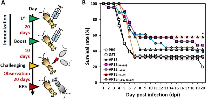
www.nature.com/scientificreports/
Figure 1. Expression of the truncated WSSV-VP15 series using the E. coli expression system. (A) Schematic
diagram representing a major nucleocapsid protein of WSSV-VP15. Four truncated VP15 constructs were
generated as illustrated and were named VP15(1–25), VP15(26–57), VP15(58–80), and VP15(1–25,58–80). The numbers
represent the amino acid position. (B) Construction of plasmids harboring the VP15 truncated series for
expression using the E. coli system and expression verification via SDS-PAGE. GST: glutathione-S-transferase
as a fusion tag and for affinity purification; Flag: DYKDDDDK epitope tag for Western blot analysis. (C)
Coomassie brilliant blue-stained SDS-PAGE gel of E. coli expressing VP15 truncated constructs and (D)
Western blot analysis of the expressed recombinant proteins. Anti-Flag antibody was applied for the detection of
the target proteins. M: marker; S: supernatant; and P: precipitate. (E) SDS-PAGE analysis of purified VP15 and
truncated VP15 series. Each recombinant protein was purified via GST affinity chromatography. The theoretical
sizes of VP15, VP15(1–25), VP15(26–57), VP15(58–80), and VP15(1–25,58–80) were 37.0, 30.8, 31.6, 30.5, and 33.4 kDa,
respectively. A main purified product of VP15(1–25), VP15(26–57), and VP15(58–80) can be observed corresponding
to the estimated size of the protein with minor bands below the main product. However, a purified product of
VP15 and VP15(1–25,58–80) has several bands below the main product. GST was loaded as a control. M indicates a
protein marker.
in Fig. 3B, with a dose of 10 µg/g shrimp of peptide or the recombinant protein and observed for 14 days after
the WSSV challenge. Our screening experiment revealed that SR11 could provide a prominent protective effect
with a final survival rate of ~ 80% (62% RPS), similar to GST-VP1526–57 at 14 dpi (62% RPS). The KR11-, SK10-,
and KK13-injected groups showed a survival rate of 54% (2% RPS) at the end of observation, similar to the PBS
group (Fig. 3C, Table 1). From this result, we interpreted that the KR11, SK10, and KK13 did not provide pro-
tective effects, as the survival rate was the same as that of the PBS control, while SR11 enhanced the resistance
against WSSV. Therefore, SR11 was considered to be an antigenic peptide against WSSV in M. japonicus.
To verify the effect of the SR11 peptide, a larger sample size of 25 individuals was tested along with the posi-
tive control, GST-VP15(26–57), and the PBS negative control group. The two experimental groups were treated
similarly as Fig. 3B with the same dose of SR11 or GST-VP15(26–57). As shown in Fig. 3D, shrimp rapidly died
from day 6 to day 10 in the PBS group, with a final survival rate of ~ 50%. However, shrimp in the group receiving
either GST-VP15(26–57) or SR11 showed survival rates of 77% (54% RPS) and 73% (46% RPS), respectively, at 20
dpi (Table 1), indicating that SR11 could generate reproducible anti-WSSV effects.
Sequence analysis, alignment, and phylogenetic analysis of MjgC1qR. The gC1qR of inverte-
brates has been suggested to be involved in innate immunity against pathogens by acting as a pathogen rec-
ognition receptor43,44. There is evidence showing that gC1qR might act as a PRR to recognize a broad range of
non-self-components such as bacteria, virus, and PAMPs and can be upregulated upon WSSV infection45,46. We
hypothesized that gC1qR from M. japonicus (MjgC1qR) might also interact with VP15. gC1qRs have been iden-
tified from many invertebrate species but not from Kuruma shrimp. We then attempted to generate and analyze
the cDNA sequence of MjgC1qR. To this end, bioinformatic analysis was performed to compare MjgC1qR with
the reported gC1qR(s) from the database. The obtained full-length cDNA of MjgC1qR was 1120 bp, consisting
of 98 bp of a 5’-untranslated region (UTR), 786 bp of an open reading frame (ORF), and 336 bp of a 3’-UTR
Scientific Reports | (2021) 11:12766 | https://doi.org/10.1038/s41598-021-92002-8 3
Vol.:(0123456789)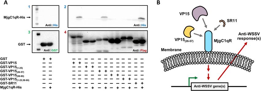
www.nature.com/scientificreports/
Figure 2. Evaluation of the E. coli-derived WSSV-VP15 and truncated VP15s for protective effects against
WSSV. (A) Time schedule of shrimp immunization, WSSV challenge, and observation. The prime-and-boost
immunization strategies for seven different groups (PBS, GST, VP15, VP15(1–25), VP15(26–57), VP15(58–80),
and VP15(1–25, 58–80)) are shown. In brief, shrimp were immune primed and boosted at day 0 and day 20 and
challenged with WSSV via intramuscular injection 10 days afterward. (B) Time course of the survival rate of
Kuruma shrimp after WSSV challenge. The mortality of shrimp was observed for 20 days at 24 h intervals. The
line marked with an asterisk is significantly higher than that of the control groups.
Treatment (prime-and-boost) Number of dead individuals Mortality (%) RPS (%)
Experiment 1
PBS (control) 16/20 80a –
GST 12/18 67 16
VP15 11/19 58 28
VP15(1–25) 13/19 68 15
VP15(26–57) 8/19 42b 48
VP15(58–80) 11/21 52 35
VP15(1–25,58–80) 11/18 61 24
Experiment 2
PBS (control) 7/15 47a –
VP15(26–57) 2/11 18b 62
KR11 6/13 46 2
SR11 2/11 18b 62
SK10 5/11 46 2
KK13 7/13 46 2
Experiment 3
PBS (control) 9/18 50a –
VP15(26–57) 5/22 23b 54
SR11 6/22 27b 46
Table 1. Mortality and RPS of VP15, truncated VP15 and SR11. a,b p < 0.05.
(including the stop codon) with one mRNA instability element (ATTTA). The ORF encodes a 262 amino acid-
long protein with an MW of 29.2 kDa and a theoretical pI of 4.74. The protein also contains an N-terminal
mitochondrial targeting sequence with a length of 44 amino acids, a mitochondrial acidic matrix (MAM33)
domain (residues 78–257) at its C-terminus, and one arginine-glycine-aspartic acid (RGD) motif (Fig. S5 of Sup-
plementary information). Comparative multiple alignment of gC1qR amino acid sequences from various species
revealed a highly conserved MAM33 domain and cell attachment RGD motif across the invertebrate species.
Scientific Reports | (2021) 11:12766 | https://doi.org/10.1038/s41598-021-92002-8 4
Vol:.(1234567890)www.nature.com/scientificreports/
Figure 3. Peptide design and evaluation of VP15-derived peptides in shrimp against WSSV. (A) De novo
design of peptides using the VP15(26–57) amino acid sequence as a template. Four peptides, named KR11
(KTKSRRGSKKR), SR11 (STTAGRISKRR), SK10 (SPSMKKRAGK), and KK13 (KRRSPSMKKRAGK) were
designed and chemically synthesized. (B) Time schedule of shrimp experiments using VP15-derived peptides
as a protective agent. (C) Screening for the effective peptide by shrimp WSSV challenge experiment (KR11
(n = 13), SR11 (n = 11), SK10 (n = 11), or KK13 (n = 13)). Shrimp mortality was observed for 14 days, and the
survival rate of each group was calculated. (D) Evaluation of SR11, an effective peptide, for enhancing shrimp
survivability against WSSV. The experiment was conducted with twice the number of individuals (n = 25). For
both experiments, VP15 (26–57) was used as a positive control, while PBS served as a negative control.
MjgC1qR also shared high identity with other invertebrate gC1qRs (93.5% identity with Penaeus vannamei, 92%
with Penaeus monodon, 90.5% with Penaeus chinensis, 74.3% with Eriocheir sinensis, 71.6% with Portunus tritu‑
berculatus, 71% with Macrobrachium rosenbergii, 70.2% with Palaemon carinicauda, 69.8% with Macrobrachium
nipponense, and 68.7% with Pacifastacus leniusculus) (Fig. S6 of Supplementary information). The phylogenetic
tree revealed that MjgC1qR had a close evolutionary relationship to crustacean gC1qRs, especially gC1qR of P.
vannamei (Fig. S7 of Supplementary information).
Expression of MjgC1qR using the silkworm‑bacmid expression vector system. The bacmid har-
boring the full-length MjgC1qR-His gene was directly injected for transfection of silkworm larvae. Hemolymph
and fat body were collected at 5 dpi. Fat bodies were homogenized and analyzed for the expressed protein via
Western blot analysis. As shown in Fig. 4 (Fig. S8 of Supplementary information), MjgC1qR was expressed as
a single protein product in silkworm larvae under the control of the polyhedrin promoter. The protein was
detected in the silkworm fat body in both a soluble and an aggregated form by the anti-His antibody. The size of
the soluble MjgC1qR was found to be smaller than that of the aggregated form, with lengths of approximately 25
and 29 kDa, respectively, possibly due to the cellular processing of the signal peptide.
VP15 and the SR11 peptide interact with MjgC1qR. The interactions of MjgC1qR with the candidate
bait proteins were observed through GST pull-down assay. In this study, the candidate baits were GST-VP15,
GST-VP15(26–57), and the GST-fused peptide GST-SR11, as these proteins and peptides could provide protective
effects in shrimp against WSSV. Before this experiment, GST-SR11 was expressed in E. coli and purified (Figs. S9,
S10 of Supplementary information)). Additionally, pull down assays between MjgC1qR and GST-VP15(1–25),
GST-VP15(58–80), or GST-VP15(1–25,58–80) were performed. As shown in Fig. 5A (Figs. S11–S14 of Supplementary
Scientific Reports | (2021) 11:12766 | https://doi.org/10.1038/s41598-021-92002-8 5
Vol.:(0123456789)www.nature.com/scientificreports/
Figure 4. Western blot analysis of MjgC1qR expressed in silkworm larvae. Western blot analysis of silkworms
was performed with an anti-His antibody. Silkworms were transfected with recombinant bacmid harboring the
MjgC1qR-His construct, and fat bodies were collected after 5 days of transfection. M: marker; S: supernatant;
and P: precipitate.
Figure 5. GST pull-down assay between MjgC1qR and GST-VP15, GST-VP15(1–25), GST-VP15(26–57),
GST-VP15(58–80), GST-VP15(1–25,58–80), GST-SR11, and a proposed antiviral mechanism. (A) The interactions
of MjgC1qR-His with GST-VP15, GST-VP15(1–25), GST-VP15(26–57), GST-VP15(58–80), GST-VP15(1–25,58–80),
GST-SR11 were observed via GST pull-down assay and confirmed by Western blot analysis with (1 and 2) anti-
His antibody, (3) anti-GST antibody, and (4) anti-Flag antibody. The symbol “ + ” indicates the proteins used for
the assay. (B) In shrimp, VP15, VP15(26–57), and SR11 interacted with the host gC1qR, hence inducing a cellular
signaling cassette that could activate the transcription of antimicrobial peptides.
Scientific Reports | (2021) 11:12766 | https://doi.org/10.1038/s41598-021-92002-8 6
Vol:.(1234567890)www.nature.com/scientificreports/
information), the recombinant MjgC1qR protein interacted with VP15, VP15(26–57), and the SR11 but not with
the control GST protein and the other tested proteins.
Discussion
VP15 of WSSV has been identified as one of the major WSSV structural proteins in the nucleocapsid fraction and
has an affinity for nucleic acids, especially supercoiled DNA41. Our aim was to identify an antigenic determinant
of VP15 that could enhance the shrimp survival rate upon WSSV infection. We designed truncated constructs
by dividing VP15 into three regions: the N-region (first 25 amino acids), middle region (32 amino acids), and
C-region (last 23 amino acids), and purified truncations from E. coli were then tested as “vaccines” in shrimp.
Purification of GST-VP15 suggested that the full-length protein might not be a stable protein, as it tended to be
fragmented, as confirmed by SDS-PAGE analysis (Fig. 1E). The full-length VP15 protein (80 aa) has a theoretical
size of approximately 35 kDa and is rich in serine (20%), arginine (20%), and lysine (21.2%). After purification,
we observed protein bands below 35 kDa and a thick protein band at > 30 kDa, which could not be observed by
Western blotting using anti-Flag antibody. Moreover, the truncated VP15s could be purified with a main band
corresponding to the predicted MW along with minor bands below the main products, except for V P15(1–25,58–80),
which resulted in several productive bands below the main product similar to the Western blot of purified pro-
teins using anti-GST antibody (Figs. 1E, S3 of Supplementary information).
To our knowledge, VP15 has barely been used as a protective agent against WSSV in penaeid shrimp, as
this protein is usually not considered a part of the “infectome,” a protein complex that comprises 14 different
proteins, including VP19, VP24, VP26, and VP28, located on the viral s urface21. VP15 was applied as a DNA
vaccine in Peneus monodon, but it could not elicit any protection against WSSV42. In our previous study, we
found that VP15, when administered to Kuruma shrimp IM at a dose of 0.04 mg/g shrimp using the prime-and-
boost method, increased the survival rate to 78% at 20 dpi. Moreover, the same GST-VP15 revealed a different
result, in which the final survival rate was approximately 43% with a dose of 0.01 mg/g shrimp using the same
protocol38. This difference can be explained by the amount of the recombinant protein delivered to the shrimp.
The dose was four times lower than the previous one. Therefore, we assumed that the protective effect of VP15
was dose dependent.
Hence, in this study, the antigenic site of VP15 was determined via in vivo vaccination studies in Kuruma
shrimp using an E. coli-derived VP15-truncated series including V P15(1–25), VP15(26–57), VP15(58–80), and
VP15(1–25,58–80). Remarkably, only the group receiving V P15(26–57) showed a promising survival rate higher than
that obtained with VP15 and was significantly different from the controls. Based on the results, we hypothesized
that VP15(26–57) might contain a specific region responsible for the anti-WSSV activity. To identify whether there
is an antigenic determinant on VP15(26–57), the amino acid sequence of VP15(26–57) (KTKSRRGSKKRSTTA-
GRISKRRSPSMKKRAGK) was taken into account. Among the designed KR11, SR11, KK13, and SK10 peptides,
only the SR11 group showed an improved survival rate at the end of observation, and this was reconfirmed in a
larger sample size group. The results were in good agreement with those of the previous experiment (Fig. 3C,D).
Both experiments were performed in comparison to the parental protein V P15(26–57). Both shrimp receiving
VP15(26–57) and those receiving SR11 showed a very similar trend during the observation periods, indicating
that SR11 could be an antiviral determinant of VP15(26–57) responsible for triggering protective effects in vivo.
gC1qR is considered a receptor for the globular head of C1q of the classical complement p athway47,48. gC1qR
has been found on a cell surface acting as a receptor for a broad range of proteins and as a pathogen recognition
receptor of innate immunity in many invertebrate species, suggesting that the protein may play a crucial role in
an innate immune response against pathogenic bacteria and viruses44,45,49. We hypothesized that MjgC1qR might
interact with VP15 as well. Therefore, a novel MjgC1qR was identified from M. japonicus and expressed with
the His-tag in the silkworm-bacmid expression system. Mammalian gC1qRs and reported crustacean gC1qRs
contain mitochondrial targeting sequences at the N-terminus and MAM33 domains at the C-terminus, which
direct the protein to the mitochondrial matrix for cleavage and maturation43,45,50–52. We also confirmed in the
current study that MjgC1qR also has similar domains, indicating that MjgC1qR belongs to the gC1qR family
and might function in the same way. Here, silkworm expression of recombinant MjgC1qR yielded a similar
result to a previous report in which recombinant mammalian gC1qR expressed in Sf9 cells was processed due to
the mitochondrial targeting s equence52. As investigated in GST pull-down assays, VP15, V P15(26–57), and SR11
interacted with MjgC1qR but not with the other truncated constructs (Fig. 5A).
As illustrated in Fig. 5B, the current study provides a possible clue that the antiviral immune response could
be triggered through an interaction between MjgC1qR and GST-VP15, GST-VP15(26–57), and GST-SR11. Based
on the in vivo experiment and in vitro GST pull-down assay, it can be concluded that SR11 might be a linear
antiviral determinant or an epitope of VP15. It can be explained that only a group of shrimps receiving SR11
showed an improvement in the survival rate. It is conceivable that shrimp might recognize “STTAGRISKRR” of
VP15 as a nonself component through the pattern recognition receptors of their innate immune system, such
as gC1qR, hence enhancing their response against the injected antigen.
Recently, “trained immunity” or “immune priming” has been evidenced in invertebrate innate immune
systems, such as those of s hrimp31,53. Immune priming in shellfish has been extensively studied as the prim-
ing effects could effectively induce immune responses by administering specific antigens or killed/deactivated
virus either septic or oral routes. Several studies have demonstrated that shrimp immune priming with inac-
tivated pathogenic bacteria (e.g., Vibrio harveyi, V. alginolyticus, and V. anguillarum) could protect them from
vibriosis54,55. In addition, evidence has indicated an improved survival rate in shrimp primed with killed or
attenuated WSSV31,56 or with WSSV structural subunits. Thus far, the priming effects can be evaluated by spe-
cific parameters from humoral and cellular responses as well as survival rate, the efficiency of pathogen clear-
ance, rate of phagocytosis, expression of immune-related genes, etc.24. Immune priming has been found to
Scientific Reports | (2021) 11:12766 | https://doi.org/10.1038/s41598-021-92002-8 7
Vol.:(0123456789)www.nature.com/scientificreports/
Name Sequence (Direction from 5′ to 3′)
pGEX-FW GAAGTTCTGTTCCAGGGGCCC
pGEX-RV AGGCAGATCGTCAGTCAGTCA
pFastBac-FW TATTCCGGATTATTCATACC
pFastBac-RV ACAAATGTGGTATGGCTGATT
pET41-RV GGTTATGCTAGTTATTGCTC
VP15(1–25)
pGEX-InvFW-Flag GACTACAAGGATGACGATGACAAGTAAG
VP15tr(aa1–25)-RV GGAGGAGCGAGCCACCATCTTCAG
VP15(58–80)
VP15tr(aa58–80)-FW AAGTCCTCCACCGTGCGT
pGEX-InvRV-ATG CATGGATCCCAGGGGC
VP15(1–25, 58–80)
VP15tr(aa58–80)-FW AAGTCCTCCACCGTGCGT
VP15tr(aa1–25)-RV GGAGGAGCGAGCCACCATCTTCAG
VP15(26–57)
pGEX-InvFW-Flag GACTACAAGGATGACGATGACAAGTAAG
VP15tr(aa26–57)-RV TTTGCCAGCGCGCTTCTT
VP15(SR–11)
pGEX-InvFW-ISKRR-FLAG ATCTCCAAGCGTCGTGACTACAAGGATGACGATGACAAG
pGEX-InvRV-STTAGR GCGGCCAGCGGTGGTGGACATGGATCCCAGGGGCCCCT
MjgC1qR
MjgC1qR-FW-BamHI ACCAGGATCCATGAGTGCCATCAGTCGTGC
MjgC1qR-RV-XhoI-NoStop TTGTCTCGAGTTTCCTCTTGACAAAGTCCT
Table 2. Primers used in this study.
promote phagocytosis, encapsulation, apoptosis, clotting, and nodulation levels of cellular responses and humoral
responses by an increase in production of various molecules, e.g., phenoloxidase, AMPs, lysozyme, and lectins
in primed a nimals26–30. The generation of immune-related molecules may require a specific recognition between
receptors and their ligands through the key-lock p rinciple57. In here, we purposed the interaction of VP15 to
gC1qR of M. japonicus might induce an immune response hence, resulting in an improvement of shrimp surviv-
ability after exposure to WSSV. gC1qR is known to inhibit RIG-I-like receptor (RLR) dependent NF-κB signaling
of mitochondrial antiviral signaling (MAVS) pathways by interacting with the MAVS p rotein58. Recently, MAVS-
related signaling molecules have been identified from invertebrate species59; however, the study is limited. The
interaction of VP15 to MjgC1qR might positively affect the MAVS pathway by interfering with the inhibitory
effect of gC1qR to MAVS protein.
In conclusion, the antigenicity and immunogenicity of recombinant truncated VP15s and VP15-derived
peptides were identified via in vivo animal experiments in Kuruma shrimp (M. japonicus) with a prime-and-
boost strategy. SR11 (STTAGRISKRR) derived from V P15(26–57) was successfully identified as an anti-WSSV
agent, showing an effective peptide to enhance shrimp persistence against WSSV.
Materials and methods
Construction of truncated WSSV‑VP15 for expression in E. coli. In a previous study, the WSSV-
VP15 gene (wsv214) fused with a C-terminal Flag-tag (DYKDDDDK) was successfully cloned into pGEX-6P-1,
named ‘pGEX-VP15′38. For the construction of a series of truncated VP15 proteins (Fig. 1A), inverse PCR was
performed with KOD-PLUS-NEO (Toyobo, Japan) using pGEX-VP15 as a template. The primers are listed in
Table 2. The amplicons were then treated with T4 DNA polymerase (NEB, Tokyo, Japan) according to the man-
ual, self-ligated, and transformed into competent E. coli DH5α by the heat-shock method. Colony PCRs were
performed using pGEX-FW and pGEX-RV for a comparison between a full-length VP15 and the truncated
VP15 series. The pGEX harboring each truncated VP15 gene was prepared from the positive clones, and DNA
sequences were verified by Sanger DNA sequencing. For the construction of pGEX-SR11 with a C-terminal
Flag-tag, pGEX-VP15 was used as a template in inverse PCR with PrimeSTAR MAX DNA polymerase (Takara,
Japan) using pGEX-InvFW-ISKRR-FLAG and pGEX-InvRV-STTAGR (Table 2). The amplicon was then self-
ligated, propagated in E. coli DH5α, and verified via DNA sequencing.
Expression and purification of recombinant proteins from E. coli. The verified plasmids were
electroporated into E. coli Rosetta gami-B (Novagen, Inc., Tokyo, Japan) before expression of the recombinant
protein. Transformed E. coli Rosetta gami-B cells were grown overnight at 37 °C in Lurie-Bertani (LB) broth
supplemented with 50 µg/ml ampicillin (LB + Amp). The inoculums were then transferred to baffled flasks
containing 250 ml of LB + Amp medium and incubated at 37 °C with shaking at 150 rpm. When the OD600
Scientific Reports | (2021) 11:12766 | https://doi.org/10.1038/s41598-021-92002-8 8
Vol:.(1234567890)www.nature.com/scientificreports/
reached 0.5, the culture was cooled on ice for 30 min, protein expression was induced by adding isopropyl β-D-
1-thiogalactopyranoside (IPTG) to a final concentration of 0.5 mM in the culture, and incubation was continued
for 18 h at 16 °C. After 18 h, the cells were collected by centrifugation (6,000 × g, 4 °C, 15 min), washed twice
with phosphate-buffered saline (PBS, pH 7.3), and stored at − 80 °C until use. Protein expression was analyzed
via sodium dodecyl sulfate–polyacrylamide gel electrophoresis (SDS-PAGE) and Western blotting using anti-
Flag antibody (1:10,000, MBL, Japan).
Before protein purification, the cells were resuspended in PBS containing 1 × proteinase inhibitor (cOmplete
Mini, EDTA-free Protease Inhibitor Cocktail, Sigma-Aldrich, Tokyo, Japan) and 10 µg/ml lysozyme. The sus-
pension was sonicated on ice (70% amplitude, 30 s on/off, 15 cycles), centrifuged (10,000 × g, 4 °C, 10 min), and
filtered through a 0.2 µm cellulose acetate membrane (Minisart NML, Sartorius, Tokyo, Japan). The GST-fused
recombinant proteins were purified by GST affinity chromatography (Glutathione Sepharose 4 Fast Flow, GE
Healthcare, Tokyo, Japan). Protein concentrations were determined by a Pierce BCA Protein Assay Kit (Thermo
Fisher Scientific, Tokyo, Japan) after dialysis against PBS using an Amicon Ultra-15 30 K Centrifugal Filter Unit
(Merck Japan, Tokyo, Japan).
Cloning of the full‑length MjgC1qR gene. Total RNA was extracted from adult Kuruma shrimp
(10.1 cm of total length and 8.1 g of body weight) using ISOGEN (Nippon Gene, Tokyo, Japan) according to
the manufacturer’s protocol. First-strand cDNA was synthesized with 5 μg of total RNA using ReverTra Ace
(Toyobo, Shiga, Japan) and was kept at − 30 °C until use. The obtained cDNA was used as a template for ampli-
fying the MjgC1qR gene without a stop codon using MjgC1qR-FW-BamHI and MjgC1qR-RV-XhoI-NoStop.
The final PCR products were cloned into pET-41a( +) (MERCK Japan) to fuse the gene with a polyhistidine-
tagged sequence at the C-terminus (designated pET41-MjgC1qR-His), and positive clones were selected and
sequenced. MjgC1qR-His was amplified from pET41-MjgC1qR-His using MjgC1qR-FW-BamHI and pET41-
RV and cloned into pFastbac-1 (Thermo Fisher Scientific K. K, Tokyo, Japan), and the product was named
pFB-MjgC1qR-His. The sequence was confirmed again via DNA sequencing. All the primers (including their
sequences) are listed in Table 2.
Sequence analysis of the MjgC1qR gene. The ORF finder (https://www.ncbi.nlm.nih.gov/orffinder/)
was used to predict the amino acid sequence. The predicted sequence was analyzed by BLAST (https://blast.
ncbi.nlm.nih.gov/Blast.cgi). The protein mass and isoelectric point were theoretically determined by computing
the pI/Mw tool (https://web.expasy.org/compute_pi/). Mitochondrial targeting sequences and protein domain
features were predicted using SMART (Simple Modular Architecture Research Tool, http://smart.embl-heide
lberg.de/)60 and MITOPROT (https://ihg.gsf.de/ihg/mitoprot.html)61, respectively. Multiple alignment analysis
of MjgC1qR was performed via Clustal Omega software (https://www.ebi.ac.uk/Tools/msa/clustalo/). A phylo-
genetic tree of gC1qR(s) was generated using the neighbor-joining method in MEGA 7.0.
Generation of a recombinant bacmid encoding MjgC1qR‑His and protein expression in silk-
worm (Bombyx mori) larvae. The positive recombinant plasmid pFB-MjgC1qR-His was used to trans-
form E. coli BmDH10Bac for the generation of a recombinant Bombyx mori nucleopolyhedrovirus (BmNPV)
bacmid62. The bacmid was then transfected into silkworm larvae. Silkworm larvae (Ehime Sansyu, Ehime,
Japan) were reared for 5 days with an artificial diet, Silkmate S2 (Nosan, Japan), under a controlled environment
(25 °C, 65 ± 5% relative humidity). Silkworm hemolymph and fat bodies were collected 5 days after bacmid injec-
tion. Hemolymph was kept at − 80 °C as a BmNPV stock for protein expression in silkworms. The fat body was
resuspended in lysis buffer (20 mM Tris–HCl, 140 mM NaCl, and 0.1% Triton X-100, pH 7.6) with proteinase
inhibitor added, sonicated, centrifuged, and clarified through a 0.45 μm filter. The clarified lysate was subjected
to protein expression analysis by Western blotting with anti-His antibody (MBL, Nagoya, Japan).
GST pull‑down assays. The GST pull-down assay was modified based on a method published by Nguyen
and Goodrich63. In the assay, glutathione Sepharose 4B resins (GE Healthcare Japan, Tokyo, Japan) were first
washed and equilibrated four times with PBS. The bait proteins GST (as a negative control), GST-VP15, GST-
VP15(1–25), GST-VP15(26–57), GST-VP15(58–80), GST-VP15(1–25,58–80) or GST-SR11 were immobilized on the resins
by adding 150 μg of each purified recombinant product and incubated for 4 h at 4 °C on a rotator shaker. After
removing the supernatant, the resins were washed two times with ice-cold TGEM (1.0) [20 mM Tris–HCl (pH
7.9), 20% glycerol, 5 mM M gCl2, 0.1% NP-40, 0.2 mM phenylmethylsulfonyl fluoride (PMSF) and 1.0 M NaCl]
and two times with ice-cold TGMC (0.1) [20 mM Tris–HCl (pH 7.9), 20% glycerol, 5 mM M gCl2, 5 mM CaCl2,
0.1% NP-40, 0.2 mM PMSF and 0.1 M NaCl]. Then, silkworm extract containing MjgC1qR-His was dialyzed
into TGMC (0.1) with 0.1% Triton X-100. MjgC1qR-His was then added to the immobilized proteins, and the
mixture was incubated overnight at 4 °C on a rotator shaker. After incubation, the resins were washed four times
with ice-cold TGEM (0.1) [20 mM Tris–HCl (pH 7.9), 20% glycerol, 5 mM M gCl2, 0.1% NP-40, 0.2 mM phe-
nylmethylsulfonyl fluoride (PMSF) and 0.1 M NaCl] to remove the unbound target protein. The immobilized
proteins on the resins were analyzed by Western blotting against GST-tag (anti-GST-tag mAb, MBL, Japan),
Flag-tag, and His-tag.
Synthesis of the peptides. Four VP15-derived peptides (KR11, SR11, SK10, and KK13) (Fig. 4A) were
commercially synthesized (GL Biochem Ltd., Shanghai, China). The peptide characteristics were analyzed via
high-performance liquid chromatography (HPLC) and electrospray ionization mass spectrometry (ESI–MS).
HPLC was employed for purification of each peptide using an Inertsil ODS-SP column (purity > 95%). The
Scientific Reports | (2021) 11:12766 | https://doi.org/10.1038/s41598-021-92002-8 9
Vol.:(0123456789)www.nature.com/scientificreports/
purity and molecular masses of purified synthetic peptides were analyzed using electrospray ionization coupled
with liquid chromatography-mass spectrometry (LC–MS/ESI, Agilent-6125B).
Shrimp and WSSV inoculum. Kuruma shrimp (Marsupenaeus japonicus) and the WSSV inoculum were
prepared according to a previous method64. In brief, shrimp with a mean body weight (MBW) of 3.1 to 6.8 g were
maintained in dechlorinated electrolyzed seawater (33.05 ± 0.13 parts per trillion) at 24 ± 1.8 °C using double-
bottomed tanks with sand beds and were fed a commercial diet (shrimp feed, Juveniles P-2, Maruha, Tokyo,
Japan) at 3% of their body weight per day.
Adult M. japonicus (MBW 78 g) were intramuscularly (IM)-inoculated with a 1 0–3 dilution of a virus prepared
from naturally WSSV-infected juvenile shrimp. The hemolymph was withdrawn after 3 days of infection and
stored at − 80 °C. Before each experiment, an aliquot of the stored virus was thawed and centrifuged at 1500 × g
at 4 °C for 10 min. The resultant supernatant was diluted in PBS to 10–4.8 for the challenge test.
Vaccination and intramuscular (IM) challenge experiment. Experiment 1: protective effect of trun‑
cated VP15s. Kuruma shrimp (MBW 3.16 g, n = 18–21) were divided into seven groups: five experimental
groups and two mock treatment groups. Each group comprised 20 individuals. Shrimp in each experimental
group were (1st-injection) IM-vaccinated with VP15, VP15(1–25), VP15(26–57), VP15(58–80), or VP15(1–25,58–80) at a
dose of 10 μg/g shrimp. The shrimp were (2nd-injection) immune-boosted again at 20-day intervals and then
IM-challenged with WSSV at a dose of 2.69 × 10⁴ DNA copies/shrimp 10 days after boosting (Fig. 2A). In the
mock treatment groups, shrimp were injected with either PBS or GST under the same procedures as in the ex-
perimental groups. Shrimp mortality was observed for 20 d at 24 h intervals, and the relative percent survival (%,
RPS) was calculated with the formula proposed by Amend65 as follows:
% mortality in immunized group
RPS = 1 − × 100.
% mortality in PBS-injectedgroup
Experiment 2: screening for an effective peptide derived from VP15. For screening of an effective peptide, juve-
nile Kuruma shrimp in each group were injected with KR11 (n = 11–13), SR11 (n = 11), SK10 (n = 11), or KK13
(n = 13) at a dose of 10 μg/g shrimp. Shrimps in each group were then again injected with the peptide to boost
their immunity at 20-day intervals. A challenge experiment was performed 10 days after the second injection by
injecting the virus at a dose of 2.69 × 10⁴ copies/shrimp, as mentioned above. Shrimps were observed for 15 days,
and RPSs were calculated (Fig. 3B). The protective effect of the peptides was compared with that of V P15(26–57)
(n = 11).
Experiment 3: evaluation of SR11, the VP15‑derived effective peptide. SR11 was found to be an effective peptide
in Experiment 2. SR11 was then verified to have protective effects against a larger shrimp group than V
P15(26–57).
Two experimental groups and one PBS control group with 18–22 individuals/group were set up. Shrimp were
injected twice with 10 μg/g shrimp of the peptide or the recombinant protein at 20-day intervals. Ten days after
a second injection, shrimp were challenged with WSSV (Fig. 3B). The mortality rate was observed for 20 days,
and the RPS was calculated.
Statistical analysis. Statistical analysis of the time-mortality relationship was performed with Kaplan–
Meier analysis (χ2 test) at a 5% confidence level.
Received: 8 January 2021; Accepted: 3 June 2021
References
1. Zwart, M. P., Dieu, B. T. M., Hemerik, L. & Vlak, J. M. Evolutionary trajectory of white spot syndrome virus (WSSV) genome
shrinkage during spread in Asia. PLoS ONE 5, e13400. https://doi.org/10.1371/journal.pone.0013400 (2010).
2. Lo, C. F. & Kou, G. H. Virus-associated white spot syndrome of shrimp in Taiwan: a review. Fish Pathol. 33, 365–371 (1998).
3. Verbruggen, B. et al. Molecular mechanisms of white spot syndrome virus infection and perspectives on treatments. Viruses 8, 23.
https://doi.org/10.3390/v8010023 (2016).
4. Stentiford, G. D. et al. Disease will limit future food supply from the global crustacean fishery and aquaculture sectors. J. Invertebr.
Pathol. 110, 141–157. https://doi.org/10.1016/j.jip.2012.03.013 (2012).
5. Lightner, D. V. et al. Historic emergence, impact and current status of shrimp pathogens in the Americas. J. Invertebr. Pathol. 110,
174–183. https://doi.org/10.1016/j.jip.2012.03.006 (2012).
6. Zhan, W. B. et al. White spot syndrome virus infection of cultured shrimp in China. J. Aquat. Anim. Health 10, 405–410. https://
doi.org/10.1577/1548-8667(1998)010%3c0405:Wssvio%3e2.0.Co;2 (1998).
7. Wang, Y. G., Hassan, M. D., Shariff, M., Zamri, S. M. & Chen, X. Histopathology and cytopathology of white spot syndrome virus
(WSSV) in cultured Penaeus monodon from peninsular Malaysia with emphasis on pathogenesis and the mechanism of white spot
formation. Dis. Aquat. Org. 39, 1–11. https://doi.org/10.3354/dao039001 (1999).
8. Chou, H. Y., Huang, C. Y., Wang, C. H., Chiang, H. C. & Lo, C. F. Pathogenicity of a baculovirus infection causing white spot
syndrome in cultured penaeid shrimp in Taiwan. Dis. Aquat. Org. 23, 165–173. https://doi.org/10.3354/dao023165 (1995).
9. Escobedo-Bonilla, C. M., Vega-Peña, S. & Mejía-Ruiz, C. H. Efficacy of double-stranded RNA against white spot syndrome virus
(WSSV) non-structural (orf89, wsv191) and structural (vp28, vp26) genes in the Pacific white shrimp Litopenaeus vannamei. J.
King Saud Univ. Sci. 27, 182–188. https://doi.org/10.1016/j.jksus.2014.11.004 (2015).
Scientific Reports | (2021) 11:12766 | https://doi.org/10.1038/s41598-021-92002-8 10
Vol:.(1234567890)www.nature.com/scientificreports/
10. Lo, C. F. et al. White spot syndrome baculovirus (WSBV) detected in cultured and captured shrimp, crabs and other arthropods.
Dis. Aquat. Org. 27, 215–225. https://doi.org/10.3354/dao027215 (1996).
11. Pradeep, B., Rai, P., Mohan, S. A., Shekhar, M. S. & Karunasagar, I. Biology, host range, pathogenesis and diagnosis of white spot
syndrome virus. Indian J. Virol. 23, 161–174. https://doi.org/10.1007/s13337-012-0079-y (2012).
12. Sánchez-Paz, A. White spot syndrome virus: an overview on an emergent concern. Vet. Res. 41, 43. https://doi.o rg/1 0.1 051/v etres/
2010015 (2010).
13. Durand, S., Lightner, D. V., Redman, R. M. & Bonami, J. R. Ultrastructure and morphogenesis of white spot syndrome baculovirus
(WSSV). Dis. Aquat. Org. 29, 205–211 (1997).
14. Nadala, E. C. J. & Tapay, L. M. Characterization of a non-occluded baculovirus-like agent pathogenic to penaeid shrimp. Dis.
Aquat. Org. 33, 221–229. https://doi.org/10.3354/dao033221 (1998).
15. Parrilla-Taylor, D. P. et al. Molecular variability and genetic structure of white spot syndrome virus strains from northwest Mexico
based on the analysis of genomes. FEMS Microbiol. Lett. https://doi.org/10.1093/femsle/fny216 (2018).
16. Oakey, H. J. & Smith, C. S. Complete genome sequence of a white spot syndrome virus associated with a disease incursion in
Australia. Aquaculture 484, 152–159. https://doi.org/10.1016/j.aquaculture.2017.11.009 (2018).
17. van Hulten, M. C. W. et al. The white spot syndrome virus DNA genome sequence. Virology 286, 7–22. https://doi.org/10.1006/
VIRO.2001.1002 (2001).
18. Sangsuriya, P. et al. Construction and application of a protein interaction map for white spot syndrome virus (WSSV). Mol. Cell.
Proteomics 13, 269–282. https://doi.org/10.1074/mcp.M113.029199 (2014).
19. Xie, X., Xu, L. & Yang, F. Proteomic analysis of the major envelope and nucleocapsid proteins of white spot syndrome virus. J.
Virol. 80, 10615–10623. https://doi.org/10.1128/JVI.01452-06 (2006).
20. Tsai, J. M. et al. Identification of the nucleocapsid, tegument, and envelope proteins of the shrimp white spot syndrome virus virion.
J. Virol. 80, 3021–3029. https://doi.org/10.1128/JVI.80.6.3021-3029.2006 (2006).
21. Huang, P. Y., Leu, J. H. & Chen, L. L. A newly identified protein complex that mediates white spot syndrome virus infection via
chitin-binding protein. J. Gen. Virol. 95, 1799–1808. https://doi.org/10.1099/vir.0.064782-0 (2014).
22. Chang, Y. S. et al. A 3D model of the membrane protein complex formed by the white spot syndrome virus structural proteins.
PLoS ONE 5, e10718 (2010).
23. Zhou, Q., Xu, L., Li, H., Qi, Y.-P. & Yang, F. Four major envelope proteins of white spot syndrome virus bind to form a complex. J.
Virol. 83, 4709–4712. https://doi.org/10.1128/jvi.02360-08 (2009).
24. Melillo, D., Marino, R., Italiani, P. & Boraschi, D. Innate immune memory in invertebrate metazoans: a critical appraisal. Front.
Immunol. 9, 1915. https://doi.org/10.3389/fimmu.2018.01915 (2018).
25. Li, C., Weng, S. & He, J. WSSV–host interaction: host response and immune evasion. Fish Shellfish Immunol. 84, 558–571. https://
doi.org/10.1016/j.fsi.2018.10.043 (2019).
26. Li, H. et al. RNAi screening identifies a new Toll from shrimp Litopenaeus vannamei that restricts WSSV infection through activat-
ing Dorsal to induce antimicrobial peptides. PLoS Pathog. 14, e1007109. https://doi.org/10.1371/journal.ppat.1007109 (2018).
27. Li, C., Wang, S. & He, J. The two NF-κB pathways regulating bacterial and WSSV infection of shrimp. Front. Immunol. 10, 1785.
https://doi.org/10.3389/fimmu.2019.01785 (2019).
28. Pope, E. C. et al. Enhanced cellular immunity in shrimp (Litopenaeus vannamei) after ‘vaccination’. PLoS ONE 6, e20960. https://
doi.org/10.1371/journal.pone.0020960 (2011).
29. Yogeeswaran, A. et al. Protection of Penaeus monodon against white spot syndrome virus by inactivated vaccine with herbal
immunostimulants. Fish Shellfish Immunol. 32, 1058–1067. https://doi.org/10.1016/j.fsi.2012.02.029 (2012).
30. Wang, L., Zhi, B., Wu, W. & Zhang, X. Requirement for shrimp caspase in apoptosis against virus infection. Dev. Comp. Immunol.
32, 706–715. https://doi.org/10.1016/j.dci.2007.10.010 (2008).
31. Chang, Y. H., Kumar, R., Ng, T. H. & Wang, H. C. What vaccination studies tell us about immunological memory within the innate
immune system of cultured shrimp and crayfish. Dev. Comp. Immunol. 80, 53–66. https://d oi.o
rg/1 0.1 016/j.d
ci.2 017.0 3.0 03 (2018).
32. Dey, B. K., Dugassa, G. H., Hinzano, S. M. & Bossier, P. Causative agent, diagnosis and management of white spot disease in shrimp:
a review. Rev. Aquac. 12, 822–865. https://doi.org/10.1111/raq.12352 (2020).
33. Witteveldt, J., Vlak, J. M. & van Hulten, M. C. W. Protection of Penaeus monodon against white spot syndrome virus using a WSSV
subunit vaccine. Fish Shellfish Immunol. 16, 571–579. https://doi.org/10.1016/j.fsi.2003.09.006 (2004).
34. Thomas, A. et al. Immunogenicity and protective efficacy of a major White Spot Syndrome Virus (WSSV) envelope protein VP24
expressed in Escherichia coli against WSSV. J. Invertebr. Pathol. 123, 17–24. https://doi.org/10.1016/j.jip.2014.08.004 (2014).
35. Satoh, J., Nishizawa, T. & Yoshimizu, M. Protection against white spot syndrome virus (WSSV) infection in kuruma shrimp orally
vaccinated with WSSV rVP26 and rVP28. Dis. Aquat. Org. 82, 89–96. https://doi.org/10.3354/dao01978 (2008).
36. Vaseeharan, B., Prem Anand, T., Murugan, T. & Chen, J. C. Shrimp vaccination trials with the VP292 protein of white spot syn-
drome virus. Lett. Appl. Microbiol. 43, 137–142. https://doi.org/10.1111/j.1472-765X.2006.01941.x (2006).
37. Ye, T., Zong, R. & Zhang, X. The role of white spot syndrome virus (WSSV) VP466 protein in shrimp antiviral phagocytosis. Fish
Shellfish Immunol. 33, 350–358. https://doi.org/10.1016/j.fsi.2012.05.017 (2012).
38. Boonyakida, J. et al. Antigenic properties of VP15 from white spot syndrome virus in kuruma shrimp Marsupenaeus japonicus.
Fish Shellfish Immunol. 101, 152–158. https://doi.org/10.1016/j.fsi.2020.03.061 (2020).
39. van Hulten, M., Reijns, M., Vermeesch, A. M., Zandbergen, F. & Vlak, J. Identification of VP19 and VP15 of white spot syndrome
virus (WSSV) and glycosylation status of the WSSV major structural proteins. J. Gen. Virol. 83, 257–265. https://doi.org/10.1099/
0022-1317-83-1-257 (2002).
40. Liu, Y., Wu, J., Chen, H., Hew, C. L. & Yan, J. DNA condensates organized by the capsid protein VP15 in White Spot Syndrome
Virus. Virology 408, 197–203. https://doi.org/10.1016/j.virol.2010.09.008 (2010).
41. Witteveldt, J. et al. Nucleocapsid protein VP15 is the basic DNA binding protein of white spot syndrome virus of shrimp. Arch.
Virol. 150, 1121–1133. https://doi.org/10.1007/s00705-004-0483-8 (2005).
42. Rout, N., Kumar, S., Jaganmohan, S. & Murugan, V. DNA vaccines encoding viral envelope proteins confer protective immunity
against WSSV in black tiger shrimp. Vaccine 25, 2778–2786. https://doi.org/10.1016/j.vaccine.2006.12.056 (2007).
43. Shen, S. et al. Characterization of a gC1qR homolog from sea cucumber Apostichopus japonicus. Fish Shellfish Immunol. 93,
216–222. https://doi.org/10.1016/j.fsi.2019.07.058 (2019).
44. Sun, B. et al. Characterization and expression analysis of a gC1qR gene from Macrobrachium nipponense under ammonia-N stress.
Aquaculture 513, 734426. https://doi.org/10.1016/j.aquaculture.2019.734426 (2019).
45. Li, X. C. et al. A novel pathogen-binding gC1qR homolog, FcgC1qR, in the Chinese white shrimp, Fenneropenaeus Chinensis.
Dev. Comp. Immunol. 36, 400–407. https://doi.org/10.1016/j.dci.2011.08.005 (2012).
46. Wang, L. et al. A C1q domain containing protein from scallop Chlamys farreri serving as pattern recognition receptor with heat-
aggregated IgG binding activity. PLoS ONE 7, e43289. https://doi.org/10.1371/journal.pone.0043289 (2012).
47. Jiang, S. et al. A C1q domain containing protein from Crassostrea gigas serves as pattern recognition receptor and opsonin with
high binding affinity to LPS. Fish Shellfish Immunol. 45, 583–591. https://doi.org/10.1016/j.fsi.2015.05.021 (2015).
48. Braun, L., Ghebrehiwet, B. & Cossart, P. gC1q-R/p32, a C1q-binding protein, is a receptor for the InlB invasion protein of Listeria
monocytogenes. EMBO J. 19, 1458–1466. https://doi.org/10.1093/emboj/19.7.1458 (2000).
49. Ye, T. et al. Characterization of a gC1qR from the giant freshwater prawn, Macrobrachium rosenbergii. Fish Shellfish Immunol. 43,
200–208. https://doi.org/10.1016/j.fsi.2014.12.030 (2015).
Scientific Reports | (2021) 11:12766 | https://doi.org/10.1038/s41598-021-92002-8 11
Vol.:(0123456789)www.nature.com/scientificreports/
50. Yang, L. et al. Characterization of complement 1q binding protein of tiger shrimp, Penaeus monodon, and its C1q binding activity.
Fish Shellfish Immunol. 34, 82–90. https://doi.org/10.1016/j.fsi.2012.10.002 (2013).
51. Ning, J., Liu, Y., Gao, F., Liu, H. & Cui, Z. Characterization and functional analysis of a novel gC1qR in the swimming crab Portunus
trituberculatus. Fish Shellfish Immunol. 84, 970–978. https://doi.org/10.1016/j.fsi.2018.11.005 (2019).
52. Dedio, J., Jahnen-Dechent, W., Bachmann, M. & Müller-Esterl, W. The Multiligand-binding protein gC1qR, putative C1q receptor,
is a mitochondrial protein. J. Immunol. 160, 3534–3542 (1998).
53. Norouzitallab, P., Baruah, K., Biswas, P., Vanrompay, D. & Bossier, P. Probing the phenomenon of trained immunity in invertebrates
during a transgenerational study, using brine shrimp Artemia as a model system. Sci. Rep. 6, 21166. https://doi.org/10.1038/srep2
1166 (2016).
54. Lin, Y. C. et al. Vaccination enhances early immune responses in white shrimp Litopenaeus vannamei after secondary exposure to
Vibrio alginolyticus. PLoS ONE 8, e69722. https://doi.org/10.1371/journal.pone.0069722 (2013).
55. Powell, A. et al. Enhanced immune defences in Pacific white shrimp (Litopenaeus vannamei) post-exposure to a vibrio vaccine. J.
Invertebr. Pathol. 107, 95–99. https://doi.org/10.1016/j.jip.2011.02.006 (2011).
56. Namikoshi, A. et al. Vaccination trials with Penaeus japonicus to induce resistance to white spot syndrome virus. Aquaculture 229,
25–35. https://doi.org/10.1016/S0044-8486(03)00363-6 (2004).
57. Schulenburg, H., Boehnisch, C. & Michiels, N. K. How do invertebrates generate a highly specific innate immune response?. Mol.
Immunol. 44, 3338–3344. https://doi.org/10.1016/j.molimm.2007.02.019 (2007).
58. West, A. P., Shadel, G. S. & Ghosh, S. Mitochondria in innate immune responses. Nat. Rev. Immunol. 11, 389–402. https://doi.org/
10.1038/nri2975 (2011).
59. Huang, B. et al. Characterization of the Mollusc RIG-I/MAVS pathway reveals an archaic antiviral signalling framework in inver-
tebrates. Sci. Rep. 7, 8217. https://doi.org/10.1038/s41598-017-08566-x (2017).
60. Letunic, I., Khedkar, S. & Bork, P. SMART: recent updates, new developments and status in 2020. Nucleic Acids Res. https://doi.
org/10.1093/nar/gkaa937 (2020).
61. Claros, M. G. & Vincens, P. Computational method to predict mitochondrially imported proteins and their targeting sequences.
Eur. J. Biochem. 241, 779–786 (1996).
62. Kato, T., Arai, S., Ichikawa, H. & Park, E. Y. Versatility of chitosan/BmNPV bacmid DNA nanocomplex as transfection reagent of
recombinant protein expression in silkworm larvae. Biotechnol. Lett. 38, 1449–1457. https://doi.org/10.1007/s10529-016-2144-x
(2016).
63. Nguyen, T. N. & Goodrich, J. A. Protein-protein interaction assays: eliminating false positive interactions. Nat. Methods 3, 135–139.
https://doi.org/10.1038/nmeth0206-135 (2006).
64. Wu, J. L., Nishioka, T., Mori, K., Nishizawa, T. & Muroga, K. A time-course study on the resistance of Penaeus japonicus induced
by artificial infection with white spot syndrome virus. Fish Shellfish Immunol. 13, 391–403. https://d oi.o
rg/1 0.1 006/f sim.2 002.0 414
(2002).
65. Amend, D. F. Potency testing of fish vaccines. International symposium on fish biologics: serodiagnostics and vaccines. Dev. Biol.
Stand. 49, 447–454 (1981).
Author contributions
J.B.: Investigation, methodology, and writing—original draft preparation. J.X.: Formal analysis, review and edit-
ing. T.N.: Methodology. J.S.: Methodology, investigation. T.M.: Methodology, Gene analysis: T.K.: Review and
editing. E.Y.P.: Conceptualization, funding acquisition, writing—revision and editing.
Funding
This work was supported by the Yanmar Environmental Sustainability Support Association (KI0201004).
Competing interests
The authors declare no competing interests.
Additional information
Supplementary Information The online version contains supplementary material available at https://doi.org/
10.1038/s41598-021-92002-8.
Correspondence and requests for materials should be addressed to E.Y.P.
Reprints and permissions information is available at www.nature.com/reprints.
Publisher’s note Springer Nature remains neutral with regard to jurisdictional claims in published maps and
institutional affiliations.
Open Access This article is licensed under a Creative Commons Attribution 4.0 International
License, which permits use, sharing, adaptation, distribution and reproduction in any medium or
format, as long as you give appropriate credit to the original author(s) and the source, provide a link to the
Creative Commons licence, and indicate if changes were made. The images or other third party material in this
article are included in the article’s Creative Commons licence, unless indicated otherwise in a credit line to the
material. If material is not included in the article’s Creative Commons licence and your intended use is not
permitted by statutory regulation or exceeds the permitted use, you will need to obtain permission directly from
the copyright holder. To view a copy of this licence, visit http://creativecommons.org/licenses/by/4.0/.
© The Author(s) 2021
Scientific Reports | (2021) 11:12766 | https://doi.org/10.1038/s41598-021-92002-8 12
Vol:.(1234567890)You can also read