Statistical optimization of experimental parameters for extracellular synthesis of zinc oxide nanoparticles by a novel haloalaliphilic ...
←
→
Page content transcription
If your browser does not render page correctly, please read the page content below
www.nature.com/scientificreports
OPEN Statistical optimization
of experimental parameters
for extracellular synthesis of zinc
oxide nanoparticles by a novel
haloalaliphilic Alkalibacillus sp.W7
Hend M. H. Al‑Kordy1, Soraya A. Sabry2 & Mona E. M. Mabrouk1*
Green synthesis of zinc oxide nanoparticles (ZnO NPs) through simple, rapid, eco-friendly and
an economical method with a new haloalkaliphilic bacterial strain (Alkalibacillus sp. W7) was
investigated. Response surface methodology (RSM) based on Box-Behnken design (BP) was used
to optimize the process parameters (ZnSO4.7H2O concentration, temperature, and pH) affecting
the size of Alkalibacillus-ZnO NPs (Alk-ZnO NPs). The synthesized nanoparticles were characterized
using UV–visible spectrum, X-ray diffraction (XRD), Scanning electron microscope-energy dispersive
X-ray spectroscopy (SEM–EDX), Transmission electron microscopy (TEM), Fourier transform infrared
spectroscopy (FTIR) and Zeta potential. The UV–Vis spectrum of ZnO NPs revealed a characteristic
surface plasmon resonance (SPR) peak at 310 nm. XRD pattern confirmed the hexagonal wurtzite
structure of highly pure with a crystallite size 19.5 nm. TEM proved the quasi-spherical shape
nanoparticles of size ranging from 1 to 30 nm. SEM–EDX showed spherical shaped and displayed a
maximum elemental distribution of zinc and oxygen. FTIR provided an evidence that the biofunctional
groups of metabolites in Alkalibacillus sp.W7 supernatant acted as viable reducing, capping and
stabilizing agents.
Nanoparticles (NPs) with at least one dimension sized from 1 to 100 n m1exhibit unique features owing to their
extremely small size and high surface area to volume ratio, which have attributed to the significant differences
in the properties over their bulk c ounterparts1,2. The increasing research in the field of nanotechnology has
influenced many areas such as energy, medicine, electronics, environment, catalysts and space i ndustries3–5.
The synthesis of nanoparticles with the help of microorganisms has no harmful impacts, does not require use
of hazardous chemicals, energy efficient, simple, clean, economical, and provides greater biocompatibility espe-
cially in the clinical and biomedical a pplications6. In addition the various natural materials in microorganisms
function as a potential biofactory for the reduction and stabilization of the n anoparticles7,8. Moreover, simple
downstream processing provides greater biocompatibility in the uses of nanoparticles for pharmaceutical and
biomedical applications and effective sources for high productivity and p urity9,10. One of the few disadvantages
for microbial synthesis of nanoparticles is the tedious purification steps and poor understanding of the mecha-
nisms controlling the shape, size, and mono dispersity.Also, scaling up the production processing for industrial
applications is a challenge for its commercialization. Another challenge is slection of biocatalyst and optimization
of reaction condition to achieve better yield and small s ize9,10.
Zinc oxide nanoparticles (ZnO NPs) are one of the most important metal oxide with many significant fea-
tures such as chemical and physical stability, high catalytic activity, effective antibacterial activity as well as
intensive ultraviolet (UV) and infrared (IR) a dsorption11. They are used in an array of products and wide range
of applications1,12,13.
Traditionally, ZnO NPs synthesized by various physicochemical methods suffer from drawbacks such as high
cost, requirement of elevated temperature, high pressure, specialized equipment, use of toxic and environmentally
hazardous chemicals; resulting in high energy consumption and the generation of large amounts of waste which
1
Botany and Microbiology Department, Faculty of Science, Damanhour University, Damanhour, Egypt. 2Botany
and Microbiology Department, Faculty of Science, Alexandria University, Alexandria, Egypt. *email: mona_
mabrouk_eg@hotmail.com
Scientific Reports | (2021) 11:10924 | https://doi.org/10.1038/s41598-021-90408-y 1
Vol.:(0123456789)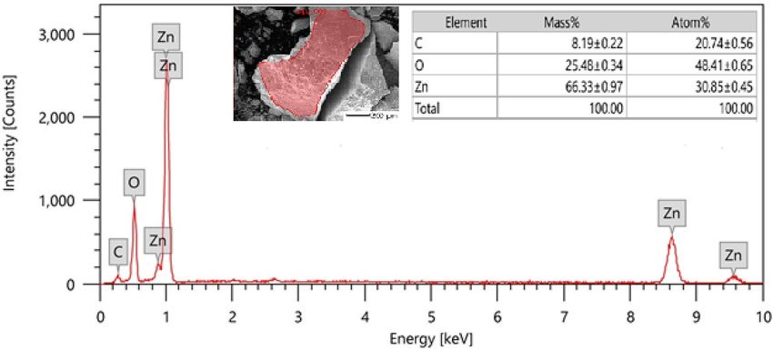
www.nature.com/scientificreports/
Figure 1. UV–vis absorption spectra of biosynthesised Alk-ZnO NPs, with maximum absorption at 334 nm.
Inset shows visual observation of color change (I) Pure precursor ( ZnSO4.7H2O), (II) reducing agent (culture
supernatant), (III) white clusters of Alk-ZnO NPs formed after mixing reducing agent and precursor.
pose environmental risks1. Microbes such as bacteria, fungi, and yeast play an important role in the biological
synthesis of metal and metal oxide NPs. Nevertheless, the biological synthesis of ZnO NPs using microbes still
remains unexplored1.
The formation of nanomaterials by extremophiles is a promising approach for the biosynthesis of metallic
nanoparticles. Extremophiles are important in the synthesis of NPs because they allow for the adjustment of the
environment to fine-tune size, shape, stability, and capping proteins. This in turn optimizes the NPs for the large
number of applications that they may fulfill14. From the literature survey, bacteria isolated from a challenged
environment like salt alkaline habitats have never been explored as potential biofactories for the synthesis and
stabilization of ZnO NPs. In an early report, Halomonas elongata IBRC-M 10214 has been recognized as a bio-
factory, capable of producing ZnO N Ps15, whereas there was no literature available on nanoparticles synthesis
by genus Alkalibacillus.
Synthesis methods that control the size and shape of nanoparticles are useful in boosting their chemico-
physical properties. It is imperative to optimize the parameters employed in the synthesis protocol to achieve
good monodispersity, greater particle stability and excellent biocompatibility16,17. The strong relationship between
size and properties of nanoparticles has provided numerous chances for many scientific a reas18.
Optimization of factors such as temperature, pH value, precursor concentration, mixture ratio, and reaction
time, and their interaction affect the biological synthesis of metal n anoparticles19 have been very poorly evalu-
ated for ZnO nanoparticles20. Statistical experiment strategy such as response surface methodology (RSM) is
used to determine the relationship between the response variable (e.g., NPs size) and the factors affecting their
size. Further, it determines the accurate optimum value (s) of the examined variables that give (s) a maximum or
minimum optimal response, depending on the product or on the process in question16. This method consists of a
number of mathematical and statistical techniques which improves and optimizes processes through evaluating
the relationship between the independent variables and r esponses21. This method was successfully applied in
many areas of biotechnology, including some recent studies for green synthesis of nanoparticles16,17,22,23.
Therefore, this innovative study explores an efficient, single step, green, environment-friendly and low-cost
method for extracellular biosynthesis of ZnO nanoparticles using Alkalibacillus sp.W7 as a reducing as well
as surface stabilizing agent. The reaction parameters including temperature, concentration of Zinc sulphate,
and pH value that affect the average diameter size of ZnO nanoparticles were statistically optimized with the
Box–Behnken experimental design method. The biosynthesized ZnO NPs were characterized and confirmed
through various techniques such as UV–visible spectroscopy followed by SEM, EDX, TEM, XRD, and FTIR.
Results and discussion
Extracellular synthesis of Alk‑ZnO NPs. To the best of our knowledge, this is the first report employing
Alkalibacillus sp. W7 as a natural nanofactory for extracellular synthesis of ZnO NPs. The preliminary detection
of nanoparticles formation was distinguished through visual observation of cloudy white clusters deposited at
the bottom of the tube compared to control (Fig. 1). The cloudy white precipitate arised as a result of reduction
of Zn+2 ions to elemental Zn0 by active metabolites in the culture filtrate and is due to the excitation of surface
plasmon resonance (SPR) of the ZnO n anoparticles24. It has been demonstrated that the proteins, enzymes
as well as other active metabolites secreted by microorganisms are the principal biomolecules involved in the
formation of metal/metal oxide n anoparticles4,25. It was found that the UV–vis absorption spectrum of biosyn-
Scientific Reports | (2021) 11:10924 | https://doi.org/10.1038/s41598-021-90408-y 2
Vol:.(1234567890)
www.nature.com/scientificreports/
Variable coded and actual values
Trial X1 pH X2 Temperature (°C) X3 ZnSO4.7H2O (mM)) Average size (nm) of ZnO NPs
1 1(9) 0 (30) 1(10) 24.03
2 0 (8) 0 (30) 0 (8) 21.4
3 1(9) 0 (30) − 1(2) 36.46
4 0 (8) 0 (30) 0 (8) 20.5
5 1(9) 1(50) 0 (8) 27.6
6 − 1(7) 1(50) 0 (8) 32.7
7 − 1(7) 0 (30) 1(10) 26.4
8 0 (8) 1(50) 1(10) 27.14
9 0 (8) 1(50) − 1(2) 33.6
10 − 1(7) 0 (30) − 1(2) 28.02
11 − 1(7) − 1(20) 0 (8) 29.4
12 0 (8) 0 (30) 0 (8) 20.7
13 0 (8) − 1(20) 1(10) 26.02
14 1(9) − 1(20) 0 (8) 25.5
15 0 (8) − 1(20) − 1(2) 31.5
Table 1. Design matrix of Box–Behnken experiments showing coded and actual values of the evaluated
independent variables along with response.
Level of
Source of Degree of Fishers’s function significance Determination
variations freedom Sum of squares Mean square F-value (P-value) coefficient R2
Regression 9 305.371 33.93 15.533 0.003783 0.96546
Residual 5 10.922 2.184
Total 14 316.293
Table 2. Analysis of variance (ANOVA) of the fitted quadratic polynomial model.
thesized Alk-ZnO NPs demonstrated SPRcharacteristic maximum absorption peak at 334 nm (Fig. 1), several
biofabricated ZnO NPs revealed comparable absorption peaks20,24,26. No other peaks were observed in the spec-
trum, indicating high purity and crystallinity of Alk-ZnO NPs. The absorption peak depends on size and shape
of synthesized NPs.
Optimization parameters of Alk‑ZnO NPs size through Box Behnken design. The average diam-
eter of metal nanoparticles can be affected by some factors including pH, salt concentration and temperature
of incubation21. Response surface methodology was applied to investigate the relation between the factors (pH,
temperature and ZnSO4.7H2O concentration) on the average size of Alk-ZnO NPs. For each trial, the average
particle size acquired from the regression equation for the 15 combinations are illustrated in Table 1.
It was observed that the smallest sized particles were produced from trials 2, 4, and 12. The determination
coefficient (R2) values provide a measure of how much variability in the observed response can be explained by
the experimental factors and their interactions. The closer the R 2 value to 1, the stronger the model is, the bet-
ter it predicts the response and has a very high c orrelation22,27. The determination coefficient ( R2) of the model
(0.9654) indicates 96.54% of variability in the response. Therefore, the present R 2-value indicates a very good
fit between the observed and predicted responses and implies that the model is reliable in the present study.
Analysis of variance (ANOVA) was used to evaluate the significance and adequacy of the regression model.
According to ANOVA results (Table 2), the model was highly appropriate; the relationship between the independ-
ent variables and the average size of nanoparticles was evident from low P-value (0.00378).
The value of adjusted-R2 (0.9033) was also very high indicating a high significance of the m odel22 and sug-
gested that the total variation of 90.33% average diameter size of nanoparticles is attributed to the independent
variables and only about 9.67% of the total variation cannot be explained by the model.
All values of model coefficients were calculated by multiple regression analysis. The significance of each
coefficient was determined by the Student’s t-test and P-value, as illustrated in Table 3. Based on the analysis
of regression coefficients of the quadratic model, among the three tested variables, the linear effects of reaction
pH (X1), and reaction temperature (X2) exhibited a negative relationship on particle size, while zinc sulphate
concentration (X3) exhibited a positive relationship.
P-value indicates the significance of each of the coefficients which is important to understand the pattern of
the mutual interaction among variables. The smaller the magnitude of the P, the more significant is the corre-
sponding coefficient. According to the degree of significance (P ≤ 0.01, significant at 99% level), the linear term
of pH (X1) had the most dramatic negative significant effect on NPs size among all linear effects. In contrast, the
Scientific Reports | (2021) 11:10924 | https://doi.org/10.1038/s41598-021-90408-y 3
Vol.:(0123456789)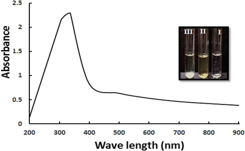
www.nature.com/scientificreports/
Variables Coefficients Main effect t Stat P value Confidence level (%)
Intercept 230.9812 461.962 4.441171 0.00000 100
X1 (pH) − 50.2081 − 100.416 − 4.0173 0.010148 99.10991
X2 (°C) − 1.00097 − 2.00194 − 2.06055 0.094367 91.39283
X3 (ZnSO4) 2.895308 5.790616 1.762809 0.138222 85.95297
X1X2 − 0.0418 − 0.08359 − 0.86907 0.424555 58.14298
X1X3 − 0.79829 − 1.59659 − 4.56962 0.006004 99.43325
X2X3 − 0.00029 − 0.00059 − 0.02589 0.980343 1.992987
X1X1 3.549259 7.098519 4.601892 0.00583 99.48717
X2X2 0.020132 0.040264 5.105393 0.003753 99.66835
X3X3 0.22403 0.448059 3.309396 0.021255 98.13707
Table 3. Regression analysis for optimizing ZnO nanoparticles size using Box–Behnken design.
Figure 2. A Pareto chart illustrating the main effects of Box-Behnken result, with the order of significance of
the variables affecting Alk-ZnO NPs size. Bars that exceed the vertical line indicate the significance of the terms
(P < 0.05).
quadratic terms for pH (X12), temperature (X22) and metal concentration (X32) had a significant positive effect;
meaning that they could be considered as limiting factors and little variation in their values will alter the size of
nanoparticles. In addition, only interaction between pH ( X1) and metal concentration ( X3) was noted to have a
negative significant effect.
The Pareto chart identified as a useful tool for determining the most significant e ffects28 was used to illustrate
the order of significance of the different factors on Alk-ZnO NPs size. Each bar length on a standardized Pareto
chart (Fig. 2) is proportional to the absolute value of its related estimated effect or regression coefficient.
Design Expert software was used to fit a second order polynomial model to the experimental results to predict
the size of NPs within the experimental constraints. The following second order polynomial equation represents
the empirical relationship between NP size and the three independent variables:
Y(NPsSize) = 230.9812 − 50.2081X1 − 1.00097X2 + 2.895308X3 − 0.0418X1 X2 − 0.79829X1 X3
− 0.00029X2 X3 + 3.549259X12 + 0.020132X22 + 0.22403X32
The coefficients sign determines the response performance. Here, coefficient with positive effect means
decrease in particle size, while a negative effect means increase in particle size. In addition, at higher absolute
value of a coefficient, the effect of variable is higher. Therefore, according to the previous equation, pH of the
reaction is the variable with the strongest influence on NPs size.
Scientific Reports | (2021) 11:10924 | https://doi.org/10.1038/s41598-021-90408-y 4
Vol:.(1234567890)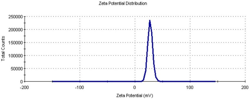
www.nature.com/scientificreports/
Figure 3. Three-dimensional response surface graphs and contour plots showing the effect of independent
variables on the average size of Alk-ZnO NPs; (A) Effect of metal concentation and temperature, (B) Effect of
metal concentation and pH and (C) Effect of temperature and pH.
From polynomial models, response surface curves were plotted using Design-Expert 7.0, a trial version
software to determine the effects of factors and their combinations that optimize the response. These three-
dimensional plots and their respective contour plots provided a visual interpretation of the interaction of two
test variables with the third one held at a constant level and facilitated the location of optimum values of the
variables for synthesis of the smallest NPs. Significance of the interaction plots between the corresponding
variables was indicated by an elliptical or circular or saddle nature of the contour p lots23. The contour plots
(Fig. 3) reveal that the optimal point for decreasing the average diameter of Alk-ZnO NPs was located inside the
experimental region, which confirms the adequacy of the independent variable concentration ranges used in this
study. The response surfaces obtained were concave, indicating that the operating conditions were well-defined
and optimum. Figure 3A represents the interaction between temperature and metal concentration and reveals
that the average diameter size decreased with gradual increasing in both metal concentration and temperature.
This might be explained that increasing metal ion concentration to a certain point allows particle growth at a
faster rate and generates NPs with smaller size. Moreover, higher metal precursor concentration in the reac-
tion mixture may increase the aggregation of the growing NPs, which resulted in the formation of larger NPs
size21,29. Phanjom &Ahmed30 reported that AgNO3 concentration up to 8 mM, resulted in the formation of a
smaller nanoparticle size, while the size increased at 9 and 10 mM concentrations. This effect was attributed to
the lack of functional groups available for the reaction when the metal precursor concentration was increased.
To achieve the smallest particle size, the middle levels of both metal concentration and temperature were used.
Higher temperature resulted in the gradual increase in the NPs size. Unsuitable temperatures lead to increased
nanoparticle size and loss of stability, as a result of denaturation, inactivation or low activity of the reductive
enzymes and other active molecules involved in the biogenesis p rocess31,32. Additionally, it was suggested that
the increase in temperature decreases NPs size which is normally due to increment in reaction rate and kinetic
energy at higher temperatures29,33. Generally, the effect of temperature on the size and stability of the synthesized
nanoparticles varies according to the microorganism used9. Figure 3B represents the mutual interaction of pH
and metal concentration on Alk-ZnO NPs biosynthesis when temperature was fixed at optimum value. The
average diameter size decreased gradually with the increment of pH but at higher pH values, the size slightly
increased. This may be due to the inactivation of biomolecules involved in the synthesis of Alk-ZnO NPs at
low and high p Hs21. Generally, pH plays a significant role in nanoparticles synthesis, mainly because it has the
ability to alter the shape of biomolecules that is responsible in capping and stabilizing the N Ps34. Also, plays a
Scientific Reports | (2021) 11:10924 | https://doi.org/10.1038/s41598-021-90408-y 5
Vol.:(0123456789)www.nature.com/scientificreports/
Figure 4. Scanning electron micrograph of Alk-ZnO NPs synthesized under optimum conditions at
35,000 × magnification.
substantial role in the oxidation state and reducing power of enzymes and secondary metabolites present in
cell-free extract35. Zhang et al.36 stated that pH of the medium solution influences the size and the texture of the
synthesized nanoparticles. Modification in pH leads to alteration in the overall charge of bioactive molecules,
which in turn facilitates their binding affinity and hence biomineralization of metal ions into nanoparticles37. The
obtained data reveal that alkaline pH and a high metal concentration are conditions that will generate smallest
size of Alk-ZnO NPs. While, a higher or lower pH generated the larger size, which indicated that the catalytic
activity of enzymes responsible for Zn2+ ions reduction appeared to be deactivated, thus causing an increase in
the size of NPs. Figure 3C shows the combined effect of pH and temperature when metal concentration value
was fixed at optimum value. It showed that higher values of pH and temperature supported the increase of Alk-
ZnO NPs size. The smallest size was obtained in the middle levels of both pH and temperature. The synthesis of
small sized ZnO NPs could be attributed to the increase in nucleation process rate as a result of a large number
of OH ions. The finding is in agreement with that reported by Mata et al.38 who indicated that higher pH favored
higher reducing power. Possible reason is that during the elevated pH, the reaction rate will be increased with
subsequent crystallization into smaller particles, which involved the nucleation and growth processes of smaller
particles from metal n uclei39.
The study of physicochemical parameters of the reaction such metal ions concentrations, pH and temperature
on Alk-ZnO NPs formation represents the effect of the direct connection between these factors on the particles
size. By solving the model regression equation using the SOLVER function of MICROSOFT EXCEL tools, the
smallest size Alk-ZnO NPs and the optimum values of these variables can be acquired. The optimal values of
the variables were temperature 33 °C, metal concentration 8 mM and pH 8. The model predicts the smallest NPs
size that can be obtained using the above optimum conditions to be 20.4 nm.
Verification of the model. In order to validate the obtained data and to evaluate the accuracy of the
applied Box-Behnken statistical design, a verification experiment with optimum concentration of examined
variables was carried out in three replicate and compared with the predicted data. The results obtained demon-
strated that the average size of nanoparticles was 17.5 nm, while the predicted value from the polynomial model
was 20.4 nm. There was a little difference between the experimental and the predicted data. The verification
revealed a high degree of accuracy of the model, indicating the model validation under the tested conditions.
The measured particle size was less than the predicted by 14.22%. Verification experiment indicated validity, and
reliability of response model and applicability of RSM to minimize the size of Alk-ZnO NPs. Additionally, in the
present study, Al. sp.W7 could synthesize 0.493 g of NPs /L of bacterial supernatant in 24 h.
Characterization of Alk‑ZnO NPs. UV–visible spectroscopy. Alk-ZnO NPs synthesized under optimal
conditions exhibited the surface plasmon resonance band shifted to a lower wavelength 310 nm, due to their
large excitation binding e nergy40. It is known from absorption spectroscopy that as particle size decreases, the
band gap energy increases. There is also an opposite ratio between band gap and the absorption wavelength41.
The corresponding band gap was found to be 4.00 eV. Our data are in good agreement with the earlier reports on
biosynthesis of ZnO N Ps25,42. Aggregation is a common problem with nanoparticles which greatly decreases the
surface area and, as a result, affects their physicochemical and biological properties43. It is worth to mention that
the nanoparticles examined after 10 months using UV–vis spectroscopy showed a peak at 310 nm, indicating
their stability (Supplementary Figure S1).
Scanning electron microscopy analysis. Scanning electron microscopy provided further insight into the mor-
phology and size of the synthesized ZnO NPs. The micrograph illustrated in Fig. 4 proved that they had nano-
sized range, smooth, irregular to nearly spherical with a uniform distribution with an average size ranging from
5 to 45 nm.
Scientific Reports | (2021) 11:10924 | https://doi.org/10.1038/s41598-021-90408-y 6
Vol:.(1234567890)www.nature.com/scientificreports/
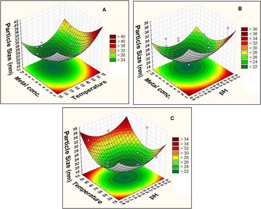
Figure 5. Energy dispersive x-ray spectroscopic analysis of Alk-ZnO NPs. Other elemental signal as carbon
was also recorded possibly from enzymes or proteins present in the culture supernatant. Inset image of scanned
area and elemental composition.
Energy‑dispersive X‑ray spectrum. The Alk-ZnO NPs were subjected to an EDX spectrum to quantify the mix-
ture of metal and oxides present in the sample. Figure 5 clearly illustrates that the EDX spectrum of the fabri-
cated NPs was recorded in the spot-profile mode from one of the densely populated nanoparticles area. Distinct
peaks obtained for zinc and oxygen atoms represent the formation of ZnO NPs. The elemental profile showed
strong higher peak at 1 keV, characteristic to Zn and confirmed the formation of ZnO NPs44,45. The weak signal
of C atoms was also recorded and likely due to X-ray emission from carbohydrates/proteins/enzymes present in
the bacterial cell free filtrate indicating their involvement in reduction and capping of the synthesized ZnO NPs.
The atomic percentages of the elements inset of Fig. 5 reveal zinc as the major element comprising more than
66.33% of total constituent along with oxygen 25.48%, which clearly also confirms the high purity of mediated
zinc oxide nanoparticles. Additionally, the optical absorption signals of zinc were observed due to surface plas-
mon resonance of ZnO NPs34,45, which is in agreement with previous r eports46,47. The impurity free nanoparti-
cles are promising in different application fields as antitumor, anti-microbial and antibiofilm. Our results are in
agreement with the results obtained by microbially synthesized ZnO NPs20,47,48, where pure form of nanoparticle
was clearly depicted through EDX imaging.
Transmission electron microscopy. Transmission electron microscopic analysis provided exact morphology and
size of Alk-ZnO NPs acquired from optimized synthesis conditions and revealed predominantly irregular to
nearly hexagonal and quasi-spherical with legitimately agglomeration (Fig. 6A,B). This is a typical phenomenon
of interaction of moisture and ZnO and inter-particle interactions (Van der Waals, electrostatic and magnetic
forces)49,50. The size distribution patterns reveal that the synthesized NPs ranged from 1–30 nm with an average
size of 17 ± 1 nm (Fig. 6C). This result is in accordance with crystallite size calculated from the X-ray diffraction.
Similar hexagonal—quasi-spherical zinc oxide nanoparticles have been observed in Rhodococcus pyridinivorans
NT246 and Lactobacillus plantarum VITES0751. It was previously reported that the smaller the size of ZnO NPs,
the greater efficacy in inhibiting the growth of bacteria52.
X‑ray diffraction analysis. X-ray diffraction is taken in order to further confirm ZnO phase of the nano-
particles. The XRD pattern of the Alk-ZnO NPs fabricated under optimum conditions provided by the RSM
having 2θ values with strong diffractions peaks appear at 31.16°(100), 33.83°(002), 35.65°(101), 46.98°(102),
56.05°(110), 62.344°(103), 65.71°(200), 67.46°(112), 68.27°(201), 72.52°(004) and 76.84°(202) (Fig. 7). The XRD
diffraction peaks were in good agreement with the reported literature and matched well with wurtzite ZnO of
the Joint Committee on Powder Diffraction Standards (JCPDS) Card number 36–1451. Thus XRD pattern shows
ZnO NPs with a fine hexagonal crystalline structure formed with significant agreement with this reference file,
and no characteristic diffraction peaks were observed other than ZnO, indicating that the bio-assisted NPs were
free from other phase impurities and significantly had a high phase p urity13,53, which was confirmed by EDX
analysis results. In addition, the strong and narrow diffraction peaks indicated that the ZnO nanoparticles were
of well crystalline s tructure46. Our results are in good agreement with various XRD diffractogram data reported
for biologically synthesized ZnO n anoparticles24,46. The average crystalline size of NPs corresponding to the
most intense diffraction peak at 2θ = 35.65o (101) as calculated by Debye–Scherrer′s equation was found to be
19.5 nm. XRD results confirmed that Alk-ZnO NPs are highly crystalline, having hexagonal wurtzite crystalline
structure which are compatible with other researchers’ w ork20,55. XRD results also reflect that the crystallization
of the bio-organic phase occurs on the surface of the ZnO NPs. The biosynthesized ZnO nanoparticles shared
accord with the crystallite size calculated from TEM.
Scientific Reports | (2021) 11:10924 | https://doi.org/10.1038/s41598-021-90408-y 7
Vol.:(0123456789)www.nature.com/scientificreports/
Figure 6. Transmission electron microscopic image of Alk-ZnO NPs (A) low magnification (X80k), (B) high
magnification (X120k), and (C) particle size distribution histogram.
Figure 7. X-ray diffraction pattern of ZnO nanoparticles synthesized by Al. sp. W7 supernatant. All peaks
indicate purity and crystalline nature. No traces of other impurity phases were detected.
Scientific Reports | (2021) 11:10924 | https://doi.org/10.1038/s41598-021-90408-y 8
Vol:.(1234567890)www.nature.com/scientificreports/
Figure 8. Zeta potential of Alk-ZnO NPs dispersed in water.
Figure 9. Fourier transforms infrared spectroscopy (FTIR) of Alk-ZnO NPs.
Zeta potential. Zeta potential of the suspension is one of the key parameters in assessing the stability of the
synthesized nanoparticle as it gives the net electrostatic potential of any particle in suspension. The biosynthe-
sized zinc oxide nanoparticles possessed a high positive potential value of 27.5 mv (Fig. 8) which indicates that
they were highly stable and prevented agglomeration due to strong electrostatic repulsive forces between them.
Nanoparticles with high negative or positive Zeta potential never aggregate due to electrostatic force of repulsion
occulate51. It is generally considered that the zeta potential values
while particles with low zeta potential tend to fl
(+ 30 mV to − 30 mV) result in good nanoparticle stability17,51, this result is comparable with zeta potential of
zinc nanoparticles synthesized by Pseudomonas hibiscicola (24.64 mV)24. Positively charged nanoparticles show
better attachment towards negatively charged cell membrane surface due to electrostatic attraction which leads
to the penetration of ZnO NPs into the cells and thus enhanced toxicity towards m icroorganism56.
Fourier transform infrared. FTIR spectrum was performed to determine the surface chemistry of the ZnO
NPs and is used to access the details of functional groups involved in the biomolecules responsible for reducing
and capping the bio-reduced ZnO NPs25,57. The FTIR spectra for ZnO NPs are presented in Fig. 9. The broad
stretching vibrational band with high intensity observed at 3369 c m–1 is characteristic for hydroxyl group (OH)
or can correspond to the protein N‒H amide I51. It is well known that protein-nanoparticle interactions can
occur either through free amine groups or cysteine residues in proteins and via the electrostatic attraction of
negatively charged carboxylate groups in e nzymes53. The absorption peak at 2922 c m−1 is attributed to C-H
stretch of the methylene groups of the protein stretching21. Bands at 2352 and 2109 cm−1 are associated with the
presence of primary amines and sulfur c ompounds44. These peaks indicate the presence of proteins and other
organic residues, which might have produced extracellularly by Al.sp. W7. Moreover, the absorption peak at
Scientific Reports | (2021) 11:10924 | https://doi.org/10.1038/s41598-021-90408-y 9
Vol.:(0123456789)www.nature.com/scientificreports/
1633 cm−1 corresponds to the stretching vibration of N–H bond of primary amines, alkyl C=C aromatic stretch-
ing and open chain amino group in proteins26. The absorption bands around 1630 cm−1 and 1384 cm−1 are due to
asymmetric and symmetric of stretching carboxylate attached to the ZnO nanoparticles during synthesis58. The
absorption peaks at 1470 and 1380 cm−1 represent the carboxylate group (COO−)59. According to Tiwari et al.60,
metal oxides exhibit absorption bands well below 1200 cm−1 arising due to interatomic vibrations. Generally
metal oxides give absorption peaks in the regions between 500 and 900 c m−154. Strong peaks observed at 578.95
cm−1 and 906 cm−1 prominently indicate Zn–O bond. These findings are in accordance with literature values20,41.
The stretching mode peaks are indicative of the successful synthesis of ZnO nanoparticles from Al.sp. W7 and
FTIR spectral analysis reveals the subtle variations in biological components of culture supernatant associated
with ZnO NPs formed. The functional groups present in secreted proteins, enzymes and secondary metabolites
in strain W7 culture supernatant are responsible for reduction of metal ions and biosynthesis of metallic nano-
particles. The significant reduction capabilities of the biofunctional groups (R-OH, R-NH2, R-COO−) would
reduce Zn2+ as electron acceptor to Zn0 resulting in the formation of ZnO NPs. These metabolites not only act
as reducing agents but also act as capping agents providing stabilization against aggregation. These results are in
agreement with previous report in green ZnO NPs synthesis13,61.
Alkalibacillus sp. W7 nanoparticles showed good antibacterial activity, inhibiting the growth of Gram-negative
and Gram-positive bacteria and also efficient photocatalytic degradation of the methylene blue and methyl orange
dye under solar i rradiation62. Moreover, biofilm inhibition activity, anticancer and antioxidant potentials of the
synthesized nanoparticles were also investigated.
Materials and methods
Microorganism and cultural conditions. Alkalibacillus sp. W7 an aerobic Gram-positive, rod-shaped
bacterium, isolated from hypersaline Lake Al- Hamra in Wadi An-Natrun, Egypt was used in this study. The 16S
rDNA sequence was submitted to NCBI GenBank with an assigned accession number L C16482963. The strain
was maintained on modified Horikoshi-I medium64, containing (g/L): glucose,10; yeast extract,5; peptone,5;
K2HPO4,1; MgSO4.7H2O,0.2; NaCl, 50; agar, 20; pH 8 and after an incubation period of 48 h at 35 °C was kept at
4 °C and subcultured every month.
Synthesis of Alk‑ZnO NPs. Seed culture was prepared firstly by cultivation of Alkalibacillus sp. W7 aerobi-
cally in 250 ml Erlenmeyer flask containing 50 mL of modified Horikoshi-1 medium and incubated in a rotatory
shaker (130 rpm) at 35 °C for 48 h ( OD600 = 1.0 ± 0.2). Sterile 50 ml of modified Horikoshi-1 medium in 250 ml
Erlenmeyer were inoculated with 2 ml of the fresh inoculum and incubated aerobically on a rotatory shaker
incubator for 48 h at 35 °C and 130 rpm. The cell free supernatant (CFS) collected by centrifugation at 5000
rpm for 15 min, then filtered with a 0.22 μm membrane filter (Millipore, USA) was used as a reducing agent
for synthesis of Alk-ZnO NPs. Aqueous metal solution of 1 mM Z nSO4.7H2O in distilled water was used as a
precursor. Ten ml of zinc sulphate solution were mixed with 5 ml of Alkalibacillus sp. W7 cell free supernatant
(2:1, V/V), and incubated for 48 h in dark under static condition at 35 °C to complete the ZnO NPs formation.
The color change was observed in the reaction mixture due to the reduction reaction. Simultaneously, control of
sterile uninoculated media mixed with precursor salt solution was also maintained at same condition along with
the experimental tubes in triplicate.
Statistical optimization of ZnO nanoparticles biosynthesis. Response surface methodology (RSM)
is a collection of mathematical and statistical techniques which has been known as a tool for modeling within
minimum number of experimental runs, determining the effects of important parameters and to identify their
prominent interactions for the best response21,23.
Response surface methodology based on Box-Behnken d esign27 was used to evaluate the relationship between
the factors that optimize the response. The combined effect of three active independent variables; pH (X1), reac-
tion temperature (X2) and Zinc sulphate concentration (X3) was considered for evaluation. Their effects were
evaluated with respect to responce (NPs size). The levels of each individual variable were selected based on the
results of preliminary experiments, involving optimization using the one-factor-at-a-time method. Table 1 shows
the experimental Box-Behnken design matrix in the coded and actual levels of selected independent variables
developed by Design Expert software (Version 7, Stat-Ease, Inc., USA) and the responce.
Statistical analysis. Hence, for a three-variable design, a total of 15 experimental runs were performed and
their observations were fitted in the second order polynomial model, which is a mathematical expression used
to determine the linear, quadratic, and cross effects of the tested independent variables to find the result and
approximate the real answer as follows:
Y = β0 + β1 x1 + β2 x2 + β3 x3 + β12 x1 x2 + β13 x1 x3 + β23 x2 x3 + β11 x21 + β22 x22 + β33 x23
where Y is the dependent variable (size of NPs), X 1, X2, and X
3 are the independent variables, β0 is the regres-
sion coefficient at center point, β1, β2, and β3 are the linear coefficient; β12, β13, and β23 are the second order
interaction coefficients, and β11, β22, and β33 are the quadratic coefficients.
Purification of Alk‑ZnO NPs. The nanoparticles were separated from the reaction mixture by high-speed
centrifugation at 15,000 rpm for 10 min, then dispersed in sterilized distilled water and centrifuged again. This
action was carried out for three times to remove the unreacted metals and the residual biological molecule impu-
rities or any residual metabolites from native nanoparticle samples to obtain purified NPs6. Finally, the obtained
Scientific Reports | (2021) 11:10924 | https://doi.org/10.1038/s41598-021-90408-y 10
Vol:.(1234567890)www.nature.com/scientificreports/
white color powder was washed with ethanol to remove ionic impurities, then dried overnight in an oven at 40
°C and characterized using different techniques.
Characterization of biogenic ZnO NPs. UV–visible spectroscopy. The biosynthesized ZnO NPs were
characterized using UV–Vis spectrophotometer (Thermo Scientific Evolution TM 300) in the wavelength region
between 200-nm and 900-nm operated at a resolution of 1 nm and maximum absorbance was d etermined65.
X‑ray diffraction. X-ray diffraction was performed using Phaser XRD-D2 powder X-ray diffractometer
(Bruker, Germany) supplied with Cu-Kα radiation over a wide range of the Bragg angles θ (10° ≤ 2θ ≤ 80°) oper-
ated at 30 kV, 10 mA. Data was compared with known standard data published by the Joint Committee on
Powder Diffraction Standards (JCPDS Card No. 36–1451). The mean of the crystallite size (nm) was determined
from the XRD line broadening measurement using Debye Scherrerr’s equation:
D = 0.89 /(βCosθ)
where λ is the wavelength of X-ray radiation, λ = 1.5406 Å, β is the full width at the half maximum (FWHM) of
the most intense diffraction peak, and θ is the diffraction angle.
Scanning electron microscopy. The shape and size of the ZnO NPs were determined using SEM JEOL IT 200,
(JOEL Corp, Tokyo, Japan). The biosynthesized powder was mixed into doubled distilled water and sonicated for
30 min. A small drop of this sample was allowed to dry on a glass slide to make a thin layer of NPs.
Energy‑dispersive X‑ray analysis. The structure of Alk-ZnO NPs was characterized by energy-dispersive analy-
sis X-ray (EDX) spectrum using X-ray micro-analyzer (Module Oxford 6587INCA X-sight) coupled with SEM–
IT 200 scanning electron microscope (JOEL Corp, Tokyo, Japan) operated at 20 kV of an accelerating voltage for
compositional analysis as well as conformation for the presence of elemental zinc.
Transmission electron microscopy. The size and shape of Alk-ZnO NPs were observed using a transmission
electron microscope TEM JEM-1400 plus, (JOEL Corp, Tokyo, Japan) operated at an accelerating voltage of 50
kV. A drop of ZnO NPs solution was placed on a carbon coated copper grid followed by water evaporation. The
average particles size and the size distribution histogram were determined from microscopic images using a
particle size analyzer programe, Image J processing and analysis software (http://imagej.nih.gov/ij/index.html).
Zeta potential measurement. The zeta potential was performed by dispersing 1 mg of ZnO NPs in 10 ml of
distilled water followed by sonication for 5 min then measurement using Malvern Zetasizer Nano ZS analyzer
(Malvern Instruments, Malvern, UK)66.
Fourier transforms infrared spectroscopy. FTIR analysis was done for identification of presumable biomolecules
n+2 ions and formation and stability of ZnO NPs. Sample was prepared by
responsible for the reduction of the Z
mixing Alk-ZnO NPs with potassium bromide KBr using hydraulic press and then dried to remove the moisture
content. Infrared spectra were recorded over a range of 450–4000 cm–1 with FTIR spectrophotometer (FTIR
spectrum Version 10.5.3, Perkin Elmer).
Conclusions
The present study, built up the first ever account of use of culture supernatant of Alkalibacillus sp. W7 towards
extracellular synthesis of zinc oxide nanoparticles. The response surface methodology was applied to optimize
the conditions influencing the Alk-ZnO NPs size. The results revealed that the optimization of the reaction
parameters was essentially needed and directly affected ZnO NPs size. The fitness of the model was verified and
predicted values were confirmed. The synthesized Alk-ZnO NPs with optimized conditions showed maximum
absorption peak at 310 nm. The XRD results confirmed the efficiency of the synthesis process, evidencing the
production of single crystalline NPs with hexagonal wurtzite structure. The average size of synthesized NPs was
17.5 nm, exhibiting quasi-spherical with smooth surface which was confirmed by TEM and SEM analyses. EDX
results confirmed the presence of zinc and oxygen with a pure phase. FTIR analysis clearly showed the formation
of ZnO NPs and verified the presence of various biomolecules secreted by Alkalibacillus sp. W7 acting as cap-
ping and stabilizing agents. This green synthesis does not need any additive stabilizer which is a positive point
in comparison with chemical methods. Our results confirm the potential of Alkalibacillus sp. W7 for ZnO NPs
biosynthesis in a simple, fast, cost-effective, convenient and ecofriendly way. Furthermore, the results confirmed
that the response surface methodology was beneficial for smaller ZnO NPs size synthesis with great stability over
time, and the reaction parameters are directly affecting their size.
Data availability
All data produced during this study are included in this published article.
Received: 22 February 2021; Accepted: 11 May 2021
Scientific Reports | (2021) 11:10924 | https://doi.org/10.1038/s41598-021-90408-y 11
Vol.:(0123456789)www.nature.com/scientificreports/
References
1. Yusof, H. M., Mohamad, R., Zaidan, U. H. & Abdul Rahman, N. Microbial synthesis of zinc oxide nanoparticles and their potential
application as an antimicrobial agent and a feed supplement in animal industry: a review. J. Anim. Sci. Biotechnol. 10, 10–57. https://
doi.org/10.1186/s40104-019-0368-z (2019).
2. Basnet, P., Chanu, T. I., Samanta, D. & Chatterjee, S. A review on bio-synthesized zinc oxide nanoparticles using plant extracts as
reductants and stabilizing agents. J. Photochem. Photobiol. B Biol. 183, 201–221. https://doi.org/10.1016/j.jphotobiol.2018.04.036
(2018).
3. Mandeep, D. & Shukla, P. Microbial nanotechnology for bioremediation of industrial wastewater. Front. Microbiol. https://doi.org/
10.3389/fmicb.2020.590631 (2020).
4. Sepahvand, M., Buazar, F. & Sayahi, M. H. Novel marine-based gold nanocatalyst in solvent-free synthesis of polyhydroquinoline
derivatives: Green and sustainable protocol. Appl. Organomet. Chem. 34, 1–11. https://doi.org/10.1002/aoc.6000 (2020).
5. Moavi, J., Buazar, F. & Sayahi, M. H. Algal magnetic nickel oxide nanocatalyst in accelerated synthesis of pyridopyrimidine deriva-
tives. Sci. Rep. 11, 6296. https://doi.org/10.1038/s41598-021-85832-z (2021).
6. Hussain, I., Singh, N. B., Singh, A., Singh, H. & Singh, S. C. Green synthesis of nanoparticles and its potential application. Biotechnol.
Lett. 38, 545–560. https://doi.org/10.1007/s10529-015-2026-7 (2016).
7. Gholizadeh, B. S., Buazar, F, Hosseini, S. M. & Mousavi, S.M. Enhanced antibacterial activity, mechanical and physical properties
of alginate/hydroxyapatite bionanocomposite film. Biomac. https://doi.org/10.1016/j.ijbiomac.2018.05.104 (2017).
8. Rezazadeh, N. H., Buazar, F. & Matroodi, S. Synergistic effects of combinatorial chitosan and polyphenol biomolecules on enhanced
antibacterial activity of biofunctionalaized silver nanoparticles. Sci. Rep. 10, 1–13. https://doi.org/10.1038/s41598-020-76726-7
(2020).
9. Guilger-Casagrande, M. & de Lima, R. Synthesis of silver nanoparticles mediated by fungi: A review. Front. Bioeng. Biotechnol.
https://doi.org/10.3389/fbioe.2019.00287 (2019).
10. Akbar, S. et al. An overview of the plant-mediated synthesis of zinc oxide nanoparticles and their antimicrobial potential. Inorg.
Nano-Met. Chem. https://doi.org/10.1080/24701556.2019.1711121 (2020).
11. Jiang, J., Pi, J. & Cai, J. The advancing of zinc oxide nanoparticles for biomedical applications. Bioinorg. Chem. Appl. https://doi.
org/10.1155/2018/1062562 (2018).
12. Buazar, F., Alipouryan, S., Kroushawi, F. & Hossieni, S. A. Photodegradation of odorous 2-mercaptobenzoxazole through zinc
oxide/hydroxyapatite nanocomposite. Appl. Nanosci. 5, 719–729. https://doi.org/10.1007/s13204-014-0368-4 (2015).
13. Buazara, F. et al. Potato extract as reducing agent and stabiliser in a facile green one-step synthesis of ZnO nanoparticles. J. Exp.
Nanosci. 11, 175–184. https://doi.org/10.1080/17458080.2015.1039610 (2016).
14. Beeler, E. & Singh, O. V. Extremophiles as sources of inorganic bionanoparticles. World J. Microbiol. Biotechnol. 32, 156. https://
doi.org/10.1007/s11274-016-2111-7 (2016).
15. Taran, M., Rad, M. & Alavi, M. Biosynthesis of T iO2 and ZnO nanoparticles by Halomonas elongata IBRC-M 10214 in different
conditions of medium. BioImpacts 8, 81. https://doi.org/10.15171/bi.2018.10 (2018).
16. Keijok, W. J. et al. Controlled biosynthesis of gold nanoparticles with Coffea arabica using factorial design. Sci. Rep. 9, 1–10. https://
doi.org/10.1038/s41598-019-52496-9 (2019).
17. Manosalva, N. et al. Green synthesis of silver nanoparticles: Effect of synthesis reaction parameters on antimicrobial activity. World
J. Microbiol. Biotechnol 35, 88. https://doi.org/10.1007/s11274-019-2664-3 (2019).
18. Balakumaran, M. D., Ramachandran, R., Balashanmugam, P., Mukeshkumar, D. J. & Kalaichelvan, P. T. Mycosynthesis of silver
and gold nanoparticles: optimization, characterization and antimicrobial activity against human pathogens. Microbiol. Res. 182,
8–20. https://doi.org/10.1016/j.micres.2015.09.009 (2016).
19. Gupta, M., Tomar, R. S., Kaushik, S., Sharma, D. & Mishra, R. K. Effective antimicrobial activity of green ZnO nano particles of
Catharanthus roseus. Front. Microbiol. 9, 2030. https://doi.org/10.3389/fmicb.2018.02030 (2018).
20. Motazedi, R., Rahaiee, S. & Zare, M. Efficient biogenesis of ZnO nanoparticles using extracellular extract of Saccharomyces cerevi‑
siae: Evaluation of photocatalytic, cytotoxic and other biological activities. Bioorg. Chem. 101, 103998. https://doi.org/10.1016/j.
bioorg.2020.103998 (2020).
21. Barabadi, H. et al. Microbial mediated preparation, characterization and optimization of gold nanoparticles. Braz. J. Microbiol.
45, 1493–1501. https://doi.org/10.1590/s1517-83822014000400046 (2014).
22. Camas, M., Celik, F., Sazak Camas, A. & Ozalp, H. B. Biosynthesis of gold nanoparticles using marine bacteria and Box–Behnken
design optimization. Part. Sci. Technol. 37, 31–38. https://doi.org/10.1080/02726351.2017.1287794 (2018).
23. Arasu, M. V. et al. One step green synthesis of larvicidal, and azo dye degrading antibacterial nanoparticles by response surface
methodology. J. Photochem. Photobiol. B Biol. 190, 154–162. https://doi.org/10.1016/j.jphotobiol.2018.11.020 (2019).
24. Punjabi, K. et al. Efficiency of biosynthesized silver and zinc nanoparticles against multi drug resistant pathogens. Front. Microbiol.
9, 2207. https://doi.org/10.3389/fmicb.2018.02207 (2018).
25. Moghaddam, A. B. et al. Biosynthesis of ZnO nanoparticles by a new Pichia kudriavzevii yeast strain and evaluation of their
antimicrobial and antioxidant activities. Molecules 22, 872. https://doi.org/10.3390/molecules22060872 (2018).
26. Kalpana, V. N. et al. Biosynthesis of zinc oxide nanoparticles using culture filtrates of Aspergillus niger: Antimicrobial textiles and
dye degradation studies. OpenNano 3, 48–55. https://doi.org/10.1016/j.onano.2018.06.001 (2018).
27. Box, G. E. P., Hunter, J. S. & Hunter, W. G. Statistics for Experimenters: Design, Innovation, and Discovery, (2nd ed.). (eds. Hunter,
J.S., Kendall, D.G.) (John Wiley and Sons, New York,Wiley, 2005).
28. Haaland, P.D. Experimental Design in Biotechnology, (Marcel Decker, New York. 105 (CRC Press, 1989).
29. Ma, L. et al. Optimization for extracellular biosynthesis of silver nanoparticles by Penicillium aculeatum Su1 and their antimicrobial
activity and cytotoxic effect compared with silver ions. Mater. Sci. Eng. C 77, 963–971. https://doi.org/10.1016/j.msec.2017.03.294
(2017).
30. Phanjom, P. & Ahmed, G. Effect of different physicochemical conditions on the synthesis of silver nanoparticles using fungal cell
filtrate of Aspergillus oryzae (MTCC No. 1846) and their antibacterial effect. Adv. Nat. Sci. Nanosci. Nanotechnol. 8, 045016. https://
doi.org/10.1088/2043-6254/aa92bc (2017).
31. Birla, S., Gaikwad, S., Gade, A. & Rai, M. Rapid synthesis of silver nanoparticles from Fusarium oxysporum by optimizing phys-
icocultural conditions. Sci. World J. 10, 1–12. https://doi.org/10.1155/2013/796018 (2013).
32. Husseiny, S. M., Salah, T. A. & Anter, H. A. Biosynthesis of size controlled silver nanoparticles by Fusarium oxysporum, their
antibacterial and antitumor activities. Beni-Seuf Univ. J. Appl. Sci. 4, 225–231. https://doi.org/10.1016/j.bjbas.2015.07.004 (2015).
33. Saxena, J., Sharma, P. K., Sharma, M. M. & Singh, A. Process optimization for green synthesis of silver nanoparticles by Sclerotinia
sclerotiorum MTCC 8785 and evaluation of its antibacterial properties. Springer Plus 5, 1–10. https://doi.org/10.1186/s40064-016-
2558-x (2016).
34. Umar, H., Kavaz, D. & Rizaner, N. Biosynthesis of zinc oxide nanoparticles using Albizia lebbeck stem bark, and evaluation of its
antimicrobial, antioxidant, and cytotoxic activities on human breast cancer cell lines. Int. J. Nanomed. 14, 87–100. https://doi.org/
10.2147/IJN.S186888 (2019).
35. Das, S. K., Das, A. R. & Guha, A. K. Microbial synthesis of multishaped gold nanostructures. Small 6, 1012–1021. https://doi.org/
10.1002/smll.200902011 (2010).
Scientific Reports | (2021) 11:10924 | https://doi.org/10.1038/s41598-021-90408-y 12
Vol:.(1234567890)www.nature.com/scientificreports/
36. Zhang, Y., Nayak, T., Hong, H. & Cai, W. Biomedical applications of zinc oxide nanomaterials. Curr. Mol. Med. 13, 1633–1645
(2013).
37. Gahlawat, G. & Choudhury, A. R. A review on the biosynthesis of metal and metal salt nanoparticles by microbes. RSC Adv. 9,
12944–12967. https://doi.org/10.1039/C8RA10483B (2019).
38. Mata, Y. N. et al. Gold (III) biosorption and bioreduction with the brown alga Fucus vesiculosus. J. Hazard. Mater. 166, 612–618.
https://doi.org/10.1016/j.jhazmat.2008.11.064 (2009)
39. Anigol, L. B., Charantimath, J. S. & Gurubasavaraj, P. M. Effect of concentration and pH on the size of silver nanoparticles syn-
thesized by green chemistry. Org. Med. Chem. IJ 3, 1–5. https://doi.org/10.19080/OMCIJ.2017.03.555622 (2017).
40. Bhuyan, D., Malakar, B., Arbuj, S. S. & Saikia, L. Flower shaped homocentric pencil like ZnO nanorod bundles: Synthesis, char-
acterisation and study of their photocatalytic activity. RSC Adv. 4, 8256–8259. https://doi.org/10.1039/c3ra45818k (2013).
41. Fakhari, S., Jamzad, M. & Kabiri Fard, H. Green synthesis of zinc oxide nanoparticles: a comparison. Green. Chem. Lett. Rev. 12,
19–24. https://doi.org/10.1080/17518253.2018.1547925 (2019).
42. Jayabalan, J. et al. Green biogenic synthesis of zinc oxide nanoparticles using Pseudomonas putida culture and its in vitro antibacte-
rial and anti-biofilm activity. Biocatal. Agric. Biotechnol. 21, 101327. https://doi.org/10.1016/j.bcab.2019.101327 (2019).
43. Sintubin, L. et al. The antibacterial activity of biogenic silver and its mode of action. Appl. Microbiol. Biotechnol. 91, 153–162.
https://doi.org/10.1007/s00253-011-3225-3 (2011).
44. Jayaseelan, C. et al. Novel microbial route to synthesize ZnO nanoparticles using Aeromonas hydrophila and their activity against
pathogenic bacteria and fungi. Spectrochim. Acta A Mol. Biomol. Spectrosc. 90, 78–84. https://doi.org/10.1016/j.saa.2012.01.006
(2012).
45. Nagarajan, S. & Kuppusamy, K. A. Extracellular synthesis of zinc oxide nanoparticle using seaweeds of gulf of Mannar, India. J.
Nanobiotechnol. 11, 39. https://doi.org/10.1186/1477-3155-11-39 (2013).
46. Kundu, D., Hazra, C., Chatterjee, A., Chaudhari, A. & Mishra, S. Extracellular biosynthesis of zinc oxide nanoparticles using Rho‑
dococcus pyridinivorans NT2: multifunctional textile finishing, biosafety evaluation and in vitro drug delivery in colon carcinoma.
J. Photochem. Photobiol. B Biol. 140, 194–204. https://doi.org/10.1016/j.jphotobiol.2014.08.001 (2014).
47. Saravanan, M. et al. Green synthesis of anisotropic zinc oxide nanoparticles with antibacterial and cytofriendly properties. Microb.
Pathog. 115, 57–63. https://doi.org/10.1016/j.micpath.2017.12.039 (2018).
48. Rajabairavi, N. et al. Biosynthesis of novel zinc oxide nanoparticles (ZnO NPs) using endophytic bacteria Sphingobacterium
thalpophilum.(ed. Ebenezar,J.) Recent Trends in Materials Science and Applications. 189, 245–254. https://doi.org/10.1007/978-3-
319-44890-9_23 (Springer, Cham. 2018).
49. Singh, B. N., Rawat, A. K. S., Khan, W., Naqvi, A. H. & Singh, B. R. Biosynthesis of stable antioxidant ZnO nanoparticles by Pseu‑
domonas aeruginosa Rhamnolipids. PLoS ONE https://doi.org/10.1371/journal.pone.0106937 (2014).
50. Al-Dhabi, N. & Valan, A. M. Environmentally-friendly green approach for the production of zinc oxide nanoparticles and their
anti-fungal, ovicidal, and larvicidal properties. Nanomaterials https://doi.org/10.3390/nano8070500 (2018).
51. Selvarajan, E. & Mohanasrinivasan, V. Biosynthesis and characterization of ZnO nanoparticles using Lactobacillus plantarum
VITES07. Mater. Lett. 112, 180–182. https://doi.org/10.1016/j.matlet.2013.09.020 (2013).
52. Farzana, R. et al. Antimicrobial behavior of zinc oxide nanoparticles and β-lactam antibiotics against pathogenic bacteria. Arch.
Clin. Microbiol. 8, 57. https://doi.org/10.4172/1989-8436.100057 (2017).
53. Khalafi, T., Buazar, F. & Ghanemi, K. Phycosynthesis and enhanced photocatalytic activity of zinc oxide nanoparticles toward
organosulfur pollutants. Sci. Rep. 9, 6866. https://doi.org/10.1038/s41598-019-43368-3 (2019).
54. Ishwarya, R. et al. Facile green synthesis of zinc oxide nanoparticles using Ulva lactuca seaweed extract and evaluation of their
photocatalytic, antibiofilm and insecticidal activity. J. Photochem. Photobiol. B Biol. 178, 249–258. https://doi.org/10.1016/j.jphot
obiol.2017.11.006 (2018).
55. Agarwal, H., Kumar, S. V. & Rajeshkumar, S. A review on green synthesis of zinc oxide nanoparticles–an eco-friendly approach.
Resource-Efficient Technologies. 3, 406–413. https://doi.org/10.1016/j.reffit.2017.03.002 (2017).
56. Agarwal, H., Menon, S., Kumar, S. V. & Rajeshkumar, S. Mechanistic study on antibacterial action of zinc oxide nanoparticles
synthesized using green route. Chem. Biol. Interact. 286, 60–70. https://doi.org/10.1016/j.cbi.2018.03.008 (2018).
57. Koopi, H. & Buazar, F. A Novel One-pot biosynthesis of pure alpha aluminum oxide nanoparticles using the macroalgae Sargassum
ilicifolium: A green marine approach. Ceram. Int. 44, 8940–8945. https://doi.org/10.1016/j.ceramint.2018.02.091 (2018).
58. Getie, S., Belay, A., Chandra Reddy, A. R. & Belay, Z. Synthesis and characterizations of zinc oxide nanoparticles for antibacterial
applications. J. Nanomed. Nanotechnol. https://doi.org/10.4172/2157-7439.S8004 (2017).
59. Chithra, M. J., Sathya, M. & Pushpanathan, K. Effect of pH on crystal size and photoluminescence property of ZnO nanoparticles
prepared by chemical precipitation method. Acta Metall. Sin. (Engl. Lett.). 28, 394–404. https://d oi.o
rg/1 0.1 007/s 40195-0 15-0 218-8
(2015).
60. Tiwari, V. et al. Mechanism of anti-bacterial activity of zinc oxide nanoparticle against carbapenem-resistant Acinetobacter bau‑
mannii. Front. Microbiol. 9, 1218. https://doi.org/10.3389/fmicb.2018.01218 (2018).
61. Buazar, F., Sweidi, S., Badri, M. & Kroushawi, F. Biofabrication of highly pure copper oxide nanoparticles using wheat seed extract
and their catalytic activity: A mechanistic approach. Green Proc. Synth. 8, 691–702. https://doi.org/10.1515/gps-2019-0040 (2019).
62. Al-Kordy, H. M. H., Sabry, S. A. & Mabrouk, M. E. M. Photocatalytic and antimicrobial activity of zinc oxide nanoparticles syn-
thesized by halophilic Alkalibacillus sp. w7 isolated from a salt lake. Egypt. J. Aquat. Biol. Fish. 24, 43–56. https://d oi.o
rg/1 0.2 1608/
EJABF.2020.94732 (2020).
63. Arayes, M. A., Mabrouk, M. E. M., Sabry, S. A. & Abdella, B. Diversity of culturable haloalkaliphilic bacteria from two distinct
hypersaline lakes in northern Egypt. Biologia 75, 751–761. https://doi.org/10.2478/s11756-020-00609-5 (2020).
64. Horikoshi, K. Alkaliphiles: some applications of their products for biotechnology. Microbiol. Mol. Biol. Rev. 63, 735–750. https://
doi.org/10.1128/MMBR.63.4.735-750.1999 (1999).
65. Mashrai, A., Khanam, H. & Aljawfi, R. N. Biological synthesis of ZnO nanoparticles using C albicans and studying their catalytic
performance in the synthesis of steroidal pyrazolines. Arab. J. Chem. 10, S1530–S1536. https://doi.org/10.1016/j.arabjc.2013.05.
004 (2017).
66. Marsalek, R. Particle size and zeta potential of ZnO. APCBEE Proc. 9, 13–17. https://doi.org/10.1016/j.apcbee.2014.01.003 (2014).
Author contributions
H.M.H.A. performed the laboratory work. M.E.M.M. supervised the study, wrote the main manuscript text,
conceived the research idea, designed the experiment, analyzed the results, design the concept and finalized the
manuscript. S. A. S. supervised the study, conceived the research idea, designed the experiment, reviewed and
edited the manuscript critically. All the authors agree to be accountable for the content of the work.
Competing interests
The authors declare no competing interests.
Scientific Reports | (2021) 11:10924 | https://doi.org/10.1038/s41598-021-90408-y 13
Vol.:(0123456789)You can also read