Chromosome-scale assembly of the Canary Island endemic spider Dysdera silvatica (Arachnida, Araneae) sheds light on the origin and genome ...

Page created by Margaret Garza
 
CONTINUE READING
Chromosome-scale assembly of the Canary Island endemic spider Dysdera silvatica (Arachnida, Araneae) sheds light on the origin and genome ...
Posted on Authorea 10 May 2021 | The copyright holder is the author/funder. All rights reserved. No reuse without permission. | https://doi.org/10.22541/au.162067941.16111128/v1 | This a preprint and has not been peer reviewed. Data may be preliminary.

                                                                                                                                                                                                                                                               Chromosome-scale assembly of the Canary Island endemic spider
                                                                                                                                                                                                                                                               Dysdera silvatica (Arachnida, Araneae) sheds light on the origin
                                                                                                                                                                                                                                                               and genome structure of chemoreceptor gene families in spiders
                                                                                                                                                                                                                                                               Paula Escuer1 , Vadim Pisarenco1 , Angel Fernández-Ruiz1 , Joel Vizueta1 , Jose
                                                                                                                                                                                                                                                               Sanchez-Herrero2 , Miquel Arnedo1 , Alejandro Sánchez-Gracia1 , and Julio Rozas1
                                                                                                                                                                                                                                                               1
                                                                                                                                                                                                                                                                   Universitat de Barcelona
                                                                                                                                                                                                                                                               2
                                                                                                                                                                                                                                                                   Institut d’Investigació en Ciències de la Salut Germans Trias i Pujol

                                                                                                                                                                                                                                                               May 10, 2021

                                                                                                                                                                                                                                                               Abstract
                                                                                                                                                                                                                                                               We present the chromosome-level genome assembly of Dysdera silvatica Schmidt, 1981, a nocturnal ground-dwelling spider
                                                                                                                                                                                                                                                               endemic from the Canary Islands. The genus Dysdera has undergone a remarkable diversification in this archipelago mostly
                                                                                                                                                                                                                                                               associated with shifts in the level of trophic specialization, becoming an excellent model to study the genomic drivers of adaptive
                                                                                                                                                                                                                                                               radiations. The new assembly (1.37 Gb; and scaffold N50 of 174.2 Mb), was performed using the chromosome conformation
                                                                                                                                                                                                                                                               capture scaffolding technique, represents a continuity improvement of more than 4,500 times with respect to the previous
                                                                                                                                                                                                                                                               version. The seven largest scaffolds or pseudochromosomes cover 87% of the total assembly size and match consistently with
                                                                                                                                                                                                                                                               the seven chromosomes of the karyotype of this species, including the characteristic large X chromosome. To illustrate the
                                                                                                                                                                                                                                                               value of this new resource we performed a comprehensive analysis of the two major arthropod chemoreceptor gene families (i.e.,
                                                                                                                                                                                                                                                               gustatory and ionotropic receptors). We identified 545 chemoreceptor sequences distributed across all pseudochromosomes, with
                                                                                                                                                                                                                                                               a notable underrepresentation in the X chromosome. At least 54% of them localize in 83 genomic clusters with a significantly
                                                                                                                                                                                                                                                               lower evolutionary distances between them than the average of the family, suggesting a recent origin of many of them. This
                                                                                                                                                                                                                                                               chromosome-level assembly is the first high-quality genome representative of the Synspermiata clade, and just the third among
                                                                                                                                                                                                                                                               spiders, representing a new valuable resource to gain insights into the structure and organization of chelicerate genomes,
                                                                                                                                                                                                                                                               including the role that structural variants, repetitive elements and large gene families played in the extraordinary biology of
                                                                                                                                                                                                                                                               spiders.

                                                                                                                                                                                                                                                               Introduction
                                                                                                                                                                                                                                                               Arthropoda is the most species-rich phylum on Earth, virtually found in all ecosystems, and includes about
                                                                                                                                                                                                                                                               80% of known animal species (Zhang, 2011, 2013). It encompasses four extant subphyla, namely Chelicerata,
                                                                                                                                                                                                                                                               Myriapoda, Crustacea (paraphyletic) and Hexapoda, and the extinct Trilobita. Among them, Chelicerata
                                                                                                                                                                                                                                                               accounts for about 10% of arthropod species, most of which belong to the class Arachnida (about 130,000
                                                                                                                                                                                                                                                               species; (Coddington, Jonathan A., Giribet, G., Harvey, M. S., Prendini, L., and Walter, 2004; Garb,
                                                                                                                                                                                                                                                               Sharma, & Ayoub, 2018; Sharma et al., 2014). Despite this great representation and special interest in
                                                                                                                                                                                                                                                               many basic and translational research areas, such as material sciences (silk from spiders), bioactive natural
                                                                                                                                                                                                                                                               compounds (venom toxins from spiders and scorpions), or pest control (mites, Acari), there are currently
                                                                                                                                                                                                                                                               very few available genome sequences of species from this group (Garb et al., 2018; Thomas et al., 2020;
                                                                                                                                                                                                                                                               Vizueta, Rozas, & Sánchez-Gracia, 2018), being most of them highly fragmented and incomplete. In fact,
                                                                                                                                                                                                                                                               only two chromosome-level spider genomes have been reported to date (Fan et al., 2021; Sheffer et al., 2021).
                                                                                                                                                                                                                                                               Spiders (order Araneae), the largest group within Arachnida, with at least ~49,000 known species (World
                                                                                                                                                                                                                                                               Spider Catalog, 2021), is a highly diverse group of predators that can be found in nearly all terrestrial

                                                                                                                                                                                                                                                                                                                               1
Chromosome-scale assembly of the Canary Island endemic spider Dysdera silvatica (Arachnida, Araneae) sheds light on the origin and genome ...
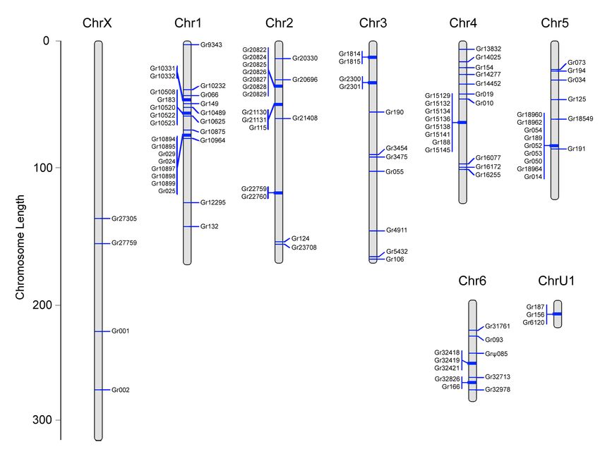
ecosystems (Figure 1). Recent studies have greatly helped to elucidate their phylogeny and delimitate its
                                                                                                                                                                                                                                                               main evolutionary lineages (Wheeler et al., 2017; Kallal et al., 2020). The nocturnal ground-dwelling genus
                                                                                                                                                                                                                                                               Dysdera Latreille 1804, which contains 286 species (World Spider Catalog, 2021), mostly with a circum-
Posted on Authorea 10 May 2021 | The copyright holder is the author/funder. All rights reserved. No reuse without permission. | https://doi.org/10.22541/au.162067941.16111128/v1 | This a preprint and has not been peer reviewed. Data may be preliminary.

                                                                                                                                                                                                                                                               Mediterranean distribution, represents nearly half of the diversity of the Dysderidae family. Approximately
                                                                                                                                                                                                                                                               50 species of this genus are endemic from the Canary Islands archipelago, representing one of the most spec-
                                                                                                                                                                                                                                                               tacular examples of diversification on islands within spiders (Arnedo, Oromı́, Múrria, Macı́as-Hernández, &
                                                                                                                                                                                                                                                               Ribera, 2007; Arnedo, Oromı́, & Ribera, 2001; Macı́as-Hernández, López, Roca-Cusachs, Oromı́, & Arnedo,
                                                                                                                                                                                                                                                               2016; Vizueta, Macı́as-Hernández, Arnedo, Rozas, & Sánchez-Gracia, 2019). Shifts in dietary preferences
                                                                                                                                                                                                                                                               have been identified as one of the main drivers of island diversification in this group (Řezáč, Pekár, Arnedo,
                                                                                                                                                                                                                                                               Macı́as-Hernández, & Řezáčová, 2021). Indeed, Dysdera includes some of the few reported cases of steno-
                                                                                                                                                                                                                                                               phagy (i.e. prey specialization) across the mostly generalists spiders (Pekár, Lı́znarová, & Řezáč, 2016), with
                                                                                                                                                                                                                                                               some species (both continental and island species) facultatively or even obligatorily specialized in feeding on
                                                                                                                                                                                                                                                               terrestrial woodlice (Crustacea: Isopoda). This trophic specialization was accompanied by morphological
                                                                                                                                                                                                                                                               (modifications of mouthparts), behavioral (unique hunting strategies) and physiological adaptations to cap-
                                                                                                                                                                                                                                                               ture woodlice and to assimilate the toxic substances and heavy metals accumulated in these usually rejected
                                                                                                                                                                                                                                                               prey (Hopkin & Martin, 1985; Řezáč, Pekár, & Lubin, 2008; Řezáč & Pekár, 2007; Toft & Macı́as-Hernández,
                                                                                                                                                                                                                                                               2017). In the Canary Islands, as in continental species, these diet shifts have occurred recurrently in different
                                                                                                                                                                                                                                                               geographic areas.
                                                                                                                                                                                                                                                               The high rates of species proliferation coupled with multiple independent eco-phenotypic shifts make Dys-
                                                                                                                                                                                                                                                               dera an excellent model for understanding the genomic basis of adaptive radiations (Vizueta et al., 2019).
                                                                                                                                                                                                                                                               With the aim of obtaining a reference genome for this genus, we sequenced the genome of Dysdera silvatica
                                                                                                                                                                                                                                                               Schmidt, 1981 (~1.37 Gb) and generated the first de novo genome assembly of this species using a hybrid
                                                                                                                                                                                                                                                               strategy (Sánchez-Herrero et al., 2019). Nevertheless, most of the assembly was based on short reads, which,
                                                                                                                                                                                                                                                               added to the high repetitive nature of the genome sequences of this species (53.8%), resulted in a very frag-
                                                                                                                                                                                                                                                               mented genome draft (N50 of 38 kb). While this first draft has been a fruitful research resource, it prevented
                                                                                                                                                                                                                                                               the study of genomic aspects requiring greater continuity, such as gene mapping across the chromosomes,
                                                                                                                                                                                                                                                               the comprehensive annotation of very long genes and gene clusters, or the identification of structural va-
                                                                                                                                                                                                                                                               riation. These features are fundamental for understanding the biological and evolutionary meaning of the
                                                                                                                                                                                                                                                               genome structure and gene organization. Some clear examples of the benefits of having a highly continuous
                                                                                                                                                                                                                                                               chromosome-level assembly are the study of the genome structure and evolution of gene families, the impact
                                                                                                                                                                                                                                                               of a number genome features (e.g., recombination, base content, distribution of genes and repetitive regions,
                                                                                                                                                                                                                                                               etc..) on adaptive processes, the analysis of impact of hybridization and divergence between populations,
                                                                                                                                                                                                                                                               or the role of chromosomal evolution in speciation (Bleidorn, 2016; Pollard, Gurdasani, Mentzer, Porter, &
                                                                                                                                                                                                                                                               Sandhu, 2018; Saha, 2019).
                                                                                                                                                                                                                                                               We present the first high-quality chromosome-level assembly of the species D. silvatica (D. silvatica genome
                                                                                                                                                                                                                                                               draft, version 2.0) . Using the first version of the genome assembly of this species (Sánchez-Herrero et
                                                                                                                                                                                                                                                               al., 2019) as a starting point, we used proximity ligation libraries (Chicago and Hi-C libraries; Dovetail
                                                                                                                                                                                                                                                               genomics), and the HiRise pipeline (Putnam et al., 2016) to obtain an improved, highly continuous assembly
                                                                                                                                                                                                                                                               of this genome. As an example of the enhanced utility of the version 2.0, we have identified and annotated
                                                                                                                                                                                                                                                               the members of the two major arthropod chemoreceptor gene families in this genome and performed a
                                                                                                                                                                                                                                                               comprehensive analysis of the physical clustering of all family members at chromosome-level scale. This new
                                                                                                                                                                                                                                                               genomic resource will foster further studies of the molecular basis underlying rapid diversification in islands
                                                                                                                                                                                                                                                               and ecological shifts by allowing comparative genomic analyses based on variation data that is inaccessible
                                                                                                                                                                                                                                                               in currently available fragmented genomes, such as structural variants, repetitive elements, and large gene
                                                                                                                                                                                                                                                               families. Additionally, this high-quality genomic data will contribute to improve our understanding of the
                                                                                                                                                                                                                                                               structure, organization, and genome evolution in chelicerates.
                                                                                                                                                                                                                                                               Material and Methods
                                                                                                                                                                                                                                                               Sampling, DNA Sequencing and Genome assembly
                                                                                                                                                                                                                                                               The new genome assembly of D. silvatica was obtained using the previous genome assembly (denoted as

                                                                                                                                                                                                                                                                                                                        2
Chromosome-scale assembly of the Canary Island endemic spider Dysdera silvatica (Arachnida, Araneae) sheds light on the origin and genome ...
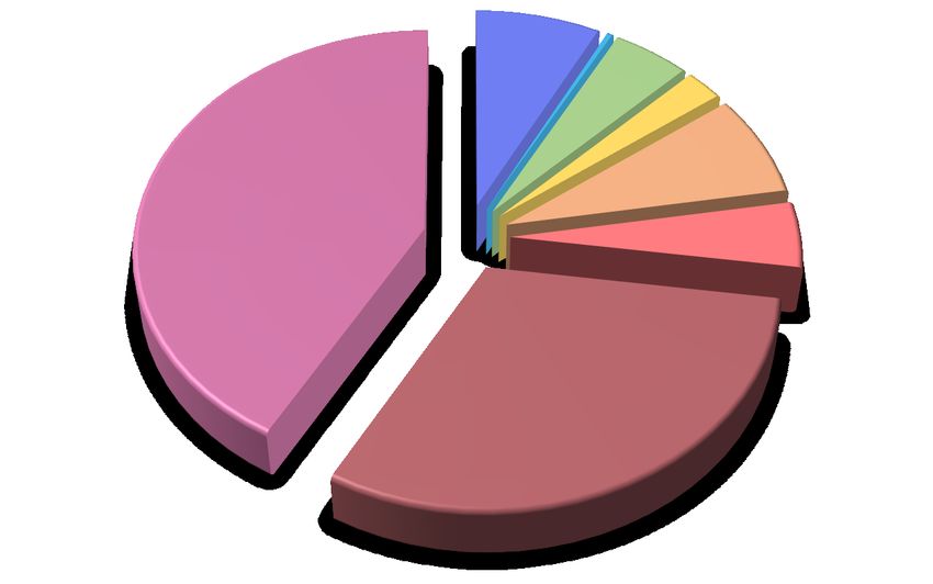
version 1; Sánchez-Herrero et al., 2019), which was further scaffolded using Chicago and Hi-C libraries.
                                                                                                                                                                                                                                                               Version 1 assembly was generated using a hybrid de novo genome assembly by combining information from
                                                                                                                                                                                                                                                               five individuals and four types of sequencing libraries; see Table 1 in Sánchez-Herrero et al. 2019. We generated
Posted on Authorea 10 May 2021 | The copyright holder is the author/funder. All rights reserved. No reuse without permission. | https://doi.org/10.22541/au.162067941.16111128/v1 | This a preprint and has not been peer reviewed. Data may be preliminary.

                                                                                                                                                                                                                                                               the latest sequencing libraries using a single female of the genus Dysdera , sampled in March 2012 in La
                                                                                                                                                                                                                                                               Gomera (Canary Islands), identified in the laboratory as D. silvatica , frozen in nitrogen liquid and stored
                                                                                                                                                                                                                                                               at -80°C until its use.
                                                                                                                                                                                                                                                               DNA extraction and sequencing were performed in Dovetail Genomics (Santa Cruz; California). The newly
                                                                                                                                                                                                                                                               generated sequence data was obtained as a combination of Chicago (one library), Hi-C (one library) and
                                                                                                                                                                                                                                                               Illumina 150-bp paired-end (HiSeq X ten platform; one lane) sequencing libraries. The final assembly (D.
                                                                                                                                                                                                                                                               silvatica reference genome version 2) was generated using the HiRise scaffolding software (Putnam et al.,
                                                                                                                                                                                                                                                               2016), and further polished with NextPolish (Hu, Fan, Sun, & Liu, 2020) using illumina short-read data
                                                                                                                                                                                                                                                               from a single individual unequivocally identified as D. silvatica (see results and discussion section). We
                                                                                                                                                                                                                                                               determined the completeness of the new assembly by applying the pipeline of Benchmarking Universal Single
                                                                                                                                                                                                                                                               Copy Orthologs (BUSCO; v. 5.0.0; Seppey, Manni, & Zdobnov, 2019), searching the arachnida (odb10; 2934
                                                                                                                                                                                                                                                               genes), arthropoda (odb10; 1013 genes) and eukaryota (odb10; 255 genes) datasets.
                                                                                                                                                                                                                                                               Repetitive elements identification
                                                                                                                                                                                                                                                               We analyzed repetitive regions in the new assembly using a combination of a de novo transposable ele-
                                                                                                                                                                                                                                                               ments identification with RepeatModeler v1.0.11 (Smit, & Hubley; RepeatModeler), and a similarity-based
                                                                                                                                                                                                                                                               search with RepeatMasker v.4.0.7 (Smit, Hubley, & Green; RepeatMasker). For the analysis we used the
                                                                                                                                                                                                                                                               repetitive elements database built for the version 1 of the D. silvatica draft genome see Sánchez-Herrero et
                                                                                                                                                                                                                                                               al. (2019) for more details), generated with RepeatModeler, plus Dfam_Consensus-20171107 and RepBase-
                                                                                                                                                                                                                                                               20181026 databases. The new assembly was masked for these repetitive sequences using the (-xsmall) option
                                                                                                                                                                                                                                                               of RepeatMasker to be used in the genome annotation steps.
                                                                                                                                                                                                                                                               Structural and functional genome annotation
                                                                                                                                                                                                                                                               Structural annotation of the soft-masked genome was accomplished with BRAKER2 (Brůna, Hoff, Lomsadze,
                                                                                                                                                                                                                                                               Stanke, & Borodovsky, 2021; Hoff, Lomsadze, Borodovsky, & Stanke, 2019; Stanke, Diekhans, Baertsch, &
                                                                                                                                                                                                                                                               Haussler, 2008; Stanke, Schöffmann, Morgenstern, & Waack, 2006), using both D. silvatica RNAseq data
                                                                                                                                                                                                                                                               (Vizueta et al., 2017) and orthologous sequences from five species of this genus (includingD. silvatica )
                                                                                                                                                                                                                                                               (Vizueta et al., 2019) as evidence (–etpmode) (Hoff et al., 2019). To perform the functional annotation of the
                                                                                                                                                                                                                                                               gene models, we used BLASTP v2.4 searches (E -value = 10-5 ) against NCBI-nr, Swiss-Prot, and an updated
                                                                                                                                                                                                                                                               version of the ArthropodDB databases (see Vizueta et al., 2017, for a detailed description of ArthropodDB).
                                                                                                                                                                                                                                                               Furthermore, we also searched the predicted peptides for specific protein domain signatures in InterProScan
                                                                                                                                                                                                                                                               v5.31.70.0 (Jones et al., 2014) and integrated all functional evidence.
                                                                                                                                                                                                                                                               Gene ontology (GO) terms (Ashburner et al, 2000) were obtained inherited from the BLAST (using top five
                                                                                                                                                                                                                                                               significant hits) and InterProScan results. We used Blast2GO to associate KEGG enzymes and pathways
                                                                                                                                                                                                                                                               to the annotated genes (Kanehisa, & Goto, 2000). We detected the transfer RNA genes (tRNAs) encoded
                                                                                                                                                                                                                                                               in the genomic sequence of D. silvaticausing the tRNAscan-SE v2.0.7 software (Chan, Lin, Mak, & Lowe,
                                                                                                                                                                                                                                                               2019).
                                                                                                                                                                                                                                                               Homology searches
                                                                                                                                                                                                                                                               We searched for homologs of D. silvatica in the genome data of representatives of a broad taxonomic range
                                                                                                                                                                                                                                                               across the order Araneae, namely Acanthoscurria geniculata (C. L. Koch, 1841) (Theraphosidae), in the sub-
                                                                                                                                                                                                                                                               order Mygalomorphae, and the representatives of the suborder Araneomorphae Loxosceles reclusa Gertsch
                                                                                                                                                                                                                                                               & Mulaik, 1940 (Sicariidae), together with D. silvatica member of the clade Synspermiata, Stegodyphus
                                                                                                                                                                                                                                                               mimosarum Pavesi, 1883 andS. dumicola Pocock, 1898 (Eresidae), and the AraneoideaParasteatoda tepidar-
                                                                                                                                                                                                                                                               iorum (C. L. Koch, 1841) andLatrodectus hesperus Chamberlin & Ivie, 1935, both in the family Theridiidae,
                                                                                                                                                                                                                                                               and the Araneidae Trichonephila clavipes (Linnaeus, 1767) and Argiope bruennichi (Scopoli, 1772) (Figure
                                                                                                                                                                                                                                                               1; Table S1), as well as all other arachnids, arthropods and ecdysozoa species surveyed in Sanchez-Herrero et

                                                                                                                                                                                                                                                                                                                       3
Chromosome-scale assembly of the Canary Island endemic spider Dysdera silvatica (Arachnida, Araneae) sheds light on the origin and genome ...
al., (2019). The search was conducted by a series of BLASTP searches (E -value cutoff < 10-3 ; we also applied
Posted on Authorea 10 May 2021 — The copyright holder is the author/funder. All rights reserved. No reuse without permission. — https://doi.org/10.22541/au.162067941.16111128/v1 — This a preprint and has not been peer reviewed. Data may be preliminary.

                                                                                                                                                                                                                                                               a filter of >30% alignment length to consider a hit as a positive). Finally, we also searched for orthogroups
                                                                                                                                                                                                                                                               and establish homology relationships amongD. silvatica and the arachnids included in the OrthoDB (v10)
                                                                                                                                                                                                                                                               (Kriventseva et al., 2019) database, namely the tick Ixodes scapularis Say, 1821 (Ullmann, Lima, Guerrero,
                                                                                                                                                                                                                                                               Piesman, & Black IV, 2005), the mite Tetranychus urticae C. L. Koch, 1836 (Grbić et al., 2011), and the
                                                                                                                                                                                                                                                               spiders Stegodyphus mimosarum (Sanggaard et al., 2014) and Parasteatoda tepidariorum(Schwager et al.,
                                                                                                                                                                                                                                                               2017).
                                                                                                                                                                                                                                                               Annotation of the chemoreceptor genes
                                                                                                                                                                                                                                                               We performed a comprehensive curation of all members of the major chemoreceptor gene families encoded
                                                                                                                                                                                                                                                               in the genome of D. silvatica , namely the Gustatory-receptor (Gr ) and Ionotropic (glutamate) receptor
                                                                                                                                                                                                                                                               (Ir/iGluR ) families (Vizueta, Escuer, Frıas-Lopez, et al., 2020; Vizueta et al., 2018). For this task, we
                                                                                                                                                                                                                                                               used the pipeline BITACORA (Vizueta, Escuer, Sánchez-Gracia, & Rozas, 2020; Vizueta, Sánchez-Gracia,
                                                                                                                                                                                                                                                               & Rozas, 2020), along with the homologous sequence data set and hidden Markov model (HMM) profiles
                                                                                                                                                                                                                                                               used in Vizueta et al. (2018) and using the annotated gene models and genome sequence as input. The
                                                                                                                                                                                                                                                               resulting identified proteins were validated, and re-annotated when necessary, in the Apollo genome browser
                                                                                                                                                                                                                                                               (Lee et al., 2013). We classified a gene as “complete” if the length of the encoded protein contains, at least,
                                                                                                                                                                                                                                                               80% of the protein domain length characteristic of the family (235 and 180 amino acids, for the GR and
                                                                                                                                                                                                                                                               IR/iGluR proteins, respectively). The remaining incomplete gene models that could not be recovered using
                                                                                                                                                                                                                                                               Apollo were classified as “partial” fragments. For each chemoreceptor family, we computed the minimum
                                                                                                                                                                                                                                                               number of chemoreceptor sequences that could be unequivocally attributed to different genes (S MIN ) as in
                                                                                                                                                                                                                                                               Vizueta et al. (2018).
                                                                                                                                                                                                                                                               Cluster definition and analysis
                                                                                                                                                                                                                                                               We determined whether the members of a given chemoreceptor gene family are physically closer (forming a
                                                                                                                                                                                                                                                               cluster) in the pseudochromosomes than expected by chance by analyzing the distribution of pairwise physical
                                                                                                                                                                                                                                                               distances between the members of a particular gene family and scaffold. We classified the paralogous copies as
                                                                                                                                                                                                                                                               “clustered” and “non clustered”. Operationally we consider that n closely linked genes from a gene family are
                                                                                                                                                                                                                                                               clustered if they are arranged within a genomic region that spans less than certain cut-offCL value following
                                                                                                                                                                                                                                                               Vieira, Sánchez-Gracia, & Rozas (2007):

                                                                                                                                                                                                                                                                                                              CL = g (n − 1)

                                                                                                                                                                                                                                                               where CL is the maximum length of a cluster that contains two or more copies of the same family, and g
                                                                                                                                                                                                                                                               is the maximum distance between two copies of a given family to consider that they are clustered. Here we
                                                                                                                                                                                                                                                               set the value of g to 100 kb. The gene density of the Ir family in the D. silvatica genome is about one copy
                                                                                                                                                                                                                                                               every 3.32 Mb (and even lower in the Gr family). Assuming a uniform distribution of gene family members
                                                                                                                                                                                                                                                               across the genome, the probability of finding by chance two (or more) Ir genes in a 100 kb stretch is p =
                                                                                                                                                                                                                                                               0.0004 (Poisson distribution, λ = 0.0301); this p -value is even lower for the Gr family. Thus, the selected g
                                                                                                                                                                                                                                                               guarantees conservativeCL lengths for the two chemoreceptor families. Pairwise physical distances between
                                                                                                                                                                                                                                                               gene family copies were processed with the R package ComplexHeatmap (Gu, Eils, & Schlesner, 2016), and
                                                                                                                                                                                                                                                               plotted as heatmaps to facilitate the visualization of gene clustering across scaffolds.
                                                                                                                                                                                                                                                               Phylogenetic and evolutionary analyses
                                                                                                                                                                                                                                                               Multiple sequence alignments
                                                                                                                                                                                                                                                               We used Mafft v. 7.475 (Katoh & Standley, 2013) to build three multiple sequence alignments (MSA)
                                                                                                                                                                                                                                                               per each gene family. First, we generated a MSA (MSAc) with only complete sequences identified in D.
                                                                                                                                                                                                                                                               silvatica using the L-INS-i algorithm with options –localpair –maxiterate 1000. Then, we built a second
                                                                                                                                                                                                                                                               MSA (MSAf) for all sequences (the full data set, comprise the complete and partial copies identified in D.
                                                                                                                                                                                                                                                               silvatica ) using the Mafft –addfragments option (–keeplength) to align fragment (partial) sequences to the

                                                                                                                                                                                                                                                                                                                     4
previous computed MSAc. Finally, we also used the L-INS-i algorithm to generate a third MSA for each
Posted on Authorea 10 May 2021 — The copyright holder is the author/funder. All rights reserved. No reuse without permission. — https://doi.org/10.22541/au.162067941.16111128/v1 — This a preprint and has not been peer reviewed. Data may be preliminary.

                                                                                                                                                                                                                                                               family, which included the complete sequences from D. silvatica and the curated members of the same family
                                                                                                                                                                                                                                                               annotated in the genome of Drosophila melanogaster Meigen, 1830 (MSAp).
                                                                                                                                                                                                                                                               Physical versus evolutionary distances
                                                                                                                                                                                                                                                               We used the best-fit amino acid substitution model found by IQ-TREE software v. 2.1.2 (Minh et al.,
                                                                                                                                                                                                                                                               2020) to estimate the evolutionary distances (measured as the number of amino acid replacements per amino
                                                                                                                                                                                                                                                               acid site) across all pairwise comparisons. The analysis was performed with MEGA-CC 10.2.4 software
                                                                                                                                                                                                                                                               (command-line version) (Kumar, Stecher, Peterson, & Tamura, 2012), using the JTT substitution model
                                                                                                                                                                                                                                                               (Jones, Taylor, & Thornton, 1992), with gamma-distributed heterogeneous rate variation among sites (5 and
                                                                                                                                                                                                                                                               7 discrete classes for the Gr and Ir families, respectively).
                                                                                                                                                                                                                                                               We investigated the relationship between physical and evolutionary distances by means of the CST statistic,
                                                                                                                                                                                                                                                               which measures the proportion of the evolutionary distance that is attributable to unclustered genes. We
                                                                                                                                                                                                                                                               computed CST independently in the two chemoreceptor families, and separately for each scaffold (or for the
                                                                                                                                                                                                                                                               whole genome). CST is estimated as:

                                                                                                                                                                                                                                                                                                                    DT − DC
                                                                                                                                                                                                                                                                                                           CST =
                                                                                                                                                                                                                                                                                                                      DT

                                                                                                                                                                                                                                                               where DT , the average of the pairwise evolutionary (amino acid replacements per site) distances between
                                                                                                                                                                                                                                                               gene family copies, is estimated as:

                                                                                                                                                                                                                                                                                                                     2    X
                                                                                                                                                                                                                                                                                                         DT =                 dij
                                                                                                                                                                                                                                                                                                                 n(n − 1) i
acid variation in the ligand-binding domain (LBD: PF00060; Mistry et al., 2021) can be used to distinguish
Posted on Authorea 10 May 2021 — The copyright holder is the author/funder. All rights reserved. No reuse without permission. — https://doi.org/10.22541/au.162067941.16111128/v1 — This a preprint and has not been peer reviewed. Data may be preliminary.

                                                                                                                                                                                                                                                               between iGluR and Ir subfamilies, we built an additional MSA (MSAp2) including only the sequences
                                                                                                                                                                                                                                                               encoding this domain inD. silvatica and in D. melanogaster . We used HMMSEARCH (Eddy, 2011) and
                                                                                                                                                                                                                                                               in-house Perl scripts to identify and cut the sequences encoding this domain in all complete Ir/iGluR genes.
                                                                                                                                                                                                                                                               We used the iTOL web server to visualize and manipulate the trees (Letunic & Bork, 2007).
                                                                                                                                                                                                                                                               Results and Discussion
                                                                                                                                                                                                                                                               A new, high-quality genome assembly and annotation
                                                                                                                                                                                                                                                               The new genome of D. silvatica , which has an assembly size of 1.37 Gb (Table 1), shows a high completeness,
                                                                                                                                                                                                                                                               with the detection of 86.3% and 92.9% of the BUSCO genes across the arachnida and eukaryota data sets,
                                                                                                                                                                                                                                                               respectively, in their genome sequence (Table 1; Table S2). Despite having 15,360 scaffolds, the N50 and
                                                                                                                                                                                                                                                               L50 values, 174.2 Mb and 4 scaffolds, respectively, also demonstrate the high continuity of the assembly.
                                                                                                                                                                                                                                                               The seven largest scaffolds (or pseudochromosomes), including the larger scaffold that likely corresponds to
                                                                                                                                                                                                                                                               the X chromosome (317.9 Mb long, nearly twice the size of the second largest scaffold), represent ˜87% of
                                                                                                                                                                                                                                                               total assembly size, matching perfectly with the haploid component of this species (6 autosomes and the X
                                                                                                                                                                                                                                                               chromosome; Bellvert & Arnedo, unpublished data; Figure 2).
                                                                                                                                                                                                                                                               The structural annotation shows that the genome of D. silvaticaencodes 33,275 coding-protein genes (35,370
                                                                                                                                                                                                                                                               transcripts) and 37,198 putative tRNAs; this annotation includes the 90% and 95% of the BUSCO arthro-
                                                                                                                                                                                                                                                               poda and eukaryote data sets, respectively, evidencing the completeness of the annotation (Table 2). Se-
                                                                                                                                                                                                                                                               quence similarity-based searches uncovered 28,904 sequences with positive hits against the surveyed protein
                                                                                                                                                                                                                                                               databases (16,241, 25,917 and 22,604 against Swiss-Prot, ArthropodDB and InterPro databases, respec-
                                                                                                                                                                                                                                                               tively). Furthermore 22,093 of these functionally annotated sequences also have at least one associated GO
                                                                                                                                                                                                                                                               term.
                                                                                                                                                                                                                                                               We identified ˜3.2 millions of repetitive sequences, which encompass 53.0% of the total assembly size (Table
                                                                                                                                                                                                                                                               2; Tables S3). The great majority of these sequences (51.6%) correspond to transposable elements, many
                                                                                                                                                                                                                                                               of them (22.1%) without detectable homologs in known databases; class II elements are the most abundant
                                                                                                                                                                                                                                                               type (16.5%), followed by retrotransposons (class I), including LINEs (10.6%) and SINEs (1.8%).
                                                                                                                                                                                                                                                               The great majority of structurally annotated genes in the new genome assembly are shared across Arthropoda
                                                                                                                                                                                                                                                               (63.5%), being 36.3% of them also present in Ecdysozoa (Figure S1; Table S1). Besides, 25.0% of these genes
                                                                                                                                                                                                                                                               are spider-specific (order Araneae), and 17.4% were identified as lineage-specific in D. silvatica . When
                                                                                                                                                                                                                                                               considering only functionally annotated genes (n = 28,904), this analysis yields equivalent results (Figure
                                                                                                                                                                                                                                                               3a), although a slightly higher fraction of genes shared within Arthropoda (72.8%) and less D. silvatica
                                                                                                                                                                                                                                                               lineage-specific genes were detected (2,200 genes: 7.6%). Remarkably, the number of lineage-specific genes
                                                                                                                                                                                                                                                               is nearly the half of those initially reported by Sánchez-Herrero et al. (2019); this feature could be partly
                                                                                                                                                                                                                                                               explained by the fact that here we have used a broader Araneae dataset for the searches, although the
                                                                                                                                                                                                                                                               higher quality of the new assembly would have allowed to identify much more proteins accurately annotated.
                                                                                                                                                                                                                                                               Homology-search results based on OrthoDB (Figure 3b) were more similar than those obtained in Sánchez-
                                                                                                                                                                                                                                                               Herrero et al. (2019), reflecting that the vast improvement in assembly continuity does not affect orthology
                                                                                                                                                                                                                                                               inference quality.
                                                                                                                                                                                                                                                               Globally, the new chromosome-scale assembly of D. silvaticarepresents a huge improvement compared with
                                                                                                                                                                                                                                                               our previous draft assembly. In terms of continuity, it implies an improvement of more than 4,500 times (the
                                                                                                                                                                                                                                                               N50 value) yielding a scaffold N50 of 174.2 Mb from the 38 kb in the previous assembly. This improvement
                                                                                                                                                                                                                                                               is also reflected in the high number of annotated genes (Table 2, Table S2), despite the number the gene
                                                                                                                                                                                                                                                               models in current version drops from 48,619 (75% of them with functional annotation) to 33,275 (87% with
                                                                                                                                                                                                                                                               functional annotation). On the other hand, the new reference sequence encompass sequence data exclusively
                                                                                                                                                                                                                                                               from D. silvatica , while that reported in version 1 was generated using information from various individuals
                                                                                                                                                                                                                                                               one of them now identified as D. enghoffi Arnedo, Oromı́ & Ribera, 1997, a phylogenetically close relative to
                                                                                                                                                                                                                                                               D. silvatica also endemic from La Gomera (Arnedo et al., 2007; see also Adrián-Serrano, Lozano-Fernandez,
                                                                                                                                                                                                                                                               Pons, Rozas, & Arnedo (2021) for the new mtDNA data). The new chromosome-level assembly of D. silvatica

                                                                                                                                                                                                                                                                                                                     6
is the first highly continuous genome of a representative of the spider clade Synspermiata, which currently
Posted on Authorea 10 May 2021 — The copyright holder is the author/funder. All rights reserved. No reuse without permission. — https://doi.org/10.22541/au.162067941.16111128/v1 — This a preprint and has not been peer reviewed. Data may be preliminary.

                                                                                                                                                                                                                                                               includes 17 families, and the third within the Araneae order (the other two are members of the superfamily
                                                                                                                                                                                                                                                               Araneoidea), an extremely poor and biased genomic representation of the taxonomic and evolutionary di-
                                                                                                                                                                                                                                                               versity of the spider tree of life (Figure 1). Our assembly, therefore, represents a valuable resource to further
                                                                                                                                                                                                                                                               conduct molecular evolutionary and functional studies in spiders and their relatives.
                                                                                                                                                                                                                                                               The chemoreceptor repertoire of D. silvatica
                                                                                                                                                                                                                                                               The chemosensory repertoire of chelicerates, particularly of spiders, is characterized by a high diversity in
                                                                                                                                                                                                                                                               terms of copy number and sequence divergence, likely resulted from a constant and prolonged gene birth and
                                                                                                                                                                                                                                                               death process, only altered by episodic bursts of gene duplication yielding lineage-specific expansions (Vizueta
                                                                                                                                                                                                                                                               et al., 2018). Here, our comprehensive analysis using the BITACORA pipeline allowed the identification in
                                                                                                                                                                                                                                                               the genome of D. silvatica of 545 chemoreceptor genes, corresponding to 134 Gr and 411 Ir (plus one homolog
                                                                                                                                                                                                                                                               to the insect Ir25a and 24 iGluR ) sequences (Figure 4; Tables 3, S4 and S5). Although these family sizes
                                                                                                                                                                                                                                                               are in agreement with the variability observed across spider genomes, the 411Ir reported here represents the
                                                                                                                                                                                                                                                               largest repertoire found for this family in chelicerates, only surpassed by the extraordinary chemoreceptor
                                                                                                                                                                                                                                                               repertoire found in the German cockroach genome (Robertson, Baits, Walden, Wada-Katsumata, & Schal,
                                                                                                                                                                                                                                                               2018). On the other hand, the number of Grs in D. silvatica is relatively low compared to other spiders (i.e. S
                                                                                                                                                                                                                                                               MIN of 634 Grs in Parasteatoda tepidariorum ), which could suggest a different contribution of the gustatory
                                                                                                                                                                                                                                                               repertories to the evolutionary, and probably adaptive history of these different spider lineages.
                                                                                                                                                                                                                                                               Noticeably, most identified chemoreceptor genes are complete (459 complete genes under the criterion consi-
                                                                                                                                                                                                                                                               dered in this article; see methods), yielding a S MIN of 540 genes (Table 3). This large proportion of complete
                                                                                                                                                                                                                                                               copies is highly unusual in reports on these chemoreceptor families across arthropod genomes (Vizueta et
                                                                                                                                                                                                                                                               al., 2018), clearly demonstrating the benefits of having a chromosome-level assembly to annotate, charac-
                                                                                                                                                                                                                                                               terize and study multigene families in animal genomes. The new high-quality assembly has uncovered, for
                                                                                                                                                                                                                                                               example, that the two studied gene families are unevenly distributed across pseudochromosomes. The X
                                                                                                                                                                                                                                                               chromosome, representing the 23.3% of the genome assembly, harbors only 3.0% and 3.2% of the Gr andIr
                                                                                                                                                                                                                                                               identified copies, respectively (Table 3); the members of these families, nevertheless, are more uniformly
                                                                                                                                                                                                                                                               distributed across the other major pseudochromosomes. Even so, the number of chemoreceptors located in
                                                                                                                                                                                                                                                               the small scaffolds, which only represent 11.8% of the total assembly, represents 23.1% and 59.6% of the
                                                                                                                                                                                                                                                               Gr and Ir repertories, respectively. This uneven distribution, however, is not observed when considering all
                                                                                                                                                                                                                                                               genes identified in the genome (Table 3). While the first feature likely hides some (unknown) particularities
                                                                                                                                                                                                                                                               of the sex chromosome, the high representation of gene family members across the minor scaffolds could be
                                                                                                                                                                                                                                                               explained by the assembly difficulties of these repetitive regions. Further studies including gene families with
                                                                                                                                                                                                                                                               different family sizes will get insights into this genomic feature.
                                                                                                                                                                                                                                                               Chemoreceptor genes are unevenly distributed across the genome of D. silvatica
                                                                                                                                                                                                                                                               Despite that not all chemoreceptor genes could be mapped on the scaffolds corresponding to the main cyto-
                                                                                                                                                                                                                                                               logical described chromosomes, the new high-quality assembly allowed us to study the genomic organization
                                                                                                                                                                                                                                                               and evolution of a great number of paralogous copies of each family. According to our criterion (see methods),
                                                                                                                                                                                                                                                               we identified 83 genomic clusters, 17 and 66 of them including Gr and Ir genes, respectively (Figures 5A,
                                                                                                                                                                                                                                                               5C, S2A and S2C). These clusters, which harbor up to 10 copies of the same family, were found in all major
                                                                                                                                                                                                                                                               scaffolds of D. silvatica.
                                                                                                                                                                                                                                                               To gain insights into the evolutionary meaning of such gene clustering structure we investigated the relati-
                                                                                                                                                                                                                                                               onship between pairwise evolutionary divergences, measured as dij (the number of amino acid substitutions
                                                                                                                                                                                                                                                               per site between two sequences), and physical distances (in kb). We found that C ST values are high in all
                                                                                                                                                                                                                                                               pseudochromosomes, ranging from 0.418 to 0.982 and from 0.428 to 0.894 for the Gr and Ir gene families,
                                                                                                                                                                                                                                                               respectively, considering all identified sequences (Table 3); these values are similar when using only the com-
                                                                                                                                                                                                                                                               plete data set (Table S4). These highC ST values translate into statistically lower evolutionary distances
                                                                                                                                                                                                                                                               among family copies included in clusters than those dispersed along the genome, both at the chromosome
                                                                                                                                                                                                                                                               (Mann–Whitney U-test, p -values < 0.05 for nearly all pseudochromosomes) but also at the whole genome

                                                                                                                                                                                                                                                                                                                      7
levels (p -values < 0.001 in all cases) (Tables 3 and S4; Figures 5 and S2). This result, jointly with the
Posted on Authorea 10 May 2021 — The copyright holder is the author/funder. All rights reserved. No reuse without permission. — https://doi.org/10.22541/au.162067941.16111128/v1 — This a preprint and has not been peer reviewed. Data may be preliminary.

                                                                                                                                                                                                                                                               large number of genomic clusters found across the D. silvatica genome, point to the recent origin of many of
                                                                                                                                                                                                                                                               the chemoreceptors in this species, and to the unequal crossing-over as a major mechanism accounting for
                                                                                                                                                                                                                                                               this origin. After gene duplication, the paralogs that are retained long enough (i.e., those that are not lost
                                                                                                                                                                                                                                                               by genetic drift or purifying selection), continuously diverged at the sequence, and likely at the functional
                                                                                                                                                                                                                                                               level (at least in terms of ligand specificity or signaling characteristics). We expect, therefore, that over
                                                                                                                                                                                                                                                               time, evolutionary distances of these retained copies increase with physical distance, just as we have found
                                                                                                                                                                                                                                                               (Figures 5B, 5D, S2B and S2D). This genomic architecture could have relevant functional and evolutionary
                                                                                                                                                                                                                                                               implications. For instance, the presence of distantly related family members within the same genomic clus-
                                                                                                                                                                                                                                                               ter, could be the hallmark of the interaction between functional and gene regulation constraints preventing
                                                                                                                                                                                                                                                               cluster breaking. A more comprehensive analysis of these specific cases deserves to be further evaluated.
                                                                                                                                                                                                                                                               Phylogenetic analysis of the chemoreceptor genes in arthropods
                                                                                                                                                                                                                                                               As commented above, having a very continuous assembly opens the door of annotating as “complete genes”
                                                                                                                                                                                                                                                               most copies of a medium to large-sized multigene families, almost outside of the scope in most of the
                                                                                                                                                                                                                                                               available, highly fragmented non-model chelicerates genomes. These improved annotations (with the inclusion
                                                                                                                                                                                                                                                               of more and longer family copies in the multiple sequence alignments) in turn, yield to much more accurate
                                                                                                                                                                                                                                                               phylogenetic analyses, increasing the evolutionary signal, and improving the tree node support. In many
                                                                                                                                                                                                                                                               cases, furthermore, these new complete copies could add very valuable information about, for instance,
                                                                                                                                                                                                                                                               recent bursts of duplication and gene retention.
                                                                                                                                                                                                                                                               Current phylogenetic analysis of the Gr and Ir families, which are based on the high-quality annotations
                                                                                                                                                                                                                                                               from the new assembly ofD. silvatica , are clear examples of these benefits. Our analysis undoubtedly reflects
                                                                                                                                                                                                                                                               the high gene turnover rates of these families in chelicerates (and, in general, in panarthropods; Vizueta,
                                                                                                                                                                                                                                                               Escuer, Frıas-Lopez, et al., 2020; Vizueta et al., 2018). However, after including the complete chemoreceptor
                                                                                                                                                                                                                                                               set, a remarkable evolutionary hallmark emerges in the D. silvatica lineage (Figures 6, 7, S3 and S4). Only
                                                                                                                                                                                                                                                               a small group of Ir genes, probably involved in some essential animal chemoreception functions, such as co-
                                                                                                                                                                                                                                                               receptors (Ir25a/8a related sequences), and the receptors involved in thermosensation and hygrosensation,
                                                                                                                                                                                                                                                               and in amino acid taste inDrosophila (i.e., Ir93a and Ir76b related sequences) (Ni, 2021), seem to be fairly
                                                                                                                                                                                                                                                               conserved between insects and spiders. Although this extreme feature is well known in arthropods, where
                                                                                                                                                                                                                                                               most family copies cluster in species-specific clades in the phylogenetic trees, current analysis is the first that
                                                                                                                                                                                                                                                               incorporate nearly complete information of most copies of these two families in a chelicerate. The quality
                                                                                                                                                                                                                                                               of the data allowed us to explore the origin and diversification trends of D. silvatica chemoreceptors with
                                                                                                                                                                                                                                                               unprecedented precision and robustness. We found, for instance, that the distribution of gene ages in the Gr
                                                                                                                                                                                                                                                               family is similar in D. silvatica and D. melanogaster , with most family members being old (likely during the
                                                                                                                                                                                                                                                               early diversification of these subphyla). In the Gr family ofD. silvatica , however, we uncovered very recent
                                                                                                                                                                                                                                                               duplication events that created (at least) one new genomic cluster in a very short period in the scaffold U29
                                                                                                                                                                                                                                                               (with at least 10 genes in the cluster).
                                                                                                                                                                                                                                                               The contrasting pattern between D. silvatica and D. melanogaster is much more pronounced in the Ir family.
                                                                                                                                                                                                                                                               Particularly noteworthy is the presence of two very recent bursts of gene duplication that originated 116
                                                                                                                                                                                                                                                               new Ir genes (83 and 33 copies, respectively; Figures 7 and S4A). Interestingly, most of these novel nearly
                                                                                                                                                                                                                                                               identical receptors map in multiple clusters in many of the smallest scaffolds; this feature suggest that they
                                                                                                                                                                                                                                                               could be indeed part of much large genomic clusters, which were not well assembled due to the high number
                                                                                                                                                                                                                                                               of repetitive Ir copies arranged in tandem in the same genomic region (Clifton et al., 2020). Such duplication
                                                                                                                                                                                                                                                               burst generating many new chemoreceptor genes could reflect some relevant evolutionary events related to
                                                                                                                                                                                                                                                               the chemosensory biology of these organisms and, consequently, they deserve to be investigated more in
                                                                                                                                                                                                                                                               depth, especially in relation to the role of selective and non-selective forces in their retention and divergence.
                                                                                                                                                                                                                                                               In fact, since current available chelicerate genomes do not allow detecting such copies accurately or they
                                                                                                                                                                                                                                                               are just annotated as different partial sequences (Vizueta, Escuer, Frıas-Lopez, et al., 2020; Vizueta et al.,
                                                                                                                                                                                                                                                               2018), they have been scarcely added to phylogenetic analyses, thus preventing the precise understanding
                                                                                                                                                                                                                                                               of the relevant events that shaped the repertoire size of large gene families at a very short time scale. Our

                                                                                                                                                                                                                                                                                                                       8
results demonstrate that new high-quality data are especially useful to conduct comprehensive studies of the
Posted on Authorea 10 May 2021 — The copyright holder is the author/funder. All rights reserved. No reuse without permission. — https://doi.org/10.22541/au.162067941.16111128/v1 — This a preprint and has not been peer reviewed. Data may be preliminary.

                                                                                                                                                                                                                                                               evolution of large multigene families.
                                                                                                                                                                                                                                                               We have also used our new assembly to test whether the LBD domain (PF00060) has enough phylogenetic
                                                                                                                                                                                                                                                               signal to classify the different subfamilies within the Ir/GluR superfamily (a strategy that we previously
                                                                                                                                                                                                                                                               used in fragmented assemblies, e.g. Vizueta et al., 2018). Here, we take advantage that the high continuity
                                                                                                                                                                                                                                                               of the assembly permitted the complete annotation of many iGluR genes, with both the ANF-receptor
                                                                                                                                                                                                                                                               (PF01094) and the LBD domains in the same gene model. The latter combination, that is characteristic of
                                                                                                                                                                                                                                                               the iGluRsubfamily, is never found in Ir genes, which lack the ANF-receptor domain (Croset et al., 2010).
                                                                                                                                                                                                                                                               This genomic structure makes it possible to unequivocally distinguish iGluR from Ir genes. Our phylogenetic
                                                                                                                                                                                                                                                               trees based on the complete sequences of this superfamily was fully consistent with those built using only
                                                                                                                                                                                                                                                               the LBD domains identified in D. silvatica (Figure S4C). This feature demonstrates that this domain, by
                                                                                                                                                                                                                                                               itself holds enough subfamily-specific information to place correctly the proteins having the ANF domain in
                                                                                                                                                                                                                                                               the phylogenetic tree (i.e., close to the D. melanogasteriGluRs and separated from the Ir sequences of both
                                                                                                                                                                                                                                                               species). In fact, the information of the LBD domain allowed us to classify correctly as iGluR some copies
                                                                                                                                                                                                                                                               of this superfamily for which we were not able to identify an ANF domain in the genomic sequences (and
                                                                                                                                                                                                                                                               that, in principle, would have been annotated asIr ).
                                                                                                                                                                                                                                                               Conclusions
                                                                                                                                                                                                                                                               The chromosome-level assembly of D. silvatica is the first high-quality continuous genome of a representative
                                                                                                                                                                                                                                                               of the Synspermiata clade, one of the major evolutionary lineages within spiders. This new assembly will
                                                                                                                                                                                                                                                               contribute to alleviate the scarce representation of spiders and chelicerates genomes within the tree of life
                                                                                                                                                                                                                                                               while represents a very useful resource to rightly characterize structural variants, repetitive elements and
                                                                                                                                                                                                                                                               large gene families involved in relevant biological functions in spiders, such as, for example, those encoding
                                                                                                                                                                                                                                                               chemosensory system proteins and venom components. An immediate application will be the comprehensive
                                                                                                                                                                                                                                                               evolutionary analysis of these genomic variants beyond single nucleotide changes to elucidate the genomic
                                                                                                                                                                                                                                                               regions and the mechanisms underlying the remarkable adaptive radiation of the genus Dysdera in Canary
                                                                                                                                                                                                                                                               Islands.
                                                                                                                                                                                                                                                               Acknowledgements
                                                                                                                                                                                                                                                               This work was supported by the Ministerio de Economı́a y Competitividad of Spain (CGL2016-75255;
                                                                                                                                                                                                                                                               CGL2016-80651; PID2019-103947GB, PID2019-105794GB) and the Comissió Interdepartamental de Recerca
                                                                                                                                                                                                                                                               I Innovació Tecnològica of Catalonia, Spain (2017SGR83, 2017SGR1287). A.S.-G. is a Serra Húnter Fellow.
                                                                                                                                                                                                                                                               P. E. was supported by an FPI grant (Ministerio de Economı́a y Competitividad of Spain, BES-2017-081740).
                                                                                                                                                                                                                                                               We acknowledge the Garajonay National Parks for granting collection permits and helping with lodging and
                                                                                                                                                                                                                                                               logistics during fieldwork.
                                                                                                                                                                                                                                                               Author contributions
                                                                                                                                                                                                                                                               J.R., A.S.-G., and M.A.A conceived the study. P.E. and J.R. drafted the manuscript. P.E. V.A.P., A.A.F,
                                                                                                                                                                                                                                                               J.V. and J.F.S.-H. performed the bioinformatics analysis. P.E. V.A.P., A.S.-G. and J. R. interpreted the
                                                                                                                                                                                                                                                               data. All authors revised and approved the final manuscript.
                                                                                                                                                                                                                                                               ORCID
                                                                                                                                                                                                                                                               Paula Escuer https://orcid.org/0000-0002-5941-0106
                                                                                                                                                                                                                                                               Vadim A. Pisarenco. https://orcid.org/0000-0002-4968-4090
                                                                                                                                                                                                                                                               Angel A. Fernández-Ruiz https://orcid.org/0000-0003-1495-0923
                                                                                                                                                                                                                                                               Joel Vizueta https://orcid.org/0000-0003-0139-3013
                                                                                                                                                                                                                                                               Jose F. Sanchez-Herrero https://orcid.org/0000-0001-6771-4807
                                                                                                                                                                                                                                                               Miquel A. Arnedo https://orcid.org/0000-0003-1402-4727

                                                                                                                                                                                                                                                                                                                     9
Alejandro Sánchez-Gracia https://orcid.org/0000-0003-4543-4577
Posted on Authorea 10 May 2021 — The copyright holder is the author/funder. All rights reserved. No reuse without permission. — https://doi.org/10.22541/au.162067941.16111128/v1 — This a preprint and has not been peer reviewed. Data may be preliminary.

                                                                                                                                                                                                                                                               Julio Rozas https://orcid.org/0000-0002-6839-9148
                                                                                                                                                                                                                                                               DATA AVAILABILITY STATEMENT
                                                                                                                                                                                                                                                               The whole-genome shotgun project has been deposited at DDBJ/ENA/GenBank under accession num-
                                                                                                                                                                                                                                                               ber QLNU00000000 and project ID PRJNA475203. The version described in this article is version QL-
                                                                                                                                                                                                                                                               NU02000000. This project repository includes raw data, sequencing libraries information, and current version
                                                                                                                                                                                                                                                               assembly. Protein sequence data of all chemoreceptor proteins (including incomplete fragments) identified in
                                                                                                                                                                                                                                                               this study are provided in the Supplementary Material online.
                                                                                                                                                                                                                                                               Tables
                                                                                                                                                                                                                                                               Table 1. Genome assembly statistics
                                                                                                                                                                                                                                                               Table 2. Genome annotation statistics
                                                                                                                                                                                                                                                               Table 3. Chromosome level statistics
                                                                                                                                                                                                                                                               Figure Legends
                                                                                                                                                                                                                                                               Figure 1. Tree of life of the order Araneae. Relationships and divergence time estimates followed (Kallal
                                                                                                                                                                                                                                                               et al., 2020). Main evolutionary lineages and taxonomic groups within spiders indicated as grey clades; the
                                                                                                                                                                                                                                                               number in brackets indicates the estimated number of included families. Phylogenetic relationships of species
                                                                                                                                                                                                                                                               with genomic information are highlighted (orange lines). Species used for the annotation and homology-based
                                                                                                                                                                                                                                                               searches of D. silvatica genome are denoted with an asterisk.
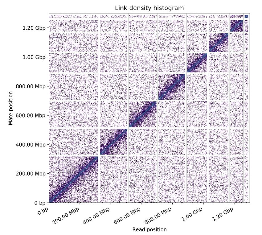
                                                                                                                                                                                                                                                               Figure 2. Long-range contact heat-map of paired-end Hi-C reads. The x and y axes show the mapping
                                                                                                                                                                                                                                                               positions of the first and second read in the read pair, respectively, grouped into bins; the color of each
                                                                                                                                                                                                                                                               square gives the number of read pairs within that bin. The seven largest scaffolds, which likely correspond
                                                                                                                                                                                                                                                               to the seven pseudochromosomes described in this species, represent the ˜87% of total assembly. The large
                                                                                                                                                                                                                                                               scaffold, which corresponds to the X chromosome, is 317.9 Mb long. In order of length, and after chromosome
                                                                                                                                                                                                                                                               6, the next scaffold is the ChrU1 (22.3 Mb long).
                                                                                                                                                                                                                                                               Figure 3. Homology-based search across different ecdysozoa species. A) Pie chart showing the taxonomic
                                                                                                                                                                                                                                                               distribution of positive BLAST hits of the functional annotation set of D. silvatica(n = 28,904 genes) across
                                                                                                                                                                                                                                                               the Araneae species illustrated in Figure 1 and the rest of the arachnida, arthropoda and ecdysozoa also ana-
                                                                                                                                                                                                                                                               lysed in Sánchez-Herrero et al. (2019). B) Homology relationships across D. silvatica (Dsil) and chelicerates
                                                                                                                                                                                                                                                               genomes available in OrthoDB v10, P. tepidariorum (Ptep), S. mimosarum (Smim),Ixodes scapularis (Isca),
                                                                                                                                                                                                                                                               and Tetranychus urticae (Turt). Red and orange bars indicate the fraction of single-copy genes identified in
                                                                                                                                                                                                                                                               all species (1:1 orthologs), and those identified in four species (missing in one species), respectively. The dark
                                                                                                                                                                                                                                                               and light green bars show orthologous relationships present in all, or in 4 species, respectively, that are not
                                                                                                                                                                                                                                                               included in the two previous categories. The blue bar shows other more complex homologous relationships.
                                                                                                                                                                                                                                                               Figure 4. Distribution of the Gr (panel A) and Ir (panel B) family members across the seven pseudochromo-
                                                                                                                                                                                                                                                               somes and the scaffold ChrU1 of D. silvatica . Genes in clusters are shown to the left of pseudochromosomes.
                                                                                                                                                                                                                                                               Figure 5. Genome organization and relationships between physical and evolutionary distances of the mem-
                                                                                                                                                                                                                                                               bers of the Gr andIr families on the D. silvatica pseudochromosome 1. A and C) Heatmaps illustrating the
                                                                                                                                                                                                                                                               distribution of physical distances (in units of 100 kb) along the pseudochromosome. B and D) Plots compa-
                                                                                                                                                                                                                                                               ring pairwise amino acid and physical (on a logarithmic scale) distances between Gr or Ir copies in the D.
                                                                                                                                                                                                                                                               silvaticapseudochromosome. Colored and grey points show distances within and outside genomic clusters,
                                                                                                                                                                                                                                                               respectively. Different clusters are depicted in different colors.
                                                                                                                                                                                                                                                               Figure 6. Phylogenetic relationships among the members of theGr family of D. silvatica and D. melanogaster
                                                                                                                                                                                                                                                               . The tree only includes the copies of D. silvatica classified as complete genes. The outer ring indicates the
                                                                                                                                                                                                                                                               chromosome (Chr; in different colors) in which the genes included in the tree are located. The inner ring

                                                                                                                                                                                                                                                                                                                      10
shows information about genomic clusters (chromosome, in the same color scale that in outer ring, genomic
Posted on Authorea 10 May 2021 — The copyright holder is the author/funder. All rights reserved. No reuse without permission. — https://doi.org/10.22541/au.162067941.16111128/v1 — This a preprint and has not been peer reviewed. Data may be preliminary.

                                                                                                                                                                                                                                                               cluster number: member number in the cluster). The scale bar refers to 1 amino acid substitution per site.
                                                                                                                                                                                                                                                               The tree was rooted in its midpoint. Minor SC, minor scaffolds. Red and green terminal branches correspond
                                                                                                                                                                                                                                                               to D. silvatica and D. melanogaster Gr , respectively. Given that the tree in this figure only includes complete
                                                                                                                                                                                                                                                               copies, some genomic clusters in table 3 and figure 4A do not appear here.
                                                                                                                                                                                                                                                               Figure 7. Phylogenetic relationships among the members of theIr/IGluR family of D. silvatica and D.
                                                                                                                                                                                                                                                               melanogaster . The tree only includes the copies classified as complete genes in this work. The light blue, dark
                                                                                                                                                                                                                                                               blue and purpura shading of gene names designate the members of the iGluR , Ir25a/8aand Ir subfamilies,
                                                                                                                                                                                                                                                               respectively. The tree was rooted considering NMDAR clade as the outgroup (Croset et al., 2010). The outer
                                                                                                                                                                                                                                                               ring indicates the chromosome (Chr) in which the genes included in the tree are located. The inner ring
                                                                                                                                                                                                                                                               shows information about genomic clusters (chromosome, in the same color scale that in outer ring, genomic
                                                                                                                                                                                                                                                               cluster number: member number in the cluster). The scale bar refers to 1 amino acid substitution per site.
                                                                                                                                                                                                                                                               Minor SC, minor scaffolds. Red and green terminal branches correspond to D. silvatica and D. melanogaster ,
                                                                                                                                                                                                                                                               respectively. The red triangles mark the D. silvatica genes with putative distant homologs in D. melanogaster
                                                                                                                                                                                                                                                               . Given that the tree in this figure only includes complete copies, some genomic clusters in table 3 and figure
                                                                                                                                                                                                                                                               4B do not appear here.
                                                                                                                                                                                                                                                               Supplementary material
                                                                                                                                                                                                                                                               Supplementary Tables
                                                                                                                                                                                                                                                               Table S1. Species analyzed in this study
                                                                                                                                                                                                                                                               Table S2. Summary of BUSCO results
                                                                                                                                                                                                                                                               Table S3. Summary of repetitive elements identified in the D. silvatica genome
                                                                                                                                                                                                                                                               Table S4. Genome organization of the members of the Gr andIr gene families across the D. silvatica pseu-
                                                                                                                                                                                                                                                               dochromosomes
                                                                                                                                                                                                                                                               Table S5. Gr and Ir/iGluR sequences identified in the new D. silvatica genome assembly. All protein sequences
                                                                                                                                                                                                                                                               (including those encoding partial genes) identified in this study and the multiple sequence alignments used
                                                                                                                                                                                                                                                               in the analyses are provided in the Supplementary Material online.
                                                                                                                                                                                                                                                               Supplementary Figures
                                                                                                                                                                                                                                                               Figure S1. Homology-based search across different ecdysozoa species. Pie chart showing the taxonomic
                                                                                                                                                                                                                                                               distribution of positive BLAST hits of the structural annotation of D. silvatica (n = 33,275 genes) across
                                                                                                                                                                                                                                                               the Araneae species illustrated in Figure 1 and the rest of the arachnida, arthropoda and ecdysozoa also
                                                                                                                                                                                                                                                               analysed in Sánchez-Herrero et al. (2019).
                                                                                                                                                                                                                                                               Figure S2. Genome organization and relationships between physical and evolutionary distances of the
                                                                                                                                                                                                                                                               members of the Gr andIr families on the D. silvatica pseudochromosomes. A and C) Heatmaps illustrating
                                                                                                                                                                                                                                                               the distribution of physical distances (in units of 100 kb) along the pseudochromosome. B and D) Plots
                                                                                                                                                                                                                                                               comparing pairwise amino acid and physical (on a logarithmic scale) distances between Gr or Ir copies
                                                                                                                                                                                                                                                               in the D. silvaticapseudochromosome. Colored and grey points show distances within and outside genomic
                                                                                                                                                                                                                                                               clusters, respectively. Different clusters are depicted in different colors.
                                                                                                                                                                                                                                                               Figure S3. Phylogenetic relationships and node support in the Gr family. A) Phylogenetic tree among all
                                                                                                                                                                                                                                                               sequences encoding members of the Gr family (including both complete and incomplete or partial genes).
                                                                                                                                                                                                                                                               Incomplete genes are marked with asterisks. The tree was rooted in its midpoint. The outer ring indicates
                                                                                                                                                                                                                                                               the chromosome (Chr) in which the genes included in the tree are located. The inner ring shows information
                                                                                                                                                                                                                                                               about genomic clusters (chromosome, in the same color scale that in outer ring, genomic cluster number:
                                                                                                                                                                                                                                                               member number in the cluster). The scale bar refers to 1 amino acid substitution per site. Minor SC, minor
                                                                                                                                                                                                                                                               scaffolds. Red and green terminal branches correspond to D. silvatica and D. melanogaster Gr, respectively.
                                                                                                                                                                                                                                                               B) Cladogram of the phylogenetic tree in Figure 6 with bootstrap node support values >90%.

                                                                                                                                                                                                                                                                                                                     11
Figure S4. Phylogenetic relationships and node support in the Ir/iGluR family. A) Phylogenetic tree among
Posted on Authorea 10 May 2021 — The copyright holder is the author/funder. All rights reserved. No reuse without permission. — https://doi.org/10.22541/au.162067941.16111128/v1 — This a preprint and has not been peer reviewed. Data may be preliminary.

                                                                                                                                                                                                                                                               all sequences encoding members of the Ir/iGluR family (including both complete and incomplete or partial
                                                                                                                                                                                                                                                               genes). Incomplete genes are marked with asterisks. The light blue, dark blue and purpura shading of gene
                                                                                                                                                                                                                                                               names designate the members of the iGluR, Ir25a/8a and Ir subfamilies, respectively. The tree was rooted
                                                                                                                                                                                                                                                               considering NMDAR clade as the outgroup (Croset et al. 2010). The outer ring indicates the chromosome
                                                                                                                                                                                                                                                               (Chr) in which the genes included in the tree are located. The inner ring shows information about genomic
                                                                                                                                                                                                                                                               clusters (chromosome, in the same color scale that in outer ring, genomic cluster number: member number in
                                                                                                                                                                                                                                                               the cluster). The scale bar refers to 1 amino acid substitution per site. Minor SC, minor scaffolds. Red and
                                                                                                                                                                                                                                                               green terminal branches correspond to D. silvatica andD. melanogaster Gr, respectively. The red triangles
                                                                                                                                                                                                                                                               mark theD. silvatica genes with putative distant homologs in D. melanogaster . B) Cladogram of the phylo-
                                                                                                                                                                                                                                                               genetic tree in figure 7 with bootstrap node support values >90%. C) Phylogenetic relationships among the
                                                                                                                                                                                                                                                               sequences encoding the LBD domain of D. silvatica and D. melanogaster . The tree only includes the LBD
                                                                                                                                                                                                                                                               domains classified as complete in this work.
                                                                                                                                                                                                                                                               Bibliography
                                                                                                                                                                                                                                                               Adrián-Serrano, S., Lozano-Fernandez, J., Pons, J., Rozas, J., & Arnedo, M. A. (2021). On the shoulder
                                                                                                                                                                                                                                                               of giants: Mitogenome recovery from non-targeted genome projects for phylogenetic inference and mole-
                                                                                                                                                                                                                                                               cular evolution studies. Journal of Zoological Systematics and Evolutionary Research , 59 (1), 5–30. doi:
                                                                                                                                                                                                                                                               10.1111/jzs.12415
                                                                                                                                                                                                                                                               Arnedo, M. A., Oromı́, P., Múrria, C., Macı́as-Hernández, N., & Ribera, C. (2007). The dark side of
                                                                                                                                                                                                                                                               an island radiation: Systematics and evolution of troglobitic spiders of the genus Dysdera Latreille (Ara-
                                                                                                                                                                                                                                                               neae:Dysderidae) in the Canary Islands. Invertebrate Systematics , 21 (6), 623–660. doi: 10.1071/IS07015
                                                                                                                                                                                                                                                               Arnedo, M. A., Oromı́, P., & Ribera, C. (2001). Radiation of the spider genus Dysdera (Araneae, Dysderidae)
                                                                                                                                                                                                                                                               in the Canary Islands: Cladistic assessment based on multiple data sets. Cladistics ,17 (4), 313–353. doi:
                                                                                                                                                                                                                                                               10.1006/clad.2001.0168
                                                                                                                                                                                                                                                               Ashburner, M., Ball, C. A., Blake, J. A., Botstein, D., Butler, H., Cherry, J. M., . . . & Sherlock, G. (2000).
                                                                                                                                                                                                                                                               Gene Ontology: tool for the unification of biology. Nature Genetics , 25 (1), 25–29.
                                                                                                                                                                                                                                                               Bleidorn, C. (2016). Third generation sequencing: technology and its potential impact on evolutionary biodi-
                                                                                                                                                                                                                                                               versity research.Systematics and Biodiversity , 14 (1), 1–8. doi: 10.1080/14772000.2015.1099575
                                                                                                                                                                                                                                                               Brůna, T., Hoff, K. J., Lomsadze, A., Stanke, M., & Borodovsky, M. (2021). BRAKER2: automatic
                                                                                                                                                                                                                                                               eukaryotic genome annotation with GeneMark-EP+ and AUGUSTUS supported by a protein database.
                                                                                                                                                                                                                                                               NAR Genomics and Bioinformatics , 3 (1), 1–11. doi: 10.1093/nargab/lqaa108
                                                                                                                                                                                                                                                               Chan, P. P., Lin, B. Y., Mak, A. J., & Lowe, T. M. (2019). TRNAscan-SE 2.0: Improved Detection and
                                                                                                                                                                                                                                                               Functional Classification of Transfer RNA Genes. BioRxiv , (6). doi: 10.1101/614032
                                                                                                                                                                                                                                                               Clifton, B. D., Jimenez, J., Kimura, A., Chahine, Z., Librado, P., Sanchez-Gracia, A., . . . Ranz, J. M.
                                                                                                                                                                                                                                                               (2020). Understanding the early evolutionary stages of a tandem drosophila melanogaster -specific gene
                                                                                                                                                                                                                                                               family: A structural and functional population study.Molecular Biology and Evolution , 37 (9), 2584–2600.
                                                                                                                                                                                                                                                               doi: 10.1093/molbev/msaa109
                                                                                                                                                                                                                                                               Coddington, Jonathan A., Giribet, G., Harvey, M. S., Prendini, L., and Walter, D. E. (2004). “Arachnida.”
                                                                                                                                                                                                                                                               in Assembling the Tree of Life.Oxford University Press , 296–318.
                                                                                                                                                                                                                                                               Croset, V., Rytz, R., Cummins, S. F., Budd, A., Brawand, D., Kaessmann, H., . . . Benton, R. (2010).
                                                                                                                                                                                                                                                               Ancient protostome origin of chemosensory ionotropic glutamate receptors and the evolution of insect taste
                                                                                                                                                                                                                                                               and olfaction. PLoS Genetics , 6 (8). doi: 10.1371/journal.pgen.1001064
                                                                                                                                                                                                                                                               Eddy, S. R. (2011). Accelerated profile HMM searches. PLoS Computational Biology , 7 (10). doi:
                                                                                                                                                                                                                                                               10.1371/journal.pcbi.1002195

                                                                                                                                                                                                                                                                                                                     12
Fan, Z., Yuan, T., Liu, P., Wang, L.-Y., Jin, J.-F., Zhang, F., & Zhang, Z.-S. (2021). A chromosome-level
Posted on Authorea 10 May 2021 — The copyright holder is the author/funder. All rights reserved. No reuse without permission. — https://doi.org/10.22541/au.162067941.16111128/v1 — This a preprint and has not been peer reviewed. Data may be preliminary.

                                                                                                                                                                                                                                                               genome of the spiderTrichonephila antipodiana reveals the genetic basis of its polyphagy and evidence of an
                                                                                                                                                                                                                                                               ancient whole-genome duplication event .GigaScience , 10 (3), 1–15. doi: 10.1093/gigascience/giab016
                                                                                                                                                                                                                                                               Garb, J. E., Sharma, P. P., & Ayoub, N. A. (2018). Recent progress and prospects for advancing arachnid
                                                                                                                                                                                                                                                               genomics. Current Opinion in Insect Science , 25 , 51–57. doi: 10.1016/j.cois.2017.11.005
                                                                                                                                                                                                                                                               Grbić, M., Van Leeuwen, T., Clark, R. M., Rombauts, S., Rouzé, P., Grbić, V., . . . Van De Peer, Y. (2011).
                                                                                                                                                                                                                                                               The genome of Tetranychus urticae reveals herbivorous pest adaptations. Nature ,479 (7374), 487–492. doi:
                                                                                                                                                                                                                                                               10.1038/nature10640
                                                                                                                                                                                                                                                               Gu, Z., Eils, R., & Schlesner, M. (2016). Complex heatmaps reveal patterns and correlations in multidimen-
                                                                                                                                                                                                                                                               sional genomic data.Bioinformatics , 32 (18), 2847–2849. doi: 10.1093/bioinformatics/btw313
                                                                                                                                                                                                                                                               Hoff, K. J., Lomsadze, A., Borodovsky, M., & Stanke, M. (2019). Whole-genome annotation with BRAKER.
                                                                                                                                                                                                                                                               Methods in Molecular Biology , 1962 (0), 65–95. doi: 10.1007/978-1-4939-9173-0 5
                                                                                                                                                                                                                                                               Hopkin, S. P., & Martin, M. H. (1985). Assimilation of zinc, cadmium, lead, copper, and iron by the spider
                                                                                                                                                                                                                                                               Dysdera crocata, a predator of woodlice. Bulletin of Environmental Contamination and Toxicology ,34 (1),
                                                                                                                                                                                                                                                               183–187. doi: 10.1007/BF01609722
                                                                                                                                                                                                                                                               Hu, J., Fan, J., Sun, Z., & Liu, S. (2020). NextPolish: A fast and efficient genome polishing tool for long-read
                                                                                                                                                                                                                                                               assembly.Bioinformatics , 36 (7), 2253–2255. doi: 10.1093/bioinformatics/btz891
                                                                                                                                                                                                                                                               Jones, D. T., Taylor, W. R., & Thornton, J. M. (1992). The rapid generation of mutation data matrices
                                                                                                                                                                                                                                                               from protein sequences.Bioinformatics , 8 (3), 275–282.
                                                                                                                                                                                                                                                               Jones, P., Binns, D., Chang, H. Y., Fraser, M., Li, W., McAnulla, C., . . . Hunter, S. (2014). InterProScan
                                                                                                                                                                                                                                                               5: Genome-scale protein function classification. Bioinformatics , 30 (9), 1236–1240. doi: 10.1093/bioinfor-
                                                                                                                                                                                                                                                               matics/btu031
                                                                                                                                                                                                                                                               Kallal, R. J., Kulkarni, S. S., Dimitrov, D., Benavides, L. R., Arnedo, M. A., Giribet, G., & Hormiga,
                                                                                                                                                                                                                                                               G. (2020). Converging on the orb: denser taxon sampling elucidates spider phylogeny and new analytical
                                                                                                                                                                                                                                                               methods support repeated evolution of the orb web. Cladistics , 1–19. doi: 10.1111/cla.12439
                                                                                                                                                                                                                                                               Kanehisa, M., & Goto, S. (2000). KEGG: Kyoto Encyclopedia of Genes and Genomes. Nucleic Acids
                                                                                                                                                                                                                                                               Research , 28 (1), 27–30.
                                                                                                                                                                                                                                                               Katoh, K., & Standley, D. M. (2013). MAFFT multiple sequence alignment software version 7: Improve-
                                                                                                                                                                                                                                                               ments in performance and usability.Molecular Biology and Evolution , 30 (4), 772–780. doi: 10.1093/mol-
                                                                                                                                                                                                                                                               bev/mst010
                                                                                                                                                                                                                                                               Kriventseva, E. V., Kuznetsov, D., Tegenfeldt, F., Manni, M., Dias, R., Simão, F. A., & Zdobnov, E. M.
                                                                                                                                                                                                                                                               (2019). OrthoDB v10: Sampling the diversity of animal, plant, fungal, protist, bacterial and viral genomes
                                                                                                                                                                                                                                                               for evolutionary and functional annotations of orthologs. Nucleic Acids Research , 47 (D1), D807–D811. doi:
                                                                                                                                                                                                                                                               10.1093/nar/gky1053
                                                                                                                                                                                                                                                               Kumar, S., Stecher, G., Peterson, D., & Tamura, K. (2012). MEGA-CC: Computing core of molecular
                                                                                                                                                                                                                                                               evolutionary genetics analysis program for automated and iterative data analysis. Bioinformatics ,28 (20),
                                                                                                                                                                                                                                                               2685–2686. doi: 10.1093/bioinformatics/bts507
                                                                                                                                                                                                                                                               Lee, E., Helt, G. A., Reese, J. T., Munoz-Torres, M. C., Childers, C. P., Buels, R. M., . . . Lewis, S. E. (2013).
                                                                                                                                                                                                                                                               Web Apollo: A web-based genomic annotation editing platform. Genome Biology ,14 (8). doi: 10.1186/gb-
                                                                                                                                                                                                                                                               2013-14-8-r93
                                                                                                                                                                                                                                                               Letunic, I., & Bork, P. (2007). Interactive Tree Of Life (iTOL): An online tool for phylogenetic tree display
                                                                                                                                                                                                                                                               and annotation.Bioinformatics , 23 (1), 127–128. doi: 10.1093/bioinformatics/btl529

                                                                                                                                                                                                                                                                                                                      13
Macı́as-Hernández, N., López, S. de la C., Roca-Cusachs, M., Oromı́, P., & Arnedo, M. A. (2016). A geo-
Posted on Authorea 10 May 2021 — The copyright holder is the author/funder. All rights reserved. No reuse without permission. — https://doi.org/10.22541/au.162067941.16111128/v1 — This a preprint and has not been peer reviewed. Data may be preliminary.

                                                                                                                                                                                                                                                               graphical distribution database of the genus Dysdera in the Canary Islands (Araneae, Dysderidae).ZooKeys
                                                                                                                                                                                                                                                               , 2016 (625), 11–23. doi: 10.3897/zookeys.625.9847
                                                                                                                                                                                                                                                               Minh, B. Q., Schmidt, H. A., Chernomor, O., Schrempf, D., Woodhams, M. D., Von Haeseler, A., . . .
                                                                                                                                                                                                                                                               Teeling, E. (2020). IQ-TREE 2: New Models and Efficient Methods for Phylogenetic Inference in the Genomic
                                                                                                                                                                                                                                                               Era.Molecular Biology and Evolution , 37 (5), 1530–1534. doi: 10.1093/molbev/msaa015
                                                                                                                                                                                                                                                               Mistry, J., Chuguransky, S., Williams, L., Qureshi, M., Salazar, G. A., Sonnhammer, E. L. L., . . . Bateman,
                                                                                                                                                                                                                                                               A. (2021). Pfam: The protein families database in 2021. Nucleic Acids Research , 49 (D1), D412–D419. doi:
                                                                                                                                                                                                                                                               10.1093/nar/gkaa913
                                                                                                                                                                                                                                                               Ni, L. (2021). The Structure and Function of Ionotropic Receptors inDrosophila . Frontiers in Molecular
                                                                                                                                                                                                                                                               Neuroscience ,13 (February), 1–11. doi: 10.3389/fnmol.2020.638839
                                                                                                                                                                                                                                                               Pekár, S., Lı́znarová, E., & Řezáč, M. (2016). Suitability of woodlice prey for generalist and specialist spider
                                                                                                                                                                                                                                                               predators: A comparative study. Ecological Entomology , 41 (2), 123–130. doi: 10.1111/een.12285
                                                                                                                                                                                                                                                               Pollard, M. O., Gurdasani, D., Mentzer, A. J., Porter, T., & Sandhu, M. S. (2018). Long reads: their
                                                                                                                                                                                                                                                               purpose and place. Human Molecular Genetics , 27 (R2), R234–R241. doi: 10.1093/hmg/ddy177
                                                                                                                                                                                                                                                               Putnam, N. H., Connell, B. O., Stites, J. C., Rice, B. J., Hartley, P. D., Sugnet, C. W., . . . Rokhsar, D. S.
                                                                                                                                                                                                                                                               (2016). Chromosome-scale shotgun assembly using an in vitro method for long-range linkage arXiv : 1502 .
                                                                                                                                                                                                                                                               05331v1 [ q-bio . GN ] 18 Feb 2015. Genome Research , 26 , 342–350. doi: 10.1101/gr.193474.115.Freely
                                                                                                                                                                                                                                                               Řezáč, M., Pekár, S., & Lubin, Y. (2008). How oniscophagous spiders overcome woodlouse armour. Journal
                                                                                                                                                                                                                                                               of Zoology , 275 (1), 64–71. doi: 10.1111/j.1469-7998.2007.00408.x
                                                                                                                                                                                                                                                               Řezáč, Milan, & Pekár, S. (2007). Evidence for woodlice-specialization in Dysdera spiders: Behavioural versus
                                                                                                                                                                                                                                                               developmental approaches.Physiological Entomology , 32 (4), 367–371. doi: 10.1111/j.1365-3032.2007.00588.x
                                                                                                                                                                                                                                                               Řezáč, Milan, Pekár, S., Arnedo, M., Macı́as-Hernández, N., & Řezáčová, V. (2021). Evolutionary insights
                                                                                                                                                                                                                                                               into the eco-phenotypic diversification of Dysdera spiders in the Canary Islands. Organisms Diversity and
                                                                                                                                                                                                                                                               Evolution , 79–92. doi: 10.1007/s13127-020-00473-w
                                                                                                                                                                                                                                                               Robertson, H. M., Baits, R. L., Walden, K. K. O., Wada-Katsumata, A., & Schal, C. (2018). Enormous
                                                                                                                                                                                                                                                               expansion of the chemosensory gene repertoire in the omnivorous German cockroach Blattella germanica
                                                                                                                                                                                                                                                               .Journal of Experimental Zoology Part B: Molecular and Developmental Evolution , 330 (5), 265–278. doi:
                                                                                                                                                                                                                                                               10.1002/jez.b.22797
                                                                                                                                                                                                                                                               Saha, S. (2019). Long range sequencing and validation of insect genome assemblies. In Humana Press (Ed.),
                                                                                                                                                                                                                                                               Methods in Molecular Biology(Vol. 1858). Springer New York. doi: 10.1007/978-1-4939-8775-7 4
                                                                                                                                                                                                                                                               Sánchez-Herrero, J. F., Frı́as-López, C., Escuer, P., Hinojosa-Alvarez, S., Arnedo, M. A., Sánchez-Gracia,
                                                                                                                                                                                                                                                               A., & Rozas, J. (2019). The draft genome sequence of the spider Dysdera silvatica (Araneae, Dysderidae): A
                                                                                                                                                                                                                                                               valuable resource for functional and evolutionary genomic studies in chelicerates. GigaScience , 8 (8), 1–9.
                                                                                                                                                                                                                                                               doi: 10.1093/gigascience/giz099
                                                                                                                                                                                                                                                               Sanggaard, K. W., Bechsgaard, J. S., Fang, X., Duan, J., Dyrlund, T. F., Gupta, V., . . . Wang, J. (2014).
                                                                                                                                                                                                                                                               Spider genomes provide insight into composition and evolution of venom and silk. Nature Communications
                                                                                                                                                                                                                                                               , 5 (May). doi: 10.1038/ncomms4765
                                                                                                                                                                                                                                                               Schwager, E. E., Sharma, P. P., Clarke, T., Leite, D. J., Wierschin, T., Pechmann, M., . . . McGregor, A.
                                                                                                                                                                                                                                                               P. (2017). The house spider genome reveals an ancient whole-genome duplication during arachnid evoluti-
                                                                                                                                                                                                                                                               on.BMC Biology , 15 (1), 1–27. doi: 10.1186/s12915-017-0399-x
                                                                                                                                                                                                                                                               Seppey, M., Manni, M., & Zdobnov, E. M. (2019). BUSCO: Assessing Genome Assembly and Annotation
                                                                                                                                                                                                                                                               Completeness BT - Gene Prediction: Methods and Protocols . Retrieved from https://doi.org/10.1007/978-
                                                                                                                                                                                                                                                               1-4939-9173-0 14

                                                                                                                                                                                                                                                                                                                        14
You can also read