One year effects of bifocal and unifocal glasses on executive functions in children with Down syndrome in a randomized controlled trial
←
→
Page content transcription
If your browser does not render page correctly, please read the page content below
www.nature.com/scientificreports
OPEN One‑year effects of bifocal
and unifocal glasses on executive
functions in children with Down
syndrome in a randomized
controlled trial
Christine de Weger1,2*, F. Nienke Boonstra1,3 & Jeroen Goossens1
Appropriate glasses can improve visual functioning of children with Down syndrome (DS), but it is
unknown if such interventions influence their cognitive impairments. In a randomized controlled
trial with 1-year follow-up. Children with DS (2–16 years) were provided either bifocal glasses (add
+2.5 Dioptres; n = 50) or unifocal glasses (n = 52). Executive functions were assessed pre- and post-
intervention with the task-based Minnesota Executive Function Scale (MEFS) and with questionnaires,
BRIEF-P and BRIEF, parents’ and teachers’ version. Intervention effects and associations between
executive functions, (near) vision and ocular alignment were analysed. Intervention improved MEFS-
Total-scores in the bifocal group (p = 0.002; Cohen’s d = 0.60) but not in the unifocal group (p = 0.191;
Cohen’s d = 0.24). Post-intervention, there was no intergroup difference (p = 0.120; Cohen’s d = 0.34).
Post-intervention, higher MEFS-scores were associated with better visual acuities (crowded near
p = 0.025; uncrowded near p = 0.019; distant p = 0.045). Pre-post changes in MEFS-scores correlated
significantly with improved ocular alignment (p = 0.040). Exploratory analysis of the questionnaires
showed improved teacher-rated BRIEF-scores in both groups (bifocals: p = 0.014, Cohen’s d = 1.91;
unifocals: p = 0.022, Cohen’s d = 1.46), with no intergroup difference (p = 0.594; Cohen’s d = 0.23). These
results demonstrate positive effects of wearing better-correcting glasses on executive functioning
in children with DS, suggesting a link between their visual and executive functioning. However, the
relative contributions of distant and near vision need further study.
Down syndrome (DS) is the most frequently occurring chromosomal anomaly; with an incidence of 14.6 in
10,000 live b irths1,2. The brain development in DS is slower and to a limited level compared to typically developing
3–6
children . As a result, children with DS have a varying degree of intellectual impairment with delayed cognitive
and motor development7. The neurological deficits, as well as the ocular disorders specific for children with DS,
hamper their visual acuity8. Reported prevalences of ocular disorders in children with DS differ between publi-
cations, but they are invariably higher than in typically developing c hildren5,8–21. In 80 to 100% of the children
with DS, reduced visual acuity (VA), poorer than 0.3 LogMAR, (near VA even more severely than distant VA)
and reduced contrast sensitivity are found. Accommodation lags (incapacity to accurately change the shape of
the ocular lens to focus the image on the retina) occur in 50 to 90% of the children with DS, strabismus (squint)
in 15 to 47%, nystagmus (involuntary eye movements) in 6 to 33%, and refractive errors (inappropriate shape
of the eye causing problems with focusing light accurately on the retina) are found in 40 to 90% (depending on
the definition of the lower limit of refractive error) and these refraction errors are larger compared to typically
developing children. The ocular disorders are mutually related and aggravate each other. For example, uncor-
rected refractive errors can hamper the development of visual acuity because there is no focussed image on the
retina. In specific refractive errors (hyperopia), accurate accommodation can focus the image on the retina,
but the accommodation is associated with convergence stimulus, which may induce strabismus which on its
1
Department of Cognitive Neuroscience, Donders Institute for Brain, Cognition and Behaviour, Radboud University
Medical Centre Nijmegen, P.O. Box 9101, 6500 HB Nijmegen, The Netherlands. 2Bartiméus, Institute for the
Visually Impaired, Van Renesselaan 309, 3703 AJ Zeist, The Netherlands. 3Royal Dutch Visio, National Foundation
for the Visually Impaired and Blind, Huizen, The Netherlands. *email: c.deweger@donders.ru.nl
Scientific Reports | (2021) 11:16893 | https://doi.org/10.1038/s41598-021-96308-5 1
Vol.:(0123456789)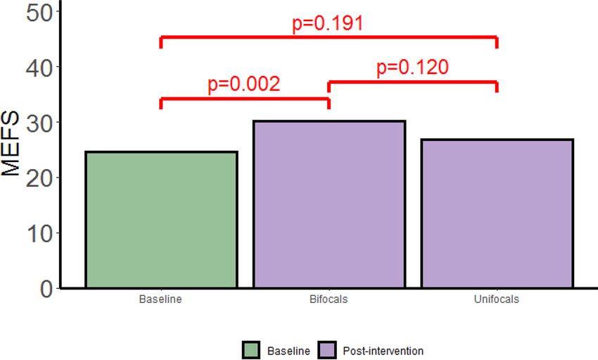
www.nature.com/scientificreports/
turn induces amblyopia (lazy eye, low visual acuity). If accommodation is poor, the attempt to accommodate
can induce strabismus and, as a result of strabismus, amblyopia. Nystagmus also hampers visual acuity, but low
visual acuity aggravates nystagmus. More information about the ocular disorders in children with DS and the
effect of bifocal on visual acuity and strabismus in children with DS is given in our previous publications21,22.
Visual impairment can hamper cognitive development too. In visual impaired children without known devel-
opmental disorders, the level of visual impairment indeed correlates with deficits in cognitive development23–27.
Cumulative debilitating consequences of early-onset visual impairment on cognitive, language and social skills
are described in other studies23,24,28. Even children with mild to moderate visual impairment show reduced adap-
tive behaviour. They have more difficulties with skills that affect development and learning than normally-sighted
typically developing c hildren28. However, in children with DS, it is still unclear whether the visual impairments
aggravate their lag in cognitive development. If this relation exists, improving their visual acuity with optimal
corrections in glasses tailored to the specific ocular disorders of children with DS could support cognitive
development. Children with DS might also benefit from higher visual acuities because studies performed in
the last two decennia show that their visuospatial memory is relatively preserved, and better than their verbal
memory29–32. In addition, the recent review by Lukowski et al.33 of studies on executive functions in children with
DS underscores the relative strength in visual spatial working memory. Deficits occur in all domains of executive
functions of children with DS—planning and goal directed behaviour, inhibitory control, cognitive flexibility
and working memory—but children with DS perform worse on verbal working memory than on visuospatial
working memory. The observed deficits in working memory are important in their own right, but its association
with academic achievement in children with DS highlight its significance f urther34.
Improving visual acuity with optimal corrections in glasses tailored to the specific ocular disorders of children
with DS could be a first step to support their visuospatial working memory and their cognitive development.
Correcting refractive errors in the way it is done in typically developing children is not optimal for children with
DS, because of their specific mixture of ocular disorders. Full correction of refractive error is required because
of the lag in accommodation. Moreover, the accommodation deficit may require different correction for looking
at far and near distances. In small scale studies with bifocals in children with DS, good results were obtained in
accommodation accuracy35,36, near visual acuity and literacy skills37,38 through the near addition. Adyanthaya
et al.39 studied the compliance with wearing bifocals. Of the children with bifocals, 89% were compliant whereas
only 50% were compliant with unifocals. Al-Bagdady et al.36 (n = 40, age range 5–14 years) found that accom-
modation was accurate in 38 (95%) children. Nandakumar et al.37,38 reported that with bifocals, visual acuity
improved more than 1.5 LogMAR and that 6 months later, literacy skills and school performance were improved
too. However, this investigation did not include a control group, and focused only on a small group of children
with DS that were pre-selected for their ability to read and write.
Triggered by these improvements in near visual acuity with b ifocals37,38, we set up a multicentre randomized
controlled trial (RCT) in the Netherlands to study the effect of bifocals in comparison to unifocals on visual func-
tions and cognitive development. Evaluated visual functions included distance visual acuity and near visual acu-
ity—both uncrowded (i.e., charts with a clear spacing between the symbols) and crowded (i.e., symbols printed
as close together as letters in a word) acuity, accommodation accuracy, strabismus, binocularity and stereopsis.
After one year, the full correction of refractive error improved distance visual acuity in both intervention groups,
but bifocals led to the largest improvement in near visual acuity and better ocular alignment (fewer children
with strabismus and smaller angles of strabismus)21,22. The improved visual acuities were a good starting point to
study the association with cognitive development too. Cognitive development was assessed by testing executive
functions—neurocognitive skills that serve as a foundation for early l earning40.
The cross sectional analysis of our baseline measurements was, to the best of our knowledge, the first study
to investigate the relation between lags in executive functions and visual impairments in children with DS6. This
analysis showed a correlation between visual acuity and the level of adaptive behaviour as previously reported for
visual impaired children without developmental d isorders23–28,41. The observed correlation between visual acuity
and the level of adaptive behaviour is a first indication that visual acuity might play a role in the development of
executive functions in children with DS. However, at baseline, the children still wore their habitual glasses, glasses
that were often not updated recently and that typically under corrected for the children’s refractive errors at the
time of inclusion. Thus, in our previous study, we did not consider the effect of best-corrected visual acuity, nor
did we analyse the 1-year cognitive development.
In the current paper, we assessed and compared the effects of two interventions improving visual acuity
in children with DS, bifocals and unifocals, on cognitive development. We analysed the executive functions
assessed in different ways and examined the relation between post-intervention visual acuity and the level of
post-intervention executive functions. We hypothesized that if visual acuity influences cognitive development,
intervention with glasses should have a larger effect on executive functioning after a whole year than shortly
after the intervention. Second, if near visual acuity is of particular importance, we would expect a larger effect
of bifocals than of unifocals. Third, we would expect a significant correlation between improvements in visual
acuity and improvements in executive functions. Note, however, that distant and near visual acuity need not
have the same effect on different measures of executive functions, as different measures of executive functions
capture different facets of executive f unctioning42,43.
Methods and participants
To study the difference between the effect of bifocals in the intervention group and the effect of unifocal glasses,
both with full correction of refractive error, we performed a multicentre randomized controlled trial in 15
participating locations in the Netherlands. Detailed descriptions of the methods and participants of this study
have been published elsewhere6,21,22. Here, we reproduce part of the methods, for completeness and clarity. The
Scientific Reports | (2021) 11:16893 | https://doi.org/10.1038/s41598-021-96308-5 2
Vol:.(1234567890)www.nature.com/scientificreports/
Figure 1. RCT time-line. Study design with the number of children tested at each point in time (n), mean
visual acuities (± standard deviation. Expressed in LogMAR), with significant short-term (T1) and 1-year (T3)
changes, and type of executive function assessments at baseline (T0), shortly after receiving new glasses (T1),
interim check for visual functions (T2) and after 1 year (T3). Randomization was stratified by age, gender and
level of verbal development. VA visual acuity, MEFS Minnesota executive function scale, a task-based executive
function test.
locations, 14 hospitals and one institute for the visually impaired, were geographically spread across the Neth-
erlands serving rural and urban populations of diverse social economic status.
The included children from the participating institutes were randomly allocated to the two intervention
groups with equal probability: bifocal group and unifocal group. The digital Web-based research data managing
system, ResearchManager (2014, a web-based electronic CRF, developed by Cloud9 Health Solutions and Isala
Academy in Zwolle, the Netherlands, according to GCP and GCDMP guidelines and 21 CFR part one of FDA
regulations) effectuated the randomization in a permuted blocks randomization schedule, stratified by gender,
age, and language development (parents report: speaking in 1 to 3 word sentences and speaking in 4 word or
longer sentences). The intervention group, to which the child was assigned, was always known to the participant,
the orthoptist and the investigator, because bifocal glasses are a visually prominent marker.
In both groups, full correction of refractive error measured using cycloplegia was applied. The bifocal seg-
ment top of the applied longline (flat-top or D-segment) bifocals with addition +2.5 dioptres, used in the bifocal
group, was placed at the pupillary centre, as used in previous s tudies35,36. In those studies, good results were
achieved in improving near vision and compliance in wearing these glasses. The children were seen on four
occasions, T0 (baseline), T1 ~ 6 weeks, T2 6 months after inclusion, and T3, the final assessments one year after
inclusion (see Fig. 1).
This project (Clinicaltrials.gov registration number NCT02241356, registration date 16/09/2014) was
approved by the Dutch Medical Ethics Committee of the Isala Hospitals (NL48288.75.14/ METC: 14.0333) and
confirmed by the local ethics committees of the participating clinics. All methods were performed in accordance
with relevant guidelines and regulations21,44,45. The sample size was calculated with G*Power 3 46 according to
results of former research37 on near vision before and after bifocals were used. To enlarge the inclusion number,
we made some amendments to the protocol shortly after the trial commenced and these were approved again
by the Medical Ethics Committee. Recruitment time, first planned for 6 months (from June to November 2015),
was extended to 9 months (to February 2016), follow-up time was shortened from 1.5 year to one year (ending
March 2017), and the age range originally limited from 2 to 14 years was extended to 18 years.
The current paper reports the effect of bifocals and unifocals, both with full correction of refractive error, on
executive functions during one year follow up. In our previous papers, we described the baseline assessments
of executive f unctions6 (Fig. 1, T0) and the effects of bifocals and unifocals on visual a cuity21, accommodation
accuracy and s trabismus22.
Participants. A total of 119 children with DS between 2 and 16 years old were included after written
informed consent was obtained from both parents of each child, and one parent in case of single parenthood.
We included children from the age of two years, the youngest age at which bifocals can be used in the appropriate
way by children with DS, because visual development takes place in the first years of life and the development of
strabismus may be avoided when corrections for hyperopia are used from young ages (to avoid excess of accom-
modation attempt, which induces convergence). At the age of two years, most children with DS can sit and per-
form a task at a table. This task performance induces a viewing direction which is needed to use bifocal glasses
in the appropriate way. All of the included children had (1) accommodation deficit, (2) not worn bifocals before,
Scientific Reports | (2021) 11:16893 | https://doi.org/10.1038/s41598-021-96308-5 3
Vol.:(0123456789)www.nature.com/scientificreports/
(3) ability to respond (verbally or non-verbally) to vision tests if they were older than 5 years, and (4) were able
to sit on a chair while doing a task. 104 children came back for testing with their newly prescribed glasses and
were included in the longitudinal analyses described in this paper.
Assessment procedures. Procedures for visual function examination and assessment of executive func-
tions were protocoled. The participating orthoptists, local investigators from the 15 participating locations in
the Netherlands, were trained to perform unfamiliar tests, to administer the MEFS as prescribed by Reflection
Sciences, LLCTM, and to use the digital research data manager ResearchManager (2014). Additionally, each
participating centre was visited by the principal investigator to review the procedures before the start and twice
by an independent research monitor during the study to verify compliance of the local investigators with the
research protocols.
A baseline visual function assessment was performed followed by executive functions assessment with the
task-based test. At the end of the first and final visit, questionnaires were handed out to the parents to be filled
out at home or were administered online by the parents or teachers, respectively.
If a child became uncooperative, testing was stopped according to the Dutch code of conduct relating to
expressions of objection by people who are incapable of giving consent, minors or mentally disabled participat-
ing in medical research45 (Code of conduct in the Netherlands 2002, NVK Code of conduct in the Netherlands
2001). Reasons for missed data, be it a lack of cooperation or otherwise, were noted.
Visual functions. Visual functions were assessed at all four time points (Fig. 1). At baseline (T0) the children
wore their habitual corrections or no corrections when they did not use glasses. At all 3 time points thereafter
(T1, T2 and T3), the children wore their newly prescribed glasses.
Visual acuity. Visual acuity was assessed with verbal or non-verbal methods at distance (5 m or 3 m, according
to the capacity of the child) and at near (40 cm) with symbol discrimination on visual acuity charts. Depending
on the child’s capacity, we used LEA s ymbols47 or Kay p
ictures48. At 40 cm, we assessed both uncrowded (sym-
bols with large spacing) and crowded (symbols arranged close to each other like letters in a word) near visual
acuity with LEA symbols with absolute spacing49,50.
Accommodation. Accommodation accuracy was assessed at 25 cm and 16.7 cm using the ‘modified Nott
method’9,51,52. The child looked at a small fixating object at the close distance. Meanwhile, the streak retinoscope
was moved closer and further away from the child’s eyes to assess the distance of the exerted accommodation.
Strabismus and binocularity. In case of (nearly) straight eye position (evaluated with corneal light reflex at
the beginning of the assessment) binocularity and stereopsis were assessed with one of several tests (TNO test
(Lameris Ootech, Nieuwegein, The Netherlands), Titmus Fly (Stereo Optical Co., Inc., Chicago, IL), Lang Ste-
reotest (Lang-Stereotest AG, Küsnacht, Switzerland), or positive base out 15 dioptre prism test), chosen by the
orthoptist according to the developmental stage and cooperation of the child.
After that, both manifest and/or latent strabismus angles were assessed with the prism cover test at 30 cm
and 5 m, the Hirschberg corneal reflex test53 and cover test at 30 cm and 5 m.
Adaptive developmental behaviour: Vineland‑S. The Vineland-Screener54,55 (Vineland-S) was used to assess
adaptive developmental behaviour at baseline (T0) only. The Vineland-S is a questionnaire for parents with 72
items. This questionnaire covers the four domains of adaptive behaviour: communication, socialization, daily
living skills, and motor skills.
Executive functions. Executive functions (EF) were assed with a task-based method and with questionnaires.
Such methods are complementary to one a nother55–58. Task-based tests are like a snapshot, a momentary assess-
ment mostly under optimal conditions. By contrast, rating based assessments provide a score of everyday execu-
tive functioning in the daily behaviour of the children in various settings.
Task‑based: Minnesota executive function scale (MEFS). At T0, baseline, T1, the assessment with newly pre-
scribed glasses after ~ 6 weeks, and at T3, the final assessments after one year, the participants themselves were
tested using the task-based Minnesota Executive Function Scale59,60 (MEFS). The MEFS is an engaging computer
card-sorting game administered on an iPad one-on-one with the child. The MEFS test, suitable for the entire
calendar age range of our participants, includes 7 levels of increasing difficulty, corresponding to the Total scores
of 10, 20, 30, 40, 50, 60 and 90.
The picture size of the MEFS test applied in our study is ~ 8 M which is visible for visual acuities of 1.5 Log-
MAR (3/100 Snellen equivalent) with an allowed viewing distance up to 15 cm.
Questionnaires: BRIEF‑P (preschool), BRIEF parents’ and BRIEF teachers’. At T0 and T3 (i.e., at baseline and
after one year), we obtained informant based ratings from the children’s parents and teachers in the Behav-
ior Rating Inventory of Executive Function (BRIEF-P or BRIEF) questionnaires61–66. The parents filled in the
BRIEF-P questionnaire or the parents’ version of the BRIEF depending on the calendar age of the child (in this
study of children with DS, younger than 8 years or eight years and older, respectively). Teachers filled in the
teachers’ version of BRIEF. These questionnaires provide an ecologically valid real-world assessment of execu-
tive functions and yield complementary information to the task-based test56. The BRIEF and its subscales can
Scientific Reports | (2021) 11:16893 | https://doi.org/10.1038/s41598-021-96308-5 4
Vol:.(1234567890)www.nature.com/scientificreports/
generally be performed in a psychometrically sound manner among school-age children with DS56,67. In our
analyses, we only considered the raw aggregated scores across domains, the raw Global Executive Composite
(GEC). Normative GEC scores in both boys and girls on the parents’ version of the BRIEF range between 72 and
216 and on the teachers’ version between 73 and 219. In BRIEF-P the normative GEC scores lie between 189 and
63 for boys and girls. Higher scores represent greater levels of executive function impairment.
Statistical analyses. Statistical analyses were performed using the statistical package for social sciences
(SPSS version 23, IBM., Chicago, IL) and the statistical software package “R” (version 3.6.2). We used mixed
effects regression models with a random intercept estimated for each participant. R-code for the mixed effects
regression analyses is made available on the data repository for this paper.
Adjustment for adaptive developmental age, assessed with the Vineland-S questionnaire, was not needed
because calendar age and adaptive developmental age assessed with the Vineland-S questionnaire were tightly
correlated (Pearson r = 0.722, p < 0.001 and r (partial adjusted for gender) = 0.724, p < 0.001).
For each intervention group, we first compared baseline MEFS, BRIEF-P and BRIEF results with post-inter-
vention scores (pooled across T1 and T3). Then, we compared the post-intervention results between the two
intervention groups. Thereafter, we analysed the relation between final visual acuities and post-intervention
MEFS scores. In addition to the test statistics and confidence intervals, we report Cohen’s d as a measure of effect
size. According to common convention, we interpret Cohen’s d effect sizes of 0.2, 0.5 and 0.8 as being small,
medium and large, respectively.
To analyze and compare the effects of the interventions on performance in the MEFS test, we used the raw
MEFS score, the Total score (further referred to as MEFS score), as opposed to its norm-referenced score, because
children with DS have cognitive and motor developmental lags and their development is heterogeneous. We ran
one mixed effects model on the MEFS scores obtained at T0, T1 and T3 with gender and age at T0 (in months)
as covariates. As the post-intervention (T1 and T3) MEFS scores were not significantly different between the T1
and T3, we pooled the data from these two time points to maximize statistical power.
Unfortunately, there were too many missing data for each of the baseline visual acuity measures to analyze
the effect of intervention-related visual acuity changes on the change in MEFS score. To make best use of the
data, we instead quantified the relation between visual acuity (i.e., visual acuity with newly prescribed glasses)
and post-intervention MEFS scores. We only considered the data of visits T1 and T3. The analysis consisted of
several steps. We first modelled the MEFS scores as a function of gender, age and intervention using the data
from subjects with no missing values. From the resulting mixed effects model, we then calculated adjusted MEFS
scores, i.e., MEFS scores adjusted for gender, age and intervention for the participants with MEFS scores at T1
and/or T3. Then, we ran three separate mixed effects models for the adjusted MEFS scores with crowded near
visual acuity, uncrowded near visual acuity and distant visual acuity as fixed effects, respectively. The missing
data of near visual acuities and distant visual acuity would otherwise introduce changes in the coefficients for
gender, age and intervention between these three analyses.
We also tested the association between the changes in ocular alignment and changes in MEFS scores between
T0 and T1 by applying the Spearman rank-correlation test. In this analysis, only children with MEFS scores
available at both T0 and T1 were included.
To analyse the results of the BRIEF-P and BRIEF questionnaires, we used the raw GEC scores (Global Execu-
tive Composit, i.e., the composite scores of all scales). We did not convert these scores to age-adjusted ‘total
scores’ for typically developing children (as described in the manuals of the questionnaires) because children
with DS have motor and cognitive delays.
Because questionnaire data were often missing, we had to limit our analyses of the BRIEF-P and the two
versions of the BRIEF to an exploratory analysis of the complete cases. A paired t-test was used to analyze the
differences between baseline scores and final scores. An unpaired t-test was used to compare these differences
between intervention groups. We calculated the lags of the children with DS compared to norm scores given
in the manual of the questionnaires, i.e., score of the child with DS minus age-matched norm score, at one year
follow-up (T3). We compared these developmental lags at final assessments to the lags found at b aseline6. For
completeness, and to be aware of possible biasing factors, we compared the group of children included in these
analyses to the group of children excluded because of missing data.
Results
At baseline, the bifocal and the unifocal group showed no statistically significant differences in calendar age,
adaptive developmental age, uncrowded and crowded near visual acuities and distant visual acuity, accommoda-
tion accuracy, strength of habitual glasses, ocular alignment and executive functions as assessed with the MEFS
and BRIEF-P or B RIEF6,21,22.
MEFS. The MEFS was successfully administered in 86 (83%) participants at baseline (T0), in 82 (79%) par-
ticipants at T1 when the children just started to use their new glasses, and in 94 (90%) participants at the final
assessments (T3). MEFS scores were missing for various reasons. Either the child was too young or it did not
understand the test (2, 5, 3 times at T0, T1 and T3, respectively), or the child was uncooperative (4, 5, 0 times
respectively) or there were technical problems with the iPad (1, 5, 3 times respectively). For the remaining cases,
no cause was described. In our analyses, only participants with baseline scores who had a follow-up score at T1
and/ or T3 were included (bifocals n = 41; unifocals n = 44).
Effect of the interventions on MEFS total scores. Post intervention MEFS score were correlated with age (See
supplementary Table S2 online) as they were at baseline6. Therefore, to investigate the effect of the treatments,
Scientific Reports | (2021) 11:16893 | https://doi.org/10.1038/s41598-021-96308-5 5
Vol.:(0123456789)www.nature.com/scientificreports/
Figure 2. Effect of the interventions on MEFS. Note the significant post-intervention improvement of MEFS
scores in the bifocal group. The intervention had no significant effect on the MEFS scores in the unifocal group.
Post-intervention values (pooled across T1 and T3) were not significantly different between the two groups.
we took into account gender and calendar age as possible confounding factors. We ran a mixed effects model
for the effect of treatment on MEFS scores adjusted for gender and calendar age at baseline across all three time
points at which MEFS scores were collected (T0, T1 and T3). In these analyses, the follow-up scores were pooled
across T1 and T3 since there were no significant differences between T1 and T3 (t(77) = − 1.459, p = 0.149; Sup-
plementary Fig. S1 online and Table S1 online). The results are shown in Fig. 2.
The mean MEFS score at baseline adjusted for gender and calendar age was 22.4 points (95% CI: 15.0, 29.7).
On the post-intervention visits, the mean MEFS score was increased to 28.0 points (95% CI: 20.5, 35.5) in the
bifocal group, and to 24.6 points (95% CI: 17.1, 32.1) in the unifocal group. In the bifocal group, the medium-
effect difference in mean MEFS scores between the post-intervention scores and baseline scores was 5.6 points
((95% CI: 2.1, 9.0); p = 0.002; Cohen’s d = 0.60 (95% CI: 0.22, 0.98)). In the unifocal group this difference was
small, only 2.26 points, and not statistically significant ((95% CI: − 1.1, 5.7); p = 0.191; Cohen’s d = 0.24 (95% CI:
− 0.12, 0.03)). The post-intervention difference in mean MEFS scores between the bifocal group and the unifocal
group of 3.3 points ((95% CI: -0.9, 7.5); p = 0.120; Cohen’s d = 0.34 (95% CI: − 0.09, 0.77)) had a small effect size
and was not statistically significant.
Thus, the effect of treatment on MEFS scores was significant between baseline and post-intervention meas-
urement in the intervention group. However, there was no significant difference in MEFS score between the two
intervention groups after the intervention. After the initial improvement with the new optical corrections in the
bifocal group, the longitudinal analysis of the MEFS scores showed no significant progression over the one-year
follow-up period. Nevertheless, we did obtain some clues that improving near vision may be helpful since the
2.5 dioptres addition for near vision in the bifocals was associated with an average medium-effect improvement
of 5.6 points in Total MEFS score with respect to baseline MEFS performance.
Cross‑sectional relation between visual acuity and the level of post‑intervention MEFS scores. At baseline, we
found no significant association between visual acuity and MEFS s cores6. However, at this point in time, most
participants were not yet wearing full corrections for their refractive errors, or the correction was out-dated21.
New prescriptions were given to both groups (bifocal and unifocal) according to the refractive error that was
measured at the start of the study. It is therefore of interest to analyze the effect of post-intervention visual acu-
ity on the level of post-intervention MEFS scores. Toward this end, we ran mixed effects models separately for
the effects of crowded near visual acuity (n = 68), uncrowded near visual acuity (n = 74) and distant visual acuity
(n = 76) on post-intervention T1 and T3 MEFS scores after adjustment of the MEFS scores for gender, calendar
age and intervention type (see Methods for details).
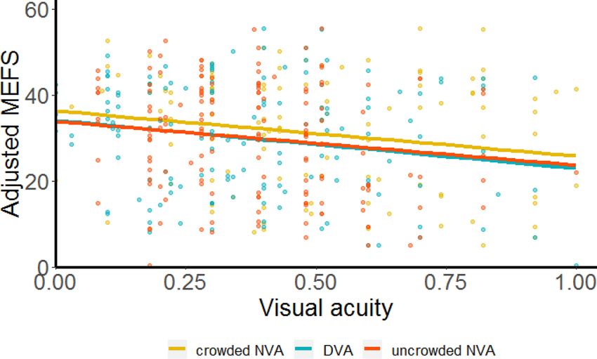
We found that the crowded near visual acuity, uncrowded near visual acuity and distant visual acuity with
newly prescribed glasses were associated significantly with post-intervention adjusted MEFS scores (Fig. 3). The
slopes for these relations were of similar magnitude and all negative (Table 1), indicating that after full correc-
tion of refractive error, better visual acuity was associated with better MEFS performance. Before pooling of the
data across T1 and T3, we verified that there was no statistically significant difference in MEFS scores between
T1 and T3.
Scientific Reports | (2021) 11:16893 | https://doi.org/10.1038/s41598-021-96308-5 6
Vol:.(1234567890)www.nature.com/scientificreports/
Figure 3. Post-intervention relations between visual acuity and MEFS scores. Post-intervention MEFS scores,
adjusted for gender, age and intervention, correlated significantly with each of the three post-intervention
visual acuity measures (LogMAR): crowded near visual acuity (orange), uncrowded near visual acuity (red) and
distant visual acuity (blue). Individual data points are from T1 and T3. Solid lines are mixed-effects regression
lines. Note that better MEFS scores are associated with better post-intervention visual acuities and vice versa.
Cohort’s mean visual acuity with newly prescribed
glasses Cohort’s mean adjusted MEFS score Slope
Crowded near visual acuity
0.51 LogMAR 31.0 points (95% CI: 28.1, 33.8) − 10.5 (95% CI: − 19.8, − 1.3) p = 0.025
Uncrowded near visual acuity
0.40 LogMAR 29.8 points (95% CI: 27.1, 32.4) − 10.2 (95% CI: − 18.6, − 1.8) p = 0.019
Distant visual acuity
0.39 LogMAR 29.7 points (95% CI: 27.1, 32.3) − 11.2 (95% CI; − 22.0, − 0.3) p = 0.045
Table 1. Post-intervention relations between visual acuity and MEFS scores. Fixed effects coefficients of
the mixed-effects regression analysis. Post-intervention MEFS scores were adjusted for gender, age and
intervention, and pooled across T1 and T3.
Association between MEFS score changes and changes in ocular alignment. Ocular alignment facilitates visual
functions as binocularity (merging the images of both eyes in the brain), accommodation endurance (eyes stay-
ing focused at the same near distance for a longer time), stereopsis (depth perception through interpretation of
the minimal differences between the image of the right and left eye) and are important in social situations for
having eye contact. Possibly, these functions may also play a role in the development of executive functions. We,
therefore, analyzed if the change in MEFS scores from baseline to T1 is associated with the change in ocular
alignment measured at T1, shortly after the children started wearing the newly prescribed g lasses22. In this analy-
sis, we found a significant positive rank-correlation between change in ocular alignment and change in MEFS
scores in the bifocal group (n = 36, rho = 0.343, p = 0.040), indicating that children with improved ocular align-
ment had improved in MEFS scores. We did not find such a significant rank-correlation in the unifocal group
(n = 37, rho = − 0.084, p = 0.620), in which the change in strabismus was not statistically significant22.
BRIEF‑P and BRIEF questionnaires. In line with our findings at baseline6, a year after the intervention,
we found no significant correlation between the informant report scores of executive functions and performance
of the children on the MEFS test (See supplementary Table S2 online). Unfortunately, all three questionnaires
suffered from large percentages of missing scores at both time points, T0 and T3, (missing questionnaires, base-
line: BRIEF-P 26%, BRIEF parents’ version 30% and BRIEF teachers’ version 43%. Final assessment: BRIEF-P
47%, BRIEF parents’ version 46% and BRIEF teachers’ version 46%). For this reason, in an attempt to still report
on all outcome measures68, we only did an exploratory analysis of complete cases on the difference between
baseline ratings (T0) and final ratings (T3). Participants with baseline and T3 data on BRIEF-P (bifocals n = 17;
Scientific Reports | (2021) 11:16893 | https://doi.org/10.1038/s41598-021-96308-5 7
Vol.:(0123456789)www.nature.com/scientificreports/
unifocals n = 14), or on BRIEF parents’ version (bifocals n = 15; unifocals n = 16) or on BRIEF teachers’ version
(bifocals n = 10; unifocals n = 13) were included in these analyses.
One year effect of bifocals and unifocals on BRIEF‑P and BRIEF scores. After one year, ratings on the teachers’
version of the BRIEF (Global executive composit, GEC) were significantly better than at baseline (change bifo-
cal group: − 21.9 points (95% CI: − 38.3, − 5.5), t(9) = 3.029, p = 0.014; Cohen’s d = 1.91 (95% CI: 0.42, 3.41));
change unifocal group: − 16.7 points (95% CI: − 30.6, 2.8), t(12) = 2.625, p = 0.022; Cohen’s d = 1.46 (95% CI: 0.23,
2.68). However, this improvement was not significantly different between the two intervention groups (t-test,
t(21) = − 0.541, p = 0.594; Cohen’s d = 0.23 (95% CI: − 0.60, 1.05)). Scores on the parents ratings BRIEF-P and
BRIEF were not significantly different between T0 and T3 (BRIEF-P: t(30) = − 0.937, p = 0.336; Cohen’s d = 0.34
(95% CI: − 1.05, 0.37; BRIEF parents version: t(30) = 0.980, p = 0.335; Cohen’s d = 0.35 (95% CI: − 1.06, 0.36)).
The mean improvement assessed by the teachers was − 19.0 points (95% CI: − 28.7, − 9.2; t(22) = 4.035, p = 0.001;
Cohen’s d = 1.68 (95% CI: 0.73, 2.63).
For completeness, we checked for differences between the children with completed teachers’ questionnaires
at both time points (baseline and final ratings) and those excluded because of missing or incomplete teachers’
questionnaires. The children included in the complete-case analysis tended to have higher adaptive developmen-
tal behaviour assessed with the Vineland-S questionnaire compared to the excluded children (49.9 months (95%
CI: 45.9, 53.9) and 45.2 months (95% CI: 41.9, 48.5) respectively, t-test, t(53) = 1.772, p = 0.082; Cohen’s d = 0.49
(95% CI: − 0.06, 1.04)). There were no differences in age, visual acuities or MEFS scores.
Because of the missingness in both the questionnaires and visual functions and the small number of children
with (improved) strabismus we were not able to analyse the associations between parent-rated and teacher-rated
executive functions with visual acuities and ocular alignment.
Discussion
The current multicentre RCT is the first longitudinal study to examine the effect of bifocals and unifocals on task-
based executive functions (assessed with MEFS) and rating-based executive functions (assessed with BRIEF-P
and BRIEF) in children with DS. The included children were 2 to 16 years old and all received full corrections of
their refractive error for distant vision. Although we found no significant differences between the interventions
after one year and shortly after the intervention, within the bifocal group there was a change. In this group, the
post-intervention MEFS scores were improved significantly compared with the participants’ baseline perfor-
mance. The effect size of this 1-year change in the bifocal group was medium, whereas the improvement of the
unifocal group was statistically small and not significant.
Unfortunately, we could not test reliably if there was an association between improvements in visual acuity
and improvements in MEFS scores (Data were too sparse to compute enough pre-post difference pairs). However,
our cross-sectional analysis of the post-intervention data showed that better post-intervention MEFS scores were
associated with better post-intervention visual acuities (crowded near visual acuity, uncrowded near visual acu-
ity, and distant visual acuity). For participants that had strabismus at baseline, improved ocular alignment with
bifocals was associated with improved MEFS scores. Exploratory analysis of the questionnaire data indicated that
improvements in executive functioning were also noted by teachers (teachers’ version of BRIEF), with a large
effect size, but not by parents (BRIEF-P and parents’ version of BRIEF). After a year, teachers reported fewer
problems with executive functioning regardless of the intervention type.
MEFS. The MEFS is a visual test in which verbal instructions are given, partly supported by visually demon-
strated instructions of swiping the picture in the right box. Thus, one might doubt about its value when testing
children with low visual acuities. Therefore, we verified the picture size (~ 8 M). The pictures presented in this
test are of good contrast and large enough to be easily discriminated by children with limited visual acuity, as
poor as 1.5 LogMAR. The mean uncrowded near visual acuity of the participants of our study was 0.58 ± 0.34
LogMAR21. None of our participants had an uncrowded near visual acuity poorer than 1.4 LogMAR at baseline.
In our previous publication on the baseline measurements of this cohort6, we checked the association of the
children’s scores with their visual acuity at baseline. We found no statistically significant association. Thus, we
could conclude that the MEFS test is suitable for children with visual impairment because the pictures are large
enough to be seen by these children, also without optical correction for near vision.
After one year, treatment effects on MEFS scores were not significantly different between the two intervention
groups and the longitudinal analysis showed no significant progression over the one-year follow-up period. Per-
haps the follow-up period was too short to find statistically significant differences between the two intervention
groups. Shortly after participants started wearing their new glasses, near visual acuity (uncrowded and crowded)
had improved on average, but it was not until a year later that the effect of bifocals on near vision exceeded the
nifocals21. Possibly, after a longer follow-up, when the near-vision differences between the intervention
effect of u
groups have developed and had more time to influence the development of executive functions, the better near
vision in the bifocal group could lead to a significant difference in the MEFS scores between the intervention
groups. Better near visual acuity might also help children with DS to sustainably enhance their visuospatial short-
term memory by training, as suggested by other a uthors69,70. However, to study the effects of better near vision
on the development of executive functions in children with DS, future studies may need longer follow-up times.
Unfortunately, we could not leverage the longitudinal design of the study to its full potential; there were too
many missing baseline measurements of visual acuity (mostly at near) to test if changes in executive functions
are directly associated with changes in a child’s visual acuity. Otherwise, we might have been able to account for
part of the between-subject variability in the intervention effects. However, we could examine the cross-sectional
association between the level of visual acuity and the level of MEFS performance observed after the interventions.
Scientific Reports | (2021) 11:16893 | https://doi.org/10.1038/s41598-021-96308-5 8
Vol:.(1234567890)www.nature.com/scientificreports/
We previously found no significant association between baseline visual acuity and baseline MEFS s cores6. We
only found an association between baseline visual acuity and adaptive developmental behaviour (assessed with
the Vineland-S questionnaire). However, at baseline, participants were not yet wearing full corrections for their
refractive errors. Our finding that better post-intervention visual acuities do correlate significantly with better
post intervention MEFS scores is in line with and extends previous cross-sectional studies in visual impaired
children without known developmental disorders23–27. It also agrees with and extends the findings of Tadic
et al.41 who compared attentional processes of visual impaired preschool children (without DS and cerebral
visual impairment (CVI)) and typically developing children with normal vision. In their cross-sectional study41,
they reported that visual impairment significantly reduces the capacity of a young child to regulate attention
between people and objects, and that in case of visual impairment, the ability to establish attention on toys and
maintaining of attention is lower than in children with normal vision.
Informant reported executive functions. After one year, only teachers reported a substantial improve-
ment of ~ 20 points. This statistically large improvement (Cohen’s d = 1.7) after either intervention represents
an ~ 50% reduction of the lag found in the baseline scores of the children with DS relative to age-matched norm
scores of typically developing children. At baseline, the mean difference in the teachers’ version of the BRIEF
scores compared to the age-matched norm scores was 40.1 points (95% CI: 32.3, 47.9)6. Although teachers
reported an improvement, parents did not report an improvement in the executive functions of their children
with DS. Such a discrepancy between parents’ and teachers’ ratings of behaviour is not u ncommon71,72. Poor
73,74 72,75
to moderate agreement was observed in children with DS , without DS , in twins with attention deficit
hyperactivity disorder42, and analysed in a review already in 198771 and more recently in 200843. Explanations for
the discrepancies include the possibility that parents and teachers are observing different behaviours and phe-
notypes, particularly given the more structured demands at school settings versus less organized home activi-
ties, placing different demands on children depending on the s etting42,43. So, different informants may validly
contribute different unique information from different perspectives. Additionally, activities at home may be
different from those at school. School activities might include more visually guided activities, which could be
more directly influenced and facilitated by better visibility and visual memory support owing to better seeing
with new glasses in both intervention groups.
The studies of Daunhauer et al.76,77 can also help understand the apparent disagreement between parents and
teachers. Their findings include that teachers do encounter the changes in executive functions and are able to rate
them in a questionnaire on executive functions. In their cross-sectional study in elementary students with DS,
aged 7.86 ± 1.75 years, they demonstrated that executive function skills scored by teachers was the only statisti-
cally significant predictor of overall school performance in elementary students with DS76. They mention the
following two implications. First, executive functions may play a more prominent role in academic contexts for
children with DS than was previously noted in literature. Second, their findings suggest that improving execu-
tive functions may be of particular use for improving overall school performance in DS. Their findings are an
additional motivation to find interventions that can improve executive functions in children with DS. Bifocals
with full corrections of refractive errors could be one of them.
Strengths and limitations. Some of the strengths of the current study are already reported in our previ-
ous publications. These include the longitudinal design and the large sample size with a relatively rare biologi-
cally well-defined condition (DS). The participants were recruited from rural and urban populations of diverse
social status and attended both regular schools and schools for children with special needs, in order to attain a
cohort that represents the general Dutch population of children with DS. Further strengths were the multimodal
and multi-informant evaluation of intervention efficacy; the robust and standardized measurements that made
data collection across multiple sites possible, the use of the combination of both task-based and informant-
based measures of executive function differentiating between parent ratings and teacher ratings. The need to
obtain reports of both types of observers is highlighted by many authors because of the difference in fundamen-
tal behaviours they observe42,43,75. Besides the need for different observers in different situations, the combina-
tion of task-based assessments (a momentary assessment mostly under optimal conditions) and rating-based
assessments (scoring everyday behaviour) is complementary in typically developing children56,57,62,75, in preterm
preschoolers58 and in children with DS78. Studies applying such a combination of task-based scores and raters’
information are scant in young children with DS, except for a few studies78,79.
Additionally, the current paper focuses on a novel question, i.e., whether visual functions (acuity and ocular
alignment) are associated with the level of cognitive performance.
A further strength is the refined visual acuity assessment used in the current study to analyse the association of
visual acuity and executive functions, instead of broad visual impairment categories, which were used in previous
studies24,25, and which do not specify visual acuity at different distances. In our study, we found different timelines
for development of uncrowded and crowded near vision and could study the differences between distant and
near vision which go unnoticed if these facets of vision are not independently measured. In DS, the difference
between distant and near visual acuity is typical if not corrected accordingly, because of their accommodation
lag and cerebral visual i mpairment21. Analyses of the correlations with other developmental measures, such as
MEFS, were possible because of the refined assessments of visual acuity.
One of the limitations of our study is the limited follow-up time of one year. In children with DS, development
is slow. Where a time lapse of one year in typically developing children is often long enough to detect develop-
ment, in children with DS it may have been too short to detect significant progress in MEFS scores in the unifocal
group or a possible difference in MEFS scores between the intervention groups. To reveal differences in slowly
developing processes, longer follow-up times are necessary. Possibly, the development of executive functions
Scientific Reports | (2021) 11:16893 | https://doi.org/10.1038/s41598-021-96308-5 9
Vol.:(0123456789)www.nature.com/scientificreports/
induced by better visual functions is one of these slow developments, which need time to reach statistically
significant differences between baseline and final assessments and between the interventions.
The large age range can also be taken as a limitation. Developmental steps of visual acuity but specially in
executive functions are not the same during one year in the youngest ages than in the older ages because of non-
linearities in the developmental curve. Especially in children with DS, development is heterogenous. In our study,
we included children from the age of two. This is the youngest age at which bifocal use could be expected in the
appropriate way. We corrected refractive errors also for near distances in the bifocal group, at the youngest ages
possible in order to stimulate the development of visual functions.
The possibility that different teachers might have completed ratings on the same child due to the nature of the
trial spanning 12 months might be another limitation. We did not monitor that, because the teachers remained
anonymous. Longer follow-up times would have exacerbated this issue even more.
The biggest limitation of our study was the large amount of missing data. For the three versions of BRIEF
questionnaires. We tried to deal with this limitation by performing an exploratory analysis of complete cases
in order to report the results for all outcome measures. The missing visual acuity data, in particular at baseline,
also limited longitudinal analyses. We could not enter all the visual acuity variables in a mixed effects model to
analyse the changes in executive functions in relation to changes of visual acuity. We therefore had to limit our
analyses to cross-sectional data from the post-intervention visits.
Overall evaluation of the interventions. After the interventions, the MEFS scores were significantly
improved in the bifocal group but not significantly in the unifocal group. Post-intervention, children with bet-
ter visual functions, crowded and uncrowded near visual acuity and distant visual acuity, showed higher MEFS
scores. Children with improvements in ocular alignment typically improved in MEFS scores.
Only explorative analyses could be performed on the BRIEF-P and BRIEF data. Teachers, but not parents,
rated improved executive functions in both intervention groups. However, these findings need replication in
larger samples with longer follow-up. Such studies could explore if the better post-intervention ratings by teach-
ers and task-based scores on executive functions in DS are a developmental phenomenon or only the result of
better visual functioning.
Notwithstanding the acuity improvements as a result of bifocals, children with DS wearing appropriate bifo-
cals still lag behind in visual acuity (far and near) compared to typically developing children6.
Conclusion
After full correction of refractive error, better distant and near vision were associated with higher executive
function scores on task-based test administered at near, the MEFS. However, there were not enough data to
test such an association with informant reported scores. Nevertheless, teachers’ ratings suggest that at school,
children show improved executive functions when wearing full corrections of their refractive error. The +2.5
addition in the bifocals with full correction of refractive error improved near vision more than the full correc-
tion of refractive error alone, and bifocals also improved the conditions to achieve better task-based executive
function scores on the MEFS.
On the basis of our findings, we suggest to optimize visual functions in children with DS by prescribing them
optimal corrections for both distant and near vision to maximize their developmental chances. We found that
good corrections for children with DS are up-to-date full corrections of refractive error in bifocals with an addi-
tion of +2.5 dioptres for near vision. Further longitudinal research is needed to investigate if improved visual
functions indeed boost the development of executive functions in DS.
Received: 13 February 2021; Accepted: 4 August 2021
References
1. Parker, S. E. et al. National Birth Defects Prevention Network. Updated national birth prevalence estimates for selected birth defects
in the United States, 2004–2006. Birth Defects Res. A. Clin. Mol. Teratol. 88, 1008–1016. https://d oi.o
rg/1 0.1 002/b
dra.2 0735 (2010).
2. van Gameren-Oosterom, H. et al. Unchanged prevalence of Down syndrome in the Netherlands: Results from an 11-year nation-
wide birth cohort. Prenat. Diagn. 32, 1035–1040. https://doi.org/10.1002/pd.3951 (2012).
3. Takashima, S., Becker, E. L., Armstrong, D. L. & Chan, F. Abnormal neuronal development in the visual cortex of the human fetus
and infant with Down’s syndrome. A quantitative and qualitative Golgi study. Brain Res. 225, 1–21. https://doi.org/10.1016/0006-
8993(81)90314-0 (1981).
4. Becker, L. E., Amstrong, D. L. & Chan, F. Dentritic atrophy in children with Down’s syndrome. Ann. Neurol. 20, 520–526. https://
doi.org/10.1002/ana.410200413 (1986).
5. Watt, T., Robertson, K. & Jacobs, R. J. Refractive error, binocular vision and accommodation of children with Down syndrome.
Clin. Exp. Optom. 98, 3–11. https://doi.org/10.1111/cxo.12232 (2015).
6. de Weger, C., Boonstra, N. & Goossens, J. Differences between children with Down syndrome and typically developing children in
adaptive behaviour, executive functions and visual acuity. Sci. Rep. 11, 7602. https://doi.org/10.1038/s41598-021-85037-4 (2021).
7. van Gameren-Oosterom, H. B. et al. Development, problem behavior, and quality of life in a population based sample of eight-
year-old children with Down syndrome. PLoS ONE 6, e21879. https://doi.org/10.1371/journal.pone.0021879 (2011).
8. Morton, G. V. Why do children with Down syndrome have subnormal vision?. Am. Orthopt. J. 61, 60–70. https://doi.org/10.3368/
aoj.61.1.60 (2011).
9. Woodhouse, J. M., Meades, J. S., Leat, S. J. & Saunders, K. J. Reduced accommodation in children with Down syndrome. Invest.
Ophthalmol. Vis. Sci. 34, 2382–2387 (1993).
10. Woodhouse, J. M. et al. Visual acuity and accommodation in infants and young children with Down’s syndrome. J. Intellect. Disabil.
Res. 40, 49–55. https://doi.org/10.1111/j.1365-2788.1996.tb00602.x (1996).
Scientific Reports | (2021) 11:16893 | https://doi.org/10.1038/s41598-021-96308-5 10
Vol:.(1234567890)You can also read