Review Article Resident Macrophages in Cystic Kidney Disease - Kidney360
←
→
Page content transcription
If your browser does not render page correctly, please read the page content below
Review Article

  Resident Macrophages in Cystic Kidney Disease
  Zhang Li,1 Kurt A. Zimmerman,2 and Bradley K. Yoder              1

  Abstract
  Interstitial inflammation is an important feature of cystic kidney disease. Renal macrophages are the most well-
  studied inflammatory cell in the kidney, and their involvement in cyst formation has been reported in different
  animal models and patients with cystic kidney disease. Originally, it was believed that renal macrophages were
  maintained from a constant supply of bone marrow–derived circulating monocytes, and could be recruited to the
  kidney in response to local inflammation. However, this idea has been challenged using fate-mapping methods, by
  showing that at least two distinct developmental origins of macrophages are present in the adult mouse kidney. The
  first type, infiltrating macrophages, are recruited from circulating monocytes and gradually develop macrophage
  properties on entering the kidney. The second, resident macrophages, predominantly originate from embryonic
  precursors, colonize the kidney during its development, and proliferate in situ to maintain their population
  throughout adulthood. Infiltrating and resident macrophages work together to maintain homeostasis and properly
  respond to pathologic conditions, such as AKI, cystic kidney disease, or infection. This review will briefly
  summarize current knowledge of resident macrophages in cystic kidney disease.
                                      KIDNEY360 2: 167–175, 2021. doi: https://doi.org/10.34067/KID.0006052020

  Macrophages in Kidney                                                resident macrophages in the kidney from birth to full
  Renal macrophages, the largest population of immune                  maturity, and found that a portion of resident macro-
  cells in kidney, play a critical role in homeostasis,                phages are actually derived from peripheral monocytes.
  surveillance, immune response, tissue injury, and re-                The idea that some kidney-resident macrophages are
  pair (1–3). Macrophages are a major constituent of the               derived from monocytes was supported by data from
  renal mononuclear phagocyte system and were be-                      Ms4a3Cre-RosaTdT fate-mapping mice, which faithfully
  lieved to arise from bone marrow–derived monocytes                   label all adult hematopoietic stem cell–derived mono-
  that could be polarized into different phenotypic sub-               cytes (19). In these studies, the authors showed that
  sets in response to environmental stimuli (4–6). How-                a significant portion of kidney-resident macrophages
  ever, they were often confused with dendritic cells due              were originated from adult hematopoietic stem cell–
  to their shared cell-surface expression of CD11c and                 derived monocytes. Liu et al. (18) also demonstrated that
  MHCII (7,8). With the advent of new lineage tracing                  both lineages of resident macrophages shared the fea-
  and single-cell RNA sequencing approaches, it is now                 ture of long-lived residency in the kidney, but had
  possible to clearly delineate macrophages and den-                   functional differences in aspects including immune re-
  dritic cells in the kidney across mammalian species                  sponse and metabolic profile in disease conditions.
  (9–11). In these studies, the authors demonstrated that              These data suggest kidney-resident macrophages can
  renal macrophages were highly heterogenous and spe-                  be derived from multiple precursor populations and
  cialized populations and were derived from two dif-                  their ontological origin may influence their function
  ferent developmental origins (12–14) (Figure 1). One                 (6,17,18).
  type, infiltrating macrophages, is derived from mono-                    In mice, infiltrating and resident macrophages can
  cyte precursors in the bone marrow and recruited to                  be distinguished on the basis of the expression of
  the kidney in response to local inflammation (15,16).                 surface markers F4/80 and CD11b, with resident mac-
  The other type, resident macrophages, maintain long-                 rophages being F4/80high, CD11blow and infiltrating
  term residency in kidney with less mobility and arise                macrophages being F4/80low, CD11bhigh (12,13,20).
  primarily during organogenesis (17). They are derived                The exact function of resident macrophages in the
  in a Myb-independent manner from erythromyeloid                      kidney is not well known, although emerging evidence
  progenitors that are first generated in the fetal yolk sac,           suggests they play an important role in kidney de-
  colonize the fetal liver, and migrate into the kidney                velopment, vascularization, and renal repair in re-
  during early development (12,13,17).                                 sponse to AKI (17,21–24). Although there are limited
     The idea that resident macrophages are a homoge-                  data on the function of resident macrophages in the
  nous population in the kidney and are exclusively                    kidney, in part due to the nonspecific approaches used
  derived from embryonic precursors has recently been                  to study these cells in the past, we may be able to gain
  challenged. Utilizing a newly generated cre-induced-                 insight into their proposed functions due to similarities
  hCD59 transgenic line, Liu et al. (18) traced the fate of            between M2-like macrophages and resident macrophages.

  1
      Department of Cell, Developmental, and Integrative Biology, University of Alabama at Birmingham, Birmingham, Alabama
  2
      Division of Nephrology, Department of Internal Medicine, University of Oklahoma Health Sciences Center, Oklahoma City, Oklahoma

  Correspondence: Dr. Bradley K. Yoder, Department of Cell, Developmental, and Integrative Biology, University of Alabama at Birmingham,
  MCLM688, 1918 University Boulevard, Birmingham, AL 35294. Email: byoder@uab.edu

www.kidney360.org Vol 2 January, 2021                                                            Copyright © 2021 by the American Society of Nephrology   167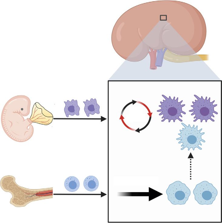
168   KIDNEY360

                               Yolk Sac
                                                                                                        Resident
                                                                     Self                              Macrophages
                                                                   renewal

                               Bone Marrow
                                                                 Recruitment                            Infiltrating
                                                                                                       Macrophages

Figure 1. | The origins of macrophages in kidney. Two types of macrophages are present in the adult mouse kidney. Resident macrophages
predominantly originate from embryonic precursors, migrate into the kidney during early development, and are maintained in the kidney
through local proliferation. Infiltrating macrophages are derived from monocyte precursors in the bone marrow, and are recruited to the kidney
in response to local inflammation. The dashed arrow indicates a limited but continuous contribution of monocyte-derived cells to the resident-
macrophage pool in adult mouse kidneys.

M2 macrophages have an anti-inflammatory and profibrotic                  evidence that macrophages could promote cyst progression in
function (3,4,25); most resident macrophages also exhibit an            animal models utilizing a phagocytic poison, liposomal clodr-
M2-like phenotype, with intrinsic anti-inflammatory prop-                onate (LC), to deplete all macrophages in the kidney. The
erties (3). In addition, renal resident macrophages share               authors showed that treatment of cystic mice with LC not only
expression of CD206 and Arg1 with M2 macrophages                        significantly decreased the number of renal macrophages (95%
(20,26), suggesting that M2 macrophages and resident mac-               reduction), but more importantly, reduced the cystic index and
rophages are similar populations of cells. It has also been             improved renal function when compared with vehicle-treated
reported that CD2061 M2 macrophages can promote tubu-                   controls (Table 1). The conclusion that macrophages could
lar regeneration by expressing growth factors during the                promote cystic kidney disease was further supported by sub-
reparative phase after AKI, which is similar to the function            sequent studies from Swenson-Fields et al. (35). In these studies,
of resident macrophages in AKI (27–29). Although these                  the authors showed that treatment with LC also reduced cystic
cells share significant functional properties, direct evidence           disease in a recessive model of cystic kidney disease. These
that M2 macrophages and resident macrophages are the                    data suggested macrophages may be involved in promoting
same cell type is still lacking.                                        cyst progression in multiple forms of cystic kidney disease.
   Overall, it is evident the renal macrophage niche in the             However, because these studies removed all phagocytic cells in
kidney is more diverse than originally appreciated. To better           the kidney, which includes infiltrating macrophages, resident
understand the role of macrophages in kidney under normal               macrophages, and dendritic cells, the contribution of each
and pathologic conditions, further studies are needed to char-          subset to cyst progression was still unknown.
acterize the respective functions of these macrophage subsets              There was a long-held belief that renal macrophages are
and the underlying molecular mechanisms involved.                       derived from, and continually replenished by, circulating mon-
                                                                        ocytes. This has led to the idea that targeting the recruitment of
                                                                        monocytes to the kidney may be therapeutically beneficial in the
Macrophages in Cystic Kidney Disease                                    context of cystic kidney disease. This hypothesis is supported by
   Seminal studies in the field of cystic kidney disease high-           data indicating that genetic deletion or pharmacologic inhibition
lighted an association between the number of renal macro-               of Ccl2 (also known as monocyte chemoattractant-1 or MCP-1)
phages and the severity of cystic disease (30–33). However, it          and macrophage migration inhibitory factor reduce the severity
was debated whether macrophages had a causative role in cyst            of cystic kidney disease (36–38). More importantly, the level of
formation or were a secondary consequence of cyst progres-              cyst reduction using macrophage-targeted therapeutics was
sion and expansion. Karihaloo et al. (34) provided the first             comparable to other inhibitors commonly used in the PKD
KIDNEY360 2: 167–175, January, 2021 Macrophages and PKD, Li et al. 169
Table 1. Effect of macrophage interventions on cystic kidney disease and their outcomes
Reference
Animal Model Intervention Method Outcomes
(Yr)
Pkd1fl/fl;Pkhd1-cre mice LC was administrated on alternate LC treatment improved renal 34 (2011)
d from PN10 to PN24. Mice were function and reduced cystic index
harvested at PN24. by 24%.
cpk/cpk mice LC was administrated at PN3 and LC treatment improved renal 35 (2013)
PN6. Mice were harvested at function and reduced cortical
PN10. cystic index by 29%.
Pck rats Bindarit (MCP-1 synthesis inhibitor) No effect on cystic index or kidney 40 (2015)
was administrated daily from 5 to function.
15 wk. Mice were harvested at 15
wk.
Hypomorphic Pkd1nl/nlmice or ISO-1 (MIF inhibitor) was ISO-1 treatment improved renal 38 (2015)
administrated daily from PN5 to function and reduced cystic index
PN27 in Pkd1nl/nl mice. Mice were by 51%.
harvested at PN28.
Mif2/2;Pkd1fl/fl;Ksp-cre mice Mif2/2;Pkd1fl/fl;Ksp-Cre mice Mif2/2;Pkd1fl/fl;Ksp-cre mice had
(DKO) (DKO) were harvested at PN7. improved renal function and
reduced cystic index by 58%.
Pkd1fl/fl;Pax8-rtTA;TetO-cre or INCB3344(CCR2 antagonist) was INCB3344 treatment improved renal 37 (2018)
administrated daily from 6 wk to function and reduced cystic index
12 wk of age in Pkd1fl/fl;Pax8- by 31%.
rtTA;TetO-Cremice.
Mcp1fl/fl;Pkd1fl/fl;Pax8-rtTA;TetO- DKO mice were induced with Mcp1fl/fl;Pkd1fl/fl;Pax8-rtTA;TetO-
cremice (DKO) doxycycline from PN28 to PN42. Cre mice had improved renal
Mice were harvested at 18 wk. function and reduced cystic index
by 60%.
Pkd1fl/fl;CAGG-creERT2 LC was administrated daily from LC treatment during Stage 1 did not 26 (2018)
PN16 to PN23 (Stage 1) or from affect renal cysts, but LC treatment
PN24 to PN32 (Stage 2) in PN10 during Stage 2 improved renal
induced Pkd1fl/fl;CAGG-CreERT2 function and reduced cystic index
mice. by 60%.
Mcp1fl/fl;Pkd1fl/fl;Pax8-rtTA;TetO- DKO were induced with Genetic deletion of Mcp1 in Pkd1fl/fl; 36 (2018)
cremice (DKO) doxycycline from PN28 to PN42. Pax8-rtTA;TetO-Cre mice
Mice were harvested at 13.5 wk. improved renal function and
reduced cyst formation.
cpk/cpk;Ccl22/2 mice cpk/cpk Ccl22/2 mice were Genetic deletion of Ccl2 did not 41 (2019)
harvested at PN18. affect kidney cysts, but prolonged
survival of mice.
Ift88fl/fl;CAGG-CreERT2 mice with GW2580 (CSF1R kinase inhibitor) GW2580 treatment reduced cystic 20 (2019)
ischemia-reperfusion (IR) injury was administrated daily from d 1 index by 60%.
to d 35 post IR injury in adult
induced Ift88fl/fl;CAGGCre ERT2
mice.
cpk/cpk mice GW2580 was administrated daily GW2580 treatment on cpk mice
from PN11 to PN21 in cpk/cpk reduced rate of growth in TKV
mice. (slope) by 37%.
Pkd1fl/fl;CAGG-CreERT2 mice with IRF5 ASO was given weekly to IRF5 ASO treatment reduced cystic 51 (2020)
UNX Pkd1fl/fl;CAGG-CreERT2 mice for 6 index by 50%.
wk post nephrectomy.
LC, liposomal clodronate; MCP, monocyte chemoattractant; MIF, migration inhibitory factor; DKO, double knockout; INCB, In-
ternational Narcotics Control Board; UNX, unilateral nephrectomy; TKV, total kidney volume; ASO, antisense oligo.
field, such as tolvaptan or rapamycin (39,40). Whereas several several decades. For example, multiple studies indicate that
studies indicate that inhibition of these macrophage ligand:re- alternatively activated M2-like macrophages expressing
ceptor interactions restricts cyst growth, other studies did not CD206 (26,35,48), a newly identified cross-species marker
report similar findings (41–43). The involvement of infiltrating of resident macrophages (11,49), could promote renal cyst
macrophages in cystic disease remains controversial, and has formation. Microarray analysis from Mrug et al. (50) found
been reviewed previously (44–47). For comparison, we include that in a nonorthologous model of autosomal recessive
a table that summarizes the outcomes from multiple studies polycystic kidney disease (Cys1cpk/cpk), the most highly
targeting macrophages in cyst severity (Table 1). expressed genes were those associated with the innate im-
mune response. Of interest, several of these innate immune
genes were recently identified as being specific for resident
Resident-Macrophage Involvement in Cystic Kidney macrophages by single-cell RNA sequencing (C1qa, Cxcl16)
Disease (11). Also, Viau et al. (36) found increased numbers of both
In retrospect, data suggested the presence and involve- infiltrating and resident macrophages during cyst progres-
ment of resident macrophages in cystic kidney disease for sion in inducible Lkb1 and Pkd1 mouse models. Although170 KIDNEY360
the authors emphasized the importance of infiltrating mac- showed that resident macrophages in wild-type mouse
rophages in controlling cyst progression in this study, they kidneys undergo a phenotypic switch (from CD11clow to
also observed an increased number of resident macrophages CD11chigh) during the postnatal period. CD11clow resident
expressing CCR2 (receptor for MCP-1) in cystic kidneys, macrophages are enriched during juvenile periods, negligible
suggesting the MCP1-CCR2 axis may also be important for in adult mice, and reappeared after renal ischemia-
resident macrophages. reperfusion injury in adult cilia mutant mice. More impor-
Zimmerman et al. (20) provided evidence for the involve- tantly, the number of CD11clow resident macrophages was
ment of resident macrophages in cystic disease using con- increased before and during cyst formation, suggesting
ditional Ift88 cilia mutant mice. These mutants develop a potential causative or at least contributing role for these
a cystic kidney disease phenotype, where the severity is cells in cyst formation. Analysis of confocal images indi-
greatly influenced by the age at which cilia loss is induced cated most resident macrophages coexpressed F4/80, Ki67,
(51). Ift88 loss induced in juvenile periods (before postnatal and CD206 in cystic regions, suggesting a reciprocal com-
day 14) leads to rapid and severe cyst formation. In contrast, munication between resident macrophages and cyst-lining
induction in adults results in slow cyst progression occur- epithelial cells (Figure 2). To understand the mechanism
ring in focal regions of the kidney. The rate of cyst formation through which resident macrophages were accumulating,
in adult-induced mutants was found to be greatly increased the authors performed parabiosis experiments by joining
and more widespread after ischemia-reperfusion injury. In the circulation of a CD45.2 control or cilia mutant mouse
correlation with these rates of cystogenesis, the authors with a congenic CD45.1 wild-type mouse and found the
Reciprocal Signaling between Resident
Macrophage and Epithelial Cell in Cystic Kidney
ECM deposition and Fibrosis
Resident Macrophage
Abnormal Vasculature CSF-1R
Inflammatary IL6, Wnts
Cytokines Signaling?
mb-CSF1
STAT3 P
STAT3 P
Epithelial
STAT3 P Cell
STAT3 P
Cyst Renal tubule
RM IM T cell Neutrophil
Epithelial Dividing Apoptotic Myo-
cell cell Cell fibroblast
Proliferation
Interstitial inflammation Cytokines Vascularture Extracellular Matrix
Apoptosis
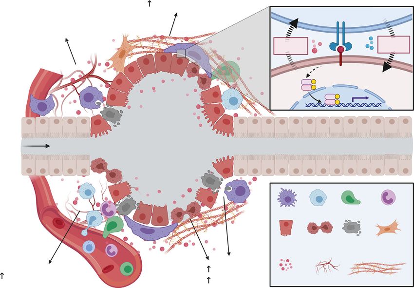
Figure 2. | Proposed functions of resident macrophages during cyst formation and expansion. Multiple processes occur during cyst formation
and expansion, including increased epithelial proliferation and apoptosis, interstitial inflammation, increased extracellular matrix deposition
and renal fibrosis, and vasculature abnormalities. Resident macrophages may be involved in controlling several of these processes directly or
indirectly. It has been proposed that resident macrophages can promote cystic epithelial proliferation by secretion of cytokines and phagocytosis
of apoptotic epithelial cells. Also, renal-resident macrophages may drive interstitial myofibroblast activation and proliferation, leading to
increased extracellular matrix deposition and renal fibrosis. Resident macrophages may serve as “first responders” in the kidney and control the
accumulation and effector function of other immune cells, such as neutrophils, infiltrating macrophages, and T cells, to indirectly regulate cyst
formation. Finally, renal resident macrophages may also regulate vasculature abnormalities through their proposed proangiogenic functions.
The inset indicates the reciprocal communication between resident macrophages and the cilia mutant epithelium via mb-CSF1/CSF-1R. ECM,
extracellular matrix; mb-CSF1, membrane-bound colony-stimulating factor-1; RM, resident macrophages; IM, infiltrating macrophages.KIDNEY360 2: 167–175, January, 2021 Macrophages and PKD, Li et al. 171 accumulation of resident macrophages in cilia mutant kid- formation (55). RNA sequencing of resident macrophages neys was independent of peripheral blood input. Analysis of after AKI indicated transcriptional reprogramming of res- cell proliferation using Ki67 indicated that accumulation of ident macrophages, including upregulation of several Wnt resident macrophages in injured cilia mutant mice was genes (Wnt4, Wnt7b, Wnt10a, and Wnt10b) (21). Wnt-induced largely driven by self-proliferation. To determine the cell b-catenin signaling can protect against epithelial apoptosis type that was responsible for driving resident macrophage and promote proliferative repair (27,56,57). In addition, Wnt proliferation, the authors flow sorted epithelial populations signaling can also drive interstitial myofibroblast activation and showed the expression of membrane-bound colony and proliferation, leading to increased matrix-protein de- stimulation factor 1 was significantly increased in the prox- position and renal fibrosis (56,58). These data suggest that imal tubule cells of the injured cilia mutant kidneys com- resident macrophage–derived Wnts promote the prolifera- pared with injured controls (Figure 2). Inhibition of CSF1R tion of cystic epithelium and drive interstitial fibrosis during kinase signaling, using GW2580 (52), reduced resident mac- cystic disease progression. rophage proliferation, prevented the accumulation of It is also possible that resident macrophages serve as “first CD11clow resident macrophages, and reduced the severity responders” in the kidney and control the accumulation and of cystic disease in the injured conditional Ift88 model and in effector function of other immune cells, such as neutrophils, the more rapidly progressing cpk mouse model. Interest- infiltrating macrophages, and T cells, which have all been ingly, GW2580 treatment did not affect the infiltrating mac- observed in patients and mouse models of cystic kidney rophage number, suggesting the effects of GW2580 were disease (47,59–62). In fact, resident macrophages are well resident-macrophage specific. suited for this role as they are able to maintain a persistent Data showing that resident macrophages could promote residence in an adult kidney through self-proliferation and cystic disease were supported by follow-up studies inves- are located directly adjacent to the tubular epithelium (63). tigating the involvement of macrophage subsets in an Thus, they may serve as sentinels in the kidney to regulate orthologous mouse model of autosomal dominant polycys- the accumulation of other immune cells that influence cyst tic kidney disease (ADPKD) (53). Using conditional Pkd1 growth and progression. Indeed, due to their residency mice with unilateral nephrectomy, Zimmerman and collea- advantage, resident macrophages can act more rapidly than gues showed the numbers of infiltrating and resident mac- neutrophils, which have always been regarded as first res- rophages were increased in conditional Pkd1 mice with ponders in kidney injury. Using a combination of intravital unilateral nephrectomy compared with sham-operated imaging and confocal multiplex microscopy, Uderhardt mice, and that the increase occurred before the onset of et al. (24) observed that resident macrophages exert a “cloak- severe cystogenesis. Furthermore, the authors showed that ing” behavior by extending pseudopods around a local IFN regulatory factor 5 (Irf5), a transcription factor known to injury, which will prevent injury-induced neutrophil acti- induce inflammatory cytokine production in macrophages vation and neutrophil-driven inflammation. Thus, it is pos- (54), was increased in infiltrating and resident macrophages sible resident macrophages serve a similar role in cystic in cystic kidneys. To identify the function of IRF5 in macro- disease. phages and its importance in cyst formation, the authors In addition, epithelial apoptosis is detected during cyst suppressed the expression of IRF5 with an antisense oligo formation (64) and macrophages are known to be profes- (ASO) treatment and found that IRF5 suppression de- sional phagocytes. Thus, resident macrophages may control creased the number of kidney-resident macrophages, re- cyst growth through phagocytosis of damaged epithelial duced inflammatory gene expression, and reduced cyst cells and subsequent activation of inflammatory signaling growth. More careful characterization of infiltrating and pathways. This is supported by data showing that resident resident macrophages showed that IRF5 ASO treatment macrophages can detect and scavenge immune complexes specifically reduced Irf5 and Il6 expression in resident mac- or foreign debris in the interstitium, and upregulate several rophages, but did not affect their expression in infiltrating inflammatory signaling pathways including NF‐kB and macrophages. More importantly, the authors found that JAK/STAT, both of which are associated with worsened IRF5 ASO-treated mice have reduced STAT3 phosphoryla- cystic disease (22,65). tion and expression of p-STAT3 target genes compared with Moreover, resident macrophages could also contribute to vehicle treated mice suggesting that IRF5-expressing resi- cyst progression through other mechanisms, including reg- dent macrophages released inflammatory cytokines (IL-6) ulation of vasculature abnormalities through their proposed stimulating STAT3 phosphorylation in the epithelium proangiogenic functions (23,66,67). It is also possible resi- thereby promoting cyst growth in mice lacking Pkd1. dent macrophages directly or indirectly regulate fluid se- The data presented in this review suggest the involve- cretion as data suggest macrophage-derived cytokines me- ment of resident macrophages in cystic kidney disease. diate the localization and activity of multiple ion channels in However, the exact mechanism by which resident macro- kidney and other tissues (68–71). All of these processes have phages influence cyst growth is largely unknown. Although been reported to be involved in cystic kidney disease (72,73). Zimmerman and colleagues provided evidence that cyto- kines such as IL6 may influence cyst growth through a STAT-dependent mechanism, other direct or indirect Targeting Resident Macrophages as a Potential mechanisms are likely involved in regulating cyst formation Therapeutic Intervention and disease progression (55) (Figure 2). For example, resi- Inhibition or reduction of resident-macrophage numbers dent macrophages control injury and repair processes in the has beneficial effects both on cyst burden and disease pro- kidney by promoting tubular epithelial cell proliferation gression in multiple preclinical models (20,53). However, and de-differentiation, which are hallmarks of renal cyst because the mechanism of resident-macrophage involvement
172   KIDNEY360

in cyst growth is unknown, and the approaches for identi-            growth. Understating the function of resident macrophages in
fying these cells across species are difficult, there have been       physiologic and pathologic conditions is important to reveal
significant limitations in targeting these cells in patients          their mechanism of action in cystic kidney disease and translate
with cystic kidney disease. Despite these limitations, it            these novel mechanisms to benefit patients with cystic kidney
should be noted that inhibition of two proinflammatory                disease.
signaling pathways that are present in resident macro-
phages, the NF‐kB and JAK/STAT pathway, has signifi-                  Disclosures
cantly ameliorated cystic severity in animal models (45,46).           All authors have nothing to disclose.
For example, a STAT3 inhibitor, S3I‐201, significantly inhibi-
ted cyst formation and growth in a neonatal PKD mouse                Funding
model (74). In addition, triptolide has well-known anti-               These studies were supported in part by University of Alabama at
                                                                     Birmingham (UAB) School of Medicine AMC21 grant (to B.K.
inflammatory effects through inhibiting NF‐kB transactiva-
tion and its beneficial effect on cystogenesis in ADPKD               Yoder); Polycystic Kidney Disease Research Foundation grant
mouse models has been reported for decades (75,76). Results          214g16a (to B.K. Yoder); National Institute of Diabetes and Di-
                                                                     gestive and Kidney Diseases grants R01DK115752 (to B.K. Yoder)
from a phase 3 clinical trial of triptolide in ADPKD
(NCT02115659) are much anticipated (https://clinical-                and K01DK119375 (to K.A. Zimmerman); a pilot and feasibility
trials.gov/ct2/show/NCT02115659).                                    grant from the Baltimore PKD Center 2P30DK090868 (to K.A.
                                                                     Zimmerman); a pilot grant from the UAB Hepato/Renal Fibrocystic
   Another caveat to understanding resident-macrophage
involvement is the difficulty of translating resident-                Disease Core Center 5P30DK074038 (to K.A. Zimmerman); a seed
macrophage–focused animal studies to humans. This dif-               grant from the Presbyterian Health Foundation (to K.A. Zimmer-
ficulty is due to the fact that markers used to delineate             man); and a pilot grant from Oklahoma Center for Microbial
                                                                     Pathogenesis and Immunity (1P20GM134973; to K.A. Zimmerman).
mouse macrophages (i.e., F4/80) are not expressed by their
human counterparts, making it challenging to identify anal-
ogous populations between species. Also, due to the lack of
                                                                     Acknowledgments
an appropriate method, there is no way to distinguish
                                                                       The authors are thankful to Dr. Courtney Haycraft and members
monocyte-derived infiltrating macrophages from embryonically
                                                                     of Dr. B.K. Yoder’s laboratory for providing insightful comments
seeded resident macrophages in the human kidney. Utilizing
                                                                     during the preparation of this manuscript. Figures in this review
the recently developed single-cell RNA sequencing technique,
                                                                     were created with BioRender.com.
Zimmerman et al. (11) identified a cross-species kidney resident-
macrophage–specific gene expression signature by sequencing
CD451 cells isolated from mouse, rat, pig, and human kidney          Author Contributions
tissue. As part of this signature, the authors identified C1QC,         Z. Li, B.K. Yoder and K.A. Zimmerman conceptualized the study,
CD81, and CD74 as novel, cross-species markers of resident           wrote the original draft, and reviewed and edited the manuscript;
macrophages. The authors went on to show these markers were          B.K. Yoder and Z. Li were responsible for funding acquisition; and
expressed in mouse resident macrophages at the protein level,        Z. Li prepared the figures.
and were coexpressed at the protein level in a population of
CD451 cells isolated from rats and humans. Thus, it is likely that
resident macrophages are present in other species and C1q,           References
CD81, and CD74 can be used to identify these cells.                   1. Duffield JS: Macrophages and immunologic inflammation of the
   The identification of resident macrophages in human                    kidney. Semin Nephrol 30: 234–254, 2010 https://doi.org/
kidneys will greatly facilitate clinically relevant translational        10.1016/j.semnephrol.2010.03.003
                                                                      2. BaekJ-H: The impact of versatile macrophage functions on acute
research from murine models to human patients. Macro-                    kidney injury and its outcomes. Front Physiol 10: 1016, 2019
phage targeting as a potential therapeutic intervention has              https://doi.org/10.3389/fphys.2019.01016
been implicated and has led to promising results in pre-              3. Chen T, Cao Q, Wang Y, Harris DCH: M2 macrophages in kidney
clinical models of inflammatory diseases and cancer (77).                 disease: Biology, therapies, and perspectives. Kidney Int 95:
However, specifically targeting resident macrophages in the               760–773, 2019 https://doi.org/10.1016/j.kint.2018.10.041
                                                                      4. Italiani P, Boraschi D: From monocytes to M1/M2 macrophages:
kidney of patients is extremely challenging, due to the lack             Phenotypical vs. functional differentiation. Front Immunol 5:
of precise approaches to deplete kidney resident macro-                  514, 2014 https://doi.org/10.3389/fimmu.2014.00514
phages from their useful counterparts in other tissues. Thus,         5. Murray PJ: Macrophage polarization. Annu Rev Physiol 79:
any resident-macrophage inhibitors would deplete resident                541–566, 2017 https://doi.org/10.1146/annurev-physiol-
                                                                         022516-034339
macrophages in all tissues where they are essential for basic         6. Lavin Y, Winter D, Blecher-Gonen R, David E, Keren-Shaul H,
biologic functions such as synapse pruning, cardiac electri-             Merad M, Jung S, Amit I: Tissue-resident macrophage enhancer
cal conduction, and preventing infections (78,79). Therefore,            landscapes are shaped by the local microenvironment. Cell 159:
it is critical to develop kidney-specific approaches to selec-            1312–1326, 2014 https://doi.org/10.1016/j.cell.2014.11.018
tively deplete resident macrophages.                                  7. George JF, Lever JM, Agarwal A: Mononuclear phagocyte sub-
                                                                         populations in the mouse kidney. Am J Physiol Renal Physiol 312:
                                                                         F640–F646, 2017 https://doi.org/10.1152/ajprenal.00369.2016
                                                                      8. Merad M, Sathe P, Helft J, Miller J, Mortha A: The dendritic cell
Conclusions and Future Perspectives                                      lineage: Ontogeny and function of dendritic cells and their
  In summary, studies have shown the involvement of renal                subsets in the steady state and the inflamed setting. Annu Rev
                                                                         Immunol 31: 563–604, 2013 https://doi.org/10.1146/annurev-
resident macrophages in cyst progression and that targeting              immunol-020711-074950
resident macrophages using genetic deletion or pharmacologic          9. Geissmann F, Manz MG, Jung S, Sieweke MH, Merad M, Ley K:
inhibition is a promising therapeutic target for reducing cyst           Development of monocytes, macrophages, and dendritic cells.
KIDNEY360 2: 167–175, January, 2021 Macrophages and PKD, Li et al. 173
Science 327: 656–661, 2010 https://doi.org/10.1126/ connections during kidney development. eLife 8: e43271, 2019
science.1178331 https://doi.org/10.7554/eLife.43271
10. Salei N, Rambichler S, Salvermoser J, Papaioannou NE, Schuchert 24. Uderhardt S, Martins AJ, Tsang JS, Lämmermann T, Germain RN:
R, Pakalniškytė D, Li N, Marschner JA, Lichtnekert J, Stremmel C, Resident macrophages cloak tissue microlesions to prevent
Cernilogar FM, Salvermoser M, Walzog B, Straub T, Schotta G, neutrophil-driven inflammatory damage. Cell 177: 541–555.e17,
Anders HJ, Schulz C, Schraml BU: The kidney contains onto- 2019 https://doi.org/10.1016/j.cell.2019.02.028
genetically distinct dendritic cell and macrophage subtypes 25. Rőszer T: Understanding the mysterious M2 macrophage through
throughout development that differ in their inflammatory prop- activation markers and effector mechanisms. Mediators Inflamm
erties. J Am Soc Nephrol 31: 257–278, 2020 https://doi.org/ 2015: 816460, 2015 https://doi.org/10.1155/2015/816460
10.1681/ASN.2019040419 26. Yang Y, Chen M, Zhou J, Lv J, Song S, Fu L, Chen J, Yang M, Mei C:
11. Zimmerman KA, Bentley MR, Lever JM, Li Z, Crossman DK, Song Interactions between macrophages and cyst-lining epithelial cells
CJ, Liu S, Crowley MR, George JF, Mrug M, Yoder BK: Single-cell promote kidney cyst growth in Pkd1-deficient mice. J Am Soc
RNA sequencing identifies candidate renal resident macrophage Nephrol 29: 2310–2325, 2018 https://doi.org/10.1681/
gene expression signatures across species. J Am Soc Nephrol 30: ASN.2018010074
767–781, 2019 https://doi.org/10.1681/ASN.2018090931 27. Lin SL, Li B, Rao S, Yeo EJ, Hudson TE, Nowlin BT, Pei H, Chen L,
12. Schulz C, Gomez Perdiguero E, Chorro L, Szabo-Rogers H, Zheng JJ, Carroll TJ, Pollard JW, McMahon AP, Lang RA, Duffield
Cagnard N, Kierdorf K, Prinz M, Wu B, Jacobsen SE, Pollard JW, JS: Macrophage Wnt7b is critical for kidney repair and re-
Frampton J, Liu KJ, Geissmann F: A lineage of myeloid cells generation. Proc Natl Acad Sci U S A 107: 4194–4199, 2010
independent of Myb and hematopoietic stem cells. Science 336: https://doi.org/10.1073/pnas.0912228107
86–90, 2012 https://doi.org/10.1126/science.1219179 28. Cao Q, Harris DC, Wang Y: Macrophages in kidney injury, in-
13. Hoeffel G, Chen J, Lavin Y, Low D, Almeida FF, See P, Beaudin AE, flammation, and fibrosis. Physiology (Bethesda) 30: 183–194,
Lum J, Low I, Forsberg EC, Poidinger M, Zolezzi F, Larbi A, Ng LG, 2015 https://doi.org/10.1152/physiol.00046.2014
Chan JK, Greter M, Becher B, Samokhvalov IM, Merad M, 29. Ferenbach DA, Sheldrake TA, Dhaliwal K, Kipari TM, Marson LP,
Ginhoux F: C-Myb(1) erythro-myeloid progenitor-derived fetal Kluth DC, Hughes J: Macrophage/monocyte depletion by
monocytes give rise to adult tissue-resident macrophages. Im- clodronate, but not diphtheria toxin, improves renal ischemia/
munity 42: 665–678, 2015 https://doi.org/10.1016/ reperfusion injury in mice. Kidney Int 82: 928–933, 2012 https://
j.immuni.2015.03.011 doi.org/10.1038/ki.2012.207
14. Hoeffel G, Ginhoux F: Ontogeny of tissue-resident macrophages. 30. Cowley BD Jr, Gudapaty S, Kraybill AL, Barash BD, Harding MA,
Front Immunol 6: 486, 2015 https://doi.org/10.3389/ Calvet JP, Gattone VH 2nd: Autosomal-dominant polycystic
fimmu.2015.00486 kidney disease in the rat. Kidney Int 43: 522–534, 1993 https://
15. Jang HS, Kim JI, Jung KJ, Kim J, Han KH, Park KM: Bone marrow- doi.org/10.1038/ki.1993.79
derived cells play a major role in kidney fibrosis via proliferation 31. Vogler C, Homan S, Pung A, Thorpe C, Barker J, Birkenmeier EH,
and differentiation in the infiltrated site. Biochim Biophys Acta Upadhya P: Clinical and pathologic findings in two new allelic
1832: 817–825, 2013 https://doi.org/10.1016/ murine models of polycystic kidney disease. J Am Soc Nephrol
j.bbadis.2013.02.016 10: 2534–2539, 1999
16. Sheng J, Ruedl C, Karjalainen K: Most tissue-resident macro- 32. Cowley BD Jr, Ricardo SD, Nagao S, Diamond JR: Increased
phages except microglia are derived from fetal hematopoietic renal expression of monocyte chemoattractant protein-1 and
stem cells. Immunity 43: 382–393, 2015 https://doi.org/10.1016/ osteopontin in ADPKD in rats. Kidney Int 60: 2087–2096, 2001
j.immuni.2015.07.016 https://doi.org/10.1046/j.1523-1755.2001.00065.x
17. Munro DAD, Hughes J: The origins and functions of tissue- 33. Phillips JK, Hopwood D, Loxley RA, Ghatora K, Coombes JD, Tan YS,
resident macrophages in kidney development. Front Physiol 8: Harrison JL, McKitrick DJ, Holobotvskyy V, Arnolda LF, Rangan GK:
837, 2017 https://doi.org/10.3389/fphys.2017.00837 Temporal relationship between renal cyst development, hyperten-
18. Liu F, Dai S, Feng D, Qin Z, Peng X, Sakamuri SSVP, Ren M, sion and cardiac hypertrophy in a new rat model of autosomal re-
Huang L, Cheng M, Mohammad KE, Qu P, Chen Y, Zhao C, Zhu F, cessive polycystic kidney disease. Kidney Blood Press Res 30:
Liang S, Aktas BH, Yang X, Wang H, Katakam PVG, Busija DW, 129–144, 2007 https://doi.org/10.1159/000101828
Fischer T, Datta PK, Rappaport J, Gao B, Qin X: Distinct fate, 34. Karihaloo A, Koraishy F, Huen SC, Lee Y, Merrick D, Caplan MJ,
dynamics and niches of renal macrophages of bone marrow or Somlo S, Cantley LG: Macrophages promote cyst growth in
embryonic origins. Nat Commun 11: 2280, 2020 https://doi.org/ polycystic kidney disease. J Am Soc Nephrol 22: 1809–1814,
10.1038/s41467-020-16158-z 2011 https://doi.org/10.1681/ASN.2011010084
19. Liu Z, Gu Y, Chakarov S, Bleriot C, Kwok I, Chen X, Shin A, Huang 35. Swenson-Fields KI, Vivian CJ, Salah SM, Peda JD, Davis BM, van
W, Dress RJ, Dutertre CA, Schlitzer A, Chen J, Ng LG, Wang H, Rooijen N, Wallace DP, Fields TA: Macrophages promote
Liu Z, Su B, Ginhoux F: Fate mapping via ms4a3-expression polycystic kidney disease progression. Kidney Int 83: 855–864,
history traces monocyte-derived cells. Cell 178: 1509–1525.e19, 2013 https://doi.org/10.1038/ki.2012.446
2019 https://doi.org/10.1016/j.cell.2019.08.009 36. Viau A, Bienaimé F, Lukas K, Todkar AP, Knoll M, Yakulov TA,
20. Zimmerman KA, Song CJ, Li Z, Lever JM, Crossman DK, Rains A, Hofherr A, Kretz O, Helmstädter M, Reichardt W, Braeg S,
Aloria EJ, Gonzalez NM, Bassler JR, Zhou J, Crowley MR, Revell Aschman T, Merkle A, Pfeifer D, Dumit VI, Gubler MC, Nitschke
DZ, Yan Z, Shan D, Benveniste EN, George JF, Mrug M, Yoder BK: R, Huber TB, Terzi F, Dengjel J, Grahammer F, Köttgen M, Busch
Tissue-resident macrophages promote renal cystic disease. J Am H, Boerries M, Walz G, Triantafyllopoulou A, Kuehn EW: Cilia-
Soc Nephrol 30: 1841–1856, 2019 https://doi.org/10.1681/ localized LKB1 regulates chemokine signaling, macrophage re-
ASN.2018080810 cruitment, and tissue homeostasis in the kidney. EMBO J 37:
21. Lever JM, Hull TD, Boddu R, Pepin ME, Black LM, Adedoyin OO, e98615, 2018 https://doi.org/10.15252/embj.201798615
Yang Z, Traylor AM, Jiang Y, Li Z, Peabody JE, Eckenrode HE, 37. Cassini MF, Kakade VR, Kurtz E, Sulkowski P, Glazer P, Torres R,
Crossman DK, Crowley MR, Bolisetty S, Zimmerman KA, Wende Somlo S, Cantley LG: Mcp1 promotes macrophage-dependent
AR, Mrug M, Yoder BK, Agarwal A, George JF: Resident mac- cyst expansion in autosomal dominant polycystic kidney disease.
rophages reprogram toward a developmental state after acute J Am Soc Nephrol 29: 2471–2481, 2018 https://doi.org/10.1681/
kidney injury. JCI Insight 4: e125503, 2019 https://doi.org/ ASN.2018050518
10.1172/jci.insight.125503 38. Chen L, Zhou X, Fan LX, Yao Y, Swenson-Fields KI, Gadjeva M,
22. Stamatiades EG, Tremblay ME, Bohm M, Crozet L, Bisht K, Kao D, Wallace DP, Peters DJ, Yu A, Grantham JJ, Li X: Macrophage
Coelho C, Fan X, Yewdell WT, Davidson A, Heeger PS, Diebold S, migration inhibitory factor promotes cyst growth in polycystic
Nimmerjahn F, Geissmann F: Immune monitoring of trans- kidney disease. J Clin Invest 125: 2399–2412, 2015 https://
endothelial transport by kidney-resident macrophages. Cell 166: doi.org/10.1172/JCI80467
991–1003, 2016 https://doi.org/10.1016/j.cell.2016.06.058 39. Kim HJ, Edelstein CL: Mammalian target of rapamycin inhibition
23. Munro DAD, Wineberg Y, Tarnick J, Vink CS, Li Z, Pridans C, in polycystic kidney disease: From bench to bedside. Kidney Res
Dzierzak E, Kalisky T, Hohenstein P, Davies JA: Macrophages Clin Pract 31: 132–138, 2012 https://doi.org/10.1016/
restrict the nephrogenic field and promote endothelial j.krcp.2012.07.002174 KIDNEY360
40. Torres VE: Vasopressin antagonists in polycystic kidney disease. 56. Zhou D, Tan RJ, Fu H, Liu Y: Wnt/b-catenin signaling in kidney
Semin Nephrol 28: 306–317, 2008 https://doi.org/10.1016/ injury and repair: A double-edged sword. Lab Invest 96:
j.semnephrol.2008.03.003 156–167, 2016 https://doi.org/10.1038/labinvest.2015.153
41. Zimmerman KA, Song CJ, Gonzalez-Mize N, Li Z, Yoder BK: 57. Whyte JL, Smith AA, Helms JA: Wnt signaling and injury repair.
Primary cilia disruption differentially affects the infiltrating and Cold Spring Harb Perspect Biol 4: a008078, 2012 https://doi.org/
resident macrophage compartment in the liver. Am J Physiol 10.1101/cshperspect.a008078
Gastrointest Liver Physiol 314: G677–G689, 2018 https://doi.org/ 58. Maarouf OH, Aravamudhan A, Rangarajan D, Kusaba T, Zhang V,
10.1152/ajpgi.00381.2017 Welborn J, Gauvin D, Hou X, Kramann R, Humphreys BD:
42. Zoja C, Corna D, Locatelli M, Rottoli D, Pezzotta A, Morigi M, Paracrine Wnt1 drives interstitial fibrosis without inflammation by
Zanchi C, Buelli S, Guglielmotti A, Perico N, Remuzzi A, tubulointerstitial cross-talk. J Am Soc Nephrol 27: 781–790, 2016
Remuzzi G: Effects of MCP-1 inhibition by bindarit therapy in https://doi.org/10.1681/ASN.2014121188
a rat model of polycystic kidney disease. Nephron 129: 52–61, 59. Prasad S, McDaid JP, Tam FW, Haylor JL, Ong AC: Pkd2 dosage
2015 https://doi.org/10.1159/000369149 influences cellular repair responses following ischemia-
43. Salah SM, Meisenheimer JD, Rao R, Peda JD, Wallace DP, Foster reperfusion injury. Am J Pathol 175: 1493–1503, 2009 https://
D, Li X, Li X, Zhou X, Vallejo JA, Wacker MJ, Fields TA, Swenson- doi.org/10.2353/ajpath.2009.090227
Fields KI: MCP-1 promotes detrimental cardiac physiology, 60. Bernhardt WM, Wiesener MS, Weidemann A, Schmitt R, Wei-
pulmonary edema, and death in the cpk model of polycystic chert W, Lechler P, Campean V, Ong AC, Willam C, Gretz N,
kidney disease. Am J Physiol Renal Physiol 317: F343–F360, Eckardt KU: Involvement of hypoxia-inducible transcription
2019 https://doi.org/10.1152/ajprenal.00240.2018 factors in polycystic kidney disease. Am J Pathol 170: 830–842,
44. Karihaloo A: Role of inflammation in polycystic kidney disease. 2007 https://doi.org/10.2353/ajpath.2007.060455
In: Polycystic Kidney Disease, edited by Li X, Brisbane (AU), 61. Kleczko EK, Marsh KH, Tyler LC, Furgeson SB, Bullock BL,
Codon Publications, 2015 https://doi.org/10.15586/ Altmann CJ, Miyazaki M, Gitomer BY, Harris PC, Weiser-Evans
codon.pkd.2015.ch14 MCM, Chonchol MB, Clambey ET, Nemenoff RA, Hopp K: CD81
45. Ta MH, Harris DC, Rangan GK: Role of interstitial inflammation T cells modulate autosomal dominant polycystic kidney disease
in the pathogenesis of polycystic kidney disease. Nephrology progression. Kidney Int 94: 1127–1140, 2018 https://doi.org/
(Carlton) 18: 317–330, 2013 https://doi.org/10.1111/nep.12045 10.1016/j.kint.2018.06.025
46. Song CJ, Zimmerman KA, Henke SJ, Yoder BK: Inflammation and 62. Zimmerman KA, Gonzalez NM, Chumley P, Chacana T, Harrington
fibrosis in polycystic kidney disease. Results Probl Cell Differ 60: LE, Yoder BK, Mrug M: Urinary T cells correlate with rate of renal
323–344, 2017 https://doi.org/10.1007/978-3-319-51436-9_12 function loss in autosomal dominant polycystic kidney disease.
47. Zimmerman KA, Hopp K, Mrug M: Role of chemokines, innate Physiol Rep 7: e13951, 2019 https://doi.org/10.14814/phy2.13951
and adaptive immunity. Cell Signal 73: 109647, 2020 https:// 63. Franken L, Schiwon M, Kurts C: Macrophages: Sentinels and
doi.org/10.1016/j.cellsig.2020.109647 regulators of the immune system. Cell Microbiol 18: 475–487,
48. Lakhia R, Yheskel M, Flaten A, Ramalingam H, Aboudehen K, Ferrè 2016 https://doi.org/10.1111/cmi.12580
S, Biggers L, Mishra A, Chaney C, Wallace DP, Carroll T, Igarashi P, 64. Zhou JX, Li X: Apoptosis in polycystic kidney disease: From
Patel V: Interstitial microRNA miR-214 attenuates inflammation and pathogenesis to treatment. In: Polycystic Kidney Disease, edited
polycystic kidney disease progression. JCI Insight 5: e133785, 2020 by Li X, Brisbane (AU), Codon Publications, 2015
https://doi.org/10.1172/jci.insight.133785 65. Roberts AW, Lee BL, Deguine J, John S, Shlomchik MJ, Barton
49. Stewart BJ, Ferdinand JR, Young MD, Mitchell TJ, Loudon KW, GM: Tissue-resident macrophages are locally programmed for
Riding AM, Richoz N, Frazer GL, Staniforth JUL, Vieira Braga FA, silent clearance of apoptotic cells. Immunity 47: 913–927.e6,
Botting RA, Popescu DM, Vento-Tormo R, Stephenson E, Cagan 2017 https://doi.org/10.1016/j.immuni.2017.10.006
A, Farndon SJ, Polanski K, Efremova M, Green K, Del Castillo 66. Puranik AS, Leaf IA, Jensen MA, Hedayat AF, Saad A, Kim KW,
Velasco-Herrera M, Guzzo C, Collord G, Mamanova L, Aho T, Saadalla AM, Woollard JR, Kashyap S, Textor SC, Grande JP,
Armitage JN, Riddick ACP, Mushtaq I, Farrell S, Rampling D, Lerman A, Simari RD, Randolph GJ, Duffield JS, Lerman LO:
Nicholson J, Filby A, Burge J, Lisgo S, Lindsay S, Bajenoff M, Kidney-resident macrophages promote a proangiogenic envi-
Warren AY, Stewart GD, Sebire N, Coleman N, Haniffa M, ronment in the normal and chronically ischemic mouse kidney.
Teichmann SA, Behjati S, Clatworthy MR: Spatiotemporal im- Sci Rep 8: 13948, 2018 https://doi.org/10.1038/s41598-018-
mune zonation of the human kidney. Science 365: 1461–1466, 31887-4
2019 https://doi.org/10.1126/science.aat5031 67. Karasawa K, Asano K, Moriyama S, Ushiki M, Monya M, Iida M,
50. Mrug M, Zhou J, Woo Y, Cui X, Szalai AJ, Novak J, Churchill GA, Kuboki E, Yagita H, Uchida K, Nitta K, Tanaka M: Vascular-
Guay-Woodford LM: Overexpression of innate immune response resident CD169-positive monocytes and macrophages control
genes in a model of recessive polycystic kidney disease. Kidney neutrophil accumulation in the kidney with ischemia-reperfusion
Int 73: 63–76, 2008 https://doi.org/10.1038/sj.ki.5002627 injury. J Am Soc Nephrol 26: 896–906, 2015 https://doi.org/
51. Sharma N, Malarkey EB, Berbari NF, O’Connor AK, Vanden 10.1681/ASN.2014020195
Heuvel GB, Mrug M, Yoder BK: Proximal tubule proliferation is 68. Norlander AE, Madhur MS: Inflammatory cytokines regulate
insufficient to induce rapid cyst formation after cilia disruption. renal sodium transporters: How, where, and why? Am J Physiol
J Am Soc Nephrol 24: 456–464, 2013 https://doi.org/10.1681/ Renal Physiol 313: F141–F144, 2017 https://doi.org/10.1152/
ASN.2012020154 ajprenal.00465.2016
52. Conway JG, McDonald B, Parham J, Keith B, Rusnak DW, Shaw E, 69. Zhang J, Rudemiller NP, Patel MB, Karlovich NS, Wu M,
Jansen M, Lin P, Payne A, Crosby RM, Johnson JH, Frick L, Lin MH, McDonough AA, Griffiths R, Sparks MA, Jeffs AD, Crowley SD:
Depee S, Tadepalli S, Votta B, James I, Fuller K, Chambers TJ, Kull Interleukin-1 receptor activation potentiates salt reabsorption in
FC, Chamberlain SD, Hutchins JT: Inhibition of colony-stimu- angiotensin II-induced hypertension via the NKCC2 Co-
lating-factor-1 signaling in vivo with the orally bioavailable cFMS transporter in the nephron. Cell Metab 23: 360–368, 2016 https://
kinase inhibitor GW2580. Proc Natl Acad Sci U S A 102: doi.org/10.1016/j.cmet.2015.11.013
16078–16083, 2005 https://doi.org/10.1073/pnas.0502000102 70. Veit G, Bossard F, Goepp J, Verkman AS, Galietta LJ, Hanrahan
53. Zimmerman KA, Huang J, He L, Revell DZ, Li Z, HsuJ-S, Fitz- JW, Lukacs GL: Proinflammatory cytokine secretion is suppressed
gibbon WR, Hazard ES, Hardiman G, Mrug M, Bell PD, Yoder BK, by TMEM16A or CFTR channel activity in human cystic fibrosis
Saigusa T: Interferon regulatory factor‐5 in resident macrophage bronchial epithelia. Mol Biol Cell 23: 4188–4202, 2012 https://
promotes polycystic kidney disease. Kidney360 1: 179–190, doi.org/10.1091/mbc.e12-06-0424
2020 https://doi.org/10.34067/KID.0001052019 71. Peteranderl C, Morales-Nebreda L, Selvakumar B, Lecuona E,
54. Weiss M, Blazek K, Byrne AJ, Perocheau DP, Udalova IA: IRF5 is Vadász I, Morty RE, Schmoldt C, Bespalowa J, Wolff T, Pleschka S,
a specific marker of inflammatory macrophages in vivo. Mediators Mayer K, Gattenloehner S, Fink L, Lohmeyer J, Seeger W,
Inflamm 2013: 245804, 2013 https://doi.org/10.1155/2013/245804 Sznajder JI, Mutlu GM, Budinger GR, Herold S: Macrophage-
55. Harris PC, Torres VE: Genetic mechanisms and signaling path- epithelial paracrine crosstalk inhibits lung edema clearance
ways in autosomal dominant polycystic kidney disease. J Clin during influenza infection. J Clin Invest 126: 1566–1580, 2016
Invest 124: 2315–2324, 2014 https://doi.org/10.1172/JCI72272 https://doi.org/10.1172/JCI83931KIDNEY360 2: 167–175, January, 2021 Macrophages and PKD, Li et al. 175
72. Huang JL, Woolf AS, Kolatsi-Joannou M, Baluk P, Sandford RN, Nephrol 19: 1659–1662, 2008 https://doi.org/10.1681/
Peters DJ, McDonald DM, Price KL, Winyard PJ, Long DA: ASN.2008030259
Vascular endothelial growth factor C for polycystic kidney dis- 77. Ponzoni M, Pastorino F, Di Paolo D, Perri P, Brignole C: Targeting
eases. J Am Soc Nephrol 27: 69–77, 2016 https://doi.org/ macrophages as a potential therapeutic intervention: Impact on
10.1681/ASN.2014090856 inflammatory diseases and cancer. Int J Mol Sci 19: 1953, 2018
73. Jafree DJ, Moulding D, Kolatsi-Joannou M, Perretta Tejedor N, https://doi.org/10.3390/ijms19071953
Price KL, Milmoe NJ, Walsh CL, Correra RM, Winyard PJ, Harris 78. Paolicelli RC, Bolasco G, Pagani F, Maggi L, Scianni M, Panzanelli
PC, Ruhrberg C, Walker-Samuel S, Riley PR, Woolf AS, Scambler P, Giustetto M, Ferreira TA, Guiducci E, Dumas L, Ragozzino D,
PJ, Long DA: Spatiotemporal dynamics and heterogeneity of renal Gross CT: Synaptic pruning by microglia is necessary for normal
lymphatics in mammalian development and cystic kidney dis- brain development. Science 333: 1456–1458, 2011 https://
ease. eLife 8: e48183, 2019 https://doi.org/10.7554/eLife.48183 doi.org/10.1126/science.1202529
74. Takakura A, Nelson EA, Haque N, Humphreys BD, Zandi-Nejad 79. Hulsmans M, Clauss S, Xiao L, Aguirre AD, King KR, Hanley A,
K, Frank DA, Zhou J: Pyrimethamine inhibits adult polycystic Hucker WJ, Wülfers EM, Seemann G, Courties G, Iwamoto Y, Sun
kidney disease by modulating STAT signaling pathways. Hum Y, Savol AJ, Sager HB, Lavine KJ, Fishbein GA, Capen DE, Da Silva
Mol Genet 20: 4143–4154, 2011 https://doi.org/10.1093/hmg/ N, Miquerol L, Wakimoto H, Seidman CE, Seidman JG, Sadreyev
ddr338 RI, Naxerova K, Mitchell RN, Brown D, Libby P, Weissleder R,
75. Leuenroth SJ, Crews CM: Studies on calcium dependence reveal Swirski FK, Kohl P, Vinegoni C, Milan DJ, Ellinor PT, Nahrendorf
multiple modes of action for triptolide. Chem Biol 12: M: Macrophages facilitate electrical conduction in the heart. Cell
1259–1268, 2005 https://doi.org/10.1016/ 169: 510–522.e20, 2017 https://doi.org/10.1016/
j.chembiol.2005.09.009 j.cell.2017.03.050
76. Leuenroth SJ, Bencivenga N, Igarashi P, Somlo S, Crews CM:
Triptolide reduces cystogenesis in a model of ADPKD. J Am Soc Received: October 13, 2020 Accepted: December 10, 2020You can also read