Review Article Classical Dichotomy of Macrophages and Alternative Activation Models Proposed with Technological Progress
←
→
Page content transcription
If your browser does not render page correctly, please read the page content below
Hindawi
BioMed Research International
Volume 2021, Article ID 9910596, 10 pages
https://doi.org/10.1155/2021/9910596

Review Article
Classical Dichotomy of Macrophages and Alternative Activation
Models Proposed with Technological Progress

          Yali Wei,1,2 Mengxi Wang,1,2 Yuwen Ma,1,2 Zhenni Que ,1,2 and Dengbo Yao1,2
          1
            Department of Oral Implantology & National Clinical Research Center for Oral Diseases & State Key Laboratory of Oral Diseases,
            West China Hospital of Stomatology, Sichuan University, Chengdu, China
          2
            Minhang Branch, Zhong Shan Hospital, Fudan University, China

          Correspondence should be addressed to Zhenni Que; quezhenni@mh-hospital.sh.cn

          Received 25 March 2021; Accepted 25 September 2021; Published 21 October 2021

          Academic Editor: Jun Lu

          Copyright © 2021 Yali Wei et al. This is an open access article distributed under the Creative Commons Attribution License,
          which permits unrestricted use, distribution, and reproduction in any medium, provided the original work is properly cited.

          Macrophages are important immune cells that participate in the regulation of inflammation in implant dentistry, and their
          activation/polarization state is considered to be the basis for their functions. The classic dichotomy activation model is
          commonly accepted, however, due to the discovery of macrophage heterogeneity and more functional and iconic exploration at
          different technologies; some studies have discovered the shortcomings of the dichotomy model and have put forward the
          concept of alternative activation models through the application of advanced technologies such as cytometry by time-of-flight
          (CyTOF), single-cell RNA-seq (scRNA-seq), and hyperspectral image (HSI). These alternative models have great potential to
          help macrophages divide phenotypes and functional genes.

1. Introduction                                                        advanced technical methods provide the potential to identify
                                                                       phenotypes and molecular markers associated with specific
Macrophages are an important part of the immune system                 disease characteristics associated with macrophages.
and can secrete cytokines and growth factors to regulate the               The purpose of this study is to clarify the argument of
occurrence and development of inflammation and can trans-               the classic dichotomy and introduce different macrophage
form their phenotype under a variety of different stimuli               activation models that have been proposed due to advanced
which is called activation or polarization [1–3]. The regula-          technologies, so that researchers can better classify macro-
tion of macrophage activation has become important in                  phages and provide a theoretical basis for interventional
immunology [4]. The classic macrophage dichotomy activa-               therapy targeting specific biomarkers of macrophages.
tion model divides macrophages into M1 and M2 in vitro
based on the type of stimulation, surface molecules, secreted          2. Classical Dichotomy Model of Macrophages
cytokines patterns, and functional characteristics [5, 6].
However, the stimulation of macrophages in the in vivo envi-           2.1. Development of Dichotomy Model. The proposal and
ronment is more complicated than in vitro experiments and,             development of the dichotomy model have been supple-
due to the emergence of macrophage heterogeneity, shows                mented by numerous studies. The earliest macrophage acti-
the limitations of the classic activation dichotomy. In recent         vation model described the behavior and gene expression
years, more information about the behavior of macrophages              changes of macrophages stimulated by interleukin 4 (IL-4)
in diseases and tissue-specific phenotypes has been obtained            as “selective activation,” while macrophages stimulated by
through different technologies, and some scholars have pro-             interferon-g (IFN-g) were described as “classic activation”
posed alternative macrophage activation models, such as                [7]. Later, Mills et al. [8] put forward the concept of
comprehensive multidimensional models and spectral                     M1/M2 dichotomy based on the difference of arginine
models. Alternative classification methods derived using                metabolism between macrophages from C57BL/6 and
2 BioMed Research International
macrophages from Balb/c mice. They believed that M1/M2 immune response can be regulated, and then tissue repair
was the inherent attribute of macrophages in the transition can be promoted [2]. Hotchkiss et al. [36, 37] have shown
from inflammation to healing, which occurred in the that macrophages are particularly important to this
absence of adaptive immune response, and appeared in the response, ultimately driving the conclusion of the inflamma-
early stages of evolution [9]. According to different activa- tory phase and recruiting mesenchymal stem cells (MSCs) to
tion scenarios and combined with the results of spectral begin the reparative phase or recruiting other inflammatory
analysis, the dichotomy has been further developed, and cells to delay the healing response [38, 39]. In fact, the polar-
M2a, M2b, and M2c have been proposed [10]. Then, Fleet- ized subtypes of macrophages have no certain advantages
wood et al. [11] observed significant differences in the tran- and disadvantages to tissue repair; for example, the forma-
scriptional expression of colony-stimulating factor 1 (CSF-1) tion of the vascular network can promote bone tissue regen-
and granulocyte-macrophage colony-stimulating factor eration, while the initiation of angiogenesis depends on M1
(GM-CSF) after growth, and they described the macro- macrophages, while M2 macrophages play a role in promot-
phages growing in GM-CSF as M1 and the macrophages in ing angiogenesis [40–42]. In addition, a too long polariza-
CSF-1 as M2 [12]. tion period of M1 macrophages will lead to an increase in
the number of M2 macrophages, resulting in increased
2.2. The Main Characteristics of M1/M2 Macrophages secretion of fibronectin, resulting in fiber wrapping on the
Proposed by the Dichotomy Model. In the classical dichot- surface of the implant and affecting the attachment of oste-
omy model, the phenotype of macrophages is determined ocytes to the surface of the implant [43–45]. Also, they have
by the environmental signal network. According to different an important role in the osseointegration of implants to the
types of stimulation, surface molecules, secreted cytokines, host recipient and determine the success of the implant [46,
and functional characteristics, activated macrophages are 47].
divided into two phenotypes: classically activated macro-
phages (M1) and alternately activated macrophages (M2) 3. Shortcomings of the Classic
[13–16]. M1 macrophages, also known as classically acti- Dichotomy of Macrophages
vated macrophages, can be activated by bacterial lipopoly-
saccharide (LPS), interferon-gamma (IFN-γ), GM-CSF, or The classical M1/M2 dichotomy model based on in vitro
tumor necrosis factor (TNF) [17–19]. M1 macrophages are provides a conceptual framework for describing the activa-
characterized by high expression of proinflammatory cyto- tion of macrophages in vivo and identifying the correspond-
kines, such as interleukin 12 (IL-12), interleukin 23 (IL- ing stimuli [13]. However, a large amount of research data
23), TNF-α, IL-1α, IL-1β, IL-6, cyclooxygenase-2 (COX-2), shows that the classical dichotomy model is too extreme to
and low expression of interleukin 10 (IL-10), and they have reflect the whole process of macrophage activation. Due to
robust antimicrobial and antitumoral activity, mediate ROS- a large number of stimuli in the environment and the inter-
induced tissue damage, impair tissue regeneration, and pro- action between stimuli, the spectrum of tissue macrophages
mote TH1 response and wound healing [20–22]. M2 macro- will show complexity and overlap [7, 28]. Recent researches
phages, also called alternately activated macrophages, can be show that the performance of macrophages under certain
further divided into four phenotypes: M2a, M2b, M2c, and special conditions is not representative, the cell surface
M2d [23–26]. M2a, named wound-healing macrophages, markers may be contradictory, and their phenotype may
can be activated by IL-4 or interleukin 13 (IL-13); M2b can change over time during the course of the disease. These
be activated upon combined exposure to immune complexes researches further illustrate the limitations of the dichotomy.
(IC) and Toll-like receptor (TLR) agonists or by IL-1R ago- In some special stages, such as embryonic macrophages,
nists; M2c, called inactivated macrophages, can be activated digestive macrophages, and macrophages from certain can-
by transforming growth factor-β (TGF-β) and cortex hor- cers, macrophages did not show a representative M1 or M2
mones; and M2d, known as tumor-associated macrophages phenotype [48]. Stables et al. [49] used zymosan to induce
(TAMs), can be activated by costimulation with TLR ligands digestive macrophages from peritonitis and compared them
and A2 adenosine receptor (A2R) agonists or by IL-6 [27, with M1/M2 macrophages derived in the vitreous. The
28]. M2 macrophages have the functions of immune regula- results showed that the digested macrophages were neither
tion, anti-inflammation, promoting wound repair, angiogen- classically activated nor alternately activated but had certain
esis, and resisting the growth of parasites and tumors [29, characteristics of the two phenotypes [49].
30] . And they have the characteristics of high IL-10, low In previous studies, M1/M2 macrophages could be dis-
IL-12, and high IL-1 decoy receptor phenotype [25, 31–33]. tinguished by unique markers expressed on the cell surface,
but many studies have shown that this classification method
2.3. Macrophages in Implant Dentistry. Macrophages are the is contradictory. Chang et al. [31] used scRNA-seq to ana-
principal cells in the innate immune reactions to implants. lyze macrophages from the aorta. They divided macrophages
When the biomaterial is implanted into the host, the host into three clusters: inflammatory, resident-like, and a differ-
will active a foreign body reaction (FBR), and the FBR can ent type of macrophages that highly expressed the triggering
regulate the tissue repair of the implanted site by releasing receptor expressed on myeloid cells 2 (TREM2). Among
of damage-associated molecular patterns from the injury to them, inflammatory macrophages highly expressed M1-
the implant site and to the material itself [34, 35]. By chang- related genes, such as IL-1, TNF, and CXC chemokine ligand
ing the characteristics of the implant, the effect of host 10 (CXCL10), and resident-like macrophages expressed M2BioMed Research International 3
genes, such as mouse macrophage mannose receptor 1 the experiments of Gosselin et al. [56], they demonstrated
(MRC1), folate receptor 2 (FOLR2), and homo sapiens coag- that environmental signals shaped the functional program
ulation factor XIII A1 polypeptide (F13A1). However, of macrophages. Therefore, the multidimensional model
MRC1, which encodes the mannose receptor CD206, is usu- can be used to evaluate macrophages from different tissues
ally used to define M2 macrophages and was also expressed in other disease environments, and the multidimensional
in a subset of inflammatory macrophages [50]. Helm et al. model can better reflect the activation state of macrophages
[51] performed a phenotypic analysis of tumor-associated than the dichotomy model. Besides, they also found that a
macrophages derived from pancreatic ductal adenocarci- large number of transcriptional regulators exhibited tran-
noma and found that tumor-associated macrophages also scriptional changes when providing different stimuli to
showed M1 (human leukocyte histocompatibility antigen- human macrophages, which reflects a subtle transcriptional
DR, IL-1, or TNF-α) and M2 (mannose receptor CD163 regulatory network in response to exogenous stimuli [56].
and IL-10) characteristics. These experiments show that As an extension of the multidimensional model, some
macrophages can exhibit anti-inflammatory and proinflam- scholars based on in vitro controlled experiments to find
matory properties at the same time. Therefore, the tradi- the precorrelation between stimuli and gene expression
tional classification of macrophages into M1 and M2 readings and proposed a stimulus-specific naming system
phenotypes cannot fully reflect the diversity of the in vivo for macrophage activation. The naming of macrophages
population [52]. stimulated outside the receptor will be designated by the
Macrophages are markedly plastic cells that can trans- inducing stimulus they receive, such as M (LPS). In vivo-
form from one phenotype to another [53]. For example, in derived macrophages will be described by multiple markers,
the case of myocardial infarction, allergic skin, and skeletal rather than directly categorizing them as M1 or M2. It can
muscle damage, the phenotype of macrophages changed as be seen that the history of macrophage activation research
the disease progresses [48]. Arnold et al. [54] studied the has evolved from a dichotomy model to a more precise sys-
phenotype and function of skeletal muscle monocytes/ma- tem linking stimuli and phenotypes. The current challenge is
crophages during the repair process. In in vitro experiments, to expand the phenotypic classification of macrophages to
injured skeletal muscle recruited proinflammatory macro- reflect their functions at specific time points and environ-
phages for phagocytosis, and then, the proinflammatory ments [12, 48].
macrophages were rapidly transformed into anti-
inflammatory macrophages, thereby stimulating muscle 4.2. Spectral Model. The researchers used different activation
generation and fiber growth [54]. The above experiments signals to stimulate human macrophages and obtained a
prove that the activation state of macrophages is not always data set of 299 macrophage transcripts. They compared
the same, and it can be reciprocally transformed under some and analyzed different stimuli set on a single microarray
peculiar stimuli. platform under highly standardized conditions, thereby
revealing the spectrum of macrophage activation states,
4. Different Activation Models and through network analysis, they identified all-important
transcriptional regulators associated with macrophage acti-
Because the classical dichotomy M1/M2 model cannot vating factors, as well as those associated with stimulating
describe the activation of macrophages sufficiently, some specific programs. Finally, they performed network model-
scholars have proposed different models of macrophage acti- ing on this data set and expanded the current M1/M2 model
vation in recent years. to a “spectral model” with at least nine different macrophage
activation programs [57]. The researchers mainly analyzed
4.1. Active Comprehensive Multidimensional Model. The the transcription process of macrophages activated by 28
comprehensive multidimensional model of activation inte- different stimuli (such as pattern recognition receptor
grates signals that act on the specific microenvironment of ligands, cytokines, and metabolic chains). Through coregu-
macrophages and presents a multidimensional view of mac- lation analysis (CRA), the overall relationship between these
rophage activation. The core view of this model is the inter- activation state data was confirmed: the activation data state
action of stress signals caused by ontogeny, local tissue forms a virtual axis where the macrophages at the baseline
microenvironment, and tissue damage to stimulate the acti- were placed between the M1 and M2 macrophages stimu-
vation of macrophages [48]. Studies have shown that the lated by INF-γ and IL-4, respectively. When other condi-
same stress signal and the same dynamics will cause different tions related to M1 or M2 activation were added, the
sources of macrophages to produce different results, and the overall M1 and M2 axes did not change, and when stimuli
first stress signal will also affect the response of macrophages that were not related to M1 or M2 activation were added,
to the stress signal at a later point in time [48]. Schultze [55] the macrophage activation signal spectrum outside the ini-
generated a mass transcriptome data set from human mac- tial bipolar axis became obvious. Besides, samples generated
rophages activated by several different stimuli and used by adding early stimulation points showed that the spectrum
mathematical and bioinformatics methods to compare the of macrophage activation was composed of a dense network
dichotomous model with the multidimensional model. of individual characteristics. Finally, by using the coordi-
Through discrete stimulation of 29 human macrophages, nates of the CRA-defined sample in the 10 clusters defined
the results showed that macrophages respond by a signal by the correlation coefficient matrix to construct the vector
input to a specific functional program and combined with sum in the three-dimensional space, the macrophage4 BioMed Research International
Monocyte
PRR
M-CSF
GM-CSF CK Spectrum model
Metabolic cues
28 Stimuli
10 Major clusters
Macrophage
299 Macrophage transcriptomes SOM, CCM
Specific Cluster Struction
Figure 1: Spectral model. Monocytes are transformed into macrophages by stimulation of M-CSF or GM-CSF. Through 28 stimuli from
PRR, cytokines, metabolic cues, etc., 299 macrophage transcription programs can be obtained. The results of the study confirm that each
stimulus can correspond to a particular structural cluster. Through the analysis of SOM and CCM, 10 main clusters can be summarized,
which is the spectral model.
activation model was described by the transcription program and memory of its response to cover the full range of func-
profile (the spectrum of the macrophage activation model) tions of activated macrophages [55, 59].
[57]. Figure 1 is shown as follows.
5. Advanced Technology for Studying
4.3. Other Views about Macrophage Activation Model. Vil- Macrophage Activation
lani et al. [58] researched the use of CSF to differentiate
peripheral monocytes into macrophages (baseline macro- In recent years, with the rapid development of technology,
phages), then they used conventional differentiation proto- we have been able to analyze the phenotype and function
cols and various standard stimuli for stimulation. Each of macrophages though obtained high-resolution data [12].
stimulus condition resulted in a specific activated macro- It helps us reveal the changes in macrophages in health
phage phenotype, and CyTOF was used to compare different and disease and also provides us with the possibility of dif-
macrophage phenotypes with baseline macrophages, finally ferent classifications.
determining the phenotypic pattern reflecting each different
activation state. For example, LPS-induced macrophages 5.1. The Macrophage Was Analyzed More Deeply and
were characterized by high levels of CD13 and CD86 and Accurately by Cytometry by Time-of-Flight. Cytometry by
low levels of CD163 and CD206. IL-4 induced differentiation time-of-flight is an advanced flow cytometry platform, and
of macrophages with high CD274 and low CD64. IFN-g it has several technological advancements. When the high-
induced macrophages with high CD64 and CD86, while parameter analysis is required, it has advantages over
IL-10 induced macrophages with high expression of CD14, fluorescence-based flow cytometry [60]. The accuracy of
CCR2, and CD163. IL-6 induced differentiation of macro- CyTOF combined with the mass spectrometric labeling of
phages with high CD11c and high CD33 [11, 58]. Murray specific ligands can detect and quantify more than 40 labels
et al. [12] believed that macrophages did not form a stable at single-cell resolution, and the 135 available detection
subpopulation but responded to a variety of factors existing channels allow the simultaneous study of additional charac-
in the tissue. And they thought the M1/M2 dichotomy teristics of complex biological systems across millions of
model was usually related to the characteristics of mature cells [61]. It enables us to have a deeper understanding of
macrophages, and activation should occur in an expanded the heterogeneity and hierarchical structure of cell popula-
macrophage family which included monocytes, myeloid- tion, cell state, multiple signaling pathways, protein hydroly-
derived dendritic cells, and multinucleated giant cells. In sates, and mRNA transcription [62]. Roussel et al. [63]
the organization, all the links were combined to produce a considered that the monocyte phagocytic system (MPS),
resulting phenotype, and no single hierarchical structure or including macrophages, was heterogeneous in phenotype
sequence could represent the biological characteristics of and function and used mass cytometry to characterize the
the cell. Therefore, while studying the activation model, it deep phenotype of the monocyte phagocytic system. They
is necessary to dynamically observe this process to consider combined a single mass cytometer panel composed of 38
the various elements in its whole body and local environ- antibodies with high-dimensional analysis methods to deci-
ment and to define the dynamics, plasticity, reversibility, pher the human MPS compartment in the original sample,BioMed Research International 5
Table 1: Different phenotypic patterns in MPS by CyTOF [63]. This table summarizes the different phenotypic patterns of macrophages
discovered by Roussel et al. using CyTOF in MPS.
Phenotype Express
M_IL-10 CD32, CD14, CCR2, CD163, CD64, and CD33 highly expressed
M_IL-4 CD274 and CD86 highly expressed; CD14, CD32, and CD33 lowly expressed
Scaffold
Foreign body response Regeneration
(FBR)
Fibrotic
Macrophage macrophage
CD64+
IL-17
CD9+
TH, response
IL-17 IL-36+
Regenerative
macrophage
IL-4 TH2 response
CD301b
CD9
CD74
Sub-population
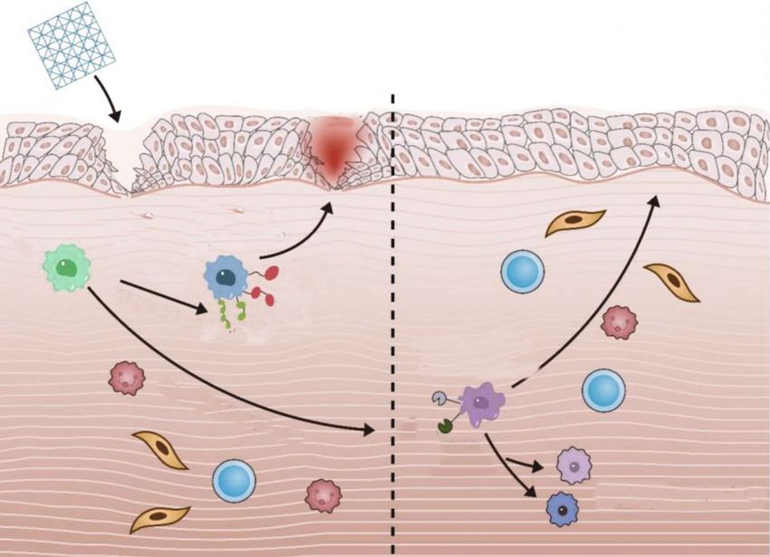
Figure 2: Differentiation of fibrotic macrophages and regenerating macrophages by scRNA-seq. When biological scaffolds are used to repair
tissue damage, they can recruit regenerated macrophages through IL-4 and TH2 responses to achieve the purpose of tissue repair, or they
can recruit fibrotic cells through IL-17 and TH1 responses to produce foreign body responses. By using scRNA-seq, the study found that
CD301b can distinguish regenerated macrophages and can distinguish their subtypes by CD9 and CD74. At the same time, fibrotic
macrophages can be distinguished by CD64+, CD9+, IL-17+, and IL-36+.
associated the results of the primary cells with the in vitro evaluate a large number of genes per cell so that the real pop-
marrow exposed to the established polarized inflammatory ulation structure can be determined unbiased, and it is pos-
factors, compared the observation results of human blood sible to identify previously unknown myeloid cell subsets
and bone marrow cells in lineage differentiation models and to understand the dynamic interaction between myeloid
and established a comprehensive reference frame for the cell subsets and other cells of the immune system more
MPS room, and described them using analysis tools such quickly [66]. Aran et al. [67] used a scRNA-seq clustering
as viSNE, SPADE, and MEM. The results showed that each calculation and unbiased annotation tool (Single) to identify
stimulation condition produced a specific activated macro- macrophages from baseline and mixed lung cell samples
phage phenotype, with no or almost no overlap between after bleomycin-induced mouse lung injury (alveolar macro-
M_IFN-g and M_LPS and M_IL-4 and M_IL-10. It is worth phages and interstitial macrophages) and applied the hierar-
noting that they found different phenotypic patterns in MPS chical clustering method to the subgroup of macrophages in
(Table 1). fibrosis. The results showed that monocyte-derived disease-
related macrophages transformed into alveoli, located in
5.2. The Heterogeneity of Macrophages Was Revealed by the fibrotic niche, and played a role in promoting fibrosis,
Single-Cell RNA-seq. Single-cell RNA-seq can be used to and the migration and proliferation of fibroblasts depended
analyze the whole genome and single-cell transcription on SiglecF+CD11c+MHCIIhi and CX3CR1+ cells secreting
map of immune cells and it can reveal immune heterogene- Pdgf-aa, indicating that the paracrine interaction between
ity in different diseases. It has become an established method these macrophages and fibroblasts maintains fibroblast pro-
to dissect cell heterogeneity, reveal cell state, and identify the liferation and tissue fibrosis. Some studies have identified a
structure of different cell subsets [64, 65]. scRNA-seq can fourth group of cardiac macrophages in the uninjured6 BioMed Research International
myocardium through scRNA-seq, and the number of these vated macrophages can mediate periprosthetic inflammation
cells will increase after injury [68]. This population is char- and make an important impact on recruitment and bone
acterized by a strong interferon-stimulated gene signature resorption [82–86]. Macrophages play an important role in
called ISG MF. However, it is currently unclear whether the early tissue healing process of bone implantation of bio-
ISG macrophages represent a unique subset of tissue macro- materials [87]. The success of biomaterial-mediated bone
phages or a part of the activation spectrum, and their role in formation depends on the effective and timely conversion
homeostasis is also unknown. This emphasizes the need for of the M1 phenotype to the M2 phenotype during the bone
researchers to develop new tools to isolate and explore this healing process, and the prolonged M1 phase may cause
population [69]. Sommerfeld et al. [70] used scRNA-seq to fibrous encapsulation and bone regeneration failure [88].
describe the relationship between macrophages isolated Osseointegration was defined as a direct structural and func-
from mouse tissue repair models and tissue environmental tional connection between ordered living bone and the sur-
fibrosis after the use of model biomaterials. They used an face of a load-carrying implant [89]. Current research
unbiased clustering algorithm to reveal the diversity of mac- believes that osseointegration is a foreign body reaction,
rophages, calculated and analyzed the phenotypic character- which can protect the implant from the tissue by forming
istics of macrophage clusters, defined phenotypic and a defense response at the interface bone [90]. The presence
functional macrophage populations, identified macrophage of oral implants stimulates higher immune participation
surface markers by flow cytometry and immunofluorescence through complement and macrophage activation, while
techniques, and identified new CD9hi+IL-36 γ + macrophage macrophage activation can affect the tissue surrounding
populations. It was found that it had the characteristics of the implant by regulating inflammation and tissue healing
type 17 immune response and autoimmunity and verified [91]. Studies have shown that the surface topographical
the ability to use surface markers to distinguish macrophage and chemical signals on the surface of titanium implants
subsets. Figure 2 is shown as follows. can regulate the polarization of macrophages, and macro-
phages can also promote the homing and osteogenic differ-
5.3. Hyperspectral Images Were Used to Detect and Classify entiation of mesenchymal stem cells on the surface of
Macrophages in an Unmarked and Noninvasive Manner. implants by producing a variety of cytokines and growth fac-
The hyperspectral image is an unlabeled and noninvasive tors, thereby regulating the healing process [91, 92]. Lee et al.
way to detect and classify living cells and has significant [93] found that the combination of bioactive ion chemistry
thermal potential. When it is applied to tissue diagnosis, and the surface morphology of nanoscale titanium can sig-
the resulting three-dimensional data hypercube can encode nificantly induce the polarization of M2 macrophages of
the properties of light-tissue interaction, such as absorption, J774.A1 cells and improve the early bone formation ability
scattering, and fluorescence [71]. Based on the spectral char- of oral implants in animal bones in clinical practice. The
acteristics of different tissues, HSI can provide quantitative activated state is the core of the executive function of macro-
diagnostic information about histopathology, morphology, phages, and it is also the key to immunology, disease patho-
and chemical composition of noncontact, noninvasive, and genesis, and anti-inflammatory [12, 94, 95]. In some
nonionized tissues [72]. Bertani et al. [73] studied human inflammatory diseases, transforming the activation state of
monocyte-derived macrophages by hyperspectral reflectance macrophages has become a treatment [96, 97]. However, in
confocal microscopy and analyzed M1 and M2 activation of recent years, some studies have proposed different macro-
hyperspectral data sets by principal component analysis. phage activation classification criteria and models based on
Then, linear discriminant analysis was used to process HSI the behavior of macrophages in the disease process, tissue-
data and semiautomatically classify macrophage activation, specific phenotypes, and high-resolution data obtained
which confirmed the possibility of single-cell level classifica- through advanced technologies [98, 99]. The heterogeneity
tion of M1 and M2 macrophages in a noninvasive and unla- of cells and different activation models show that the activa-
beled manner. tion of macrophages is not two extreme changes as described
in the classic dichotomy but takes on different forms as envi-
6. Conclusion ronmental stimuli change. The improvement of the macro-
phage activation model also enables the optimization of
Macrophages are myeloid immune cells, which can be found immune-based therapeutic measures. These alternative acti-
in almost every tissue of the human body [74, 75]. Their vation models will provide the possibility of treating oral dis-
main functions are to participate in host defense, maintain eases in the future.
the stability of the tissue environment, remove cell debris,
recover apoptotic cells, help tissue regeneration and repara-
tion by secreting cytokines and growth factors, and secrete Data Availability
some proteins, such as extracellular matrix proteins, to take
No datasets were generated or analyzed during the current
part in cell adhesion [76–81]. According to the activation of
study.
macrophages by in vitro signals, the classical polarization
model divides macrophages into two states. When they are
activated as proinflammatory phenotypes and release some Conflicts of Interest
cytokines, they can mediate the balance between bone salt
deposition, osteogenesis, and osteoclast; for example, acti- The authors declare that they have no conflicts of interest.BioMed Research International 7
Authors’ Contributions [16] M. Kang, C. C. Huang, Y. Lu et al., “Bone regeneration is medi-
ated by macrophage extracellular vesicles,” Bone, vol. 141, arti-
Yali Wei, Mengxi Wang, and Yuwen Ma contributed equally cle 115627.
to this work. [17] A. Mantovani, A. Sica, and M. Locati, “Macrophage polariza-
tion comes of age,” Immunity, vol. 23, no. 4, pp. 344–346,
2005.
References [18] X. Shi and S. L. Shiao, “The role of macrophage phenotype in
regulating the response to radiation therapy,” Translational
[1] L. Johnson, C. L. C. Almeida-da-Silva, C. M. Takiya et al., Research, vol. 191, pp. 64–80, 2018.
“Oral infection of mice with Fusobacterium nucleatum results [19] C. Chu, J. Deng, C. Cao, Y. Man, and Y. Qu, “Evaluation of
in macrophage recruitment to the dental pulp and bone epigallocatechin-3-gallate modified collagen membrane and
resorption,” Biomedical Journal, vol. 41, no. 3, pp. 184–193, concerns on Schwann cells,” BioMed research international,
2018. vol. 2017, Article ID 9641801, 2017.
[2] K. M. Hotchkiss, K. T. Sowers, and R. Olivares-Navarrete, [20] M. de Gaetano, D. Crean, M. Barry, and O. Belton, “M1- and
“Novel in vitro comparative model of osteogenic and inflam- M2-type macrophage responses are predictive of adverse out-
matory cell response to dental implants,” Dental Materials, comes in human atherosclerosis,” Frontiers in immunology,
vol. 35, no. 1, pp. 176–184, 2019. vol. 7, p. 275, 2016.
[3] L. Panahipour, E. Kochergina, A. Kreissl, N. Haiden, and [21] J. Chylikova, J. Dvorackova, Z. Tauber, and V. Kamarad, “M1/M2
R. Gruber, “Milk modulates macrophage polarization macrophage polarization in human obese adipose tissue,” Bio-
in vitro,” Cytokine X, vol. 1, no. 2, p. 100009, 2019. medical Papers of the Medical Faculty of the University Palacky,
[4] A. C. Aschenbrenner and J. L. Schultze, “New “programmers” Olomouc, Czech Republic, vol. 162, no. 2, pp. 79–82, 2018.
in tissue macrophage activation,” Pflugers Archiv : European [22] M. J. Feito, R. Diez-Orejas, M. Cicuéndez, L. Casarrubios, J. M.
journal of physiology, vol. 469, no. 3-4, pp. 375–383, 2017. Rojo, and M. T. Portolés, “Characterization of M1 and M2
[5] U. Juhas, M. Ryba-Stanisławowska, P. Szargiej, and polarization phenotypes in peritoneal macrophages after treat-
J. Myśliwska, “Different pathways of macrophage activation ment with graphene oxide nanosheets,” Colloids and Surfaces.
and polarization,” Postepy higieny i medycyny doswiadczalnej, B, Biointerfaces, vol. 176, pp. 96–105, 2019.
vol. 69, pp. 496–502, 2015. [23] A. Mazzoni and D. M. Segal, “Controlling the Toll road to den-
[6] J. Muñoz, N. S. Akhavan, A. P. Mullins, and B. H. Arjmandi, dritic cell polarization,” Journal of Leukocyte Biology, vol. 75,
“Macrophage polarization and osteoporosis: a review,” Nutri- no. 5, pp. 721–730, 2004.
ents, vol. 12, no. 10, 2020. [24] F. O. Martinez, S. Gordon, M. Locati, and A. Mantovani,
[7] F. O. Martinez and S. Gordon, “The M1 and M2 paradigm of “Transcriptional profiling of the human monocyte-to-
macrophage activation: time for reassessment,” F1000Prime macrophage differentiation and polarization: new molecules
Reports, vol. 6, p. 13, 2014. and patterns of gene expression,” Journal of Immunology,
[8] C. D. Mills, K. Kincaid, J. M. Alt, M. J. Heilman, and A. M. Hill, vol. 177, no. 10, pp. 7303–7311, 2006.
“M-1/M-2 macrophages and the Th1/Th2 paradigm,” Journal [25] W. Noël, G. Raes, G. Hassanzadeh Ghassabeh, P. De Baetselier,
of Immunology, vol. 164, no. 12, pp. 6166–6173, 2000. and A. Beschin, “Alternatively activated macrophages during
[9] C. D. Mills, “M1 and M2 macrophages: oracles of health and parasite infections,” Trends in Parasitology, vol. 20, no. 3,
disease,” Critical Reviews in Immunology, vol. 32, no. 6, pp. 126–133, 2004.
pp. 463–488, 2012. [26] Y. Wang, W. Smith, D. Hao, B. He, and L. Kong, “M1 and M2
[10] S. K. Biswas and A. Mantovani, “Macrophage plasticity and macrophage polarization and potentially therapeutic naturally
interaction with lymphocyte subsets: cancer as a paradigm,” occurring compounds,” International Immunopharmacology,
Nature Immunology, vol. 11, no. 10, pp. 889–896, 2010. vol. 70, pp. 459–466, 2019.
[11] A. J. Fleetwood, H. Dinh, A. D. Cook, P. J. Hertzog, and J. A. [27] W. Hu, J. Lin, X. Lian et al., “M2a and M2b macrophages pre-
Hamilton, “GM-CSF- and M-CSF-dependent macrophage dominate in kidney tissues and M2 subpopulations were asso-
phenotypes display differential dependence on type I inter- ciated with the severity of disease of IgAN patients,” Clinical
feron signaling,” Journal of Leukocyte Biology, vol. 86, no. 2, Immunology, vol. 205, pp. 8–15, 2019.
pp. 411–421, 2009. [28] L. X. Wang, S. X. Zhang, H. J. Wu, X. L. Rong, and J. Guo,
[12] P. J. Murray, J. E. Allen, S. K. Biswas et al., “Macrophage acti- “M2b macrophage polarization and its roles in diseases,” Jour-
vation and polarization: nomenclature and experimental nal of Leukocyte Biology, vol. 106, no. 2, pp. 345–358, 2019.
guidelines,” Immunity, vol. 41, no. 1, pp. 14–20, 2014. [29] A. Sica and A. Mantovani, “Macrophage plasticity and polari-
[13] M. C. Bosco, “Macrophage polarization: reaching across the zation: in vivo veritas,” The Journal of Clinical Investigation,
aisle?,” The Journal of Allergy and Clinical Immunology, vol. 122, no. 3, pp. 787–795, 2012.
vol. 143, no. 4, pp. 1348–1350, 2019. [30] J. Ji, D. Shu, M. Zheng et al., “Microbial metabolite butyrate
[14] D. Zhou, C. Huang, Z. Lin et al., “Macrophage polarization facilitates M2 macrophage polarization and function,” Scien-
and function with emphasis on the evolving roles of coordi- tific reports, vol. 6, no. 1, 2016.
nated regulation of cellular signaling pathways,” Cellular Sig- [31] Z. Chang, Y. Wang, C. Liu, W. Smith, and L. Kong, “Natural
nalling, vol. 26, no. 2, pp. 192–197, 2014. products for regulating macrophages M2 polarization,” Cur-
[15] J. Luo, Y. He, F. Meng, N. Yan, Y. Zhang, and W. Song, “The rent Stem Cell Research & Therapy, vol. 15, no. 7, pp. 559–
role of autophagy in M2 polarization of macrophages induced 569, 2020.
by micro/nano topography,” International Journal of Nano- [32] A. Mily, S. Kalsum, M. G. Loreti et al., “Polarization of M1 and
medicine, vol. 15, no. 9, pp. 7763–7774, 2020. M2 human monocyte-derived cells and analysis with flow8 BioMed Research International
cytometry upon Mycobacterium tuberculosis infection,” JoVE [47] S. Y. Kim and M. G. Nair, “Macrophages in wound healing:
(Journal of Visualized Experiments), vol. 164, no. 14, 2020. activation and plasticity,” Immunology & Cell Biology,
[33] R. ARB, S. ECO, A. PMC, S. T. Souza, F. EJDS, and E. Barreto, vol. 97, no. 3, pp. 258–267, 2019.
“Application of Raman spectroscopy for characterization of [48] F. Ginhoux, J. L. Schultze, P. J. Murray, J. Ochando, and S. K.
the functional polarization of macrophages into M1 and M2 Biswas, “New insights into the multidimensional concept of
cells,” Spectrochimica Acta Part A: Molecular and Biomolecu- macrophage ontogeny, activation and function,” Nature
lar Spectroscopy, vol. 265, no. 27, 2021. Immunology, vol. 17, no. 1, pp. 34–40, 2016.
[34] R. Trindade, T. Albrektsson, S. Galli, Z. Prgomet, and [49] M. J. Stables, S. Shah, E. B. Camon et al., “Transcriptomic anal-
P. Tengvall, “Suppress bone resorption during the first 4 weeks yses of murine resolution-phase macrophages,” Blood,
after implantation,” Clinical Implant Dentistry and Related vol. 118, no. 26, pp. e192–e208, 2011.
Research, vol. 20, no. 1, pp. 82–91, 2018. [50] C. Cochain, E. Vafadarnejad, P. Arampatzi et al., “Single-cell
[35] E. Mariani, G. Lisignoli, R. M. Borzì, and L. Pulsatelli, “Bioma- RNA-Seq reveals the transcriptional landscape and heteroge-
terials: foreign bodies or tuners for the immune response?,” neity of aortic macrophages in murine atherosclerosis,” Circu-
International journal of molecular sciences, vol. 20, no. 3, lation Research, vol. 122, no. 12, pp. 1661–1674, 2018.
p. 636, 2019. [51] O. Helm, J. Held-Feindt, E. Grage-Griebenow et al., “Tumor-
[36] K. M. Hotchkiss, G. B. Reddy, S. L. Hyzy, Z. Schwartz, B. D. associated macrophages exhibit pro- and anti-inflammatory
Boyan, and R. Olivares-Navarrete, “Titanium surface charac- properties by which they impact on pancreatic tumorigenesis,”
teristics, including topography and wettability, alter macro- International Journal of Cancer, vol. 135, no. 4, pp. 843–861,
phage activation,” Acta Biomaterialia, vol. 31, pp. 425–434, 2014.
2016. [52] J. W. Williams, C. Giannarelli, A. Rahman, G. J. Randolph, and
[37] J. O. Abaricia, A. H. Shah, M. N. Ruzga, and R. Olivares- J. C. Kovacic, “Macrophage biology, classification, and pheno-
Navarrete, “Surface characteristics on commercial dental type in cardiovascular disease: JACC macrophage in CVD
implants differentially activate macrophages in vitro and series (part 1),” Journal of the American College of Cardiology,
in vivo,” Clinical Oral Implants Research, vol. 32, no. 4, vol. 72, no. 18, pp. 2166–2180, 2018.
pp. 487–497, 2021. [53] A. Shapouri-Moghaddam, S. Mohammadian, H. Vazini et al.,
[38] C. W. Wang, Y. Hao, R. Di Gianfilippo et al., “Machine “Macrophage plasticity, polarization, and function in health
learning-assisted immune profiling stratifies peri-implantitis and disease,” Journal of Cellular Physiology, vol. 233, no. 9,
patients with unique microbial colonization and clinical out- pp. 6425–6440, 2018.
comes,” Theranostics, vol. 11, no. 14, pp. 6703–6716, 2021. [54] L. Arnold, A. Henry, F. Poron et al., “Inflammatory monocytes
[39] H. Zhang, X. Wu, G. Wang et al., “Macrophage polarization, recruited after skeletal muscle injury switch into antiinflam-
inflammatory signaling, and NF-κB activation in response to matory macrophages to support myogenesis,” The Journal of
chemically modified titanium surfaces,” Colloids and Surfaces Experimental Medicine, vol. 204, no. 5, pp. 1057–1069, 2007.
B: Biointerfaces, vol. 166, pp. 269–276, 2018. [55] J. L. Schultze, “Transcriptional programming of human mac-
[40] M. Baseri, F. Radmand, R. Hamedi, M. Yousefi, and H. S. Kafil, rophages: on the way to systems immunology,” Journal of
“Immunological aspects of dental implant rejection,” BioMed Molecular Medicine, vol. 93, no. 6, pp. 589–597, 2015.
Research International, 2020. [56] D. Gosselin, V. M. Link, C. E. Romanoski et al., “Environment
[41] B. Liang, H. Wang, D. Wu, and Z. Wang, “Macrophage Drives Selection and Function of Enhancers Controlling Tis-
M1/M2 polarization dynamically adapts to changes in micro- sue- Specific Macrophage Identities,” Cell, vol. 159, no. 6,
environment and modulates alveolar bone remodeling after pp. 1327–1340, 2014.
dental implantation,” Journal of Leukocyte Biology, vol. 110, [57] J. Xue, S. V. Schmidt, J. Sander et al., “Transcriptome-based
no. 3, pp. 433–447, 2021. network analysis reveals a spectrum model of human macro-
[42] A. Insua, A. Monje, H. L. Wang, and R. J. Miron, “Basis of phage activation,” Immunity, vol. 40, no. 2, pp. 274–288, 2014.
bone metabolism around dental implants during osseointegra- [58] A. C. Villani, R. Satija, G. Reynolds et al., “Single-cell RNA-seq
tion and peri-implant bone loss,” Journal of Biomedical Mate- reveals new types of human blood dendritic cells, monocytes,
rials Research, vol. 105, pp. 2075–2089, 2017. and progenitors,” Science, vol. 356, no. 6335, 2017.
[43] Y. Wang, Y. Zhang, A. Sculean, D. D. Bosshardt, and R. J. [59] A. Mantovani, A. Sica, S. Sozzani, P. Allavena, A. Vecchi, and
Miron, “Macrophage behavior and interplay with gingival M. Locati, “The chemokine system in diverse forms of macro-
fibroblasts cultured on six commercially available titanium, phage activation and polarization,” Trends in Immunology,
zirconium, and titanium-zirconium dental implants,” Clinical vol. 25, no. 12, pp. 677–686, 2004.
Oral Investigations, vol. 23, no. 8, pp. 3219–3227, 2019. [60] A. W. Kay, D. M. Strauss-Albee, and C. A. Blish, “Application
[44] L. Zhou, Y. Han, J. Ding et al., “Regulation of an antimicrobial of mass cytometry (CyTOF) for functional and phenotypic
peptide GL13K-modified titanium surface on osteogenesis, analysis of natural killer cells,” Methods in Molecular Biology,
osteoclastogenesis, and angiogenesis base on osteoimmunol- vol. 1441, pp. 13–26, 2016.
ogy,” ACS Biomaterials Science & Engineering, 2021. [61] T. R. Matos, H. Liu, and J. Ritz, “Research techniques made
[45] S. Franz, S. Rammelt, D. Scharnweber, and J. C. Simon, simple: mass cytometry analysis tools for decrypting the com-
“Immune responses to implants–a review of the implications plexity of biological systems,” The Journal of Investigative Der-
for the design of immunomodulatory biomaterials,” Biomate- matology, vol. 137, no. 5, pp. e43–e51, 2017.
rials, vol. 32, no. 28, pp. 6692–6709, 2021. [62] T. R. Matos, H. Liu, and J. Ritz, “Research techniques made
[46] Z. Chen, T. Klein, R. Z. Murray et al., “Osteoimmunomodula- simple: experimental methodology for single-cell mass cytom-
tion for the development of advanced bone biomaterials,” etry,” The Journal of Investigative Dermatology, vol. 137, no. 4,
Materials Today, vol. 19, no. 6, pp. 304–321, 2016. pp. e31–e38, 2017.BioMed Research International 9
[63] M. Roussel, P. B. Ferrell Jr., A. R. Greenplate et al., “Mass [80] Y. Okabe and R. Medzhitov, “Tissue-specific signals control
cytometry deep phenotyping of human mononuclear phago- reversible program of localization and functional polarization
cytes and myeloid-derived suppressor cells from human blood of macrophages,” Cell, vol. 157, no. 4, pp. 832–844, 2014.
and bone marrow,” Journal of Leukocyte Biology, vol. 102, [81] Y. Qiu, K. D. Nguyen, J. I. Odegaard et al., “Eosinophils and
no. 2, pp. 437–447, 2017. type 2 cytokine signaling in macrophages orchestrate develop-
[64] K. Street, D. Risso, R. B. Fletcher et al., “Slingshot: cell lineage ment of functional beige fat,” Cell, vol. 157, no. 6, pp. 1292–
and pseudotime inference for single-cell transcriptomics,” 1308, 2014.
BMC Genomics, vol. 19, no. 1, p. 477, 2018. [82] M. G. Tu, K. T. Sun, T. H. Wang et al., “Effects of mineral tri-
[65] K. N. Natarajan, Z. Miao, M. Jiang et al., “Comparative analy- oxide aggregate and bioceramics on macrophage differentia-
sis of sequencing technologies for single-cell transcriptomics,” tion and polarization _in vitro_,” Journal of the Formosan
Genome Biology, vol. 20, no. 1, p. 70, 2019. Medical Association, vol. 118, no. 10, pp. 1458–1465, 2019.
[66] D. A. Jaitin, E. Kenigsberg, H. Keren-Shaul et al., “Massively [83] C. Chu, J. Deng, X. Sun, Y. Qu, and Y. Man, “Collagen mem-
parallel single-cell RNA-seq for marker-free decomposition brane and immune response in guided bone regeneration:
of tissues into cell types,” Science, vol. 343, no. 6172, recent progress and perspectives,” Tissue Engineering. Part B,
pp. 776–779, 2014. Reviews, vol. 23, no. 5, pp. 421–435, 2017.
[67] D. Aran, A. P. Looney, L. Liu et al., “Reference-based analysis [84] D. M. Mosser and J. P. Edwards, “Exploring the full spectrum
of lung single-cell sequencing reveals a transitional profibrotic of macrophage activation,” Nature Reviews. Immunology,
macrophage,” Nature Immunology, vol. 20, no. 2, pp. 163–172, vol. 8, no. 12, pp. 958–969, 2008.
2019. [85] S. C. Funes, M. Rios, J. Escobar-Vera, and A. M. Kalergis,
[68] S. A. Dick, J. A. Macklin, S. Nejat et al., “Self-renewing resident “Implications of macrophage polarization in autoimmunity,”
cardiac macrophages limit adverse remodeling following myo- Immunology, vol. 154, no. 2, pp. 186–195, 2018.
cardial infarction,” Nature Immunology, vol. 20, no. 1, pp. 29– [86] A. Saradna, D. C. Do, S. Kumar, Q. L. Fu, and P. Gao, “Macro-
39, 2019. phage polarization and allergic asthma,” Translational
[69] S. A. Dick, R. Zaman, and S. Epelman, “Using high- Research, vol. 191, pp. 1–14, 2018.
dimensional approaches to probe monocytes and macro- [87] H. Terheyden, N. P. Lang, S. Bierbaum, and B. Stadlinger,
phages in cardiovascular disease,” Frontiers in Immunology, “Osseointegration–communication of cells,” Clinical Oral
vol. 10, p. 2146, 2019. Implants Research, vol. 23, no. 10, pp. 1127–1135, 2012.
[70] S. D. Sommerfeld, C. Cherry, R. M. Schwab et al., “Single cell [88] C. Chu, Y. Wang, Y. Wang et al., “Evaluation of
RNA-seq in regenerative and fibrotic biomaterial environ- epigallocatechin-3-gallate (EGCG) modified collagen in
ments defines new macrophage subsets,” 2019. guided bone regeneration (GBR) surgery and modulation
[71] J. Yoon, J. Joseph, D. J. Waterhouse et al., “A clinically translat- of macrophage phenotype,” Materials Science & Engineer-
able hyperspectral endoscopy (HySE) system for imaging the ing. C, Materials for Biological Applications, vol. 99,
gastrointestinal tract,” Nature Communications, vol. 10, pp. 73–82, 2019.
no. 1, p. 1902, 2019. [89] M. B. Guglielmotti, D. G. Olmedo, and R. L. Cabrini,
[72] R. Sucher, A. Athanasios, H. Köhler et al., “Hyperspectral “Research on implants and osseointegration,” Periodontology
imaging (HSI) in anatomic left liver resection,” International 2000, vol. 79, no. 1, pp. 178–189, 2019.
Journal of Surgery Case Reports, vol. 62, pp. 108–111, 2019. [90] T. Albrektsson and A. Wennerberg, “On osseointegration in
[73] F. R. Bertani, P. Mozetic, M. Fioramonti et al., “Classification relation to implant surfaces,” Clinical Implant Dentistry and
of M1/M2-polarized human macrophages by label-free hyper- Related Research, vol. 21, Supplement1, pp. 4–7, 2019.
spectral reflectance confocal microscopy and multivariate [91] R. Trindade, T. Albrektsson, S. Galli, Z. Prgomet, P. Tengvall,
analysis,” Scientific Reports, vol. 7, no. 1, p. 8965, 2017. and A. Wennerberg, “Osseointegration and foreign body reac-
tion: titanium implants activate the immune system and sup-
[74] F. Geissmann, M. G. Manz, S. Jung, M. H. Sieweke, M. Merad,
press bone resorption during the first 4 weeks after
and K. Ley, “Development of monocytes, macrophages, and
implantation,” Clinical Implant Dentistry and Related
dendritic cells,” Science, vol. 327, no. 5966, pp. 656–661, 2010.
Research, vol. 20, no. 1, pp. 82–91, 2018.
[75] M. N. Artyomov, A. Sergushichev, and J. D. Schilling, “Inte-
[92] M. A. Alfarsi, S. M. Hamlet, and S. Ivanovski, “Titanium sur-
grating immunometabolism and macrophage diversity,” Sem-
face hydrophilicity modulates the human macrophage inflam-
inars in Immunology, vol. 28, no. 5, pp. 417–424, 2016.
matory cytokine response,” Journal of Biomedical Materials
[76] R. Curi, R. de Siqueira Mendes, L. A. de Campos Crispin, G. D. Research. Part A, vol. 102, no. 1, pp. 60–67, 2014.
Norata, S. C. Sampaio, and P. Newsholme, “A past and present
[93] C. H. Lee, Y. J. Kim, J. H. Jang, and J. W. Park, “Modulating
overview of macrophage metabolism and functional out-
macrophage polarization with divalent cations in nanostruc-
comes,” Clinical Science, vol. 131, no. 12, pp. 1329–1342, 2017.
tured titanium implant surfaces,” Nanotechnology, vol. 27,
[77] T. A. Wynn, A. Chawla, and J. W. Pollard, “Macrophage biol- no. 8, article 085101, 2016.
ogy in development, homeostasis and disease,” Nature,
[94] F. Ginhoux and S. Jung, “Monocytes and macrophages: devel-
vol. 496, no. 7446, pp. 445–455, 2013.
opmental pathways and tissue homeostasis,” Nature Reviews.
[78] R. Gentek, K. Molawi, and M. H. Sieweke, “Tissue macrophage Immunology, vol. 14, no. 6, pp. 392–404, 2014.
identity and self-renewal,” Immunological Reviews, vol. 262, [95] O. Waidmann, F. Brunner, E. Herrmann, S. Zeuzem, A. Piiper,
no. 1, pp. 56–73, 2014. and B. Kronenberger, “Macrophage activation is a prognostic
[79] I. Rhee, “Diverse macrophages polarization in tumor microen- parameter for variceal bleeding and overall survival in patients
vironment,” Archives of Pharmacal Research, vol. 39, no. 11, with liver cirrhosis,” Journal of Hepatology, vol. 58, no. 5,
pp. 1588–1596, 2016. pp. 956–961, 2013.10 BioMed Research International
[96] M. Court, M. Malier, and A. Millet, “3D type I collagen envi-
ronment leads up to a reassessment of the classification of
human macrophage polarizations,” Biomaterials, vol. 208,
pp. 98–109, 2019.
[97] M. Stein, S. Keshav, N. Harris, and S. Gordon, “Interleukin 4
potently enhances murine macrophage mannose receptor
activity: a marker of alternative immunologic macrophage
activation,” The Journal of Experimental Medicine, vol. 176,
no. 1, pp. 287–292, 1992.
[98] Y. Lavin, D. Winter, R. Blecher-Gonen et al., “Tissue-resident
macrophage enhancer landscapes are shaped by the local
microenvironment,” Cell, vol. 159, no. 6, pp. 1312–1326, 2014.
[99] A. A. Tarique, J. Logan, E. Thomas, P. G. Holt, P. D. Sly, and
E. Fantino, “Phenotypic, functional, and plasticity features of
classical and alternatively activated human macrophages,”
American Journal of Respiratory Cell and Molecular Biology,
vol. 53, no. 5, pp. 676–688, 2015.You can also read