Spontaneous Neoplasms in Amphibia: A Review and Descriptions of Six New Cases* - Cancer Research
←
→
Page content transcription
If your browser does not render page correctly, please read the page content below
Spontaneous Neoplasms in Amphibia: A Review and
                     Descriptions of Six New Cases*
                                                    MICHAELBALLS
                         (Station de Zoologie expérimentale,
                                                           Route de Malagnou 154, Geneva, Switzerland)

                                                          SUMMARY
             Fifty-two previous accounts of neoplastic growths in Amphibia are listed and sum
          marized. Six new cases, including five of lymphosarcoma, are described. The diversity,
          sites, and distribution of amphibian tumors are discussed.

    The last detailed review of neoplasms occurring               in amphibians are listed chronologically in Table
 spontaneously in amphibia was that of Schlum-                    1. Short abstracts of those original publications
 berger and Lücke(38), who summarized seven                      not summarized by Schlumberger and Lücke(38)
 cases in Urodeles and 22 in Anurans. The same                    follow.
 authors (22) also discussed the literature on the
experimental induction of tumors in these cold                       ABSTRACTS OF REPORTS OF TUMORS
 blooded vertebrates, as did Leone (18). Schlum-                               IN AMPHIBIA
 berger and Lückeconsidered that, in spite of the                            TUMORSIN ANUBA
 small number of tumors, there was no good reason                     Llambés  and Garcia (20) : described the spontaneous appear
for believing amphibians to be less susceptible to                ance of multiple tumors on the left leg, the abdomen, and be
neoplastic growth than fish or other vertebrates.                  tween the eyes of a frog (Rana catesbiana). The tumors were
                                                                   diagnosed as adenoepitheliomas of skin. A tumor fragment
This view has been upheld by the increase in our                  grew rapidly after transference to the anterior eye chamber of
 knowledge of amphibian tumors as a result of                     another frog.
recent research.                                                      Stolk (48) : found a spherical encapsulated tumor of kidney
    A completely comprehensive review is made                     in Bufo bufo.
                                                                      Rose (85): transplanted fragments of a fat-body tumor of
difficult by both the isolated occurrence of animals              a Rana pipiens. The original tumor had invaded one lung and
with neoplastic disease and the varied journals in                retained the ability to store oil. The transplants took in all
which the details are published. Lists of cases have              cases, but oil storage was not a property of further serially
more recently been given by Leone (18) and Stolk                  transplanted fragments.
 (49). Schlumberger (37) and Reichenbach-Klinke                       Rostand (36) : found a female Rana temporaria bearing in
                                                                  the anal region a melanic tumor covered with rounded pro
 (33) both comment on the high incidence of cu                    tuberances.
taneous tumors in the cases reported and the lack                     Stolk has investigated a number of tumors in Anura :
of visceral neoplasia. The author considers that                      (a) (49). An adenoma of parotid gland in Bufo marinus.
this position has been so altered by the work of                  This was a white, nodular structure composed of cystic and
Inoue (16, 17), Mori (24), Schlumberger (37), El-                 noncystic tissue. No infiltration, metastasis, or parasites were
                                                                  found. The tumor cells had large, vesicular, and polymorphic
kan (10), and the occurrence of the six new cases                 nuclei, some being acidophilic and having cytoplasmic inclu
to be described below that there is a fresh op                    sions.
portunity to discuss the distribution of spontane                     (6) (50). Multiple adenoma of skin in fiana artalis. The
                                                                  nodules consisted of stratified squamous epithelium cells ar
ous tumors in the Amphibia.                                       ranged as tubules, acini, or solid sheets separated by a delicate
   The known reports of spontaneous neoplasms                     stroma. The nuclei were large, vesicular, and polymorphic, and
    * This investigation has been carried out in the Embryology   the cytoplasm contained some special inclusions. There was no
                                                                  infiltration or metastasis, and the tumor was apparently benign.
Laboratory, Department of Zoology, Oxford University, and             (c) (51). Hemangioma of heart in Hyla arborea. A dark-red
at the Institut de Zoologie, Geneva University. The work
                                                                  mass was found on the side of the ventriculus, being composed
has been supported by grants from the Medical Research Coun
                                                                  of a network of dilated blood vessels separated by a stroma.
cil (U.K.) and the Fonds National Suisse pour la Recherche
                                                                  Proliferation of the endothelial cells had proceeded to such an
Scientifique.                                                     extent that some blood vessels were occluded.
   Received for publication May 29, 1962.                             (d) (54). Erythrophoroma of skin in Dendrobatestypographi-

                                                              1142

Downloaded from cancerres.aacrjournals.org on February 23, 2021. © 1962 American Association for Cancer
                                              Research.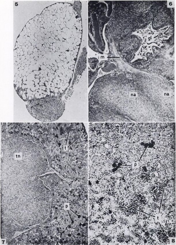
TABLE 1
                                       REPORTSOFSPONTANEOUS
                                                          NEOPLASMS
                                                                  IN AMPHIBIA

           ANIMALSAnuraEberth,
  DATE186818981902190519061908191219121918191419191928192819291930193219341934193419481948194819491950195219571957195819581959195919591960196019601

           J.Ohlmacher,
                   C.                     sp.Rana
           P.Vaillant,    H.              virescensRana            sarcomaFibromaAdenocarcinomaCarcinomaAdenocarcinomaAdenomaAdenocareinomaHypernephroma(
           A.Smallwood,L., & Pettit,      esculentaRana                                        cavityKidneyOvarySkinSkinSkinKidneySkinSkinSkinLeg,
           M.Plehn,       W.              pipiensRana
           M.Murray,                      esculentaRana
           A.Pavlovsky,
                   J.                     sp.Rana
           N.Pavlovsky, E.                sp.Rana
           N.Carl,      E.                sp.Rana
           W.Pentimalli,                  esculenteRana
                                                                   carcinoma)AdenomaAdenomaAdenocarcinomaFibrosarcomaAdenocarcinomaSarcomaAdenocarcinomaA
                                                                       =
           F.Secher,                      sp.Rana
           K.Masson,                      esculentaRana
           Schwarz,E.Volterra,
                     P., &                esculentaCeratophrus

           M.Duany,                       ornataRana                                           liverSkinLegKidneySkinLegKidneySacral
           J.Gheorghiu,,                  catesbìanaRana
           [6)Downs,     I.               esculentaRana
           W.Pirlot, A.                   pipiensRana
           M.Pirlot,
                  J., & Welsch,           fuscaRana
           M.Lücke,
                   J., & Welsch,          fuscaRana
           47]Schlumberger,
                     B. [32,              pipiensRana
             &Lücke,        H.,          catesbianaRana                                       plexusTailLiverSkinKidneyFat-body,
           B.Schlumberger,
             &Lücke,        H.,          clamitans(tadpole)Rana
           B.Willis,
           A.Llambés,
                 R.                       esculentaRana
             Garcia,J. J. J., &           catesbianaBufo
           M.Stolk,
           A.Rose,                        bufoRana                 tumorCarcinomaAdenomaAdenomaMelanomaHaemangiomaErythrophoromaGuanophoromaXanthoplior
           M.Stolk,
                 S.                       pipiensRufo                                         lungParotid
           A.Stolk,                       marinusRana                                         glandSkinSkinHeartViscera
           A.Rostand,                     arvalisRana
           J.Stolk,                       temporariaHuÃ-a
           A.Stolk,                       arboreaDendrobates
           A.Stolk,                       typo-graphicusHuÃ-a                                  skinSkinSkinLungKidneyPelvisFaceSkinUnder
                                                                                                        &

           A.Stolk,                       meri-dionalisIlyla
                                               arborea
           A.Elkan,                       arboreaRufo
           E.Elkan,                       calamitaXenopus
           E.Elkan,                       laevisXenopus
           E.Elkan,                       laevisXenopus
           E.Stolk,                       laevisBufo
        A.asesBalls,                      japónicasXenopus
                                                bufo

  c196219621962196219621962AUTHORSSPECIESTUMORSITENo.
             M.utÃ-uu((Rana                laevis lae                                          skinVisceraVisceraVisceraHead
                                  visXenopus
                                            vic-torianus/
                                                     laevis
                                            'Xeno-pus
                                          laevisXenopus
                                                  laevis
                                          fraseriXenopus
                                          fraseriXenopus
                                                    laevis lae                                        muscle & vis
                                          visXenopus                                           ceraHead
                                             lae-oaAdenomaOsteogenic
                                                    laevis                          & leu             muscle, vis
                                                                     kemiaSkinFemurBuccal         cera & blood1111112111111111171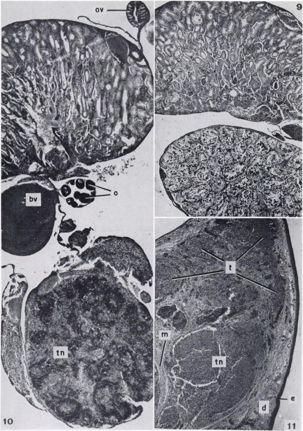
1144                                                   Cancer Research                           Vol. 22, November 1962

                                                      TABLE 1—Continued

            .\sni\L3UrodelaVaillant,
  DATE190219031908191619201923193519471948195019531954195419581958AUTHORSSPECIESTDUOBSITENo.

            A.Pick, L., & Pettit,                                                              footTestisSkin-glandsSkinSkinUnder
                                                                                                     of
            H.Murray,
                L., & Poll,
            A.Krontovsky,
                     J.                   cristatusSiredon
            [12JTeutschlaender,
                         A. A.            mexicanumSiredon
            O.Schwarz,                    mexicanumMegalobatrachusmaximusTriturus
            E.Champy,                                                                          skinSkinSkinSkinSkinSkinVisceraLiverSkinKidney11111131
            Champy,C.Broz,
                    C., &                 alpestri»Triturus
            O.Sheremetieva-Brunst,        teaniatusSiredon
               E.,&                       mexicanumTriturus
            V.Rickenbacher,
                 Brunst,
            J.Sheremetieva-Brunst,        alpestri»Siredon
            E.Inoue,                      mexicanumTriturus
            S.Mori,                       pyrrhogas-terTriturus

            H.Stolk,                      pyrrhogas-terTriturus

            A.Schlumberger,           taeniatusNecturus
                                                maculatitiFibromaCarcinomaAdenocarcinomaMelanomaMelanosarcomaFibromaCarcinomaChondromaM
                          H. G.MegalobatrachusmaximusMegalobatrachusmaximusTrituras

 cus. The animal had a red, irregular mass on the trunk and       many tumors near the external nares, stopped feeding and died.
 red nodules on the liver, spleen, and kidneys. These consisted   Sections showed no evidence of malignancy, but a widespread
 of fusiform cells containing small pigment granules and with     destruction of the maxillary region of the skull, which, no
 vacuolar cytoplasm. An erythrophoroma is a tumor arising         doubt, prevented feeding.
 from the red pigment-containing erythrophores.                       Stolk (59-61): observed on the dorsal surface of eight adult
     (e) (55). Guanophoroma of skin in Hyla arboreameridionalis.  toads (Bufo bufo japonicus] circumscribed nodular swellings
This tumor was found between the eye and tympanic mem             consisting of fusiform fibroblasts surrounded by interlacing
 brane and, when sectioned, had a silver-white sheen. Micro       bundles of connective tissue fibers. The tumors were similar
scopically, it was made up of spindle-shaped cells containing     to those described in Triturus taeniatus (Stolk, 1958, etc.) in
 guanin crystals. Vascularization was scanty, and necrosis and    that the same four phases of development were apparent, and
 infiltration were absent.                                        transplantation experiments and those involving the inhibitory
     (f) (56). Xanthophoroma in Hyla arborea. The animal had      effect of colchicine gave similar results.
an orange mass behind the right tympanic membrane. The
 tumor was made up of large epithelial cells and single or multi-                      TUMORSIN URODELA
 nucleate giant cells, all of which contained orange-yellow pig        Teutschlaender1(62) : mentioned a melanosareoma of skin in
 ment in the cytoplasm. The tumor was abundantly vascular-
                                                                  Siredon mexicanum.
 ized and showed hemorrhage and local necrosis. Stolk was             Broz (3) : discovered a female newt (Triturus taeniatus) with
 uncertain whether this was a true neoplasm or a granulomatous    islets of cartilage-like tissue in the stratum spongiosum corii
reaction to injury.                                               on the side of the trunk, behind the head, and on the right hind
     Elkan (10): considered four cases of Anuran cancer.          limbs. The blets appeared to be tumorous, probably chondro-
     (a) Pulmonary carcinoma in Bufo calamita. An adult female mas, and were benign. No métastaseswere found in other
refused food, became emaciated, and died. White nodules, 1-2
                                                                  organs.
mm. in diameter, were found in both lungs, and sections re            Rickenbacher (34) : described an ulcerated pseudo-adenoma-
vealed an adenocarcinoma invading the alveoli and filling tous carcinoma in a female newt (Triturus alpestris). The tu-
them.
     (¿>)
         Renal carcinoma in Xenopus laevis. The animal stopped        1Since Teutschlaender (62) mentions the melanosareoma of
feeding, and its abdomen became swollen. On dissection a large    the Axolotl only in his Table I and II, it is possible that this
 tumor was found, lying retroperitoneally, in continuation with   is mistaken reporting of the melanoma of Axolotl reported by
the left kidney and pushing the intestines forward. Sections      Krontovsky (1916, see Finkelstein [12]), whose work is listed
showed the lesion to be a typical adenocarcinoma which had        in the references but not cited in the text or tables of
destroyed most of the kidney.                                     Teutschlaender's    paper.
     (c) Pelvic fibroma in Xenopus laevis. A large adult female       Teutschlaender also mentioned the following neoplasia, but
died and was found to contain a firm, pale tumor near the         without giving any details:
bladder and attached to the pelvic wall. It had little cellular            Anura: Hyla sp.—carcinoma of skin-glands.
structure, consisting of layers of poorly staining tissue with             Urodela: Newt—carcinoma of testis.
some nuclei at the periphery and none at the center. Elkan                            Salamander—fibroma of heart.
wondered if the tumor had arisen as a defense against cercarÃ-a.      These last three cases are not included in the tables in the
     (d) Facial fibromata mXenopus laevis. An adult female, with  present paper.

Downloaded from cancerres.aacrjournals.org on February 23, 2021. © 1962 American Association for Cancer
                                              Research.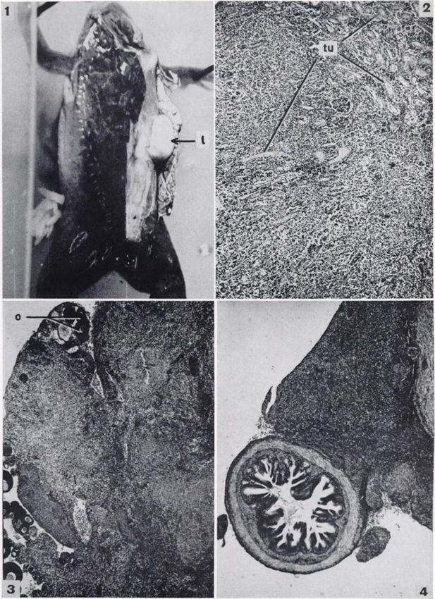
BALLS—SpontaneousNeoplasms in Amphibia 1145
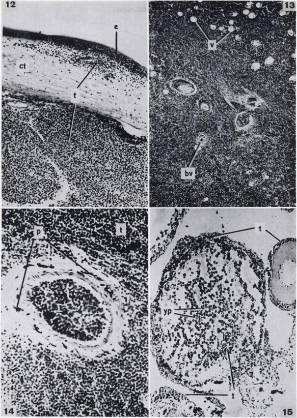
mor, situated on the head, invaded the skin and the right OS one of granuloma, and six of neoplastic disease.
nasale. The tumor was of slow growth and there were no mé Tumors were found in Xenopus fraseri, Xenopus
tastases. The author considered that the neoplasm originated laevis laevis, and in two animals created by em-
in the skin glands.
Sheremetieva-Brunst (48, 44): found a richly vascularized bryological technics. The six tumors were identi
tumor on the upper jaw of an adult black axolotl (Siredon fied as a subcutaneous lipoma and five cases of
mexicanum). The tumor, an epithelioma, grew rapidly until the lymphosarcoma, one with lymphocytic leukemia.
animal died 2 months later, having refused food and lost
weight. Fragments were transferred into 21 animals, in fourteen
The animals in question were fixed in formalin,
Worcester's fluid or Zenker's fluid. Paraffin sec
of which tumors developed. Pieces of the most rapidly growing
tumor were transferred into albino axolotls and increased in
tions were cut at 5-12 ^ and stained with Mayer's
size in 85 per cent of them. The most actively growing tumors Acid Haemalum and Eosin.
caused loss of appetite in their hosts, but no métastaseswere
found in the viscera at autopsy. CASENUMBER1 : Xenopus laevis laevis.—
Inoue (16, 17): found sarcomas in an adult male newt
(Triturus pyrrhogasler), consisting of a large tumor of the liver,
Lipoma
measuring 1.1 X 0.7 X 0.4 cm., and 37 smaller nodules in the A large adult female imported from Cape Town
liver and spleen. The tumor was made up of a stroma and cells developed a prominent, dorsal lump. On dissection
with large polymorphic or smaller round nuclei. The tumor a subcutaneous mass was found, measuring about
had invaded blood vessels. Transplantation experiments were
successful when: (a) fragments were put into the abdominal 2 cm. in diameter and having the appearance of
cavity of other newts; (6) centrifuged supernatant of implants fat-body tissue (Fig. 1). The mass was encapsu
was injected into other newts; (c) tumor fragments, stored in lated, little vascularized, and joined to the right
glycerine/saline for 34 days at room temperature, were ground, fat-body by a connection passing through the
centrifuged, and injected into other newts.
Mori (24) : records post-mortem examinations of two newts dorsal muscles of the body wall and into the ab
(Triturila pyrrhogaster) containing liver sarcomas. In one case
dominal cavity. The left fat-body and viscera
were found a large white mass, 8 cm. in diameter, and eight
were normal. The animal was fixed in Zenker's
smaller nodules. The neoplastic cells were smaller than normal fluid.
liver cells, and their cytoplasm was less stainable. Sections confirmed that the growth consisted of
Stolk (52, 53, 57, 58): observed in adult newts (Tritunis fat-body tissue. Such a growth could have arisen
taeniatus) firm, irregular nodules consisting of fusiform fibro-
blasts and collagenous connective tissue fibers. Four phases from a developmental hamartoma, by acquired
were distinguishable in the development of these fibromas: hyperplasia, or as a result of traumatic damage.
(a) enlargement of the nodular swellings of the adenoepidermal However, since the animal appeared normal on
reticular network; (6) continued enlargement of the nodular
swellings and concentration of fibroblasts in the vicinity of the
arrival, it seemed more likely that this was a true
swellings; (c) formation of new fibrils from the swellings, so lipoma. Neither of the special features mentioned
that round about the fibroblasts a densely structured network by Willis (67) were apparent, viz.: (a) mixture
is formed; and (d) fusion of the concentrated masses of fibro with fibroblastic or other mesenchymal tissue, or
blasts to form the small tumor nodules. (6) incorporation of other tissues within the
Small pieces of tumor tissue of nineteen newts were each
transferred to a normal area of the same animal. In thirteen tumor.
of these newts a distinct tumor of the same histológica!type Tumors of adipose tissue are relatively common
developed after 3 weeks at the site of transplantation. In six in animals. Feldman (11) mentions examples in
cases, the transplant was resorbed. A further series of experi almost all domestic animals, and Willis writes of
ments showed that colchicine had an inhibitory effect on the
growth of similarly transplanted tumor fragments. two cases in dogs and one in a parrot. Schlum
Schlumberger (37) : described a lobed tumor measuring 3 X berger and Lücke(38) reviewed seven reports of
4 cm. and found on the ventral part of the kidney in Necturus lipoma in fishes, including four subcutaneous
maculosus. On microscopic examination, the tumor was found lipomas, one within the ventral trunk musculature,
to be an adenocarcinoma. one projecting into the posterior body cavity, and
NEW CASES2 another on the lower border of the liver.
MATERIALSANDMETHODS CASENUMBER2 : Xenopus laevis victorianus/
In spite of recent publications on the pathology Xenopus laevis laevis
of the Amphibia (Schlumberger [37], Elkan [10], The first case of lymphosarcoma was found in
and Reichenbach-Klinke [33]), little is known of an animal produced by Dr. J. B. Gurdon (15), who
the anatomy and histology of nonparasitic disease transferred a blástula nucleus of Xenopus laevis
in these animals. The cases to be described oc victorianus into an enucleated egg of Xenopus
curred in the colony of animals of the genus laevis laevis. An apparently normal frog developed.
Xenopus of the Embryology Laboratory, Depart When 10 months old, the animal suddenly became
ment of Zoology, Oxford University. During 14 edematous in the head, abdomen, and legs, and
months, 28 animals died of disease, including two 2 Four of the cases have been briefly summarized elsewhere
cases of vascular hamartoma, two of hematoma, (Balls [1]), and are here numbered differently.
Downloaded from cancerres.aacrjournals.org on February 23, 2021. © 1962 American Association for Cancer
Research.1146 Cancer Research Vol. 22, November 1962
died. Dissection showed that this female had many 774) considers that the occurrence of lympho-
eggs in one ovary and few in the other. The kidney sarcomatous invasion in sites other than the
was much enlarged, and nodules were scattered on lymphoid structures themselves—i.e., the liver,
the outer surfaces of both lungs. In addition, two lungs, heart, kidneys, or skin—is abundant evi
round lumps were found in the mesentery near the dence that metastasis occurs, but that studies of
stomach. The body was fixed in 5 per cent neutral early lesions show that tumors also arise multi-
formalin. Histological study gave the following focally. In any event, the infiltration of liver,
information : lungs, and blood vessels in the present case sug
Kidney.—The kidneys were very swollen and gests that metastasis had occurred, the tumor
were being destroyed by the widespread infiltra originating in the kidneys or the mesenteric
tion of lymphoid cells (Fig. 2). These cells were of nodules.
a small, uniform size, spheroidal, and with little CASENUMBER3 : Xenopus fraseri—
cytoplasm but round nuclei staining deeply with
the Haemalum. The left kidney contained few Lymphosarcoma
intact tubules ; those of the right kidney persisted Juvenile animals imported from the Congo in
only in the periphery and were widely spaced, but the summer of 1960 were later identified as Xeno
fragments of the destroyed tubules were found pus fraseri (Boulenger). One female died in March,
among the lymphoid cells. Few red blood cor 1961, and was found to have many white nodules
puscles were present as compared with those in on the ovaries and fat-bodies. The viscera were
fixed in Worcester's fluid.
normal kidney, which might explain the wide
spread necrosis within the lymphoid tissue. In Microscopic evidence of neoplastic change was
vasion of blood vessels was also observed. found in the ovary, gut, and fat-body. These
Lung.—The lungs contained many cells similar organs will be discussed in turn.
to those replacing the normal tissue of the kidneys. Ovary. (Fig. 3).—The main ovarian nodule
These cells were destroying the lung epithelium measured 0.5 cm. in diameter and was found to
and enlarging the organ as a whole. Erythrocytes consist of a mass of uniform, small lymphocyte-like
were conspicuously few, and necrosis was more cells with round nuclei and little cytoplasm. Only
widespread than in the kidneys. the cells around the few fairly large collections of
Liver.—Sections of the liver showed very large erythrocytes seemed viable; otherwise there were
numbers of tumor cells mixed with liver paren huge areas of necrosis, no doubt because the tu
chyma cells. Pigment, a striking feature of the mor was outgrowing the blood supply. On one side
normal liver, was almost completely absent. of the mass were egg cells surrounded by the tu
Erythrocytes were common and uniformly spread mor cells. The tumor mass also contained many
throughout the tissue, but many of the lymphoid polymorphonuclear leukocytes.
cells were necrotic. Gut. (Fig. 4).—The duodenal region of the
Mesenteric nodules.—The two mesenterio nod alimentary canal showed a mass of tumor cells in
ules were each about 3 mm. in diameter, composed vading between layers of the circular muscle
of lymphoid cells with foamy cytoplasm, and along about one-third of the circumference of the
showed much necrosis. Erythrocytes and odd section. A large piece of circular muscle had been
patches of pigment were also present. The nodules pushed to the outside of the tumor mass, which
appeared to have no limiting capsule. also contained pancreatic cells. The tumor cells
A small piece of fat-body was incidentally sec were similar to those in the ovarian mass, but
tioned with the kidneys and contained tumor there was much less necrosis, many groups of
cells, although the invasion was not so marked as erythrocytes being uniformly spread throughout
in cases 3 and 4. the tissue.
The widespread occurrence of the round uni Fat-body. (Fig. 5).—The nodules on the fat-
form cells, similar to mature lymphocytes, show bodies illustrated very well the diffuse infiltrative
ing diffuse infiltration and destruction of the nor
growth of the lymphosarcoma. Figure 5 shows
mal tissues of the kidneys, lungs, and liver, led to
the conclusion that this was a case of malignant three nodules on the outside of one of the branches
of the fat-body. The tumor cells were spreading
lymphosarcoma. A notable feature of the tumor
cells was the lack of mitosis and the frequent oc into the organ from the outside, moving between
currence of necrosis. The question whether the the fat-storing cells accompanied by erythrocytes.
presence of lymphosarcomas in many tissues is Macroscopical examination had shown that the
due to multifocal origin or the metastatic spread fat-bodies were covered with such nodules. The
of cells has been much discussed. Willis (67, p. only previously described tumor affecting the fat-
Downloaded from cancerres.aacrjournals.org on February 23, 2021. © 1962 American Association for Cancer
Research.BALLS—Spontaneous Neoplasms in Amphibia 1147
body of an amphibian was the transplantable sar with erythrocytes. Two large portions of the
coma of Rana pipiens described by Rose (35). muscularis externa remained, and further frag
The normal tubular structure of the kidney was ments were seen among the tumor cells. Necrotic
unchanged. lymphoid cells were found toward the center of
The difference in viability between the tumor the nodules, but those near the groups of blood
cells of the ovarian mass and those invading the cells appeared to be very viable.
intestine and fat-bodies suggests that the tumor A lower region of the intestine had a tumor
may have originated in or near the ovary and nodule on one side, cells from which had replaced
that the other areas were affected as a result of the nearby muscularis externa and the entire
metastasis. In the light of the above histological mucous membrane and submucosa, but had left
findings the tumor was diagnosed as a malignant the muscularis externa intact for the other three
lymphosarcoma. parts of its circumference. Within the lumen were
loose tumor cells and erythrocytes, together with
CASENUMBER4 : Xenopus fraseri—
cellular detritus.
Lymphosarcoma Sections in the rectal region showed that
A second Xenopus fräsen died and was dis lymphoid cells had replaced the normal rectum, of
sected. The intestine, liver, kidneys, and fat- which only a few muscular and epithelial remnants
bodies were covered in a multitude of white remained. An unidentified parasite was found in
swellings of various sizes. The viscera were fixed in the lumen, but was not thought to be directly con
Worcester's fluid.
nected with the general pathological condition of
Macroscopical examination of the viscera of the animal.
this animal clearly indicated advanced tumorous Fat-body.—Nodules on the outside of the fat-
growth. The intestine was, from stomach to rec bodies were seen to have given rise to neoplastic
tum, completely covered with white lumps and in cells invading between the fat-storing cells. This
places was swollen to twice the normal size. The condition was similar to that described in Case 3
liver bore five white masses, which seemed to be and illustrated by Figure 5.
spreading over its surface, and fifteen smaller Liver.—Part of the liver bearing a large, white
nodules. The fat-bodies were covered with many nodule showed that the nodule consisted of a mass
similar nodules. Adjacent to the kidneys were four of lymphoid tumor cells which were invading the
large, white vascularized bodies in the position normal liver tissue (Fig. 7). Also present were
normally occupied by the testes, yet oviducts small groups of tumor cells throughout the rest of
were also clearly present. The results of histo the liver, a demonstration of the diffuse, infiltrat
logical examination were as follows: ing spread of this type of tumor (Fig. 8). The liver
Alimentary canal.—Transverse sections in the contained much less pigment than occurs in the
stomach region showed a mass of small, uniform, normal liver of Xenopus, and the pigment present
lymphocyte-like cells similar to those described in was confined to the relatively normal tissue, being
cases 2 and 3. These cells, mixed with pancreatic absent from the tumor nodules (Fig. 7).
cells, formed a mass equal in size to the stomach Lung.—The lung contained large numbers of
and adjacent to it. The stomach itself appeared tumor cells, both in the lumen and infiltrating
relatively normal. from the periphery and destroying the muscular
The duodenum was seen to have its normal basis of the lung epithelium. The center of the lung
structure on the whole, but some tumor cells were contained many erythrocytes, but tumor cells
contained within the villi. Outside the muscularis were predominant toward the outside.
externa was a large mass loosely attached to the Kidney-gonad region.—This region was of spe
duodenum. This mass contained many of the cial interest in view of the presence of four, white,
lymphoid cells together with pancreatic cells and testis-like bodies as well as oviducts. Micro
one large group and several smaller groups of scopical examination of the stained preparations
blood cells. The blood cells were not limited by immediately confirmed that the animal was a
a cellular wall but did not contain large numbers hermaphrodite. The bodies below the kidneys were
of the lymphoid cells. found to be two testes and two enormous nodules
A further part of the intestine was found to composed of tumor cells and of a diameter equal to
have been completely replaced by tumor cells that of the kidney. Figure 9 shows part of the
(Fig. 6). The mucous membrane and submucosa posterior half of the left kidney together with an
had been destroyed, leaving only the lymphoid apparently normal, sperm-containing testis. The
cells maintaining the shape of the villi and frag kidney was relatively normal, but groups of tu
ments of the epithelium inside the lumen, together mor cells were present near the periphery. Figure
Downloaded from cancerres.aacrjournals.org on February 23, 2021. © 1962 American Association for Cancer
Research.1148 Cancer Research Vol. 22, November 1962
10 shows the anterior part of the right kidney, a but the glands of the dermis were absent or
piece of ovary containing oocytes, an oviduct, and ruptured, perhaps by the pressure of the rapid
one of the tumor nodules. This part of the kidney growth beneath it. Below the remains of the der
was less normal, tumor cells invading from the mis was a red mass of small, lymphocyte-like cells
glomerular region toward the tubules, though the (Fig. 11) similar to those described in cases 2, 3,
blood vessels did not appear to have been invaded. and 4. The nuclei of the cells were round and
The nodule of tumor cells showed greater differ about the size of erythrocyte nuclei, but sur
entiation than in any of the other previously de rounded by much less and more faintly staining
scribed regions. The density of the lymphoid cells cytoplasm. The lymphoid cells were arranged in
varied in different parts of the nodule to give a large groups containing few erythrocytes and were
follicular appearance somewhat like a mam surrounded by less dense tissue with many more
malian lymph node (Fig. 10). Circumscribed erythrocytes and the remains of the replaced
nodes of lymphoid tissue, however, are not muscle. Two particularly large groups of erythro
normally found in amphibians (see "Discussion"). cytes contained many of the lymphoid cells.
Other regions of the gonads showed invasion fr) Tumor posterior to the right eye and destroy
and less normality in the testicular tissue, that the ing the pterygoides and temporalis muscles.—The
ovarian and testicular tissue were connected, but destruction of the muscle tissue was more com
that the gametes were not intermingled. plete in this region than in region a, there being
The above histological description clearly indi many large groups of tumor cells with little muscle
cates that this was a further case of lymphosar- and few erythrocytes. The skin above the tumor
coma. From the information given by the sections was intact and normally colored.
of the alimentary canal, fat-body, liver, and kid c) Tumor at left jaw angle.—Thetumor at the
ney, it would appear that the distribution of the left jaw angle was found to have destroyed the
tumor throughout the visceral organs was the re normal tissue from the skin below the jaw through
sult of metastatic spread, the tumor cells infiltrat the submaxillary muscle to the lining of the
ing inward from the outside of the organs. Early mouth. The ventral skin was ulcerated in about
stages in this spread could be seen in the liver (Fig. fifteen places, the edges of which were covered
8), fat-body, kidney, stomach, and duodenum. with pigment cells. The mucous membrane of the
The eventual result was shown by the mid-gut floor of the buccal cavity had about ten similar
(Fig. 6) and the rectum. The large size and greater ulcérations,and in places the tumor cells could
differentiation of the two nodules near the kidney be seen invading the outer epithelial layer (Fig.
might be taken to suggest that this was the original 12). The general appearance of the tumor between
site of the tumor, especially since the mid- and the skin and mouth lining was similar to that of
hind-gut were the regions where invasion had pro tumor 6. The tumor cells were grouped in very
ceeded to the greatest extent and were situated large numbers, among which were the remains of
immediately ventral to the nodules. the muscle.
It is impossible to say whether the hermaph- Microscopical examination revealed no ab
roditism was in any way connected with the normality in the lung, ovary, ventricle, spleen,
presence of neoplastic cells throughout the viscera. or intestine. The liver sections showed many
small groups of lymphoid cells among the liver
CASENUMBER5 : Xenopus laevis laevis— parenchyma, but replacement of the normal tissue
Lymphosarcoma was not advanced. The extent of invasion of the
A female Xenopus laevis laevis containing Xeno kidney was variable. The glomeruli contained
pus laevis victorianus germ-cells was produced by erythrocytes and lymphocytes in equal numbers.
Dr. A. \V. Blackler (2). When about 17 months Some tubules were surrounded or replaced by
old, the animal developed three rapidly growing lymphoid cells.
tumors anterior and posterior to the right eye Thus from histological observations the tumor
and at the left angle of the jaw.
was diagnosed as a lymphosarcoma affecting the
Dissection of the abdomen showed that the
viscera were of normal appearance. The head with muscles of the head, and the liver and kidney. In
the tumors was fixed in Zenker's fluid, and the view of the very rapid growth of the head tumors
rest of the animal was preserved in 5 per cent and the variable degree of infiltration of the kid
formalin. Examination of the head tumors gave ney, it would appear that the tumor originated in
the following information. either of these regions and that the invasion of the
a) Tumor anterior to right eye.—The epidermis liver had just begun. The tumor proved to be
above the tumor was found to be relatively normal, transplantable, as will be discussed elsewhere.
Downloaded from cancerres.aacrjournals.org on February 23, 2021. © 1962 American Association for Cancer
Research.BALLS—SpontaneousNeoplasms in Amphibia 1149
CASE NUMBER 6: Xenopus laevis laevis— lymphocytes, which make up the white pulp of the
Lymphosarcoma normal organ, but both the red pulp and the blood
A female Xenopus laevis laevis was found to vessels contained a predominance of lymphoid
have rapidly growing swelling on the side of the cells instead of erythrocytes.
head behind the right eye. The skin had many Ovary.—Sections of the ovary showed that
light patches, but that in the region of the tumor lymphoid cells were present both in the stroma
appeared normal. When the skin was removed, the and replacing the follicle cells around the oocytes.
swelling was seen as a white mass of firm con In some cases (Fig. 15) oocytes had been invaded
sistency, which was replacing the temporalis and and contained lymphoid cells and erythrocytes
depressor mandibularis muscles on the right side. among the yolk platelets.
When the abdominal cavity was opened, the Ventricle.—The muscle of the ventricle had not
viscera appeared normal, although the liver was been invaded by the lymphoid tumor cells, but the
unusually large and the fat-bodies were small. The blood present within the heart was composed of
region containing the tumor was fixed in Zenker's approximately equal numbers of erythrocytes
fluii!, and the rest of the animal was preserved in and lymphocytes.
5 per cent formalin. Thus, examination of the viscera showed wide
Microscopic examination of the abnormal spread infiltration of and destruction by the lymph
region of the head showed that the muscular oid cells. The tumor cells of the viscera appeared
tissue had been completely replaced by lymphoid to have entered via the blood-system, since the
cells of a uniform, small size, with large round capsules of these organs were intact and without
nuclei and little cytoplasm. The tumor showed the white nodules described in cases 3 and 4.
little sign of differentiation (Fig. 13), the large Furthermore, the blood itself was highly abnormal.
spaces among the lymphoid cells not being limited Counts of the blood within the ventricle and the
by an epithelium. Sparse fragments of muscle re blood vessels of the head tumor, liver, lung, and
mained, and a few erythrocytes were scattered kidney gave a ratio of 1.2 lymphocytes to one
throughout the tissue. Particularly interesting was erythrocyte. This is outstandingly different from
the fact that most of the erythrocytes present were the normal ratio in Anurans, which is one leuko
contained in arteries which had not been invaded cyte: more than 300 erythrocytes, lymphocytes
but contained many tumor cells. Figure 14 shows representing 19-50 per cent of the leukocytes
such a vessel surrounded by spheroid tumor cells. (Schlumberger [37], p. 773).
This suggested that the tumor cells had entered As a result of the presence of malignant lymph
the blood vessel during its passage through an ocytes in the circulating blood, this case was
other part of the tumor or that blood-borne me diagnosed as one of lymphosarcoma with lympho-
tastasis had occurred. The results of the micro cytic leukemia. Since the condition was so ad
scopic examination of the viscera were as follows : vanced at the time of dissection, it is impossible to
Liver.—Sections of the posterior part of the say whether the tumor originated at any one site
right lobe indicated that the normal liver tissue of blood formation or simultaneously in many
had been almost completely destroyed by lymph places. In fact, it would seem better to call this a
case of "lymphosarcomatosis"—a generalized and
oid cells. The remnants of the liver parenchyma
were seen between large groups of neoplastic widespread involvement of lymphoid tissue
cells, but the usual, large amount of pigment was throughout the body (Willis [67], p. 769). As far as
present. The blood vessels contained many lymph we are aware, no case of lymphosarcoma with
oid cells as well as erythrocytes. lymphocytic leukemia has been previously re
Kidney.—The destruction of the kidney had ported in an amphibian.
proceeded to a state intermediate between those DISCUSSION
observed in cases 2 and 4. The tubules were still Tumors of lymphoid tissue are among the most
clearly present but were surrounded by lymphoid common tumors of many animal species. Furth
cells which could be seen replacing the cells lining et al. (13) have demonstrated a close resemblance
the tubules. between the lymphoid tumors of man and those of
Lung.—Although the outer muscle of the lung other mammals, but Willis (67) is doubtful of the
was intact, the whole respiratory epithelium was analogy between avian and mammalian lymphoid
covered by a mass of lymphoid cells with some diseases.
erythrocytes. Large groups of lymphoid cells were Fish and amphibians have no lymph nodes as
seen in the lumen. such, circumscribed collections of lymphoid tissue
Spleen.—The spleen contained large groups of being confined to mammals and some birds.
Downloaded from cancerres.aacrjournals.org on February 23, 2021. © 1962 American Association for Cancer
Research.1150 Cancer Research Vol. 22, November 1962
Hematopoiesis occurs mainly in the spleen, kid fraseri having been kept together—and one has
ney, and intestinal submucosa in these lower no evidence of any infective agent. The destruction
vertebrates, but the bone marrow may also be a of normal organs in the Xenopus fraseri was par
source of red cells in amphibia, as, for example, in ticularly advanced. In one animal the gut, ovary,
Rana temporaria (Young, 68). Therefore, much and fat-bodies were being invaded, the gut, liver,
caution is necessary when comparing the lymph fat-bodies, kidneys, and gonads in the other. Since
oid diseases of amphibia and man. the viscera were being invaded from the outer
Schlumberger and Lücke (38) mention some peritoneal covering, it would appear that the
twenty cases of lymphosarcoma in fishes but con lymphoid cells were carried in the fluid of the body
sider that many other cases of sarcoma may have cavity. This is clearly different from case number
been tumors of lymphoid tissue. Fifteen were lym- 6, where the blood was the probable means of trans
phosarcomas of kidney, with a tendency to metas- portation, leading to equally extensive infiltration
tasize to the liver and spleen. The others were of the visceral organs.
tumors in the branchial region, peritoneum, orbit, Table 2 summarizes the sites affected by lym
and under the skin. phosarcoma in the five animals. Four of the
animals were female, and one was hermaphrodite,
TABLE 2 although the stock of Xenopus contains a large
SITESAFFECTEDIN THEFIVE number of males. The most frequently affected
CASESOFLYMPHOSARCOMA organs were the kidney and liver.
Any discussion of neoplasms in amphibians
CABENü must consider certain facts which dominate the
SITESkinMuscle subject and are as yet unexplained. They are as
follows :
1. The small number of reported cases. Schlum
berger and Lücke (38) considered some 300 re
headBuccal
of
cavityLungHeartLiverGutKidneyFat ports of tumors in fishes, but only 29 in am
phibians.
2. The small number of species in which tumors
have been observed.
3. The lack of observations of tumors in
-bodiesGonadSpleenNodules
urodeles as compared with anurans.
4. The high rate of skin as compared with
in abdomi visceral tumors.
cavityBlood2——+?+—+———+S————?—+—++——~4———+?+++++—+~t+++——+—+————_a—+—+—+—+â€
nal
5. The lack of tumors of hemopoietic tissues or
of pigment cells.
? = unknown; —= negative; + = positive. We believe that the six new cases described and
important work of Stolk, Elkan, Inoue, and others,
The only previous record of spontaneous lym published since the 1948 review, add so much to
phosarcoma in an amphibian is that of Inoue (16, our knowledge of amphibian tumors that a unique
17). Nodules were found in the liver and spleen of opportunity of discussing the above points is now
an adult newt (Triturus pyrrhogaster), and, on the presented.
basis of transplantation experiments, Inoue de 1. The small number of reports.—The rarity of
cided that the tumor was a kind of lympho tumors in amphibians has been widely discussed,
sarcoma. and many explanations have been put forward.
Tumors of hemopoietic tissue have been in Some consider that the amphibians are merely less
duced, however, by chemical carcinogens. Leone susceptible to tumor formation than the other
(19) induced lymphosarcomas of the liver and classes of vertebrates. Others have suggested that
spleen by placing methylcholanthrene crystals the regenerative ability of newts may prevent un
under the skin of newts (Triturus cristatus). The controlled cancer growth, the retention into adult
tumors showed metastasis to the limbs and viscera life of highly potent morphogenetic fields forcing
and were transplantable. aberrant cells to differentiate by the organizer
The occurrence within 1 year of five cases of effect of adjacent tissues. Skapier (45), having
lymphosarcoma in a stock of frogs of the genus dissected 30,000 toads without finding a single
Xenopus was surprising, in view of the sole previ spontaneous tumor, suggested that the toad
ous report of Inoue. The five animals were kept in venom might give immunity against spontaneous
four separate aquaria—only the two Xenopus or induced tumors. Yet one considers that none of
Downloaded from cancerres.aacrjournals.org on February 23, 2021. © 1962 American Association for Cancer
Research.BALLS—SpontaneousNeoplasms in Amphibia 1151
these explanations is adequate, but rather that the other organs, but Table 4 shows that 27 tumors
other biological factors and a general lack of ob have now been described in the skin and 42 in the
servation are involved. other organs. The high incidence of cutaneous tu
The 1948 review contained discussions of 29 mors could be due to a special susceptibility in
reports of tumors in amphibians, and the present this region which, especially in the amphibia, is
one considers 58—a twofold increase. Of the 29 the center of so much metabolic activity and is in
new reports, nineteen result from the work of three close contact with the external environment. On
authors, which suggests that tumors do occur but the other hand, it may merely be that abnormali-
are seldom identified and described.
TABLE 8
Age and maximum length of life must also be
taken into account. Reichenbach-Klinke men SPECIESDISTRIBUTION
OF
tions estimations that the maximum lengths of AMPHIBIANTUMORS
life of amphibians range from 10 to 40 years ac
cording to species, but that one Megalobatrachus SPECIES
No.
maximus lived in Amsterdam Zoo for 52 years. REPORTS
Cancer, however, is mainly a disease of the aged,
but few frogs would live to old age in the natural Anura
environment with the hazards of prédation,para
sitism, and other diseases. In this connection it is Rana sp.
interesting that many of the known amphibian Rana virescens
Rana esculenta
tumors have occurred in animals kept in zoo Rana pipiens
logical gardens. Furthermore, one suspects that Rana caiesbìana
few of the animals used in laboratories all over the Rana temporaria
Rana fusca
world are kept after their useful lifetime and that Rana clamitans
few workers are able to turn from their special Rana arvalis
Ceratophrys ornata
research to the study of pathology. Ilyla arborea
2. The species distribution of amphibian tu Hyla arborea merìdionalis
mors.—Theearly descriptions were mainly of tu Dendrobates typographicus
Xenopus fraseri
mors in Rana esculenta or unidentified Rana Xenopus laevis laevis
species, but more recent publications, especially Xenopus laevis victorianus/Xenopus
those of Stolk, have shown that neoplasms occur laevis laevis
Bufo bufo
in a wide range of amphibian species. The present Bufo bufo japonicus
paper contains the first descriptions of tumors in Bufo marinus
Bufo calamitÃ
Xenopus fraseri (Boulenger) and in animals cre
ated by embryological technics.
The species distribution of amphibian neo Urodela
plasms is summarized in Table 3. In this table
(as also in Tables 4-6) each report of tumors is Triturus cristatus
counted as one under the appropriate heading Triturus alpestris
Triturus pyrrhogaster
regardless of the number of animals concerned. Triturus taeniatus
The new cases described, however, are counted Siredon mexicanum
Megalobatrachus maximus
separately in view of the range of species, tumors, Necturus maculatila
and sites involved.
3. The apparent rarity of tumors in urodeles.—
Hitherto one knows of fifteen reports of tumors in ties of the skin are so much more easily noticed in
urodeles and 43 in anurans. The view of Schlum- the living animal.
berger and Lücke,viz., that the difference in tu It is interesting that the kidney should be the
mor incidence is due more to lack of investigation most common site of visceral tumors, the fourteen
than to any biological dissimilarity, is supported reports including adenocarcinomas of kidney in
by the fact that attempts at the experimental in Xenopus laevis and Necturus maculatus, as well as
duction of cancer with chemical substances has in Rana pipiens. Of the five lymphosarcomas de
been far more successful in urodeles than in scribed above, four involved the kidney, which is
anurans (Leone [18—Table2, 19]). the main site of hemopoiesis, whereas the other
4. The comparative incidence of skin and visceral case and that of Inoue did not. The two lympho
tumors.—The 1948 review showed that more neo sarcomas which were destroying the muscles of
plasms had been reported in the skin than in all the head region were both of very rapid growth.
Downloaded from cancerres.aacrjournals.org on February 23, 2021. © 1962 American Association for Cancer
Research.1152 Cancer Research Vol. 22, November 1962
This argues against the opinion that amphibian ported to date more were malignant than were
tumors are of slow growth as a result of their low benign. Most of the malignant tumors were de
metabolic rate. scribed as invasive, but less than one-third of
5. Tumors of hemopoietic tissues and of pigment them showed metastasis. Many of the animals
cells.—Notumors of hemopoietic tissues had been concerned were found only after death. Attempts
found in amphibia up to the time of the Schlum-
TABLE 5
berger and Lückereview. This position has been
markedly altered by the work of Inoue, Leone, and TUMORS
OFAMPHIBIA
the reports in the present paper. Indeed, it is clear
that, when tumors of lymphoid tissue occur in REPORTSAnura791121411111151111111UrodelaZ3131112
amphibia, they are especially invasive and destruc GBOUPTumors
Ti M. .H
tive.
Melanosarcomas have often been found in
of epithe
Siredon mexicanum and are notable because of tissuesTumors
lial
TABLE4
SITESOFAMPHIBIAN
TUMORS
nonhe-mopoietic
of
mes-encbymal
SITESkinMuscles tissuesTumors
oftotal
cent oftotal
cent
no. no. sarcomaLipomaSarcomaChondromaLymphosarcomaNeurosarcomaMelanoma
reporta172211311115172132331Per
reports25.53.03.01.51.54.51.51.51.51.57.51.510.53.019.53.04.54.51.5URODELANo.reports10121111Per
reports58.85.911.85.95.95.95.9
headBuccal
of
cavityFaceParotid of hemo
tissuesTumors
poietic
glandLegFemurPalmTail
neuraltissuesPigment-cell
of
(tadpole)PelvisSacral tu
morsAngiomasOther
plexusLungHeartLiverGutKidneySpleenFat-bodyTestisOvaryBloodANCHANo.
tumorNo.
tumorsTOMOBAdenomaAdenocarcinomaHepatomaHypernephromaCarcinomaAdenoepitheliom
TABLE 6
SUMMARY
OFPRESENT
KNOWLEDGE
OFAMPHIBIAN
TUMORS
their genetic significance (Sheremetieva-Brunst
and Brunst [41]). No tumors of pigment cells were i432419198325
recorded in anurans, however, until Stolk de
scribed an erythrophoroma in Dendrobates typo- Total number of
reportsMalignant
graphicus, a guanophoroma in Hyla arborea meri- tumorsBenign
tumorsTumors
dionalis, and a xanthophoroma in Hyla arborea, invasionTumors
showing
and Rostand (36) found a melanic tumor in Rana metastasisTransplantation:Not
showing
temporaria. attemptedSuccessful
Table 5 gives a classification of tumors re attemptswith (+D*3i (+2)*81
(+1)*5(+1)*523Urodela159682104
corded in amphibians, based on that of Willis tumorswith
malignant
tumorsUnsuccessful
benign (+D*Total5833252710429
(+2)*523
(67, p. 17). When compared with that of Schlum- attemptswith
berger and Lücke,this table clearly illustrates the tumorswith
malignant
benign tumorsAnuÃ-.
increase in our knowledge of amphibian tumors
since 1948. * N.B. Figures in parentheses refer to the experiments of
Table 6 is an attempt to summarize the spon Stolk involving transplantation to different parts of the same
taneous neoplasms in Amphibia. Of the tumors re animal bearing the original tumor.
Downloaded from cancerres.aacrjournals.org on February 23, 2021. © 1962 American Association for Cancer
Research.BALLS—SpontaneousNeoplasms in Amphibia 1153
at transplantation, when possible, have been 24. MORI, H. Observation of the Liver Sarcoma in the Newt,
reasonably successful. Triturus pyrrhogaster. Sci. Repts. Tohoku Imp. Univ., 20:
187-88, 1954.
25. MURRAY,J. A. The Zoological Distribution of Cancer.
ACKNOWLEDGMENTS Sci. Rep. Int. Cancer Research Fund, 3:41-60, 1908.
The author is particularly grateful to Professor M. Fisch- 26. OHLMACHER,H. P. Several Examples Illustrating the
berg for his guidance and for his interest in this work, and to Comparative Pathology of Tumours. Bull. Ohio Hosp.
both him and to Professor A. W. Blackler for reading the Epilep., 1:223-36, 1898.
manuscript. The author is pleased to thank Dr. N. F. C. Gow- 27. PAVLOVSKY,E. N. Zur Kasuistik der Tumoren beim
ing of the Royal Marsden Hospital, London, without whose Frosch. Zentr. allgem. Pathol. u. pathol. Anat., 23:94,
advice and interest this paper would not have been written. 1912.
Miss M. Petersen has provided valuable technical assistance. 28. PENTIMALLI,F. Über Geschwülstebei Amphibien. Z.
Krebsforsch., 14:623-32, 1914.
REFERENCES 29. PICK, L., and POLL, H. Ãœbereinige bemerkenswerthe
1. BALLS,M. Spontaneous Neoplasms in Amphibians. Rev. Tumorbildungen aus der Thierpathologie, insbesondere
Suisse Zool., 69:285-86, 1962. über gutartige und krebsige Neubildungen bei Kaltblutern.
2. BLACKLER,A. W. Transfer of Primordial Germ-Cells be Klin. Wochnschr., 40:572-74, 1903.
tween Two Sub-species of Xenopus laevis. J. Embryol. 30. PIRLOT,J., and WELSCH,M. Etude anatomique et expéri
Exp. Morphol. (in press). mentale de quelques tumeurs chez la Grenouille Rousse
3. Buoz, O. Mnohocetne "chondromy" v. kuzi Triton taeniatus. (Rana fusca). Arch, intern, niéd. exp., 9:341-65, 1934.
Vestnik Csl. zoologicke spolecnosti, 11:89-91, 1947. 31. PLEHN,M. ÜberGeschwülstebei Kaltblütern.Z. Krebs-
4. CARL,\V. Ein Hypernephrom beim Frosch. Zentr. allgem. forsch., 4:525-64, 1906.
Pathol. u. pathol. Anat., 24:436-38, 1913. 32. RAFFERTY,K. A., and RAFFERTY,N. S. High Incidences of
5. CHAMPV,C'., and CHAMPV,C. Epithelioma transmissible
Transmissible Kidney Tumors in Uninoculated Frogs
du Triton. Bull, assoc. franc, étudecancer, 24:206-20, Maintained in the Laboratory. Science, 133:702-3, 1961.
1935. 33. REICHENBACH-KLINKE, H-H. Krankheiten der Amphibien.
6. CRAMER,W. Cancer Review, 6:406, 1930. 100 pp. Stuttgart: Fischer Verlag. 1961.
7. DOWNS,A. W. Adenocarcinoma of Kidney in Rana pipiens. 34. RICKENBACHER, J. Ãœberein spontan enstandenes Carcinom
Nature, 130:778, 1932. bei Triton alpestris. Rev. suisse pathol. gén.bacteriol.,
8. DUANT, J. Un epithelioma glándulaen una Rana, Bull 13:497-503, 1950.
Frog. Arch. Soc. Etud. Clin. Habana, 29:186-95, 1929.
35. ROSE,S. M. The Interaction of Tumour Agents and Nor
9. EBERTH,C. J. Multiple Adénomeder Froschaut. Virch. mal Cellular Components in Amphibia. Ann. N.Y. Acad.
Arch. f. Path. u. Anat., 44:12-22,1868. Sci., 54:1110-19, 1952.
10. ELKAN, E. A. Some Interesting Pathological Cases in 36. ROSTAND,J. Anomalies des Amphibiens Anoures. Paris:
Amphibia. Proc. Zool. Soc. London, 134:375-96, 1960.
S.E.D.E.S., 1958.
11. FELDMAN,W. H. Neoplasms of Domesticated Animals. 37. SCHLUMBERQER, H. G. Krankheiten der Fische, Amphibien
Philadelphia, 1932. und Reptilien. In: P. COURS,R. JAFFÉ, and H. MEESEN.
12. FINKELSTEIN,E. A. Tumor Growth in Invertebrates and Pathologie der Laboratoriumstiere II. Berlin-Göttingen-
Lower Vertebrates. Uspekhi. Sovremennoi Biol., 17:320-
Heidelberg: Springer, 1958.
48, 1949. 38. SCHLUMBEHGER, H. G., and LÃœCKE, B. Tumors of Fishes,
13. FURTH,J.; FERRIS,H. W.; and REZNIKOFF,P. Relation Amphibians and Reptiles. Cancer Research, 8:657-754,
of Leukaemia of Animals to Leukaemia of Man. J.A.M.A.,
106:1824-30, 1935. 1948.
14. GHEORGHIÜ, I. Contribution à l'étudedu cancer de la 39. SCHWARZ,E. Überzwei Geschwülstebei Kaltblütern.Z.
Krebsforsch., 20:353-57, 1923.
grenouille. Compt. Rend. Soc. Biol., 103:280-81, 1930.
15. GURDON,J. B. The Transplantation of Nuclei between Two 40. SECHER,K. Kasuistische Beiträgezur Kenntnis der Ge
Sub-species of Xenopus laevis. Heredity, 16:305-15, 1961. schwülste bei Tieren. Z.Krebsforsch., 16:297-313,1917-19.
16. INOUE, S. On the Transplantable Spontaneous Visceral 41. SHEREMETIEVA-BRUNST, E. A. A Further Investigation of
Tumour in the Newt, Triturus pyrrhogaster. Sei. Repts. Melanotic Tumors in thé Axolotl (Siredon mexicanum).
Tohoku Imp. Univ., 20:226-36, 1954. Cancer Res., 12:296, 1952.
17. . Experiments on the Potency of the Tumour Super 42. SHEREMETIEVA-BRUNST, E. A., and BRUNST,V. V. Origin
natant and the Peritoneal Exúdate of the Newt (Triturus and Transplantation of a Melanotic Tumor. In: The Biol
pyrrhogaster) bearing Spontaneously Originated Visceral ogy of Melanosarcomas. Spec. Pub. N.Y. Acad. Sci., 4:
Tumour. Gunma J. Med. Sci., 3:269-78, 1954. 269-87, 1948.
18. LEONE,V. Ricerche e considerazioni sulla cancerizzazione 43. . An Epithelioma in théAxolotl. Proc. Am. Assoc.
negli anfibi. Tumori, 39:420-42, 1953. Cancer Research, l:No. l, 51, 1953.
19. . Tumori da metilcolantrene in tritoni. 1st. Lom 44. . Further Investigation of Epithelioma in Axolotl.
bardo Sci. Lett. Rendiconti Sci. (B), 92:220-40, 1957. Ibid., No. 2, 44, 1954.
20. LLAMEES,J. J., and GARCÕA, J. M. Apariciónespontanea 45. SKAPIER,J. Cancer-free Species. Acta UnióIntern. Contra
de tumores multiples en la Rana catesbiana. Rev. med. Cancrum, 6:65-67, 1948-50.
Cubana, 60:26-32, 1949. 46. SMALLWOOD, W. M. Adrenal Tumors in the Frog Kidney.
21. LÃœCKE, B. A Neoplastic Disease of the Kidney of the Anat. Anz., 26:652-58, 1905.
Leopard Frog. Am. J. Cancer, 20:352-79, 1934. 47. STEWART,H. L.; SNELL, K. C.; DUNHAM,L. J.; and
22. LÜCKE, B., and SCHLÜ.MBERGER, H. G. Neoplasia in Cold SCHLYEN,S. M. Transplantable and Transmissible Tumors
blooded Vertebrates. Physiol. Rev., 29:91-126, 1949. of Animals. Atlas of Tumor Pathology Sect. 12, Fase. 40,
23. MASSON,P., and SCHWARZ, E. Un cas d'épitheliomacutané Washington: Armed Forces Institute of Pathology, 1959.
chez la grenouille verte. Bull. Assoc. Franc. ÉtudeCancer, 48. STOLK,A. Enige gevallen van gezwellen en ontstekingen
12:719-25, 1923. bij Poikilotherme vertebraten een bijdrage tot de Vergelij-
Downloaded from cancerres.aacrjournals.org on February 23, 2021. © 1962 American Association for Cancer
Research.You can also read