Overcoming Resistance to Anabolic Selective Androgen Receptor Modulator (SARM) Therapy in Experimental Cancer Cachexia with Histone Deacetylase ...
←
→
Page content transcription
If your browser does not render page correctly, please read the page content below
bioRxiv preprint first posted online Nov. 6, 2017; doi: http://dx.doi.org/10.1101/214155. The copyright holder for this preprint
(which was not peer-reviewed) is the author/funder, who has granted bioRxiv a license to display the preprint in perpetuity.
                             It is made available under a CC-BY-NC-ND 4.0 International license.

      Overcoming Resistance to Anabolic Selective Androgen
    Receptor Modulator (SARM) Therapy in Experimental Cancer
        Cachexia with Histone Deacetylase Inhibitor AR-42
    Yu-Chou Tseng1,*, Sophia G. Liva2,*, Anees M. Dauki2, Michael Sovic2, Sally E. Henderson3, Yi-
      Chiu Kuo1, Jason A. Benedict4, Samuel K. Kulp2, Moray Campbell2, Tanios Bekaii-Saab5,
                 Mitchell A. Phelps2, Ching-Shih Chen1,6 and Christopher C. Coss2

   1
     Division of Medicinal Chemistry and Pharmacognosy, College of Pharmacy, The Ohio State
   University, Columbus, OH
   2
     Division of Pharmaceutics and Pharmaceutical Chemistry, College of Pharmacy, The Ohio
   State University, Columbus, OH
   3
     Department of Veterinary Biosciences, College of Veterinary Medicine, Ohio State University,
   Columbus OH
   4
     Center for Biostatistics, Department of Biomedical Informatics, The Ohio State University,
   Columbus, OH
   5
     Mayo Clinic Cancer Center, Phoenix, AZ
   6
     Department of Medical Research, China Medical University Hospital, China Medical University,
   Taichung 40447, Taiwan

   *Equally contributing authors

   Running title: Combined SARM and HDAC inhibitor therapy for cancer cachexia

   Keywords: cachexia, androgen, HDAC inhibitor, AR-42, GTx-024, Enobosarm, STAT3, C-26,
   SARM, TFM-4AS-1, WNT, Beta-catenin, GP130

   Corresponding Author: Christopher C. Coss, Ph.D., Mailing Address: The Ohio State
   University College of Pharmacy, 500 W.12th Ave, Columbus OH, 43210. Phone: 614-688-1309;
   Fax: 614-292-7766; E-mail:coss.16@osu.edu

   Financial support: This work was supported in part by NCI/NIH K12-CA133250-07 (C. Coss),
   Eli Lilly Fellowship (S. Liva) and Pelotonia Idea Award (OSU Comprehensive Cancer Center, C.
   Coss).
   Conflict of interest: C.-S. Chen is an inventor of AR-42, which was licensed to Arno
   Therapeutics, Inc., for clinical development by The Ohio State University Research Foundation.
   C.C. Coss is a former employee of GTx, Inc., owner of GTx-024, but has no financial
   relationship with or any equity in GTx, Inc. Arno Therapeutics, Inc. and GTx, Inc. were not
   involved in any way with the financing, design, or interpretation of the reported studies. All other
   authors have no conflicts of interest to declare.

                                                                       1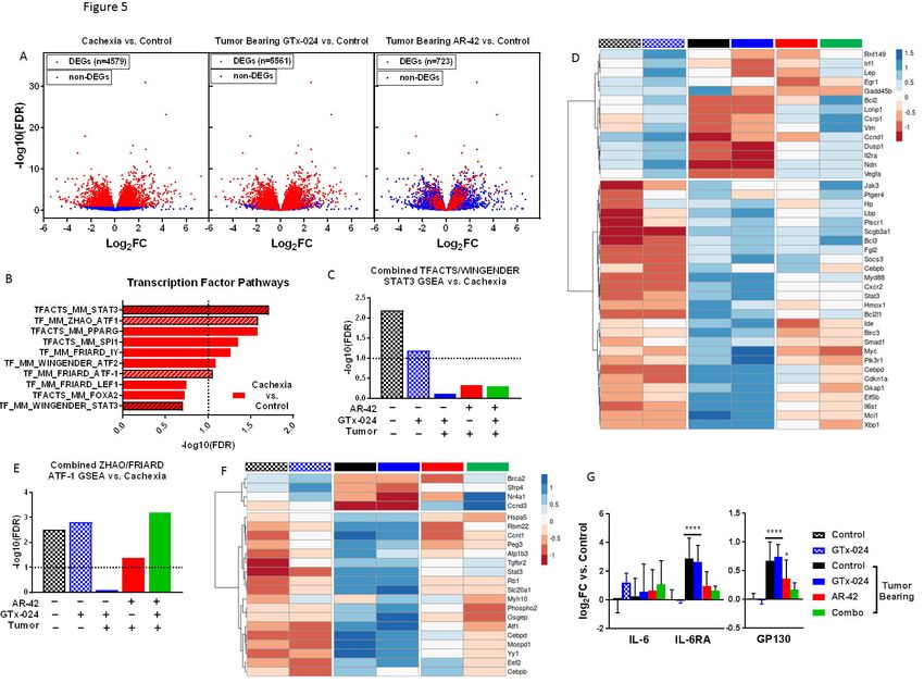
bioRxiv preprint first posted online Nov. 6, 2017; doi: http://dx.doi.org/10.1101/214155. The copyright holder for this preprint
(which was not peer-reviewed) is the author/funder, who has granted bioRxiv a license to display the preprint in perpetuity.
                             It is made available under a CC-BY-NC-ND 4.0 International license.

   TRANSLATIONAL RELEVANCE: Our data provide insight into the recent late stage clinical

   failure of SARM anti-cachectic therapy by characterizing anabolic resistance in the C-26 model

   of cancer cachexia. We show that fully anabolic doses of multiple androgens provide no anti-

   cachectic efficacy and that SARM-mediated WNT activation in skeletal muscle is disrupted by

   cachectic signaling. To the best of our knowledge, this is the first study to characterize WNT-

   mediated anabolic resistance in experimental cachexia. We further show that the novel HDACi

   AR-42 suppresses the IL-6/GP130/STAT3 axis within skeletal muscle to provide anti-cachectic

   benefit that is additionally associated with improved anabolic response to co-administered

   SARM. Our data support combined SARM/AR-42 administration as improved anti-cachectic

   therapy.

                                                                       2
bioRxiv preprint first posted online Nov. 6, 2017; doi: http://dx.doi.org/10.1101/214155. The copyright holder for this preprint
(which was not peer-reviewed) is the author/funder, who has granted bioRxiv a license to display the preprint in perpetuity.
                             It is made available under a CC-BY-NC-ND 4.0 International license.

   ABSTRACT

   Purpose

   The common colon-26 mouse (C-26) model of experimental cachexia mimics recent late stage

   clinical failures of anabolic anti-cachexia therapy, and does not respond to the anabolic

   selective androgen receptor modulator (SARM) GTx-024. Based on the demonstrated anti-

   cachectic efficacy of the histone deacetylase inhibitor (HDACi) AR-42 in this model, we

   hypothesized that combined SARM/AR-42 would provide improved anti-cachectic efficacy.

   Design

   In the C-26 model, we determined a reduced efficacious dose of AR-42 which was combined

   with anabolic SARM therapy and evaluated for anti-cachectic efficacy. The effects of treatment

   and tumor burden on anabolic and catabolic signaling occurring in skeletal muscle were

   characterized using muscle performance parameters and RNA-seq.

   Results

   Anabolic anti-cachexia therapy with diverse androgens had no impact on cachectic outcomes in

   the C-26 model. A reduced dose of the HDACi AR-42 alone provided limited anti-cachectic

   benefits, but when combined with the SARM GTx-024, significantly improved bodyweight

   (p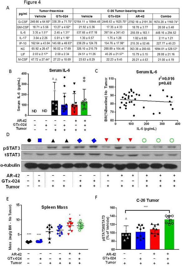
bioRxiv preprint first posted online Nov. 6, 2017; doi: http://dx.doi.org/10.1101/214155. The copyright holder for this preprint
(which was not peer-reviewed) is the author/funder, who has granted bioRxiv a license to display the preprint in perpetuity.
                             It is made available under a CC-BY-NC-ND 4.0 International license.

   clinically evaluated HDACi, represents a promising approach to improve anabolic response in

   cachectic patient populations.

                                                                       4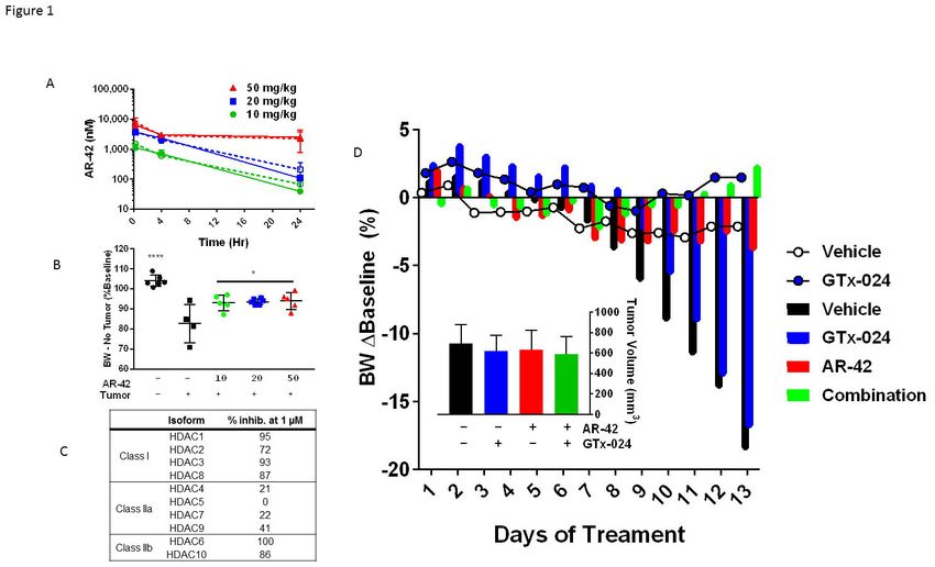
bioRxiv preprint first posted online Nov. 6, 2017; doi: http://dx.doi.org/10.1101/214155. The copyright holder for this preprint
(which was not peer-reviewed) is the author/funder, who has granted bioRxiv a license to display the preprint in perpetuity.
                             It is made available under a CC-BY-NC-ND 4.0 International license.

   INTRODUCTION

             Cancer cachexia is a multifactorial syndrome characterized by the involuntary loss of

   muscle mass occurring with or without concurrent losses in adipose tissue. The progressive

   loss of lean mass associated with cachexia results in decreased quality of life, decreased

   tolerance of chemotherapy and reduced overall survival [1]. It is estimated that 50-80% of all

   cancer patients experience cachexia symptoms and up to 20% of all cancer-related deaths are

   attributable to complications arising from cachexia-mediated functional decline [2]. A multitude

   of tumor and host factors are recognized as contributors to the multi-organ system dysfunction

   in cancer cachexia, presenting a considerable therapeutic challenge. Diverse cachexia

   treatment strategies have been evaluated in patients with few offering effective palliation and

   none gaining FDA approval for this devastating consequence of advanced malignancy [3].

   Among the complex sequelae associated with cachectic progression, compromised muscle

   function associated with reduced muscle mass is viewed as a primary contributor to patient

   morbidity and mortality [1]. Recognizing this feature of cancer cachexia, regulatory agencies

   require the demonstration of meaningful improvements in physical function in addition to

   improvements in patient body composition for successful registration of novel cachexia

   therapies [4]. Anabolic androgenic steroids or steroidal androgens are among the most well

   recognized function-promoting therapies [5] and, as such, have been extensively evaluated in

   muscle wasting of diverse etiology [6]. Despite meeting FDA approval criteria in other wasting

   diseases [6], steroidal androgens are yet to demonstrate clinical benefit in cancer cachexia.

   However, the continued development of novel androgens for the treatment of wasting diseases

   suggests confidence in this therapeutic strategy remains [3].

             In addition to their well characterized anabolic effects on skeletal muscle, steroidal

   androgens elicit a number of undesirable virilizing side effects and can promote prostatic

   hypertrophy which limits their widespread clinical use [7]. Recently developed, non-steroidal,

   selective androgen receptor modulators (SARMs) offer a number of improvements over

                                                                       5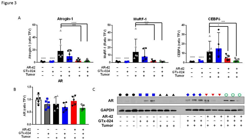
bioRxiv preprint first posted online Nov. 6, 2017; doi: http://dx.doi.org/10.1101/214155. The copyright holder for this preprint
(which was not peer-reviewed) is the author/funder, who has granted bioRxiv a license to display the preprint in perpetuity.
                             It is made available under a CC-BY-NC-ND 4.0 International license.

   steroidal androgens including prolonged plasma exposures and oral bioavailability with greatly

   reduced side effects (virilization, etc.), while maintaining full agonism in anabolic tissues like

   skeletal muscle [8]. With once daily dosing, the SARM GTx-024 (enobosarm) showed

   promising gains in fat-free mass in both male and female cancer patients, but ultimately failed to

   demonstrate a clear functional benefit in pivotal phase III trials in a cachectic non-small cell lung

   cancer (NSCLC) population [9]. GTx-024 has a strong safety profile and proven effects on

   skeletal muscle, but is no longer being developed for cancer cachexia.

             Hypogonadism is a feature of advanced malignancy and experimental cachexia that

   worsens multiple cachectic sequelae, including decreased skeletal muscle mass, providing a

   rationale for therapeutic exogenous androgen administration [10, 11]. Though the relationship

   between androgen status and body composition is well established, the exact molecular basis

   by which androgens modulate skeletal muscle mass is not completely characterized but

   involves the repression of several atrogenes, induction of PI3K/AKT/mTOR signaling and direct

   stimulation of muscle satellite cells (MUSCs) [12]. Despite demonstrating the clear ability to

   attenuate orchiectomy- and glucocorticoid-mediated muscle loss [13], in our hands, SARMs

   displayed essentially no impact on muscle wasting associated with the common colon-26 (C-26)

   mouse model of experimental cancer cachexia (unpublished). In these mice, androgen-

   mediated gene transcription was severely muted in the presence of a cachectic burden, and

   fully anabolic doses of SARM were unable to normalize muscle E3-ligase expression to

   effectively combat the catabolic decline driven by the C-26 tumor. We hypothesized that a

   better understanding of the failure of SARMs in the C-26 model would offer key insights into the

   limitations of androgens in cachectic cancer patients. Furthermore, we recently demonstrated

   the effectiveness of a novel class I/IIB HDAC inhibitor (HDACi, AR-42), currently under clinical

   evaluation in hematologic malignancies [14] and solid tumors, as anti-cachexia therapy in the C-

   26 model [15]. AR-42 administration in these mice spared body weight and was associated with

   improvements, but not complete rescue, of skeletal muscle mass relative to controls. Notably,

                                                                       6
bioRxiv preprint first posted online Nov. 6, 2017; doi: http://dx.doi.org/10.1101/214155. The copyright holder for this preprint
(which was not peer-reviewed) is the author/funder, who has granted bioRxiv a license to display the preprint in perpetuity.
It is made available under a CC-BY-NC-ND 4.0 International license.
AR-42 differed from other approved HDACis in its ability to fully suppress tumor-mediated
atrogin-1 and MuRF1 induction and prolong survival in the C-26 model. Unlike androgens, AR-
42 does not promote skeletal muscle hypertrophy in tumor-free animals, and AR-42’s anti-
cachectic efficacy was highly dependent on early initiation of treatment, suggesting AR-42’s
dramatic anti-cachectic efficacy in the C-26 model is primarily associated with its anti-catabolic
effects [15]. Given SARMs’ established anabolic potential, but clear inability to attenuate tumor-
driven wasting in the C-26 model, and AR-42’s effects on tumor-mediated catabolic signaling,
but apparent lack of anabolic effects on skeletal muscle, we hypothesized that co-administration
of AR-42 with SARMs in the C-26 model would result in improved anabolic response and overall
anti-cachectic efficacy. Before evaluating combination therapy, we first explored reduced anti-
cachectic doses of AR-42 given the need to minimize side-effects attributable to anti-cachectic
therapy.
MATERIALS AND METHODS
Information on reagents and chemicals, antibodies, cell culture, animals, and methodological
details for grip strength measurement, luteinizing hormone analysis, pharmacokinetics, western
blot analysis and gene expression analyses are included in the Supplementary Materials and
Methods. All animal studies were conducted according to protocols approved by The Ohio State
University Institutional Animal Care and Use Committee.
Animal studies using the C-26 colon adenocarcinoma cachexia model
These studies were performed as previously described [15] with modifications. Tumors were
established in the right flank by subcutaneous injection of C-26 cells (0.5 x 10⁶ cells in 0.1 mL).
AR-42, GTx-024 and their vehicles were administered orally by gavage. TFM-4AS-1 (a potent
experimental SARM), dihydrotestosterone (DHT) and vehicles were administered by
subcutaneous injection.
7bioRxiv preprint first posted online Nov. 6, 2017; doi: http://dx.doi.org/10.1101/214155. The copyright holder for this preprint
(which was not peer-reviewed) is the author/funder, who has granted bioRxiv a license to display the preprint in perpetuity.
It is made available under a CC-BY-NC-ND 4.0 International license.
AR-42 dose-response study: Male CD2F1 mice were stratified by body weight and then
randomly assigned into 5 groups of 6 animals each. C-26 tumors were established in four of the
groups, while those in the fifth group, serving as tumor-free controls, were injected with sterile
saline. Six days later, animals with palpable tumors were treated with AR-42 once daily at 10
(n=5) and 20 mg/kg (n=6), and every other day at 50 mg/kg (n=5), or vehicle control (n=4) for 13
days. Upon sacrifice on study day 18, when the majority of tumor-bearing control mice met
euthanasia criteria, the left gastrocnemius muscle was excised, flash frozen in liquid nitrogen
and stored at -80°C for subsequent analyses. Carcass weights were corrected for tumor weight
by assuming a tumor density equivalent to water (1 g/cm3).
Initial AR-42/GTx-024 Combination Study (Study 1): Male CD2F1 mice were stratified by body
weight and then randomly assigned into 6 groups of 6 animals each. Historically, 6 animals per
group provided sufficient power to detect treatment-mediated differences in tumor-bearing
treated animals compared to controls. Tumors were established in four of the groups, while the
fifth and sixth groups served as tumor-free controls. Six days later, animals with palpable
tumors were treated twice daily for 13 days. AR-42 and its vehicle were administered in the
mornings, and GTx-024 and its vehicle in the afternoons. Treatments included vehicles for AR-
42 and GTx-024 (n=5), GTx-024 (15 mg/kg; AR-42 vehicle; n=5), AR-42 (10 mg/kg; GTx-024
vehicle; n=5), or the AR-42+GTx-024 combination (10 and 15 mg/kg, respectively; n=5). The
remaining tumor-free groups received either vehicles (n=6) or GTx-024 (15 mg/kg; AR-42
vehicle; n=6). Body weight, tumor volume, and feed consumption were monitored every other
day. Upon sacrifice on day 18, sera were collected and hind limb skeletal muscles, heart,
spleen and epididymal adipose tissues were harvested, weighed, flash frozen, and stored for
subsequent analyses.
Confirmatory AR-42/GTx-024 Combination Study (Study 2): This confirmatory study was
performed exactly as Study 1 with expanded animal numbers. Tumor-free control groups were
maintained at 6 animals each, whereas 10 animals were included in each of the tumor-bearing
8bioRxiv preprint first posted online Nov. 6, 2017; doi: http://dx.doi.org/10.1101/214155. The copyright holder for this preprint
(which was not peer-reviewed) is the author/funder, who has granted bioRxiv a license to display the preprint in perpetuity.
It is made available under a CC-BY-NC-ND 4.0 International license.
groups. Six days after cell injection, animals with palpable tumors were treated as in Study 1
with vehicles (n=7), GTx-024 (n=10), AR-42 (n=9), or the combination (n=9). Grip strength was
measured on study days 0 (baseline) and 16. Due to rapid model progression, this study was
terminated after only 12 days of treatment.
Combined Androgen and AR-42 Study (Study 3): Similar to Study 2, the tumor-free control
group was maintained at 6 animals, whereas 10 animals were included in each of the 6 tumor-
bearing groups. Six days after cell injection, animals with palpable tumors were treated once
daily for 13 days with vehicles for AR-42 and TFM-4AS-1/DHT (n=9), AR-42 (10 mg/kg; TFM-
4AS-1/DHT vehicle; n=10), TFM-4AS-1 (10 mg/kg; AR-42 vehicle; n=9), DHT (3 mg/kg; AR-42
vehicle; n=10), the combination of AR-42 and TFM-4AS-1 (10 mg/kg each; n=9), or the
combination of AR-42 (10 mg/kg) and DHT (3 mg/kg; n=10). Grip strength was measured and
tissues collected as in the previous studies.
AR-42 Plasma and Tissue Pharmacokinetics
Pharmacokinetic studies were performed as previously described [16] with modifications
described in Supplementary Materials and Methods.
Western Blot Analyses
All Western blots were performed on gastrocnemius muscle from representative animals in each
study using standard methods as described in Supplementary Materials and Methods.
Gene Expression Analyses
All gene expression experiments were performed on gastrocnemius muscles from
representative animals. Expression levels were estimated by both traditional q-RT-PCR and
sequencing of polyA-selected RNAseq libraries. Details of the methods for data generation and
associated analyses are described in detail in Supplementary Materials and Methods.
9bioRxiv preprint first posted online Nov. 6, 2017; doi: http://dx.doi.org/10.1101/214155. The copyright holder for this preprint
(which was not peer-reviewed) is the author/funder, who has granted bioRxiv a license to display the preprint in perpetuity.
It is made available under a CC-BY-NC-ND 4.0 International license.
Statistical Methodology
Plotting and statistical analyses were performed using GraphPad Prism Version 7 (GraphPad
Software, La Jolla, CA). The specific statistical tests employed are outlined in detail within the
figure legends.
RESULTS
AR-42 administration demonstrates anti-cachectic effects at a reduced 10 mg/kg dose level
We recently characterized the anti-cachectic effects of AR-42 (50 mg/kg via oral gavage
every other day) in C-26 tumor-bearing mice [15]. This dose represented the maximally
tolerated dose in mice, which was used to observe its anti-tumor effects in different xenograft
tumor models. To better understand the disposition of AR-42 following oral administration in
mice, we performed a limited pharmacokinetic study of single oral doses of 50, 20 and 10 mg/kg
of AR-42 (Figure 1A). Plasma exposure following oral administration of 50 mg/kg was 74.3
µM*h (Supplementary Figure S1), which exceeded the well tolerated plasma exposure in
humans of 8.5 µM*h by 8.7-fold [14]. Consequently, we evaluated the anti-cachectic effects of
lower doses of AR-42 in a dose-response study in the C-26 model. Similar to six total 50 mg/kg
doses (administered q2d), thirteen daily oral doses of 20 or 10 mg/kg AR-42 reversed C-26
tumor-mediated reductions in tumor-corrected body weight (Figure 1B). AR-42 readily
distributed into gastrocnemius muscle tissue (Figure 1A) and, at the lowest efficacious dose of
10 mg/kg, muscle concentrations remained above 700 nM for 4 hours consistent with the ability
of AR-42 at this dose to inhibit Class I and IIb HDACs for a portion of the dosing interval in
muscle tissue based on its in vitro HDAC inhibition profile (Figure 1C). The plasma exposure
resulting from the 10 mg/kg dose (10.9 µM*h, Supplementary Figure S1) compares more
favorably to well-tolerated exposures in patients while providing anti-cachectic efficacy and was
therefore utilized in subsequent combination studies.
10bioRxiv preprint first posted online Nov. 6, 2017; doi: http://dx.doi.org/10.1101/214155. The copyright holder for this preprint
(which was not peer-reviewed) is the author/funder, who has granted bioRxiv a license to display the preprint in perpetuity.
It is made available under a CC-BY-NC-ND 4.0 International license.
Combination GTx-024 and AR-42 administration results in improved anti-cachectic efficacy
To evaluate our hypothesis that combining HDAC inhibition with SARM administration
would improve anti-cachectic activity, we designed a series of three studies combining AR-42
with androgen/SARM in the C-26 model. In Study 1, similar to results from this and other
laboratories [15, 17], vehicle-treated tumor-bearing animals lost approximately 20% of their
body weight prior to meeting euthanasia criteria (Figure 1D). This severe tumor-induced weight
loss (Figure 2A, 80.4±9.1% of baseline) was accompanied by parallel reductions in
gastrocnemius and quadriceps masses (Figure 2B, 86±12.4 and 88±12.0%, relative to tumor-
free controls, respectively). Consistent with previous findings in this model (unpublished),
SARM monotherapy had no apparent anti-cachectic efficacy in C-26 tumor-bearing mice. GTx-
024 at 15 mg/kg did not spare body weight (Figure 1D, 2A) or the mass of gastrocnemius and
quadriceps muscles (Figure 2B). At this dose, GTx-024 was well tolerated in xenografted mice
[18] and, in this study, did not cause body weight loss in tumor-free controls (Figure 1D, 2A).
Furthermore, GTx-024 was reported to be fully anabolic at doses as low as 0.5 mg/kg/day in
rodents (reported as S-22 in Kim et al. [19]) and compared favorably to the less potent structural
analog S-23 [20], which reversed orchiectomy- and glucocorticoid-mediated wasting [13]. A
separate control study in tumor-free CD2F1 mice confirmed that, in our hands, 15 mg/kg GTx-
024 was capable of increasing body weight, gastrocnemius and quadriceps mass, and grip
strength in orchiectomized (ORX) mice relative to vehicle-treated ORX controls (Supplementary
Figure S2). Importantly, GTx-024 suppressed serum luteinizing hormone, a very well
characterized pharmacological effect of potent androgen administration [21], demonstrating that
GTx-024 administered to C-26 tumor-bearing mice was active (Supplementary Figure S3A).
Consistent with the preliminary dose-response study, 10 mg/kg AR-42 alone significantly
spared body weight (Figure 1D and 2A, 93.6±7.7% of baseline) relative to tumor-bearing
vehicle-treated controls. However, these changes were not translated into significant
11bioRxiv preprint first posted online Nov. 6, 2017; doi: http://dx.doi.org/10.1101/214155. The copyright holder for this preprint
(which was not peer-reviewed) is the author/funder, who has granted bioRxiv a license to display the preprint in perpetuity.
It is made available under a CC-BY-NC-ND 4.0 International license.
improvements in gastrocnemius and quadriceps mass (Figure 2B). In contrast to monotherapy,
C-26 tumor-bearing mice receiving both GTx-024 and AR-42 started to gain body weight relative
to baseline after nearly two weeks of treatment, whereas all other treated tumor-bearing groups
lost body weight (Figure 1D). This combination exhibited a striking ability to consistently protect
body weight (99.9±0.8% of baseline, corrected for tumor weight) relative to either agent alone
(Figure 2A). Furthermore, the effects of combined therapy completely spared gastrocnemius
(102.4±3.8%) and quadriceps (99.9±5.5%) mass relative to tumor-free controls (Figure 2B).
The effects of combined therapy on total body weight or amelioration of cachectic
symptoms were not due to any overt impact on tumor burden as no significant differences in
tumor volumes were apparent at the end of the study (Figure 1D, inset). Food consumption was
monitored to account for potential anti-anorexic effects of treatment on the cachectic sequela
following C-26 cell inoculation. GTx-024-treated tumor-free control animals, as well as the
combination-treated group, demonstrated small increases in per animal food consumption
relative to other groups between day 14 and 16 (Supplementary Figure S3B), which are unlikely
to account for differences in body weight apparent by study day 14 (treatment day 9), as well as
end of study differences in skeletal muscle masses (Figure 2B).
These promising results prompted us to repeat the experiment with expanded animal
numbers and the use of forelimb grip dynamometry as a measure of muscle function. In this
confirmatory study (Study 2), the model was more aggressive resulting from significantly larger
tumors relative to Study 1, though no differences within treatment groups were apparent
(Supplementary Figure S4A). As a result, the study was terminated early, after only 12 days of
treatment. In accordance with this increased tumor burden, tumor-corrected body weights were
more consistently reduced and to a larger degree in tumor-bearing controls (77.0±5.7% and
80.4± 9.1% of baseline in the second and first studies, respectively; Supplementary Figure
S4B), and larger losses in gastrocnemius (76.3±8.1%) and quadriceps (69.1±7.6%) mass
relative to tumor-free controls were noted (Supplementary Figure S4C). In the face of this more
12bioRxiv preprint first posted online Nov. 6, 2017; doi: http://dx.doi.org/10.1101/214155. The copyright holder for this preprint
(which was not peer-reviewed) is the author/funder, who has granted bioRxiv a license to display the preprint in perpetuity.
It is made available under a CC-BY-NC-ND 4.0 International license.
severe cachexia, only combined AR-42 and GTx-024 administration significantly spared body
weight (90.3±4.3 of baseline), though not to the degree realized in the first study
(Supplementary Figure S4B vs Figure 2A), while both AR-42 alone and the combination
significantly spared gastrocnemius and quadriceps mass (Supplementary Figure S4C). C-26
tumors were accompanied by large reductions in forelimb grip strength (Figure 2C, 63.8±15.3%
versus 83.8±10.7% of baseline in tumor-bearing and tumor-free controls, respectively), but,
consistent with the improvements in hind limb skeletal muscle mass, AR-42 alone and in
combination with GTx-024 improved grip strength over vehicle-treated tumor-bearing controls.
Unlike the adipose-sparing effect of the higher 50 mg/kg dose of AR-42 [15], the lower dose of
10 mg/kg had no impact on adipose or heart mass (Supplementary Figure S4D). As androgens
are thought to actively prevent adipogenesis [22], SARM administration was not expected to
protect against C-26 tumor-mediated fat losses. Indeed, no treatment mediated effects on
abdominal adipose were apparent (Supplementary Figure S4D). The data show heart mass
was significantly improved by combination therapy, but this result is likely due to the effects of a
single outlier animal.
Multiple androgens demonstrate improved anti-cachectic efficacy when combined with AR-42
To confirm that the improvement of GTx-024’s anti-cachectic efficacy in the C-26 model
by co-administration with AR-42 was not a drug-specific phenomenon, tumor-bearing animals
were treated with the SARM TFM-4AS-1 [23] and the potent endogenous androgen DHT alone
and in combination with AR-42 (Study 3). Similar to the 15 mg/kg dose of GTx-024, TFM-4AS-1
was administered at a previously characterized fully anabolic dose (10 mg/kg), but, as a
monotherapy, did not spare body weight (Figure 2D) or mass of gastrocnemius or quadriceps
(Figure 2E).
13bioRxiv preprint first posted online Nov. 6, 2017; doi: http://dx.doi.org/10.1101/214155. The copyright holder for this preprint
(which was not peer-reviewed) is the author/funder, who has granted bioRxiv a license to display the preprint in perpetuity.
It is made available under a CC-BY-NC-ND 4.0 International license.
AR-42 alone resulted in significant attenuation of body weight loss (93.5±4.8 of
baseline), but was less effective than in combination with TFM-4AS-1 (99.5±4.4 of baseline) or
DHT (106.0±5.4 of baseline). The DHT/AR-42 combination significantly improved bodyweights
(pbioRxiv preprint first posted online Nov. 6, 2017; doi: http://dx.doi.org/10.1101/214155. The copyright holder for this preprint
(which was not peer-reviewed) is the author/funder, who has granted bioRxiv a license to display the preprint in perpetuity.
It is made available under a CC-BY-NC-ND 4.0 International license.
agents on genes whose function has been previously associated with C-26 tumor-mediated
wasting (Figure 3A). As expected for this model, the muscle-specific E3 ligases atrogin-1
(FBXO32) and MuRF-1(TRIM63) were induced in skeletal muscles of tumor-bearing animals
[15, 17] as was the STAT3 target gene and regulator of atrogin-1 and MuRF-1, CEBPδ(CEBPD)
[24]. Consistent with the absence of any anti-cachectic effects of GTx-024 monotherapy, this
treatment had no significant impact on atrogin-1, MuRF-1, or CEBPδ expression. Ten mg/kg
AR-42 alone and in combination with GTx-024 significantly reduced the expression of each
atrogene relative to tumor-bearing controls, returning them to near baseline levels. AR-42’s
effects on E3 ligase expression were consistent with results from animals receiving the higher
dose of 50 mg/kg [15] further supporting the importance of AR-42’s ability to reverse induction of
these key enzymes to its overall anti-cachectic efficacy.
To determine the effect of tumor burden and treatment on androgen receptor (AR) levels
in skeletal muscle, which could influence response to androgen therapy, gastrocnemius AR
levels were characterized. Neither tumor nor treatment had a significant impact on AR mRNA
(Figure 3B). AR protein expression in gastrocnemius was low in tumor-free controls and
increased in response to GTx-024 administration irrespective of tumor burden (Figure 3C)
consistent with androgen agonist binding and stabilization of the AR [25]. In contrast, AR-42
treatment did not have a marked impact on AR expression.
Anti-cachectic efficacy of AR-42 is associated with STAT3 inhibition but not general immune
suppression
Previously reported ingenuity pathway analyses of AR-42-regulated genes in
gastrocnemius muscle revealed that 66 genes associated with muscle disease or function were
significantly regulated by AR-42 relative to C-26 tumor-bearing vehicle-treated controls [15]. In
an effort to enrich previously reported differentially regulated genes (n=548) for transcripts
15bioRxiv preprint first posted online Nov. 6, 2017; doi: http://dx.doi.org/10.1101/214155. The copyright holder for this preprint
(which was not peer-reviewed) is the author/funder, who has granted bioRxiv a license to display the preprint in perpetuity.
It is made available under a CC-BY-NC-ND 4.0 International license.
critical to the anti-cachectic efficacy of AR-42, these data were intersected with previously
published differentially regulated genes from the quadriceps of moderate and severely wasted
C-26 tumor-bearing mice [17] (n=700, Supplementary Figure S6A). Using this approach, the
likely biological relevance of the 147 overlapping genes is increased when it is considered that
these transcripts represent genes regulated by AR-42 that are associated with C-26-induced
wasting from two different muscles (gastrocnemius and quadriceps), detected by two different
technologies (RNA-seq and microarray) and reported by two different research laboratories.
Pathway analyses performed on this pool of 147 genes revealed IL-6 signaling and immune
system pathways, along with other gene sets regulated subsequent to cytokine stimulation,
implicating AR-42’s effects on cytokine and immune signaling in its anti-cachectic efficacy
(Supplementary Figure S6B).
In agreement with the present pathway analyses, we previously reported that the higher
50 mg/kg dose of AR-42 reduced serum IL-6 levels, as well as gastrocnemius IL-6 receptor
mRNA abundance in tumor-bearing mice suggesting AR-42’s efficacy may be related to its
suppression of systemic IL-6 activation which is thought to drive muscle wasting in the C-26
model [15]. In this study, the impact of C-26 tumor burden and treatment with AR-42, GTx-024
or combination therapy on a panel of circulating cytokines, including IL-6, was assessed (Figure
4A, Supplementary Table S1). Consistent with our previous report, multiple pro-cachectic
factors, including G-CSF, IL-6, and LIF, were significantly elevated by the presence of C-26
tumors [15]. Unlike the 50 mg/kg dose, 10 mg/kg AR-42 did not significantly impact IL-6 family
cytokine levels (i.e. IL-6 or LIF) alone or in combination with GTx-024. Furthermore, 10 mg/kg
AR-42 monotherapy did not significantly reduce circulating levels of any evaluated cytokine,
despite demonstrating clear anti-cachectic effects across the multiple studies presented here
(Figure 1D, Figure 2, Supplementary Figure S4). An ELISA analysis confirmed our findings that
AR-42 treatment did not affect circulating IL-6 levels (Figure 4B), and demonstrated serum IL-6
16bioRxiv preprint first posted online Nov. 6, 2017; doi: http://dx.doi.org/10.1101/214155. The copyright holder for this preprint
(which was not peer-reviewed) is the author/funder, who has granted bioRxiv a license to display the preprint in perpetuity.
It is made available under a CC-BY-NC-ND 4.0 International license.
levels were not associated with body weight in treated, C-26 tumor-bearing mice at sacrifice
(Figure 4C).
When significant effects on circulating cytokines were not apparent, we hypothesized
AR-42 might be acting downstream of the IL-6 receptor on critical mediators of cytokine
signaling. One well characterized effector of cytokine-induced signaling shown to be central to
tumor-induced wasting in a number of models is signal transducer and activator of transcription
(STAT)3 [11, 17]. Notably, STAT3 activation is associated with the severity of wasting in both
the C-26 and Apcmin/+ models of cancer cachexia, and AR-42 was previously shown to suppress
the IL-6/GP130/STAT3 signaling axis in multiple myeloma cells [26]. Thus, we evaluated AR-
42’s effects on phospho-STAT3 (pSTAT3) in gastrocnemius muscle from C-26 tumor-bearing
animals (Figure 4D, Supplementary Figure S7). As expected, the presence of the C-26 tumor
resulted in increased pSTAT3 abundance. GTx-024 treatment had no apparent effect on
pSTAT3, consistent with its inability to spare body weight or lower limb skeletal muscle mass as
a monotherapy. AR-42 monotherapy reduced pSTAT3 but not equally in all animals, whereas
the combination treatment exhibited the most consistent suppression, concordant with its
marked anti-cachectic efficacy. Furthermore, treatment-mediated effects on the well
characterized STAT3 target gene CEBPδ [24] closely paralleled those on STAT3 activation
(Figure 3A).
In addition to skeletal muscle STAT3 activation, C-26 tumor-bearing mice exhibit
splenomegaly as a result of increased systemic inflammation [27]. Consistent with increased
circulating cytokine levels, C-26 tumor-bearing animals in both Study 1 and 2 demonstrated
large increases in spleen mass across all treatment groups relative to tumor-free controls
(Supplementary Figure S3C and Figure 4E, respectively). Similar to findings with 50 mg/kg AR-
42 [15], spleen mass was either unchanged or slightly increased by AR-42 alone or in
combination with GTx-024. As a gross measure of the systemic effects of treatment on immune
17bioRxiv preprint first posted online Nov. 6, 2017; doi: http://dx.doi.org/10.1101/214155. The copyright holder for this preprint
(which was not peer-reviewed) is the author/funder, who has granted bioRxiv a license to display the preprint in perpetuity.
It is made available under a CC-BY-NC-ND 4.0 International license.
function, these spleen mass results suggest AR-42 is not generally immunosuppressive and its
activity is distinct from inhibitors of the JAK/STAT pathway in this context [28]. Unlike in
gastrocnemius tissue, AR-42 treatment did not significantly suppress pSTAT3 signaling within
the C-26 tumors (Figure 4F). Taken together, these multiple lines of evidence suggest that the
anti-cachectic efficacy of AR-42 involves the inhibition of the IL-6/GP130/STAT3 axis in skeletal
muscle tissue, but not systemic suppression of IL-6 or general immune signaling.
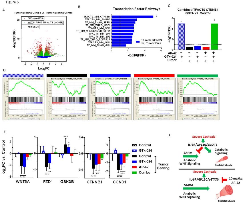
Transcriptomic analyses of AR-42’s anti-cachectic effects in skeletal muscle
To further characterize AR-42’s anti-cachectic effects at the reduced dose of 10 mg/kg,
RNA-seq analyses were performed on all gastrocnemius tissues from Study 1 (Figure 2A, B).
This resulted in 31 evaluable samples across treatment groups (Supplementary Figure S8) after
removal of two samples due to insufficient sequencing yield/quality. We detected 4,579
differentially expressed genes (DEGs; FDR < 0.1) in cachectic versus control muscle, whereas
treatment of cachectic mice with GTx-024 or AR-42 alone resulted in 5,561 and 723 DEGs,
respectively, consistent with their corresponding anti-cachectic efficacies (Figure 5A,
Supplementary Figure S9A and B). Given the ability of HDAC inhibitors and androgens to
modulate transcription, initial functional analyses were focused on curated Mus musculus
transcription factor (TF) target gene sets, and revealed multiple over-represented TF targets in
cachectic versus control muscle (Figure 5B). STAT3 and activation of transcription-1 (ATF1)
gene sets were each represented twice in the top ten pathways following GSEA supporting their
potential relevance in cachectic signaling. The two STAT3 target gene sets were combined and
GSEA was repeated with the combined set for all treatment groups. In contrast to pSTAT3
activation (Figure 4D), this analysis demonstrated the inability of any treatment in tumor-bearing
mice to significantly limit the importance of STAT3 target-gene regulation relative to cachectic
18bioRxiv preprint first posted online Nov. 6, 2017; doi: http://dx.doi.org/10.1101/214155. The copyright holder for this preprint
(which was not peer-reviewed) is the author/funder, who has granted bioRxiv a license to display the preprint in perpetuity.
It is made available under a CC-BY-NC-ND 4.0 International license.
controls (Figure 5C and Supplementary Figure S10). However, when analysis is focused on
individual genes within the combined set that are differentially expressed in at least one
comparison, clear cachexia-dependent regulation is apparent that responds only to AR-42
treatment (Figure 5D). A similar analysis with combined ATF-1 data sets revealed the ability of
AR-42, but not GTx-024 treatment, to significantly impact ATF-1 target gene regulation in tumor-
bearing mice implicating AR-42’s ability to modulate ATF-1 activation in its anti-cachectic
efficacy (Figure 5E and Supplementary Figure S11). Of note, STAT3 and CEPBδ are among
the differentially expressed ATF-1 target genes induced by cachexia that respond only to AR-42
treatment (Figure 5F).
We further evaluated the expression of genes within the IL-6 pathway as IL-6-mediated
STAT3 target gene regulation is well characterized in the C-26 model [17], and IL-6-mediated
increases in skeletal muscle cyclic AMP (cAMP), a primary driver of ATF-1 activation [29], have
also been reported[30]. Unlike circulating IL-6 cytokine, IL-6 mRNA in gastrocnemius muscle
was not induced by cachexia, nor was it modulated by any treatment (Figure 5G). However,
expression of both IL-6 receptor (IL-6RA) and the key effector GP130 were elevated in
cachectic mice and required AR-42 (IL-6RA) or combination treatment (GP130) to restore to
non-cachectic control levels.
Considerable overlap exists between the transcriptomes of cachectic gastrocnemius
muscles from mice treated with 10 or 50 mg/kg AR-42 such that high fold-change DEGs
identified by Tseng et al. [17] and in the current study are all regulated in the same direction (n=
209, Supplementary Figure 12A-B). Similar to previous analyses (Supplementary Figure S5),
functional interrogation of the genes within this overlap further support the importance of AR-
42’s ability to modulate immune and extracellular matrix signaling in eliciting its anti-cachectic
effects (Supplementary Figures S12C). Taken together these findings support the ability of the
19bioRxiv preprint first posted online Nov. 6, 2017; doi: http://dx.doi.org/10.1101/214155. The copyright holder for this preprint
(which was not peer-reviewed) is the author/funder, who has granted bioRxiv a license to display the preprint in perpetuity.
It is made available under a CC-BY-NC-ND 4.0 International license.
reduced 10 mg/kg dose of AR-42 to generate anti-cachectic effects by reducing procachectic IL-
6RA/GP130/STAT3 signaling in skeletal muscle.
Transcriptomic analyses of GTx-024’s anabolic effects in skeletal muscle
To better understand GTx-024’s contribution to the efficacy apparent in combination-
treated mice, the transcriptome of combination-treated gastrocnemius muscle was compared to
cachectic controls revealing 2,026 DEGs (or 50.6% of all DEGs) not solely attributable to AR-42
treatment (Figure 6A). We hypothesized that GTx-024-mediated anabolic signaling detectable
in GTx-024-treated tumor-free controls would be diminished in tumor-bearing GTx-024-treated
animals in the absence of AR-42. Though very few DEGs were apparent in GTx-024-treated
tumor-free controls (n = 27, Supplementary Figure S13), GSEA focused on TF pathways
revealed abundant coordinated signaling with regulation of β-catenin (CTNNB1) target genes
providing the most significant overlap (FDR < 1e-5, Figure 6B). Coordinate regulation of β-
catenin target genes was not apparent in cachectic controls or following GTx-024 or AR-42
monotherapy, but was again among the most prominent pathways detected by GSEA in
combination-treated mice (FDR < 1e-5, Figure 6C). GSEA plots demonstrate a robust pattern
of GTx-024-mediated activation of β-catenin target genes requiring AR-42 co-administration in
cachectic mice (Figure 6D, leftmost panel compared to rightmost panel). Analysis of overlap of
the leading edge genes revealed a large number of CTNNB1 target genes regulated by both
GTx-024 and cachexia versus tumor free controls but in different directions (n=49 middle, 17
bottom left; Supplementary Figure S14A). Many fewer leading edge genes were regulated by
AR-42 monotherapy but also in an opposite direction to GTx-024 (n= 2 middle, 23 top middle;
Supplementary Figure S14B). However, combined therapy results in a larger leading edge
gene set overlap that is regulated in a similar direction to GTx-024 monotherapy (n= 29 middle,
20bioRxiv preprint first posted online Nov. 6, 2017; doi: http://dx.doi.org/10.1101/214155. The copyright holder for this preprint
(which was not peer-reviewed) is the author/funder, who has granted bioRxiv a license to display the preprint in perpetuity.
It is made available under a CC-BY-NC-ND 4.0 International license.
37 of 47 top middle; Supplementary Figure S14C). This pattern of β-catenin target gene
regulation is also apparent when DEG’s within the TFACTS_CTNNB1 gene set are visualized
across treatment groups (Supplementary Figure S15).
Expression of the canonical skeletal muscle WNT agonist Wnt5a (WNT5A), canonical
WNT receptor Fzd1 (FZD1) and β-catenin itself (CTNNB1) were all reduced with C26 tumor
burden, whereas the negative regulator of β-catenin, GSK3B, was up-regulated (Figure 6E). In
each case, GTx-024 monotherapy in tumor-bearing mice failed to restore expression to tumor-
free control levels. However, with the exception of β-catenin, AR-42 treatment effectively
reversed tumor-induced regulation. Furthermore, combination treatment alone restored β-
catenin and the well-characterized β-catenin target gene cyclin D1 (CCND1) [31] expression to
tumor-free control levels. Taken together these data provide strong support for: 1) the
dependence of GTx-024’s anabolic effects in skeletal muscle on functional WNT/β-catenin
signaling; 2) C-26 tumor burden’s ability to disrupt WNT/β-catenin signaling in skeletal muscle;
and 3) AR-42’s ability to restore WNT/β-catenin responsiveness to treatment with GTx-024.
DISCUSSION
Anti-cachectic efficacy of reduced dose 10 mg/kg AR-42
AR-42 is currently under clinical evaluation as a direct anti-tumor agent (NCT02282917,
NCT02795819, NCT02569320). Recent clinical experience suggested the previously described
50 mg/kg anti-cachectic dose in mice would be poorly tolerated and inconsistent with
administration to already heavily treated cachectic cancer patients. We describe a 5-fold AR-42
dose reduction that retained anti-cachectic efficacy across multiple studies (Figure 2 and
Supplementary Figure S4). Preliminary enrichment analyses of AR-42-regulated transcripts in
muscle implicated IL-6 and immune system signaling in the anti-cachectic efficacy of AR-42
(Supplementary Figure S6). However, at this reduced dose of AR-42, circulating cytokines were
21bioRxiv preprint first posted online Nov. 6, 2017; doi: http://dx.doi.org/10.1101/214155. The copyright holder for this preprint
(which was not peer-reviewed) is the author/funder, who has granted bioRxiv a license to display the preprint in perpetuity.
It is made available under a CC-BY-NC-ND 4.0 International license.
not significantly modulated by AR-42 treatment (Figure 4A and Supplementary Table S1). By
contrast, activation of STAT3, an essential mediator of IL-6 family cytokine-derived immune
signals, was AR-42-sensitive in skeletal muscle (Figure 4D). Seto et al. have recently
demonstrated that IL-6 family cytokine signaling through STAT3-dependent, as opposed to
FOXO-, NF-κB-, SMAD- or C/EBP-dependent transcription, drives C2C12 myotube atrophy in
response to C-26 cell conditioned media [32]. In agreement with the critical role of STAT3
activation in C-26-mediated cachexia, both genetic manipulation and pharmacological inhibition
of STAT3 mitigate C-26 tumor-induced losses in skeletal muscle [24, 32]. Transcriptomic
analyses of gastrocnemius muscle confirmed the ability of reduced dose AR-42 to markedly
impact cachexia-associated transcription (Figure 5A) and substantiated STAT3 and ATF-1
transcriptional programs as cachectic drivers (Figure 5B). ATF-1 is a member of the cAMP
response element–binding protein (CREB) family of TFs whose activation is associated with
fibroblast proliferation and transformation, but has no described role in muscle wasting [33].
AR-42 treatment reduced the mRNA expression of IL-6RA and the effector protein GP130
(Figure 5G) similar to reports of AR-42 activity in multiple myeloma cells [26] and the activity of
pan-HDACi’s in naïve CD4+ T cells [34]. Tissue-specific HDACi-mediated muting of IL-6R
and/or GP130 induction following cachectic challenge provides a plausible mechanism for the
reversal of IL-6 family cytokine-driven ATF-1/STAT3 transcription (Figure 5D and 5F) we
detected in the absence of broader systemic immune effects (Figure 4). Determining the precise
mechanism by which AR-42 treatment, but not treatment with other HDACi’s[15], mediates anti-
cachectic efficacy will require further study, but our data support the continued evaluation of AR-
42 as a compelling anti-cachectic agent.
Impact of C-26 tumor burden on androgen signaling
22bioRxiv preprint first posted online Nov. 6, 2017; doi: http://dx.doi.org/10.1101/214155. The copyright holder for this preprint
(which was not peer-reviewed) is the author/funder, who has granted bioRxiv a license to display the preprint in perpetuity.
It is made available under a CC-BY-NC-ND 4.0 International license.
A key finding of this report is the extent of resistance to anabolic androgen administration in
the C26-model of cancer cachexia. We utilized fully anabolic doses of two SARMS and a potent
steroidal androgen, administered orally (GTx-024) and parenterally (TFM-4AS-1, DHT), which
resulted in no detectable anti-cachectic efficacy (Figure 2), despite demonstrated anabolic
capability (Supplementary Figure S2) and evidence of systemic hormonal activity
(Supplementary Figure S3A). Our results reflect the dearth of published reports of anabolic
therapeutic efficacy in this common model. At the time of manuscript preparation, we identified
a single demonstration of anti-cachectic anabolic therapy in C-26 mice, despite anabolic agents
representing the most advanced clinical development programs in cancer wasting [35].
Androgens have a well characterized ability to normalize skeletal muscle catabolic gene
expression associated with either glucocorticoid (dexamethasone)- or hypogonadism
(castration)-induced atrophy [13, 36, 37]. We hypothesized that the inability of androgens to
reverse C-26 tumor-mediated atrogene expression underlies their lack of efficacy (Figure 3A).
Consistent with this hypothesis, inflammatory cytokine-driven catabolic signaling in the C-26-
model, which is mechanistically distinct from androgen-responsive wasting, appears completely
insensitive to androgen administration (Figure 5). A plausible explanation for androgens’
ineffectiveness as a monotherapy is a cachexia-mediated direct disruption of AR signaling.
However, the response of the hypothalamic-pituitary-gonadal axis (Supplementary Figure S3A),
several cytokines (Figure 4A) and gastrocnemius transcriptome (Supplementary Figure S13) to
androgen, along with no obvious effects of tumor burden on AR mRNA or protein (Figure 3B
and C), suggests the AR’s ability to respond to androgen in skeletal muscle remains intact.
Nonetheless, catabolic signaling through the IL-6/GP130/STAT3 axis appears refractory to
diverse androgen administration.
In addition to mitigating catabolic proteasomal signaling, androgens have well-
characterized direct anabolic effects on skeletal muscle that include targeting MUSCs and
23bioRxiv preprint first posted online Nov. 6, 2017; doi: http://dx.doi.org/10.1101/214155. The copyright holder for this preprint
(which was not peer-reviewed) is the author/funder, who has granted bioRxiv a license to display the preprint in perpetuity.
It is made available under a CC-BY-NC-ND 4.0 International license.
pluripotent mesenchymal progenitor cells to promote muscle hypertrophy [12]. We
hypothesized that compromised androgen-mediated anabolic signaling might contribute to GTx-
024’s lack of anti-cachectic efficacy. For consistency across studies, all of our mechanistic
analyses focused on gastrocnemius muscle which, like most skeletal muscles, has scant AR
expression (Figure 3C), but readily responds to androgen administration [37]. As such, GTx-
024 treatment in tumor-free mice resulted in very few DEGs (Supplementary Figure S13), but
GSEA, which is designed to detect patterns within whole transcriptomes, as opposed to
individual DEGs [38], revealed a robust induction of β-catenin target gene regulation (Figure 6B-
D). Our results are consistent with androgen-mediated β-catenin activation reported in the
context of whole muscle tissue [39], and as a requirement for myogenic differentiation of
pluripotent mesenchymal cells [40]. Notably, GTx-024-mediated β-catenin target gene
regulation is completely abrogated in the context of C26-tumor burden (Figure 6C-D), which
corresponds with coordinated suppression of canonical WNT pathway effectors (Figure 6E).
GTx-024-mediated β-catenin activation was only restored in the presence of AR-42 which, as a
monotherapy, normalized WNT effector expression. Elucidating AR-42-responsive cachectic
signals governing WNT suppression warrants further interrogation.
To the best of our knowledge, this is the first report of dysfunctional skeletal muscle
WNT signaling in experimental cachexia. Cachexia was associated with suppression of
canonical WNT effectors (Figure 6E) and an inability to respond to androgen-mediated WNT
signals (Figure 6D). Multiple β-catenin target genes did respond to cachectic signaling
(Supplementary Figure S14 and S15), suggesting that components of WNT-mediated β-catenin
target gene regulation remain intact despite the suppression of upstream WNT effectors.
Importantly, both constitutive activation and genetic abrogation of WNT signaling impair proper
adult MUSC function in response to injury [41-43]. Our data suggest tightly controlled WNT-
signaling is lost in tumor-bearing mice. This is consistent with other reports of MUSC
dysfunction in the C-26 model [44]. Intriguingly, β-catenin-mediated follistatin induction is
24bioRxiv preprint first posted online Nov. 6, 2017; doi: http://dx.doi.org/10.1101/214155. The copyright holder for this preprint
(which was not peer-reviewed) is the author/funder, who has granted bioRxiv a license to display the preprint in perpetuity.
It is made available under a CC-BY-NC-ND 4.0 International license.
required to promote MUSC differentiation following stimulation with WNT ligands[45] and
androgens [46]. Given the clear effects of exogenous androgen administration on MUSC
activation [47], it is plausible that cachexia-mediated disruption of WNT signaling represents a
functional blockade of androgenic anabolism in skeletal muscle (Figure 6G). Furthermore,
intact WNT signaling is required for proper MUSC function irrespective of androgen
administration, suggesting the dysfunctional WNT signaling reported here might be linked more
broadly to the important clinical problem of cancer-induced anabolic resistance [48].
We recognize that our experimental paradigm is limited in a number of ways. We have
evaluated a single rapid model of experimental cachexia, which necessarily limits the broader
interpretation of our findings. The short treatment window (bioRxiv preprint first posted online Nov. 6, 2017; doi: http://dx.doi.org/10.1101/214155. The copyright holder for this preprint
(which was not peer-reviewed) is the author/funder, who has granted bioRxiv a license to display the preprint in perpetuity.
It is made available under a CC-BY-NC-ND 4.0 International license.
show improved total body weight (Figure 2A, D), lower limb skeletal muscle mass (Figure 2B,
E), and grip strength (Figure 2C, F) for two different SARMs when combined with AR-42 over
tumor-bearing controls and SARM monotherapy. Transcriptome characterization of skeletal
muscle tissue revealed the ability of AR-42, but not GTx-024, to ameliorate IL-6/GP130/STAT3-
mediated catabolic signaling, whereas GTx-024, but not AR-42, stimulated anabolic canonical
WNT signaling. Strikingly, GTx-024’s ability to effectively stimulate WNT signaling required AR-
42 co-treatment in cachectic mice. Notably, when AR-42 was combined with DHT, terminal
body weights were significantly improved compared to single agent AR-42 treatment (Figure
2D). Our mechanistic support for beneficial signaling in muscle following SARM and HDACi co-
administration along with DHT’s in vivo efficacy suggests that similar results are possible with
optimized combination SARM regimens.
Despite established efficacy in diverse patient populations [50, 51], GTx-024 failed to
provide anabolic benefit in advanced NSCLC patients [52]. Though weight loss was not
required for enrollment in GTx-024’s registration trials, roughly half of all patients reported >5%
unexplained weight loss at initiation of chemotherapy suggesting a high prevalence of cachexia
at diagnosis. In a similar cohort receiving anabolic ghrelin mimetic anamorelin therapy,
subgroup analyses revealed patients with body mass indicesbioRxiv preprint first posted online Nov. 6, 2017; doi: http://dx.doi.org/10.1101/214155. The copyright holder for this preprint
(which was not peer-reviewed) is the author/funder, who has granted bioRxiv a license to display the preprint in perpetuity.
It is made available under a CC-BY-NC-ND 4.0 International license.
ACKNOWLEDGEMENTS
We thank Arno Therapeutics, Inc., for generously providing AR-42. We also thank the
Molecular Carcinogenesis and Chemoprevention Program (OSU Comprehensive Cancer
Center), as well as Jiang Wang and the Pharmacoanalytical Shared Resource and Genomic
Shared Resource in The Ohio State University Comprehensive Cancer Center which is
supported by NCI/NIH Grant P30-CA016058. We are grateful to Dr. Appaso Jadhav and Ms.
Uma Subrayan (The Ohio State University College of Pharmacy) for verification of GTx-024
purity and technical assistance in preparation of tissue samples for analysis. The LH assays
were performed by The University of Virginia Center for Research in Reproduction Ligand
Assay and Analysis Core which is supported by the Eunice Kennedy Shriver NICHD/NIH
(NCTRI) Grant P50-HD28934. This work was also supported in part by NCI/NIH K12-
CA133250-07 (Dr. Coss), Eli Lilly Fellowship (S. Liva) and Pelotonia Idea Award (OSU
Comprehensive Cancer Center, Dr. Coss).
27You can also read