BRD7 Promotes Cell Proliferation and Tumor Growth Through Stabilization of c-Myc in Colorectal Cancer - Frontiers
←
→
Page content transcription
If your browser does not render page correctly, please read the page content below
ORIGINAL RESEARCH
                                                                                                                                                  published: 24 May 2021
                                                                                                                                          doi: 10.3389/fcell.2021.659392

                                            BRD7 Promotes Cell Proliferation
                                            and Tumor Growth Through
                                            Stabilization of c-Myc in Colorectal
                                            Cancer
                                            Ran Zhao 1,2,3† , Yukun Liu 1,2,3† , Chunchun Wu 1,2,4† , Mengna Li 2,4 , Yanmei Wei 2,4 ,
                                            Weihong Niu 2,4 , Jing Yang 2,4 , Songqing Fan 5 , Yong Xie 5 , Hui Li 5 , Wei Wang 3 ,
                                            Zhaoyang Zeng 1,2,4 , Wei Xiong 1,2,4 , Xiaoling Li 1,2,4 , Guiyuan Li 1,2,4 and Ming Zhou 1,2,4,6*
                                            1
                                             NHC Key Laboratory of Carcinogenesis, Hunan Cancer Hospital and The Affiliated Cancer Hospital of Xiangya School
                                            of Medicine, Central South University, Changsha, China, 2 Cancer Research Institute and School of Basic Medical Sciences,
                                            Central South University, Changsha, China, 3 Department of Pathology, Affiliated Hospital of Jining Medical University, Jining
                              Edited by:
                                            Medical University, Jining, China, 4 The Key Laboratory of Carcinogenesis and Cancer Invasion of the Chinese Ministry
                        Zhenqiang Sun,
                                            of Education, Central South University, Changsha, China, 5 Department of Pathology, The Second Xiangya Hospital, Central
 First Affiliated Hospital of Zhengzhou
                                            South University, Changsha, China, 6 Hunan Key Laboratory of Oncotarget Gene, Hunan Cancer Hospital and The Affiliated
                       University, China
                                            Cancer Hospital of Xiangya School of Medicine, Central South University, Changsha, China
                       Reviewed by:
                          Ceshi Chen,
  Kunming Institute of Zoology, China       BRD7 functions as a crucial tumor suppressor in numerous malignancies. However,
                     Julie Pannequin,
                                            the effects of BRD7 on colorectal cancer (CRC) progression are still unknown. Here,
    Centre National de la Recherche
        Scientifique (CNRS), France         based on the BRD7 knockout (BRD7−/− ) and BRD7flox/flox (BRD7+/+ ) mouse models
                         Heran Wang,        constructed in our previous work, we established an azoxymethane/dextran sodium
     Central South University, China
                                            sulfate (AOM/DSS)-induced mouse model. BRD7+/+ mice were found to be highly
                  *Correspondence:
                          Ming Zhou
                                            susceptible to AOM/DSS-induced colitis-associated CRC, and BRD7 significantly
            zhouming2013@csu.edu.cn         promoted cell proliferation and cell cycle G1/S transition but showed no significant
 † These   authors share first authorship   effect on cell apoptosis. Furthermore, BRD7 interacted with c-Myc and stabilized c-Myc
                                            by inhibiting its ubiquitin–proteasome-dependent degradation. Moreover, restoring the
                    Specialty section:
         This article was submitted to      expression of c-Myc in BRD7-silenced CRC cells restored cell proliferation, cell cycle
                   Molecular Medicine,      progression, and tumor growth in vitro and in vivo. In addition, BRD7 and c-Myc
                a section of the journal
  Frontiers in Cell and Developmental
                                            were both significantly upregulated in CRC patients, and high expression of these
                                Biology     proteins was associated with clinical stage and poor prognosis in CRC patients.
           Received: 27 January 2021        Collectively, BRD7 functions as an oncogene and promotes CRC progression by
            Accepted: 29 March 2021         regulating the ubiquitin–proteasome-dependent stabilization of c-Myc protein. Targeting
             Published: 24 May 2021
                                            the BRD7/c-Myc axis could be a potential therapeutic strategy for CRC.
                               Citation:
    Zhao R, Liu Y, Wu C, Li M, Wei Y,       Keywords: BRD7, oncogene, c-Myc, deubiquitination, colorectal cancer
    Niu W, Yang J, Fan S, Xie Y, Li H,
 Wang W, Zeng Z, Xiong W, Li X, Li G
                                            Abbreviations: AOM, azoxymethane; BRD7, bromodomain-containing protein 7; bHLHZ, basic helix-loop-helix leucine
 and Zhou M (2021) BRD7 Promotes
                                            zipper; CRC, colorectal cancer; CAC, colitis-associated carcinogenesis; CCK-8, Cell Counting Kit-8; CHX, cycloheximide;
  Cell Proliferation and Tumor Growth
                                            Co-IP, co-immunoprecipitation; DAI, disease activity index; DSS, dextran sodium sulfate; DMEM, Dulbecco’s modified
        Through Stabilization of c-Myc      Eagle medium; EdU, 5-ethynyl-2’-deoxyuridine; FBS, fetal bovine serum; FL, full length; H&E, hematoxylin and eosin; IF,
                   in Colorectal Cancer.    immunofluorescence; IHC, immunohistochemistry; OS, overall survival; ORF, open reading frame; PVDF, polyvinylidene
       Front. Cell Dev. Biol. 9:659392.     fluoride; qPCR, quantitative real-time PCR; RT-PCR, reverse transcription PCR; SDS-PAGE, sodium dodecyl sulfate–
      doi: 10.3389/fcell.2021.659392        polyacrylamide gel; TAD, transcription activation domain.

Frontiers in Cell and Developmental Biology | www.frontiersin.org                   1                                               May 2021 | Volume 9 | Article 659392
Zhao et al.                                                                                                 BRD7/c-Myc Axis in CRC Progression

INTRODUCTION                                                              tumor-suppressive role, BRD7 exerts an anti-inflammatory effect
                                                                          in the early stage of dextran sodium sulfate (DSS)-induced
Colorectal cancer (CRC) is the third most common                          colitis in a BRD7 knockout mouse model (Zhao et al., 2017).
gastrointestinal cancer and the fourth most deadly cancer                 However, the functional role and mechanism of BRD7 in CRC
worldwide (Dekker et al., 2019). Over the past decade, CRC                initiation and development are still unknown and need to be
incidence has continued to decline by approximately 2%                    further studied.
per year, which is largely attributed to premalignant lesion                 In this study, we further employed a well-established
excision and the practice of colonoscopy (Saunders and                    azoxymethane (AOM)/DSS model of colitis-associated cancer
Tsiamoulos, 2016; Wolf et al., 2018; Siegel et al., 2019). However,       with BRD7 knockout mice and determined the functional
owing to the complicated pathological mechanisms of CRC                   role of BRD7 in colitis-associated CRC. Subsequently, we
initiation and progression, declines in mortality from CRC                confirmed the tumor-promoting properties of BRD7, including
have markedly slowed during recent years (Arnold et al., 2017).           cell proliferation, cell cycle progression, and tumor growth,
Therefore, it is necessary to further clarify the mechanism               in vitro and in vivo. Moreover, we determined the structural
of pathogenesis of CRC and identify sensitive and specific                basis of the interaction between BRD7 and c-Myc and the impact
biomarkers and potential therapeutic targets for the diagnosis            of BRD7 on the ubiquitin–proteasome-mediated degradation
and treatment of CRC.                                                     of c-Myc protein. Furthermore, we explored the role of the
   c-Myc functions as a key transcription factor and is                   BRD7/c-Myc axis in cell proliferation in vitro and in vivo
involved in the regulation of over 15% genes in the human                 and the clinical significance of BRD7 and c-Myc in CRC
transcriptome (Dang et al., 2006; Kress et al., 2015), which              patients. Our study demonstrated that BRD7 promotes CRC
affects almost all cellular processes and is essential for                progression through stabilizing the c-Myc protein and protecting
normal cellular homeostasis (Scognamiglio et al., 2016; Perez-            it from ubiquitin–proteasome-mediated degradation and that
Olivares et al., 2018; Sanchez-Arevalo Lobo et al., 2018).                BRD7 functions as a promising prognostic factor and a potential
Under physiological conditions, c-Myc protein stability and               therapeutic target for CRC.
activity are tightly controlled via the ubiquitin–proteasome
system (Li et al., 2015; Sun et al., 2015). In normal cells,
c-Myc is an extremely unstable protein with a half-life of                MATERIALS AND METHODS
20–30 min, and its rapid turnover is triggered primarily
by the posttranslational modification of the T58 and S62                  Ethics Statement
phosphorylation (Wang W. et al., 2016; Dingar et al., 2018).              Ethical approval for this study was obtained from the Ethics
Increasing evidence has shown that enhanced stabilization of              Review Committees/Institutional Review Boards of Central
the c-Myc protein contributes to its ectopic expression and               South University and Affiliated Hospital of Jining Medical
deregulated control of cell proliferation and, subsequently, cancer       University. All in vivo procedures were conducted in accordance
initiation and development (Zhang P. et al., 2016; Zhang                  with protocols approved by the Institutional Animal Care and
et al., 2018). Consistently, c-Myc is aberrantly overexpressed            Use Committee (IACUC) of Central South University.
or hyperactivated in more than 50% of malignancies and
exerts its pro-oncogenic effects through accelerating cell
cycle progression, promoting metastasis, and reprogramming                Human Colorectal Cancer Tissues
metabolism (Zwolinska et al., 2012).                                      Human CRC samples were collected at the Second Xiangya
   BRD7 is a ubiquitously expressed bromodomain-containing                Hospital of Central South University (Changsha, China) and
protein, which generally interacts with numerous chromatin                the Affiliated Hospital of Jining Medical University (Shandong,
remodeling factors and transcription factors (Drost et al., 2010;         China). Written informed consent was obtained from all
Harte et al., 2010; Pan et al., 2018). Consequently, BRD7 plays           patients. Clinical data are shown in Table 1. All of the 180
crucial roles in many aspects of cellular processes, such as cell         patients had valid follow-up data. Overall survival (OS) was
proliferation, apoptosis, differentiation, and glucose metabolism         defined as the time from diagnosis to the date of death or
(Zhou et al., 2004; Park S. W. et al., 2014; Wang et al., 2017; Liu       the date that the patient was last known to be alive. All
et al., 2019); and its abnormal expression is broadly associated          specimens were confirmed by pathological diagnosis according
with the development of many diseases (Xu Y. et al., 2015;                to the criteria of the American Joint Committee on Cancer
Wang H. et al., 2016; Zhao et al., 2017; Wei et al., 2018),               (AJCC, 8th edition).
including cancers. To date, considerable evidence has shown
that BRD7 is commonly downregulated in many malignancies,                 Cell Lines and Culture
such as nasopharyngeal carcinoma, epithelial ovarian cancer,              HEK293, HEK293T, NCM460, HCT116, SW480, SW620, and
and breast cancer, and BRD7 functions as a tumor suppressor               HT29 cell lines were preserved in our laboratory. HEK293
(Park Y. A. et al., 2014; Liu et al., 2018; Ma et al., 2019).             and HEK293T cells were cultured in Dulbecco’s modified
In our previous studies, we found that BRD7 inhibits cell                 Eagle medium (DMEM) supplemented with 10% fetal bovine
proliferation and tumor growth by maintaining cell cycle G1/S             serum (FBS) and antibiotics including 1% penicillin and 1%
phase arrest in nasopharyngeal carcinoma cells (Zhou et al.,              streptomycin (all purchased from Gibco, Invitrogen, Paisley,
2004; Liu et al., 2016, 2018). Moreover, consistent with its              United Kingdom). The other cell lines were cultured in

Frontiers in Cell and Developmental Biology | www.frontiersin.org     2                                     May 2021 | Volume 9 | Article 659392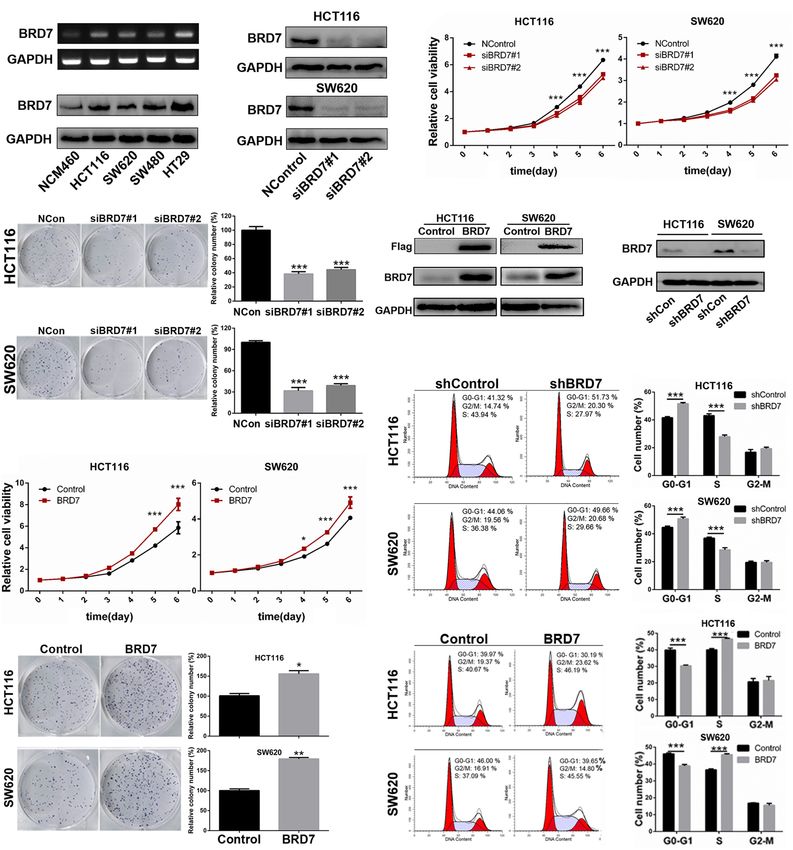
Zhao et al.                                                                                                                 BRD7/c-Myc Axis in CRC Progression

TABLE 1 | Association between the expression of BRD7, c-Myc, and CRC clinical pathological features (N = 180).

Characteristics (N)                            BRD7                                         c-Myc                                     BRDT/c-Myca

                              H (%)            L (%)                P         H (%)           L (%)              P       H–H (%)          L–L (%)            P

Age (year)
Zhao et al.                                                                                                                    BRD7/c-Myc Axis in CRC Progression

  FIGURE 1 | BRD7 promotes azoxymethane/dextran sodium sulfate (AOM/DSS)-induced colitis-associated carcinogenesis in mice. (A) Schematic representation of
  the AOM/DSS-induced colitis-associated colorectal cancer (CRC) protocol. (B) disease activity index (DAI) score of BRD7+/+ and BRD7−/− mice following
  AOM/DSS treatment, n = 20. (C) The number of BRD7+/+ and BRD7−/− mice harboring tumor nodules. (D) Representative images of the colorectum of BRD7+/+
  and BRD7−/− mice on day 126. (E) Hematoxylin and eosin (H&E) staining of colorectal tissues of BRD7+/+ and BRD7−/− mice. Images in the top panel were
  representative of H&E staining. Scale bar: 50 µm. Images in the bottom panel were magnified from the black box area in the images in the top panel, respectively.
  Scale bar: 20 µm. (F) The number and (G) volume of colorectal tumors in BRD7+/+ and BRD7−/− mice. The results are presented as the mean ± SD. *P < 0.05,
  **P < 0.01, ***P < 0.001.

(GV115/shBRD7) and shRNA control (shControl) from                                    normalized relative to those of GAPDH using the 2−11Ct
GeneChem (Shanghai, China). HCT116 and SW620 cells were                              method. PCR was performed using PCR Mix (BioTeke, Beijing,
infected with the BRD7 shRNA lentiviral vector or shControl                          China). The PCR products were analyzed by electrophoresis
vector. Subsequently, the infected cells were screened using                         through a 1.5% agarose gel. Primer pair sequences for
fluorescence-activated cell sorting with flow cytometry (BD                          BRD7 were 50 -GCTGTTGCACTCAGGAATGA-30 (forward)
Biosciences, MD, United States) to obtain a stable pool of clones.                   and 50 -ACTCTTGAAGGCGTGTGCTT-30 (reverse). Primer pair
                                                                                     sequences for GAPDH were 50 -CGAGATCCCTCCAAAAT
                                                                                     CAA-30 (forward) and 50 -TTCACACCCATGACGAACAT-30
RNA Extraction, qPCR, and Reverse                                                    (reverse). PCRs of each sample were conducted in triplicate.
Transcription-PCR
Total RNA was extracted from the cultured cells using
TRIzol (Invitrogen). cDNA was synthesized from the total
                                                                                     Cell Proliferation (Cell Counting Kit-8),
RNA using a reverse transcription system (Fermentas, Glen                            5-Ethynyl-2’-Deoxyuridine Incorporation,
Burnie, MD, United States) according to the manufacturer’s                           and Colony Formation Assays
instructions. GAPDH was amplified in parallel as an internal                         Cells were seeded onto 96-well plates (1,000 cells/well). A cell
control. The expression level of each gene was quantified                            growth curve was constructed using a Cell Counting Kit-8
by measuring the cycle threshold (Ct) values, which were                             (CCK-8; Bimake, Houston, TX, United States). Absorbance

Frontiers in Cell and Developmental Biology | www.frontiersin.org                4                                              May 2021 | Volume 9 | Article 659392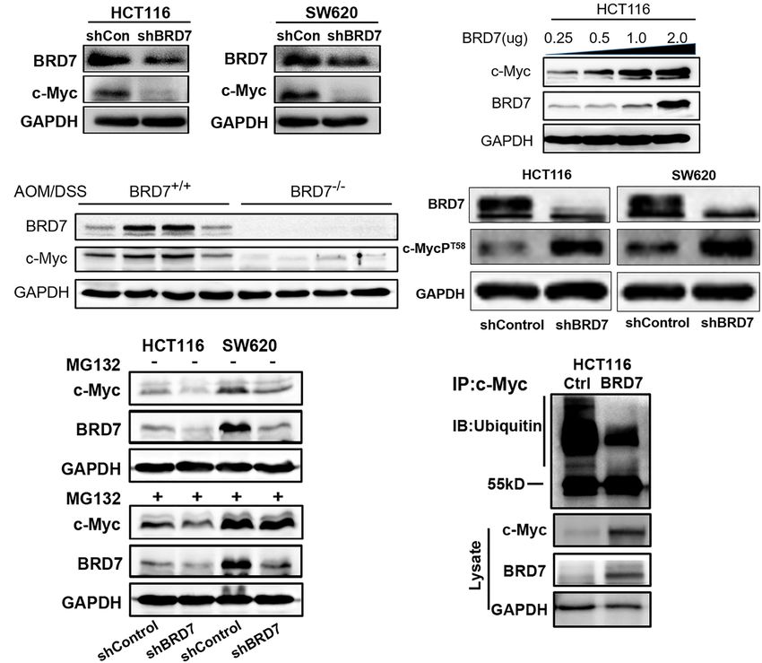
Zhao et al.                                                                                                BRD7/c-Myc Axis in CRC Progression

was measured at 450 nm using a microplate spectrometer.                 immunoprecipitation (IP) buffer and then were sonicated,
For EdU cell proliferation assays, EdU incorporation                    and the lysates were centrifuged. The supernatant was
assays were performed using a Cell-Light EdU Apollo567                  subjected to IP assay with anti-c-Myc (CST). Subsequently,
in vitro Kit (RiboBio, Guangzhou, China) according to the               the sample was subjected to western blotting with an
manufacturer’s protocol.                                                anti-ubiquitin antibody (CST).
   For colony formation, 500 cells were seeded into a 6-well
plate, with three wells per sample. After a 14-day incubation,          Immunofluorescence and
the cells were fixed with 4% paraformaldehyde and stained with          Co-Immunoprecipitation
Giemsa solution (Beyotime, Beijing, China). Colonies containing         For immunofluorescence (IF), BRD7-overexpressing HEK293
more than 50 cells were counted as 1 positive colony. The               and HCT116 cells were seeded on glass coverslips, fixed with
plate clone formation efficiency was calculated as (number of           4% paraformaldehyde for 15 min at room temperature, and
colonies/number of cells inoculated) × 100%. All experiments            then permeabilized with 0.3% Triton X-100 for 5 min at room
were performed in triplicate.                                           temperature. Cells were washed three times with TBS, incubated
                                                                        for 15 min in 5% goat serum, and then incubated at 4◦ C overnight
Flow Cytometry for Cell Cycle and                                       with the anti-c-Myc primary antibody (dilution 1:800; CST).
Apoptosis                                                               After three 5-min washes with TBS, cells were incubated for
The cell cycle and apoptosis analysis was described in our              1 h at 37◦ C in the dark with the Alexa Fluor 568 donkey
previous work (Liu et al., 2016). Briefly, cells were harvested         anti-Rabbit IgG (H + L) secondary antibody (dilution 1:1,000;
and fixed in 70% ethanol for 24 h at -20◦C, then treated with           Invitrogen). Coverslips were washed with TBS and visualized
RNase A, and stained with 25 µg/ml of propidium iodide (PI)             using an epifluorescence microscope.
(Beyotime). For cell apoptosis analysis, the ratio of apoptotic            For Co-IP, HEK293T or HCT116 cells were transfected with
cells was determined using an Annexin V-PE/7-AAD double                 pIRESneo3/2Flag-c-Myc or pCMV-HA-BRD7 expression vector
staining kit (BD). Samples were all analyzed using a MoFlo              and their truncated mutants together, respectively. Cells were
XDP High-Performance Cell Sorter (Beckman Coulter, CA,                  lysed with cell lysis buffer for western blotting and IP (Beyotime,
United States), and the data were analyzed using Summit v.5.2           Beijing, China). They were then premixed with protease inhibitor
software according to the manufacturer’s protocol. At least three       cocktail (Roche) on ice for 20 min and centrifuged at 12,000 g for
independent experiments were performed.                                 15 min. The supernatants were incubated at 4◦ C overnight with
                                                                        30 µl of anti-Flag or anti-HA magnetic beads (Biomake) and anti-
Western Blot Analysis                                                   mouse IgG (Santa Cruz). The immunocomplexes were isolated
Briefly, cells were lysed in radioimmunoprecipitation assay             and purified according to the manufacturer’s protocol and then
(RIPA) buffer containing both Protease Inhibitor Cocktail               subjected to western blot analysis.
and PhoSTOP (Roche, Basel, Switzerland). The total proteins
were quantified using the BCA Protein Assay Kit (Pierce                 HCT116 and SW620 Tumor Xenograft
Biotechnology, Rockford, IL, United States), separated by sodium        Model
dodecyl sulfate–polyacrylamide gel (SDS-PAGE) electrophoresis,          A total volume of 100 µl of BRD7 stable knockdown HCT116
and transferred onto polyvinylidene fluoride (PVDF) membranes           and SW620 cells or control cells (5 × 10⁶ cells) was inoculated
(Millipore, Billerica, MA, United States). The membranes were           subcutaneously into the right flanks of 6-week-old female
blocked with 5% non-fat milk in Tris-buffered saline and then           nude mice. Mice were checked every 4 days, and tumor
incubated with primary antibodies at 4◦ C overnight. The primary        nodules were measured with a caliper. Tumor volume was
antibodies used were anti-BRD7 (dilution 1:500; ProteinTech,            evaluated as described in our previous work (Liu et al., 2016).
Wuhan, China), anti-c-Myc (dilution 1:1,000; CST, Danvers,              Tumor growth curves were calculated. Mice in the three
MA, United States), anti-Flag (dilution 1:2,000; Sigma, MO,             experimental groups were killed after 24 days. All tumors
United States), anti-HA (dilution 1:2,000; ProteinTech), anti-          were excised, weighed, harvested, fixed, and embedded in
GAPDH (dilution 1:5,000; ProteinTech), anti-Phospho-c-Myc               paraffin. The anti-Ki-67 antibody (dilution 1:100; Bioworld
(Thr58) (dilution 1:1,000; CST), anti-CDC25A (dilution 1:500;           Technology, Atlanta, GA, United States) was used to detect
ProteinTech), anti-CCND1 (dilution 1:500; Santa Cruz), and              the proliferation marker Ki-67 using immunohistochemistry
anti-CDK4 (dilution 1:500; Santa Cruz). The membranes were              (IHC) procedures. Samples were observed using an Olympus
then washed three times in TBST solution for 10 min each                microscope (Olympus, Tokyo, Japan).
time and then incubated with secondary antibodies. Signals were
detected with an enhanced chemiluminescence detection system            Immunohistochemistry Staining and
(Bio-Rad, Hercules, CA, United States).                                 Scores
                                                                        IHC staining was described in our previous work (Zhao
Deubiquitination Assay of c-Myc Protein                                 et al., 2017). Briefly, the sections were rehydrated, and antigen
BRD7-overexpressing HCT116 cells or control cells were                  retrieval was performed by boiling in 0.01 M of citrate buffer
treated with the proteasome inhibitor MG132 (Selleck,                   (pH 6.0). After inhibition of endogenous peroxidase activity,
Houston, United States) for 3 h. The cells were lysed with              the sections were blocked with 2% bovine serum albumin

Frontiers in Cell and Developmental Biology | www.frontiersin.org   5                                       May 2021 | Volume 9 | Article 659392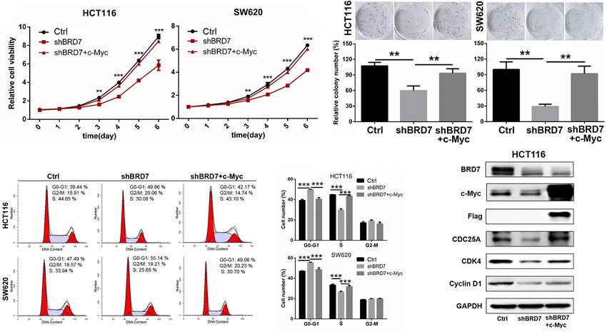
Zhao et al.                                                                                                 BRD7/c-Myc Axis in CRC Progression

in phosphate-buffered saline (PBS) and then incubated with              a quick reduction in the DAI until the mice were euthanized
anti-BRD7 (dilution 1:100; ProteinTech), anti-c-Myc (dilution           (Figure 1B); these results demonstrated that BRD7+/+ mice
1:100; Millipore), anti-CDK4 (dilution 1:100; Santa Cruz), anti-        suffered more severe colitis after the 14th day, suggesting that the
CCND1 (dilution 1:100; Santa Cruz), and anti-CDC25A (dilution           deficiency of BRD7 plays a protective role in the middle-late stage
1:100; ProteinTech) antibodies. The immune complex was                  of CAC. More surprisingly, 70% of BRD7+/+ mice displayed
visualized with the MaxVison HRP-polymer IHC Kit Detection              tumor nodules in the colorectum, while no tumor nodules were
System, Peroxidase/DAB, Rabbit/Mouse (MaxVison, Fuzhou,                 observed in the BRD7−/− group (Figures 1C,D). Furthermore,
China) according to the manufacturer’s protocol. The nuclei             we performed H&E staining and found that BRD7−/− mice
were counterstained with hematoxylin (Beyotime). Positive               developed low-grade dysplasia, while BRD7+/+ mice displayed
control slides were included in every experiment in addition            high-grade dysplasia characterized by loss of epithelial polarity
to the internal positive control. The specificity of the antibody       (Figure 1E) and an increased tumor multiplicity in the colons,
was determined using a matched IgG isotype antibody as a                measured as the sum of tumor numbers and tumor volume,
negative control.                                                       respectively (Figures 1F,G). To exclude the genetic inactivation
   For IHC scores, IHC staining was evaluated at                        caused by chemical-induced mutations, we detected the ORF
200 × magnification using light microscopy and independently            sequences of BRD7 in tumors of BRD7+/+ mice and confirmed
assessed by two pathologists who were blinded to the                    that there were no mutants in the BRD7 ORF as shown in
clinicopathological data. The score of BRD7 and c-Myc                   Supplementary Figure 1, which suggest that the mechanism
protein in nucleus compartment was calculated according to              that BRD7 promotes tumor development and that growth is
the staining intensity and extent of staining using a method            not dependent on the loss of a tumor-suppressive role resulted
described in our previous work (Liu et al., 2018).                      from AOM/DSS-driven BRD7 mutation but exerts oncogenic
                                                                        roles. Taken together, all of the findings above revealed that
Statistical Analysis                                                    BRD7+/+ mice are highly susceptible to colitis-associated CRC
The correlation between the expression of BRD7 and c-Myc                and that BRD7 plays a crucial cancer-promoting role in colorectal
protein and the clinicopathological characteristics of CRC              carcinogenesis in mice.
patients were evaluated using the chi-square test. Spearman’s
rank correlation coefficient was used to assess the significance        BRD7 Promotes Cell Proliferation by
of the association between the expression of BRD7 and c-Myc
in CRC. Kaplan–Meier analysis was performed to generate OS              Accelerating the G1/S Phase Transition
curves, and statistical significance was assessed using the log-        in Human Colorectal Cancer Cells
rank test. The differences between the groups were analyzed             Then, we quantified the expression of BRD7 in the human CRC
using Student’s t-test when there were only two groups or using         cell lines HCT116, SW620, SW480, and HT29 and the normal
one-way ANOVA when there were more than two groups. All                 colon mucosal epithelial cell line NCM460. The RT-PCR and
statistical analyses were performed using SPSS software (SPSS,          western blotting results showed that the mRNA and protein
Chicago, IL, United States). A two-tailed value of P < 0.05 was         expression of BRD7 were both markedly upregulated in CRC cell
considered statistically significant.                                   lines when compared with NCM460 (Figure 2A), and HCT116
                                                                        and SW620 cells were therefore chosen as the cell models for
                                                                        investigation of BRD7 function and mechanism based on their
RESULTS                                                                 moderate expression among these CRC cell lines. To further
                                                                        confirm the role of BRD7 in CRC progression, we transiently
Knockout of BRD7 Inhibits                                               transfected HCT116 and SW620 cell lines with BRD7-siRNAs
Tumorigenesis and Tumor Growth in                                       (siBRD7#1 and siBRD7#2) and determined the endogenous
                                                                        levels of BRD7 mRNA and protein, respectively. The results
Azoxymethane/Dextran Sodium                                             showed that the levels of endogenous BRD7 expression were
Sulfate-Induced Colorectal Cancer                                       significantly downregulated in both HCT116 and SW620 cell
Model                                                                   lines (Supplementary Figure 2A and Figure 2B). Subsequently,
Previous studies have reported that BRD7 is aberrantly of low           we performed CCK-8 and colony forming assays to determine
levels of expression in many malignancies and significantly             the effect of BRD7 knockdown on cell proliferation, and the
associated with malignant features and poor prognosis (Park             results showed that BRD7 knockdown significantly inhibited
Y. A. et al., 2014; Liu et al., 2018; Ma et al., 2019). To              cell proliferation and colony formation in both HCT116 and
explore the functional role of BRD7 in CRC progression, we              SW620 cell lines (Figures 2C,D). In contrast, ectopic expression
investigated the effect of BRD7 knockout on AOM/DSS-induced             of BRD7 markedly promoted cell proliferation in the two cell
colitis-associated carcinogenesis (CAC) in mice. BRD7+/+ and            lines (Figures 2E–G).
BRD7−/− mice were injected with AOM and then subjected to                  To further illuminate the cell proliferation-promoting role
three rounds of 2% DSS exposure to induce CRC (Figure 1A).              of BRD7 in CRC cells, we further constructed BRD7 stable
As a result, BRD7−/− mice show an increased DAI before the              knockdown HCT116 and SW620 cell lines based on the
14th day than did BRD7+/+ mice, which is consistent with our            most efficient siRNA (siBRD7#2) (Supplementary Figure 2B
previous results (Zhao et al., 2017), but this was followed by          and Figure 2H) and examined the effect of stable BRD7

Frontiers in Cell and Developmental Biology | www.frontiersin.org   6                                       May 2021 | Volume 9 | Article 659392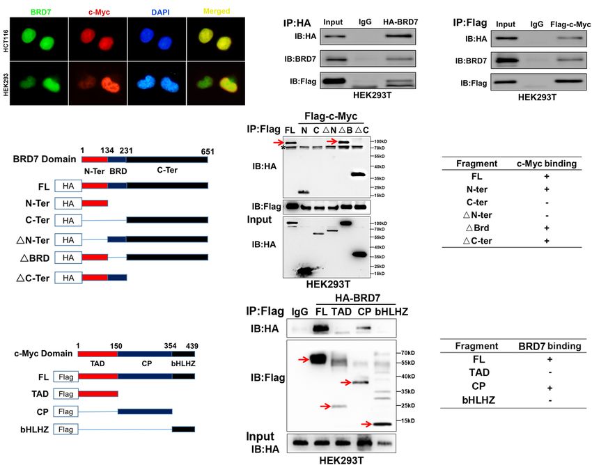
Zhao et al.                                                                                                                     BRD7/c-Myc Axis in CRC Progression

  FIGURE 2 | Effect of BRD7 on the cell proliferation and cell cycle of colorectal cancer (CRC) cells. (A) BRD7 mRNA and protein expression levels were detected by
  RT-PCR and western blotting in HCT116, SW620, SW480, and HT29 CRC cell lines and the normal colon mucosal epithelial cell line NCM460. (B) Western blotting
  confirmed the interference efficiency of BRD7 siRNAs on endogenous BRD7 protein levels in HCT116 and SW620 cells. (C,F) The effect of BRD7 knockdown and
  overexpression on cell viability detected by CCK-8 assays. (D,G) The effect of BRD7 knockdown and overexpression on the colony formation of HCT116 and
  SW620 cells. (E,H) Western blot analysis confirmed the BRD7 protein expression levels in HCT116 and SW620 cells with stable BRD7 overexpressing or
  knockdown, respectively. (I,J) The effect of BRD7 stable knockdown or overexpression on cell cycle progression, as determined by flow cytometry. The results are
  presented as the mean ± SD. *P < 0.05, **P < 0.01, ***P < 0.001.

knockdown and overexpression on cell cycle progression in                             or SW620 cells (Supplementary Figures 2E,F). Taken together,
these two cell lines. The results showed that BRD7 knockdown                          these results demonstrated that BRD7 functions as a promoting
caused significant G0/G1 phase arrest (Figure 2I), while ectopic                      factor in CRC cell proliferation by accelerating the G1/S phase
expression of BRD7 markedly promoted G1/S phase transition                            transition of the cell cycle.
(Figure 2J). Furthermore, we performed EdU incorporation
assays to confirm the role of BRD7 in promoting cell cycle
progression, and the results showed that BRD7 knockdown
                                                                                      BRD7 Increases the Stability of c-Myc
significantly reduced EdU incorporation in both HCT116                                Protein by Decreasing the Ubiquitination
and SW620 cell lines (Supplementary Figure 2C) and that                               of c-Myc
BRD7 overexpression played the opposite role (Supplementary                           As c-Myc is aberrantly overexpressed or hyperactivated and
Figure 2D). In addition, BRD7 knockdown or overexpression                             exerts its pro-oncogenic roles in more than 50% of malignancies
had no significant effect on cell apoptosis in either HCT116                          (Zwolinska et al., 2012), including CRC, we detected the

Frontiers in Cell and Developmental Biology | www.frontiersin.org                 7                                             May 2021 | Volume 9 | Article 659392
Zhao et al. BRD7/c-Myc Axis in CRC Progression
FIGURE 3 | BRD7 enhances the stability of the c-Myc protein by inhibiting its ubiquitination. (A) Western blot analysis detected the effect of BRD7 knockdown on
c-Myc protein expression in HCT116 and SW620 cell lines. (B) The effect of BRD7 overexpression on c-Myc protein in HCT116 cells at different doses. (C) The
expression of BRD7 and c-Myc protein in the colorectal tissues of azoxymethane/dextran sodium sulfate (AOM/DSS)-induced colorectal cancer (CRC) mice (N = 4).
(D) Western blot analysis showed the effect of BRD7 knockdown on the phosphorylation levels of c-Myc protein at T58 sites in both HCT116 and SW620 cells.
(E) Western blot analysis showed the effect of BRD7 knockdown on ubiquitin–proteasome-mediated protein stability of c-Myc in MG132-treated HCT116 and
SW620 cells. MG132, proteasome inhibitor MG132. (F) Immunoprecipitation (IP) and western blot assays confirmed the effect of BRD7 on the ubiquitination of
c-Myc protein.
effect of BRD7 on c-Myc expression. The expression of c-Myc the cells were treated with proteasome inhibitor, MG132, in
protein was significantly downregulated in BRD7 knockdown both BRD7 knockdown HCT116 and SW620 cells (Figure 3E).
HCT116 and SW620 cells (Figure 3A), and the half-life of Moreover, ectopic expression of BRD7 significantly reduced the
c-Myc protein was markedly reduced by BRD7 knockdown level of c-Myc ubiquitination (Figure 3F). Taken together, our
(Supplementary Figure 3). Accordingly, ectopic expression of findings demonstrated that BRD7 can stabilize c-Myc protein by
BRD7 increased c-Myc protein expression in a dose-dependent inhibiting its ubiquitination dependent on its phosphorylation at
manner (Figure 3B), while there were no significant changes the T58 site. In addition, we examined whether FBW7 mediates
in the mRNA level of c-Myc (Supplementary Figure 4). In the degradation of c-Myc after BRD7 knockdown. We found
addition, the expression of c-Myc protein was significantly that BRD7 knockdown decreases c-Myc protein level, which
upregulated in BRD7+/+ group than the BRD7−/− group is consistent with the previous results, but when FBW7 was
of AOM/DSS-induced CRC model (Figure 3C). These results depleted in BRD7 knockdown HCT116 cells, the stability of
indicate that BRD7 might be involved in the regulation of c-Myc is clearly recovered (Supplementary Figure 5). The results
c-Myc stability. As c-Myc T58 phosphorylation is tightly suggested that FBW7 is involved in the stabilization of c-Myc
correlated to the degradation of c-Myc through the proteasome mediated by BRD7.
system (Sun et al., 2015; Wang W. et al., 2016), we therefore
detected the phosphorylation level of c-Myc protein, and
the immunoblotting results showed that BRD7 knockdown
BRD7 Interacts With c-Myc in a Manner
significantly increased the c-Myc T58 phosphorylation levels Dependent on the N-Terminus of BRD7
(Figure 3D). To further determine the effect of BRD7 on and the CP-Domain of c-Myc
c-Myc protein stability, we explored the functional role of Since BRD7 increased c-Myc protein stability through the
BRD7 in c-Myc protein degradation mediated by ubiquitin– ubiquitin–proteasome pathway, we investigated whether they
proteasome. As a result, knockdown of BRD7 decreased the interacted with each other. IF and nuclear/cytosol fractionation
protein level of c-Myc consistent with the previous results, assays showed that both BRD7 and c-Myc were predominantly
while c-Myc protein degradation was obviously hindered after colocalized in the nucleus in both HEK293 and HCT116 cells
Frontiers in Cell and Developmental Biology | www.frontiersin.org 8 May 2021 | Volume 9 | Article 659392Zhao et al. BRD7/c-Myc Axis in CRC Progression
FIGURE 4 | BRD7 interacts with c-Myc in a manner dependent on the N-terminus of BRD7 and the CP-domain of c-Myc. (A) Immunofluorescence assays showed
the localization of BRD7 and c-Myc in HCT116 and HEK293 cells. (B,C) Co-immunoprecipitation (Co-IP) confirmed the interaction between BRD7 and c-Myc in
HEK293T cells. (D,G) The schematic diagram of different truncated mutants of BRD7 and c-Myc domains. (E,F,H,I) Co-IP assays revealed the structural basis of the
interaction between BRD7 and c-Myc. The antibody used for IP detection in (E,H) is anti-Flag antibody, and the antibodies used for IB detection are anti-HA and
anti-Flag antibodies, respectively. Of them, Flag was used to tag c-Myc and its mutants, while HA was used to tag BRD7 and its mutants. FL, full length; TAD,
transcription activation domain; CP, central portion flanked by TAD and bHLHZ; bHLHZ, basic helix-loop-helix leucine zipper; N-Ter/N, N-terminal; C-Ter/C,
C-terminal; 1N-Ter/1N, N-terminal deletion; 1BRD, bromodomain deletion; 1C-Ter/1C, C-terminal deletion. The arrow indicates the destination bands, and
* indicates non-specific bands.
(Figure 4A and Supplementary Figure 6). In addition, Co-IP and colony formation assays showed that the restoration of c-Myc
assays showed that BRD7 could interact with c-Myc in HEK293T levels could significantly rescue the cell viability and colony
or HCT116 cells (Figures 4B,C and Supplementary Figure 7). To formation inhibition induced by BRD7 knockdown in HCT116
further determine the structural basis of the protein interaction and SW620 cell lines when compared with that in the control
between BRD7 and c-Myc, we constructed domain-truncated groups (Figures 5A,B). Moreover, the flow cytometry results
mutants of BRD7 and c-Myc (Figures 4D,G) and then further revealed that restoring c-Myc expression could markedly reverse
confirmed the interaction between different domains of BRD7 the inhibitory effect of BRD7 knockdown on G1/S cell cycle
and c-Myc. The Co-IP results showed that BRD7 interacts with arrest (Figure 5C). Increasing evidence has shown that CDC25A,
c-Myc in a manner dependent on N-terminus of BRD7 and the CDK4, and cyclin D1, as c-Myc downstream targets, play crucial
CP-domain of c-Myc (Figures 4E,F,H,I). roles in cell cycle progression, especially in the G1/S phase
transition (Macdonald and Bennett, 1999; Bouillez et al., 2016).
Restoring c-Myc Levels Reverses the Therefore, we detected the expression of some critical molecules
Effect of BRD7 Knockdown on the involved in the G1/S phase transition of the cell cycle, and the
immunoblotting results showed that the protein expression of
Inhibition of Cell Proliferation and Cell CDC25A, CDK4, and cyclin D1 was significantly downregulated
Cycle Progression in Colorectal Cancer in BRD7 knockdown HCT116 cells compared with control cells,
Cells while the restoration of c-Myc expression markedly rescued the
To further confirm our hypothesis that BRD7 promotes cell levels of CDC25A and CDK4 in BRD7 knockdown HCT116 cells
proliferation through stabilizing c-Myc protein, we investigated (Figure 5D). Taken together, BRD7 promotes cell proliferation
the effect of restored expression of c-Myc on BRD7 knockdown- and cell cycle G1/S transition at least partially by stabilizing
induced cell proliferation inhibition and cell cycle arrest. CCK-8 c-Myc protein.
Frontiers in Cell and Developmental Biology | www.frontiersin.org 9 May 2021 | Volume 9 | Article 659392Zhao et al. BRD7/c-Myc Axis in CRC Progression
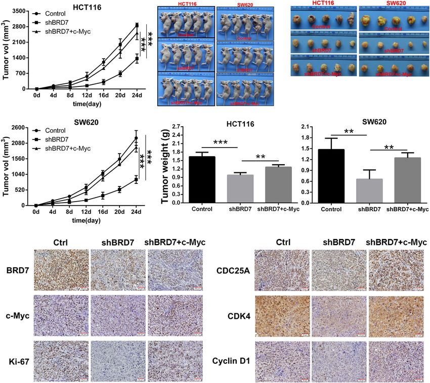
FIGURE 5 | Restoring the expression of c-Myc reverses BRD7 knockdown-induced effects on cell proliferation and cell cycle progression in colorectal cancer (CRC)
cells. (A) The effect of restoring c-Myc expression on BRD7 knockdown-induced cell proliferation inhibition, (B) cell colony formation, and (C) cell cycle G1/S phase
arrest. (D) Western blotting confirmed the key molecules regulated by the BRD7/c-Myc axis. The results are presented as the mean ± SD. **P < 0.01, ***P < 0.001.
Targeting c-Myc Attenuates the Effect of in CRC tissues derived from 180 patients with different clinical
BRD7 on Tumor Growth in vivo stages (Table 1). Most patients exhibited enhanced expression
of BRD7, which was significantly associated with differentiation
To further confirm the functional roles of the BRD7/c-Myc
grade (Table 1). Additionally, the expression of BRD7 and
axis in tumor growth and progression, we established a
c-Myc was markedly associated with the clinical stage of CRC
xenograft model in nude mice with BRD7 knockdown and
patients (Table 1 and Figure 7A). The expression of c-Myc was
BRD7 knockdown with simultaneous c-Myc restoration in
significantly positively correlated with the expression of BRD7
HCT116 and SW620 cells. We found that BRD7 knockdown
in CRC patients (P = 0.006, r = 0.205, Table 2). Moreover, the
significantly inhibited tumor growth in both HCT116 and
survival analysis showed that CRC patients with high levels of
SW620 cells (Figure 6A). In addition, the tumor weight was
BRD7 or c-Myc expression had poor prognoses (Figures 7B,C).
markedly reduced in the BRD7 knockdown group compared
In addition, CRC patients with high expression of both BRD7 and
with the control group (Figures 6B–D), while restoring c-Myc
c-Myc had unfavorable prognoses (Figure 7D). These findings
expression reversed BRD7 knockdown-mediated tumor growth
indicated that both BRD7 and c-Myc play critical roles in CRC
inhibition in both HCT116 and SW620 cells (Figures 6A–D).
progression and that the BRD7/c-Myc axis could serve as a
These results demonstrated that BRD7 promotes CRC tumor
potential biomarker and therapeutic target for the early diagnosis
growth at least partially by targeting c-Myc stability in vivo.
and treatment of CRC patients.
In addition, we further determined the expression of key
molecules downstream of the BRD7/c-Myc axis, and the results
showed that BRD7 knockdown significantly downregulated
the expression of c-Myc, CDC25A, CDK4, and cyclin D1
DISCUSSION
(Figure 6E), while restoring c-Myc expression attenuated BRD7 Increasing evidence shows that BRD7 is widely downregulated
knockdown-induced downregulation of CDC25A, CDK4, and and contributes to tumorigenesis in many malignancies (Chiu
cyclin D1 (Figure 6E). Taken together, our findings demonstrated et al., 2014; Gao et al., 2016; Yu et al., 2016; Hu et al., 2019).
that targeting c-Myc attenuates the effect of BRD7 on tumor BRD7 functions as a tumor suppressor, and low expression of
growth and cell cycle progression in vivo. BRD7 serves as an independent factor for prognosis and is
positively associated with the clinical stage of cancer progression
A High Level of BRD7 Is Positively (Liu et al., 2016, 2018). In our previous study, we investigated the
impact of BRD7 knockout (BRD7−/− ) on DSS-induced colitis
Associated With c-Myc Expression, and found that BRD7 plays an anti-inflammatory role during
Clinical Stage, and Poor Prognosis in early acute colitis in mice (Zhao et al., 2017); the results showed
Colorectal Cancer Patients that DAI was significantly increased in DSS-induced acute colitis
To explore the clinical significance of the BRD7/c-Myc axis, the in BRD7−/− mice from 1 to 5 days, consistent with its tumor-
expression of BRD7 and c-Myc was assessed by IHC staining suppressive function in carcinogenesis. In this study, we further
Frontiers in Cell and Developmental Biology | www.frontiersin.org 10 May 2021 | Volume 9 | Article 659392Zhao et al. BRD7/c-Myc Axis in CRC Progression FIGURE 6 | Restoring c-Myc expression reverses BRD7 silencing-induced inhibition of tumor growth. (A) Growth curve of HCT116 and SW620 xenograft tumors from nude mice. (B) Images of the HCT116 and SW620 xenograft tumors from nude mice. (C) Tumor images (N = 5). (D) Tumor weight quantification (N = 5). (E) Immunohistochemistry (IHC) confirmed the expression of key molecules downstream of the BRD7/c-Myc axis in vivo. Scale bar: 50 µm. The results are presented as the mean ± SD. **P < 0.01, ***P < 0.001. employed a well-established AOM/DSS model of CAC in BRD7 which is also a novel finding for BRD7 functions. Actually, more knockout mice. Unexpectedly, DAI was markedly reduced in and more genes with dual functions as a tumor suppressor or the late stage of AOM/DSS-induced CRC in BRD7−/− mice, an oncogene in cancer development and progression have been and 70% of BRD7+/+ mice developed CAC, while there were identified, such as Yin Yang 1 (YY1) (Sarvagalla et al., 2019) no tumors in the BRD7−/− group on day 126, indicating that and TGF-β (Xu J. et al., 2015), and what functions they play BRD7 plays a pro-inflammatory role and potentially plays an may depend on the type of tumors as well as the genotype and oncogenic role in the late stage of AOM/DSS-induced CRC expression of some critical genes in tumor cells. For example, YY1 in mice. Moreover, BRD7 was markedly upregulated in CRC was identified as a critical transcription factor, which has been cells, and BRD7 knockdown inhibited cell proliferation, colony implicated as a major driver of many cancers, while recent reports formation, and G1/S phase progression in two CRC cell lines. The indicated that YY1 also functions as a tumor suppressor, and in vivo assays also revealed that BRD7 promotes cell proliferation the mechanism by which YY1 brings out opposing outcome in and tumor growth through facilitating the G1/S phase transition tumor growth vs. suppression is not completely clear (Sarvagalla of the cell cycle. All of these findings demonstrate that BRD7 et al., 2019). Therefore, it could be understood that BRD7 has dual could significantly inhibit the occurrence of early inflammation functions as a tumor suppressor and an oncogene. and exert a tumor suppressor role but significantly promote BRD7 belongs to the bromodomain-containing protein the process of colorectal carcinogenesis in the late stage. It is family; it is primarily localized in the cell nucleus (Zhou et al., suggested that BRD7 has a significant “functional duality” in 2006; Wang et al., 2014) and serves as a crucial transcriptional different stages of the occurrence and development of CRC, and regulatory factor (Burrows et al., 2010; Xu et al., 2016; Liu et al., it is different from the role of tumor inhibition in other tumors, 2017). Accumulating evidence has revealed that BRD7 is involved Frontiers in Cell and Developmental Biology | www.frontiersin.org 11 May 2021 | Volume 9 | Article 659392
Zhao et al.                                                                                                                     BRD7/c-Myc Axis in CRC Progression

TABLE 2 | BRD7 expression were positively correlated with c-Myc expression in         and promising therapeutic target. Unfortunately, although the
CRC patients (N = 180).
                                                                                      functional role and mechanism of c-Myc is well-defined in
                                  BRD7 expression                                     promoting cell proliferation in many cancers, no anti-cancer
                                                                                      strategies that directly target c-Myc protein are available for
                                  High       Low       P             r                clinical cancer treatment (Koh et al., 2016), owing to its
c-Myc expression                                                                      “undruggable” protein structure (Bachmann and Geerts, 2018;
                          High    52         33        0.006         0.205            Yoshida, 2018; Wang et al., 2019). Interestingly, we found that
                          Low     44         51                                       BRD7 knockdown significantly downregulates c-Myc protein
                                                                                      levels and reduces the half-life of c-Myc protein. Mechanistically,
CRC, colorectal cancer.
                                                                                      BRD7 interacts with c-Myc protein through the N-terminus of
                                                                                      BRD7 and the CP domain of c-Myc and therefore potentially
in cell cycle arrest via regulation of Ras/Raf/MEK/ERK, Rb/E2F,                       competitively inhibits T58 phosphorylation of c-Myc triggered
and AKT signaling in tumorigenesis and cancer progression                             by GSK3β, whose activity is regulated by PI3K/Akt signaling.
(Zhou et al., 2004; Liu et al., 2016; Zhang Q. et al., 2016). In the                  Then dephosphorylation of T58 stabilizes c-Myc by inhibiting
present study, our results show that c-Myc downstream targets                         T58 phosphorylation-dependent ubiquitination and degradation
CDC25A, CDK4, and cyclin D1 are significantly downregulated                           through the proteasome system (Hann, 2006; Sun et al., 2015).
in BRD7 knockdown CRC cells. Furthermore, the restoration                             In addition, FBW7 was reported to be the E3 ubiquitin ligase
of c-Myc expression markedly reversed the suppressive effects                         involved in this process of T58 dephosphorylation stabilizing
of BRD7 knockdown on the proliferation of CRC cell lines                              c-Myc protein (Welcker et al., 2004; Yada et al., 2004). Therefore,
in vitro and in vivo. These findings demonstrate that BRD7                            we speculated that FBW7 might be the E3 ubiquitin ligase
exerts its oncogenic role at least partially through c-Myc signaling                  involved in the stabilization of c-Myc mediated by BRD7; the
and that the BRD7/c-Myc axis plays an important role in                               detail mechanism needs to be further confirmed in the future
CRC progression.                                                                      work. Importantly, both BRD7 and c-Myc are significantly
   c-Myc, which serves as a crucial transcription factor, regulates                   upregulated in CRC patients, and the expression of BRD7
over 15% of genes of the human transcriptome (Dang et al., 2006;                      is positively associated with the expression of c-Myc protein;
Kress et al., 2015) and is ectopically expressed or hyperactivated                    high expression of these proteins indicates advanced clinical
in a wide variety of malignancies (Wolf et al., 2015; Xiao                            stage and poor prognosis in CRC patients. Therefore, based on
et al., 2017; Jung et al., 2018; Li et al., 2019), including CRC;                     our research, the development of anti-tumor small molecule
and c-Myc knockdown significantly inhibited cell proliferation                        inhibitors targeting BRD7/c-Myc axis could be an attractive
and tumor growth (Niu et al., 2015; Li et al., 2017; Wang                             approach in solving the problem of c-Myc “undruggable” and the
et al., 2018), revealing that c-Myc could be a broadly applicable                     clinical targeted therapy of CRC.

  FIGURE 7 | The expression and clinical significance of BRD7 and c-Myc in colorectal cancer (CRC) patients. (A) The expression of BRD7 and c-Myc protein in
  patients with different clinical stages of CRC, as detected by immunohistochemistry (IHC). Scale bar: 100 µm. (B–D) Kaplan–Meier analysis was used to plot the
  overall survival (OS) curve of CRC patients according to the expression of BRD7 and c-Myc.

Frontiers in Cell and Developmental Biology | www.frontiersin.org                12                                             May 2021 | Volume 9 | Article 659392
Zhao et al. BRD7/c-Myc Axis in CRC Progression
In summary, our study demonstrated that BRD7 exerts JY, HL, and YX collected the samples. RZ and YL wrote the
an oncogenic role and promotes CRC progression through manuscript. MZ, RZ, YL, YX, HL, ZZ, WX, XL, and GL reviewed
inhibiting ubiquitin–proteasome-mediated degradation of the and polished the manuscript. RZ, YL, and SF analyzed the data.
c-Myc protein. Both BRD7 and c-Myc are significantly All authors read and approved the final manuscript.
upregulated in CRC patients, and the expression of BRD7 is
positively associated with high levels of c-Myc protein; the high
expression of these proteins indicates advanced clinical stage and FUNDING
poor prognosis in CRC patients. More importantly, a potent
and selective inhibitor for BRD7 was reported recently (Clark This work was supported by the Grants from the National
et al., 2015), which provides a promising therapeutic option Natural Science Foundation of China (81772990, 81802786, and
for BRD7-driven cancers, revealing that targeting the BRD7/c- 81902815); the Natural Science Foundation of Hunan Province
Myc axis could be an attractive approach for the treatment of (2019JJ40343); the Natural Science Foundation of Shandong
c-Myc-driven cancers. Province (ZR2018BH025 and ZR2019BH044); the Shandong
Medical and Healthy Science Technology Development Program
(2017WS138); the Supporting Fund for Teachers’ research of
DATA AVAILABILITY STATEMENT Jining Medical University (JYFC2018FKJ005); and the “111”
The original contributions presented in the study are included project (111-2-12).
in the article/Supplementary Material, further inquiries can be
directed to the corresponding author/s.
ACKNOWLEDGMENTS
ETHICS STATEMENT We thank the Shanghai Research Center for Biomodel Organisms
(Shanghai, China) for their excellent technical assistance in
The animal study was reviewed and approved by the Institutional generating the BRD7−/− mice.
Animal Care and Use Committee (IACUC) of Central
South University.
SUPPLEMENTARY MATERIAL
AUTHOR CONTRIBUTIONS
The Supplementary Material for this article can be found
MZ designed the project and supervised the research. RZ, YL, online at: https://www.frontiersin.org/articles/10.3389/fcell.2021.
CW, ML, WN, and YW performed the experiments. WW, SF, 659392/full#supplementary-material
REFERENCES Dingar, D., Tu, W. B., Resetca, D., Lourenco, C., Tamachi, A., de Melo, J.,
et al. (2018). Myc dephosphorylation by the Pp1/Pnuts phosphatase complex
Arnold, M., Sierra, M. S., Laversanne, M., Soerjomataram, I., Jemal, A., and Bray, F. regulates chromatin binding and protein stability. Nat. Commun. 9:3502. doi:
(2017). Global patterns and trends in colorectal cancer incidence and mortality. 10.1038/s41467-018-05660-0
Gut 66, 683–691. doi: 10.1136/gutjnl-2015-310912 Drost, J., Mantovani, F., Tocco, F., Elkon, R., Comel, A., Holstege, H., et al. (2010).
Bachmann, A. S., and Geerts, D. (2018). Polyamine synthesis as a target of Myc Brd7 is a candidate tumour suppressor gene required for P53 function. Nat. Cell
oncogenes. J. Biol. Chem. 293, 18757–18769. doi: 10.1074/jbc.TM118.003336 Biol. 12, 380–389. doi: 10.1038/ncb2038
Bouillez, A., Rajabi, H., Pitroda, S., Jin, C., Alam, M., Kharbanda, A., et al. (2016). Gao, Y., Wang, B., and Gao, S. (2016). Brd7 acts as a tumor suppressor gene in lung
Inhibition of Muc1-C suppresses Myc expression and attenuates malignant adenocarcinoma. PLoS One 11:e0156701. doi: 10.1371/journal.pone.0156701
growth in Kras mutant lung adenocarcinomas. Cancer Res. 76, 1538–1548. Hann, S. R. (2006). Role of post-translational modifications in regulating C-Myc
doi: 10.1158/0008-5472.CAN-15-1804 proteolysis, transcriptional activity and biological function. Semin. Cancer Biol.
Burrows, A. E., Smogorzewska, A., and Elledge, S. J. (2010). Polybromo-associated 16, 288–302. doi: 10.1016/j.semcancer.2006.08.004
Brg1-associated factor components Brd7 and Baf180 are critical regulators of Harte, M. T., O’Brien, G. J., Ryan, N. M., Gorski, J. J., Savage, K. I., Crawford,
P53 required for induction of replicative senescence. Proc. Natl. Acad. Sci. N. T., et al. (2010). Brd7, a subunit of Swi/Snf complexes, binds directly to
U.S.A. 107, 14280–14285. doi: 10.1073/pnas.1009559107 Brca1 and regulates Brca1-dependent transcription. Cancer Res. 70, 2538–2547.
Chiu, Y. H., Lee, J. Y., and Cantley, L. C. (2014). Brd7, a tumor suppressor, doi: 10.1158/0008-5472.CAN-09-2089
interacts with P85alpha and regulates Pi3k activity. Mol. Cell 54, 193–202. Hu, K., Wu, W., Li, Y., Lin, L., Chen, D., Yan, H., et al. (2019).
doi: 10.1016/j.molcel.2014.02.016 Poly(Adp-Ribosyl)ation of Brd7 by Parp1 confers resistance to DNA-
Clark, P. G., Vieira, L. C., Tallant, C., Fedorov, O., Singleton, D. C., Rogers, damaging chemotherapeutic agents. EMBO Rep. 20:e46166. doi: 10.15252/
C. M., et al. (2015). Lp99: discovery and synthesis of the first selective Brd7/9 embr.201846166
bromodomain inhibitor. Angew. Chem. Int. Ed. Engl. 54, 6217–6221. doi: 10. Jung, J. H., Jung, D. B., Kim, H., Lee, H., Kang, S. E., Srivastava, S. K., et al. (2018).
1002/anie.201501394 Zinc finger protein 746 promotes colorectal cancer progression via C-Myc
Dang, C. V., O’Donnell, K. A., Zeller, K. I., Nguyen, T., Osthus, R. C., and Li, stability mediated by glycogen synthase kinase 3beta and F-Box and Wd repeat
F. (2006). The C-Myc target gene network. Semin. Cancer Biol. 16, 253–264. domain-containing 7. Oncogene 37, 3715–3728. doi: 10.1038/s41388-018-
doi: 10.1016/j.semcancer.2006.07.014 0225-0
Dekker, E., Tanis, P. J., Vleugels, J. L. A., Kasi, P. M., and Wallace, M. B. (2019). Koh, C. M., Sabo, A., and Guccione, E. (2016). Targeting Myc in cancer therapy:
Colorectal Cancer. Lancet 394, 1467–1480. doi: 10.1016/S0140-6736(19)3 RNA processing offers new opportunities. Bioessays 38, 266–275. doi: 10.1002/
2319-0 bies.201500134
Frontiers in Cell and Developmental Biology | www.frontiersin.org 13 May 2021 | Volume 9 | Article 659392Zhao et al. BRD7/c-Myc Axis in CRC Progression
Kress, T. R., Sabo, A., and Amati, B. (2015). Myc: connecting selective Siegel, R. L., Miller, K. D., and Jemal, A. (2019). Cancer statistics, 2019. CA Cancer
transcriptional control to global RNA production. Nat. Rev. Cancer 15, 593– J. Clin. 69, 7–34. doi: 10.3322/caac.21551
607. doi: 10.1038/nrc3984 Sun, X. X., He, X., Yin, L., Komada, M., Sears, R. C., and Dai, M. S. (2015).
Li, M., Liu, Y., Wei, Y., Wu, C., Meng, H., Niu, W., et al. (2019). Zinc-finger The nucleolar ubiquitin-specific protease Usp36 deubiquitinates and stabilizes
protein Yy1 suppresses tumor growth of human nasopharyngeal carcinoma by C-Myc. Proc. Natl. Acad. Sci. U.S.A. 112, 3734–3739. doi: 10.1073/pnas.
inactivating C-Myc-mediated microrna-141 transcription. J. Biol. Chem. 294, 1411713112
6172–6187. doi: 10.1074/jbc.RA118.006281 Wang, H., Zhao, R., Guo, C., Jiang, S., Yang, J., Xu, Y., et al. (2016). Knockout of
Li, S., Jiang, C., Pan, J., Wang, X., Jin, J., Zhao, L., et al. (2015). Regulation of Brd7 results in impaired spermatogenesis and male infertility. Sci. Rep. 6:21776.
C-Myc protein stability by proteasome activator Reggamma. Cell Death Differ. doi: 10.1038/srep21776
22, 1000–1011. doi: 10.1038/cdd.2014.188 Wang, H., Zhao, R., Zhou, M., Guo, C., Liu, Y., Jiang, S., et al. (2014). Preparation of
Li, X. X., Shi, L., Zhou, X. J., Wu, J., Xia, T. S., Zhou, W. B., et al. (2017). The role polyclonal antibody highly specific for mouse Brd7 protein and its application.
of C-Myc-Rbm38 loop in the growth suppression in breast cancer. J. Exp. Clin. Acta Biochim. Biophys. Sin. 46, 163–166. doi: 10.1093/abbs/gmt131
Cancer Res. 36:49. doi: 10.1186/s13046-017-0521-5 Wang, Q., Zhou, Y., Rychahou, P., Harris, J. W., Zaytseva, Y. Y., Liu, J., et al.
Liu, T., Zhao, M., Liu, J., He, Z., Zhang, Y., You, H., et al. (2017). Tumor (2018). Deptor is a novel target of Wnt/beta-catenin/C-Myc and contributes
suppressor bromodomain-containing protein 7 cooperates with Smads to to colorectal cancer cell growth. Cancer Res. 78, 3163–3175. doi: 10.1158/0008-
promote transforming growth factor-beta responses. Oncogene 36, 362–372. 5472.CAN-17-3107
doi: 10.1038/onc.2016.204 Wang, W., Liao, P., Shen, M., Chen, T., Chen, Y., Li, Y., et al. (2016). Scp1
Liu, Y., Zhao, R., Wang, H., Luo, Y., Wang, X., Niu, W., et al. (2016). Mir-141 regulates C-Myc stability and functions through dephosphorylating C-Myc
is involved in Brd7-mediated cell proliferation and tumor formation through Ser62. Oncogene 35, 491–500. doi: 10.1038/onc.2015.106
suppression of the Pten/Akt pathway in nasopharyngeal carcinoma. Cell Death Wang, X. M., Wang, Y. C., Liu, X. J., Wang, Q., Zhang, C. M., Zhang, L. P.,
Dis. 7:e2156. doi: 10.1038/cddis.2016.64 et al. (2017). Brd7 mediates hyperglycaemia-induced myocardial apoptosis
Liu, Y., Zhao, R., Wei, Y., Li, M., Wang, H., Niu, W., et al. (2018). Brd7 expression via endoplasmic reticulum stress signalling pathway. J. Cell. Mol. Med. 21,
and C-Myc activation forms a double-negative feedback loop that controls the 1094–1105. doi: 10.1111/jcmm.13041
cell proliferation and tumor growth of nasopharyngeal carcinoma by targeting Wang, X. N., Su, X. X., Cheng, S. Q., Sun, Z. Y., Huang, Z. S., and Ou, T. M. (2019).
oncogenic Mir-141. J. Exp. Clin. Cancer Res. 37:64. doi: 10.1186/s13046-018- Myc modulators in cancer: a patent review. Expert Opin. Ther. Pat. 29, 353–367.
0734-2 doi: 10.1080/13543776.2019.1612878
Liu, Z., Yan, M., Liang, Y., Liu, M., Zhang, K., Shao, D., et al. (2019). Nucleoporin Wei, Z., Yoshihara, E., He, N., Hah, N., Fan, W., Pinto, A. F. M., et al. (2018).
Seh1 interacts with Olig2/Brd7 to promote oligodendrocyte differentiation and Vitamin D switches BAF complexes to protect beta cells. Cell 173, 1135–
myelination. Neuron 102, 587–601.e7. doi: 10.1016/j.neuron.2019.02.018 1149.e15. doi: 10.1016/j.cell.2018.04.013
Ma, J., Niu, W., Wang, X., Zhou, Y., Wang, H., Liu, F., et al. (2019). Welcker, M., Orian, A., Jin, J., Grim, J. E., Harper, J. W., Eisenman, R. N.,
Bromodomaincontaining protein 7 sensitizes breast cancer cells to paclitaxel et al. (2004). The Fbw7 tumor suppressor regulates glycogen synthase kinase 3
by activating Bcl2-antagonist/killer protein. Oncol. Rep. 41, 1487–1496. doi: phosphorylation-dependent C-Myc protein degradation. Proc. Natl. Acad. Sci.
10.3892/or.2018.6951 U.S.A. 101, 9085–9090. doi: 10.1073/pnas.0402770101
Macdonald, K., and Bennett, M. R. (1999). Cdc25a is necessary but not sufficient Wolf, A. M. D., Fontham, E. T. H., Church, T. R., Flowers, C. R., Guerra, C. E.,
for optimal C-Myc-induced apoptosis and cell proliferation of vascular smooth LaMonte, S. J., et al. (2018). Colorectal cancer screening for average-risk adults:
muscle cells. Circ. Res. 84, 820–830. doi: 10.1161/01.res.84.7.820 2018 guideline update from the American cancer society. CA Cancer J. Clin. 68,
Niu, Z., Liu, H., Zhou, M., Wang, H., Liu, Y., Li, X., et al. (2015). Knockdown 250–281. doi: 10.3322/caac.21457
of C-Myc inhibits cell proliferation by negatively regulating the Cdk/Rb/E2f Wolf, E., Lin, C. Y., Eilers, M., and Levens, D. L. (2015). Taming of the beast:
pathway in nasopharyngeal carcinoma cells. Acta Biochim. Biophys. Sin. 47, shaping Myc-dependent amplification. Trends Cell Biol. 25, 241–248. doi: 10.
183–191. doi: 10.1093/abbs/gmu129 1016/j.tcb.2014.10.006
Pan, D., Kobayashi, A., Jiang, P., Ferrari de Andrade, L., Tay, R. E., Luoma, A. M., Xiao, Z. D., Han, L., Lee, H., Zhuang, L., Zhang, Y., Baddour, J., et al. (2017). Energy
et al. (2018). A major chromatin regulator determines resistance of tumor cells stress-induced Lncrna FILNC1 represses C-Myc-mediated energy metabolism
to T cell-mediated killing. Science 359, 770–775. doi: 10.1126/science.aao1710 and inhibits renal tumor development. Nat. Commun. 8:783. doi: 10.1038/
Park, S. W., Herrema, H., Salazar, M., Cakir, I., Cabi, S., Basibuyuk Sahin, F., s41467-017-00902-z
et al. (2014). Brd7 regulates Xbp1s’ activity and glucose homeostasis through Xu, J., Acharya, S., Sahin, O., Zhang, Q., Saito, Y., Yao, J., et al. (2015). 14-3-3zeta
its interaction with the regulatory subunits of Pi3k. Cell Metab. 20, 73–84. turns Tgf-beta’s function from tumor suppressor to metastasis promoter in
doi: 10.1016/j.cmet.2014.04.006 breast cancer by contextual changes of Smad partners from P53 to Gli2. Cancer
Park, Y. A., Lee, J. W., Kim, H. S., Lee, Y. Y., Kim, T. J., Choi, C. H., et al. (2014). Cell 27, 177–192. doi: 10.1016/j.ccell.2014.11.025
Tumor suppressive effects of bromodomain-containing protein 7 (Brd7) in Xu, K., Xiong, W., Zhou, M., Wang, H., Yang, J., Li, X., et al. (2016).
epithelial ovarian carcinoma. Clin. Cancer Res. 20, 565–575. doi: 10.1158/1078- Integrating Chip-sequencing and digital gene expression profiling to identify
0432.CCR-13-1271 Brd7 downstream genes and construct their regulating network. Mol. Cell.
Perez-Olivares, M., Trento, A., Rodriguez-Acebes, S., Gonzalez-Acosta, D., Biochem. 411, 57–71. doi: 10.1007/s11010-015-2568-y
Fernandez-Antoran, D., Roman-Garcia, S., et al. (2018). Functional Interplay Xu, Y., Cao, W., Zhou, M., Li, C., Luo, Y., Wang, H., et al. (2015). Inactivation
between C-Myc and Max in B lymphocyte differentiation. EMBO Rep. of Brd7 results in impaired cognitive behavior and reduced synaptic plasticity
19:e45770. doi: 10.15252/embr.201845770 of the medial prefrontal cortex. Behav. Brain Res. 286, 1–10. doi: 10.1016/j.bbr.
Sanchez-Arevalo Lobo, V. J., Fernandez, L. C., Carrillo-de-Santa-Pau, E., Richart, 2015.02.031
L., Cobo, I., Cendrowski, J., et al. (2018). C-Myc downregulation is required for Yada, M., Hatakeyama, S., Kamura, T., Nishiyama, M., Tsunematsu, R., Imaki, H.,
preacinar to acinar maturation and pancreatic homeostasis. Gut 67, 707–718. et al. (2004). Phosphorylation-dependent degradation of C-Myc is mediated
doi: 10.1136/gutjnl-2016-312306 by the F-box protein Fbw7. EMBO J. 23, 2116–2125. doi: 10.1038/sj.emboj.
Sarvagalla, S., Kolapalli, S. P., and Vallabhapurapu, S. (2019). The two sides of Yy1 7600217
in cancer: a friend and a foe. Front. Oncol. 9:1230. doi: 10.3389/fonc.2019.01230 Yoshida, G. J. (2018). Emerging roles of Myc in stem cell biology and novel tumor
Saunders, B. P., and Tsiamoulos, Z. P. (2016). Endoscopic mucosal resection therapies. J. Exp. Clin. Cancer Res. 37:173. doi: 10.1186/s13046-018-0835-y
and endoscopic submucosal dissection of large colonic polyps. Nat. Rev. Yu, X., Li, Z., and Shen, J. (2016). Brd7: a novel tumor suppressor gene in different
Gastroenterol. Hepatol. 13, 486–496. doi: 10.1038/nrgastro.2016.96 cancers. Am. J. Transl. Res. 8, 742–748.
Scognamiglio, R., Cabezas-Wallscheid, N., Thier, M. C., Altamura, S., Reyes, A., Zhang, L., Zhou, H., Li, X., Vartuli, R. L., Rowse, M., Xing, Y., et al. (2018). Eya3
Prendergast, A. M., et al. (2016). Myc depletion induces a pluripotent dormant partners with Pp2a to induce C-Myc stabilization and tumor progression. Nat.
state mimicking diapause. Cell 164, 668–680. doi: 10.1016/j.cell.2015.12.033 Commun. 9:1047. doi: 10.1038/s41467-018-03327-4
Frontiers in Cell and Developmental Biology | www.frontiersin.org 14 May 2021 | Volume 9 | Article 659392You can also read