Technical Report Mycoplasma testing of biopharmaceuticals and ATMP: Current regulations, challenges and trends
←
→
Page content transcription
If your browser does not render page correctly, please read the page content below
Technical Report

Mycoplasma testing                              Preface
of biopharmaceuticals and
ATMP: Current regulations,                      Mycoplasma contamination in the manufacturing process
                                                of ‘classical‘ biopharmaceutical products (also known as
challenges and trends                           biologics or large molecules) and of cell-based medicinal
From traditional culture methods to automated   products (also known as cell therapy products or advanced-
high-throughput NAT-based methods               therapy medicinal products, ATMP) poses a potential health
                                                risk to patients. Mycoplasmas can affect virtually every cell
                                                culture parameter with often only minor visible effects,
                                                creating an uncontrollable environment that is undesirable
                                                in the pharmaceutical and cell therapy industry. Therefore,
                                                regulatory agencies require manufacturers to test their
                                                biopharmaceutical products and ATMP to ensure the absence
                                                of mycoplasmas in released products. Most regulatory
                                                agencies have issued guidelines that provide protocols for
                                                mycoplasma testing, and some give recommendations for
                                                the validation of rapid NAT (nucleic acid amplification
                                                techniques) testing methods. These recommendations for
                                                rapid mycoplasma testing, however, are not harmonized,
                                                making establishment of such tests a challenge for
                                                manufacturing companies.

                                                3rd updated edition

                                                February 2021
                                                Sebastian Machado Weber, Dr. Andreas Rohwer,
                                                Alexander Bartes, Christina Krause, Raphael Greiner
                                                Roche CustomBiotech
                                                Prof. Dr. Renate Rosengarten
                                                University of Veterinary Medicine Vienna
                                                Mycosafe ® Consulting
Preface
This document aims to provide a guideline with recommendations on how to validate and implement the Roche CustomBiotech
MycoTOOL Mycoplasma Real-Time PCR (polymerase chain reaction) Assay (MycoTOOL qPCR – http://go.roche.com/mycotool)
in combination with the Roche MagNA Pure 96 or MagNA Pure 24 Sample Preparation System as a rapid, automated,
NAT-based mycoplasma testing method for biopharmaceutical products and ATMP.
Content Overview About us
1. A brief introduction about mycoplasmas is given. Further-
more, the relevance of mycoplasmas in biopharmaceutical Roche CustomBiotech
and ATMP manufacturing processes is discussed. Leveraging the know-how of Roche Diagnostics and
Roche Pharmaceuticals, CustomBiotech delivers high
quality raw materials, instrumentation and services for
2. An overview, and examples, of existing non-compendial biopharmaceutical, cell therapy, or in vitro diagnostics
and compendial mycoplasma testing methods are provided. companies, customized to your unique quality and
The two compendial methods, the Culture Method and the regulatory needs.
Indicator Cell Culture Method are handled in more detail,
and the rapid NAT-based method is introduced as a For more information:
compendial testing method. (http://go.roche.com/custombiotech)
3. Regulatory aspects to be considered during the implemen
tation of a rapid mycoplasma testing method are addressed.
Regulatory guidelines such as the European Pharmacopoeia
(EP 2.6.7.)1, the United States Pharmacopeia (USP )2,
and the Japanese Pharmacopoeia (JP 17th Ed.) 3, are
discussed. Mycosafe ® Consulting
With her internationally recognized mycoplasma
4. The fourth section addresses the potential design of a expertise and more than 40 years of experience in
product-specific validation and implementation project mycoplasma detection, prevention and control,
and the recommended step-by-step approach. It touches Prof. Dr. Renate Rosengarten serves since many
upon each step in the process of implementation, from years as an independent mycoplasma expert,
supplier due diligence and conducting a feasibility and opinion leader and consultant to biopharma,
validation study, to performing routine testing. biotech and cell therapy companies concerning
all mycoplasma-related questions.
5. A summary and outlook are provided in the last section
addressing long-term prospects offered by NAT-based Contact:
rapid mycoplasma testing methods. The competitive office@mycosafe-consulting.com
advantages of these rapid methods when implemented as
early warning systems and for lot release testing, as well as
the revolutionary opportunities offered by these alternative
testing systems for the biopharmaceutical and cell therapy
industry, are discussed.
2 | Technical Report
Table of contents

Table of contents

Introduction ........................................................................................................................ 4
Mycoplasmas in Cell Cultures ........................................................................................... 5
Mycoplasmas in Biopharmaceutical Manufacturing Processes ................................. 6
Mycoplasma Testing Methods Overview ................................................................. 8
Non-compendial Testing Methods ................................................................................... 8
Compendial Testing Methods ............................................................................................ 9
NAT-based Methods ......................................................................................................... 13
Regulatory Overview ..................................................................................................... 17
Step-by-step Validation and Implementation of MycoTOOL qPCR ............. 19
Summary and Outlook .................................................................................................. 22
Appendix ............................................................................................................................ 23
Glossary ............................................................................................................................. 24
References ........................................................................................................................ 26

                                                                                                                                            Technical Report | 3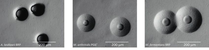
Introduction
Introduction
The term ‘mycoplasmas’ is often used as a trivial name for all Depending on species, mycoplasmas can grow in liquid media,
members of the bacterial class Mollicutes (lat. mollis = „soft“, either as single cells (Mycoplasma arthritidis) or in aggregates
cutis = „skin“) (Figure 1). Mollicutes are characterized (Acholeplasma laidlawii, Mycoplasma pneumoniae, Mycoplasma
by the lack of a cell wall and a small genome size (0.5 –2.2 fermentans).4 The lack of a cell wall makes mycoplasmas
megabase pair) with low GC (guanine-cytosine) content resistant to cell wall-targeting antibiotics such as penicillin.
(20–40 mol%). Due to their small genome, mycoplasmas are Furthermore, some mycoplasmas can form biofilms on solid
host-dependent and live as commensals or infectious agents in surfaces in liquid media, such as glass or plastic surfaces, which
or on a variety of hosts, including humans, other vertebrates, provides another level of resistance, namely to disinfecting
plants, and insects. These microorganisms can multiply under agents and environmental stress conditions. 5 The first
aerobic or anaerobic conditions. They have a pleomorphic cell mycoplasma species was cultured at the Institut Pasteur in
morphology, with the exception of spiroplasmas, which have a 1896; it was isolated from cattle with pleuropneumonia and
spiral shape, and some mycoplasmas of the genus Mycoplasma, much later described as Mycoplasma mycoides subsp.
which have a flask-like shape due to a terminal (tip) structure mycoides SC (small colony type).6
(Mycoplasma gallisepticum, Mycoplasma pneumoniae).
Class Mollicutes
Order Mycoplasmatales Achoplasmatales Entomoplasmatales Anaeroplasmatales
Family Mycoplasmataceae Achoplasmataceae Spiroplasmataceae Entoplasmataceae Anaeroplasmataceae
Genus Mycoplasma* Ureaplasma* Acholeplasma* Spiroplasma Entomoplasma Mesoplasma Anaeroplasma Asteroleplasma
Figure 1: Taxonomy of the bacterial class Mollicutes. The red boxes indicate genera with relevant species in biopharmaceutical manufacturing processes.
*Genera containing mycoplasma species that are prevalent in humans. Source: Authors, 2017.
Due to their genome reduction, mycoplasmas lack several For a long time, mycoplasmas were largely underestimated
metabolic pathways either completely or partially, forcing them as pathogens. For that reason, there was a lack of suitable
to acquire necessary nutrients (amino acids, nucleobases, and molecular diagnostic approaches. This initial situation has
fatty acids) from the environment and to exert a parasitic life changed considerably in recent years, and there has been
style.7 greater acceptance and improvement of culture-based and
molecular methods for mycoplasma detection. 8
4 | Technical Report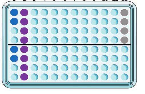
Introduction
Mycoplasmas in Cell Cultures
Biopharmaceuticals
In addition to their growing clinical significance mycoplasmas
Biopharmaceuticals (also known as biologics) are the
have gained great attention in the context of cell cultures.
‘classical’ medicinal products manufactured in and
As they naturally reside in plant and animal tissues, every cell
extracted from biological sources such as bacteria,
culture medium containing plant- or animal-derived supple
yeast, mammalian cell lines, or mammals. Vaccines,
ments is prone to contamination by mycoplasmas. Due to their
purified blood components and recombinant proteins
small size of only 0.1–0.8 μm on average (Figure 2), and their
fall into this category. They can consist of nucleic acids,
variable shape resulting from the missing cell wall, mycoplasmas
proteins, sugars and complex combinations of these
can pass through standard sterilizing filters and enter cell
and are either identical or similar to molecules naturally
cultures with culture media or raw material-derived additives.
occurring in the human body.
The two most common sources of contamination are labor atory
In contrast to chemically synthesized drugs (often
personnel and already contaminated cell cultures, from which
referred to as small molecules), biopharmaceuticals are
the contaminant is passed on by cross-contamination. 9 Since
much larger with a molecular weight 100 times that of
mycoplasmas are not visible with standard light microscopy
small molecules. Biopharmaceutical manufacturing in
setups and usually barely affect the obvious state of the cell
mammalian cell lines typically involves the development
culture, they often remain undetected. Nevertheless, they
of a genetically engineered eukaryotic cell line (such
impact cell growth and metabolism, and consequently, the
as CHO or HEK293) to express the biopharmaceutical,
therapeutic proteins expressed by host cells.
and subsequent harvesting, purification, and drug
formulation. Also refer to Figure 3.
Due to the complex manufacturing process, biopharma
ceuticals face unique manufacturing and product release
challenges. Firstly, cell lines may be contaminated with
mycoplasmas, requiring mycoplasma testing prior to lot
release. Secondly, they are sterilized by filtering, which
has the potential risk that mycoplasmas or viruses pass
through the filter. Thirdly, cell culture contamination may
be introduced by raw materials. This is why mycoplasma
testing methods, especially early warning systems (also
known as in-process control), are essential to detect a
Yeast cell contamination as fast and as early as possible.
Mycoplasma cell (3-10 μm)
(0.1-0.8 μm)
Eukaryotic cell
(10-100 μm)
Virus
(0.05-0.1 μm)
Bacterial cell
(1-10 μm) Figure 2: Relative size of
different microorganisms.
Source: Authors, 2017. This figure
is a graphical illustration by the
authors of this Technical Report
and provided under the terms
of the Creative Commons
Public License CC BY 3.0
(http://creativecommons.org/
0.1 μm 1 μm 10 μm 100 μm licenses/by/3.0/), and can be
used under the terms of such
Eye license notwithstanding any
Light microscope
rights that may exist with
Electron microscope respect to the document it is
embedded in.
Technical Report | 5
Introduction
Despite the negative effects of mycoplasma contamination,
ATMP
cell cultures are rarely monitored, even though testing for
Advanced therapy medicinal products (ATMP) are a
mycoplasma contamination is a necessary quality control
new class of therapeutics that are based on genes
procedure. Studies have shown a contamination rate of about
(gene therapy), somatic cells (cell therapy), or tissue
5–35% of existing cell lines available worldwide.10, 11, 12 The only
(tissue engineering). These advanced therapies herald
meaningful safety precaution to maintain mycoplasma-free cell
novel treatments of a number of diseases and thus, a
cultures is to regularly test for mycoplasmas.
huge potential for patients is expected. Autologous cell
therapy, for instance, typically involves cell dissociation
Only well-established routine mycoplasma testing during
from an individual patient, cell culture outside the
the ongoing process can minimize the risk of a concealed
human body, and subsequent injection of cells back
contamination that can lead to serious problems.13 Because
into the patient.
the testing procedure and subsequent results interpretation
require solid training and experience, mycoplasma testing
Compared to the ‘classical’ large molecule
cannot always be carried out in-house. Outsourcing myco
biopharmaceuticals, cell therapy products face additional
plasma testing to a trustworthy and experienced contract
manufacturing and release challenges. Firstly, most of
research organization (CRO) is an alternative that brings
them cannot be sterilized at all. Secondly, storage may
several advantages. Firstly, qualified results can be obtained
be challenging as cell therapy products sometimes face
in the shortest time possible. Secondly, an in-house testing
a short shelf life and need to be injected into patients
facility does not need to be maintained, which frees resources
immediately. Thirdly, the batch size often consists of one
to concentrate on the core business. Last but not least, the
dose rate, and volumes are usually very small. In such
potential risk of mycoplasma contamination associated with
cases, rapid mycoplasma testing methods are favorable
introducing necessary positive controls into a facility are
over the conventional culture-based mycoplasma
avoided.
testing methods because they require smaller volumes
and release manufacturing batches much faster than
Mycoplasmas in Manufacturing Processes of
conventional mycoplasma testing methods. Different
Biopharmaceuticals and ATMP
from the ‘classical’ biopharmaceuticals, there is also
Especially in the biopharmaceutical and cell therapy industry,
the need to evaluate the possible risk in terms of entry
the effects of mycoplasma contamination are devastating,
and growth of uncommon mycoplasma species during
as entire production batches must be discarded and the
the manufacturing process due to new cell types and
manufacturing plant must stop production.14 International
production conditions.
regulatory authorities have published guidelines to demonstrate
that biological products intended for preventive or therapeutic
clinical use and prepared in cell culture substrates must be free
of mycoplasmas to ensure product safety, purity and potency.
Therefore, early detection of mycoplasmas is essential for
smooth processes in manufacturing of biopharmaceutical and
cell therapy products. Figure 3 depicts common testing points
in the manufacturing process of ‘classical’ biopharmaceuticals.
Given these multiple check points, numerous different methods
for mycoplasma testing have been developed and will be
covered in the following section.
6 | Technical Report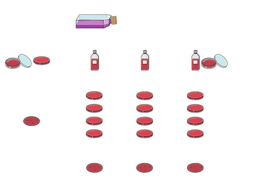
Introduction

                        Buffer          Working cell           Seed           Fermentation
Raw materials                                                                       +              Harvest           Purification        Formulation
                     preparation          culture             culture         Clarification

  Industrially                           Additional              In-process control            Prescribed by the
   necessary                             test point                recommended                regulatory authority

Figure 3: Testing points for mycoplasma contamination in the manufacturing process of biopharmaceuticals. After raw materials have been
tested for contamination (grey loop), and solutions like buffer and media have been applied to working cultures, it is necessary to check for contamination
(blue loop), as mycoplasma contamination can also be introduced by the cell line and lab staff. It is also recommended to carry out in-process controls during
the seed culture and the actual fermentation (light purple loops). The final and prescribed test point is the endpoint of the fermentation, the harvest
(purple loop). Once the mycoplasma-free state of the harvest has been proven, further test points are usually no longer necessary since the purification
of the products is carried out without living organisms. Source: Roche CustomBiotech, 2017.

Mycoplasma species frequently or potentially detected as                         the manufacturing process can lead not only to reduced
contaminants in cell cultures and in manufacturing of                            product quality, but also to lower expression levels and
biopharmaceuticals are listed in Table 1. For ATMP, additional                   consequently reduced production yields. In addition, poor
mycoplasma species might be product-relevant, depending on                       quality and contamination with mycoplasmas may trigger
the starting material. A mycoplasma species spectrum analysis                    serious side effects in patients.15, 16
is therefore recommended. The effects of contamination in

                                           Primary isolation source                   Frequent cell
                                           (relevant for products                     culture contaminant
                                           where raw materials of the                 based on published               Potential contamination
Mycoplasma species                         following origins are used)                reports                          source
Acholeplasma laidlawii                     Bovine, porcine, avian, plant                          Yes                  Other cell line, bovine sera, nutrient
                                                                                                                       broth powders
Mycoplasma arginini                        Bovine, ovine, caprine, porcine                        Yes                  Other cell line, bovine sera
Mycoplasma bovis                           Bovine                                                 Yes                  Other cell line, bovine sera
Mycoplasma fermentans                      Human                                                  Yes                  Other cell line, personnel
Mycoplasma gallisepticum                   Avian                                                  No                   Other cell line, embryonated eggs
Mycoplasma hyorhinis                       Porcine                                                Yes                  Other cell line, porcine trypsin
Mycoplasma orale                           Human                                                  Yes                  Other cell line, personnel
Mycoplasma salivarium                      Human                                                  Yes                  Other cell line, personnel
Mycoplasma synoviae                        Avian                                                  No                   Other cell line, embryonated eggs
Spiroplasma citri                          Plant                                                  No                   Other cell line

Table 1: Mycoplasma species which are frequently, occasionally or potentially detected in cell cultures and in biopharmaceutical processes.

                                                                                                                                         Technical Report | 7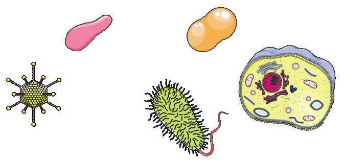
Mycoplasma Testing Methods Overview
Mycoplasma Testing Methods Overview
Detection of mycoplasmas presents a challenge for quality because the prolonged conventional Culture Method can delay
control of cell cultures and biopharmaceuticals because pharmaceutical product release, which is often associated with
especially low-grade contaminations can only be identified high costs. The EP guidelines permit substitution of the
through expertise and experience. Two conventional methods conventional culture methods by NAT if they achieve equivalent
have been used for regulatory mycoplasma testing in recent sensitivity as the traditional methods and prove to be as robust
decades, as they have proven to be sensitive and reliable: (i) and specific.
the Culture Method (also referred to as agar and broth
method) and (ii) the Indicator Cell Culture Method. This section briefly summarizes the non-compendial and
Non-compendial methods, such as enzyme-based and compendial methods used for mycoplasma detection.
immunology-based assays, are easy and fast to apply but NAT-based methods are described in more detail, with special
most do not reach the level of sensitivity of the culture-based focus on the MycoTOOL qPCR. This test method has passed all
methods. Therefore, pharmacopoeial monographs do not necessary validation criteria formulated by the EP mycoplasma
consider these tests acceptable substitutes for regulatory NAT validation guideline to substitute both the Indicator Cell
testing. The necessity for fast yet sensitive and robust Culture Method and the Culture Method.
detection of mycoplasmas has increased over the past years,
Non-compendial Testing Methods
Non-compendial tests for mycoplasma detection often lack the An example of an enzymatic assay is the luciferase-based myco
sensitivity to detect the level of contamination in a sample that plasma detection assay.18, 19 Although the assay is fast (< 20 min),
is required by regulatory monographs. Moreover, results are relatively easy to handle (two luminescence readings), and the
sometimes difficult to interpret if a contamination is at low interpretation of results is easy, no such test has yet been shown
level. These tests are prone to giving false-negative results. to reach the limit of detection that is required from a compendial
test (≤ 10 CFU (colony forming units)/ml). Most mycoplasma
Direct DNA Staining species are detected only at a high titer of 10 4 to 10 5 CFU/ml. 20
Direct staining of cultures with a DNA (Deoxyribonucleic
acid)-specific fluorescent dye is sensitive, but not Mycoplasma PCR-ELISA
recommended for the purpose of detecting mycoplasma An application that combines PCR with a subsequent ELISA
contaminations. Although the test reliably detects heavily (enzyme-linked immunosorbent assay) is the mycoplasma
contaminated cultures, interpretation of low-grade PCR-ELISA, a photometric enzyme immunoassay that detects
contaminations is often difficult because DNA from the cell PCR-amplified mycoplasma DNA in cell culture. 21 During
culture may give rise to small points of fluorescence that can the PCR reaction, digoxigenin-labeled nucleotides are
mimic mycoplasmas.17 incorporated into the amplicons, allowing their detection in
a subsequent ELISA assay. The mycoplasma PCR-ELISA
Enzyme-based Method test is claimed to have a detection sensitivity of 1–3 myco
Enzyme-based assays are selective biochemical tests that plasma “particles” for particular mycoplasma species (e.g.,
exploit the activity of mycoplasma enzymes. A prerequisite for M. fermentans and A. laidlawii). 22 However, since for others the
such a test to be applicable for routine mycoplasma testing is Limit of Detection (LOD) was 1000 “particles” per ml sample,
that the enzymatic activity measured must ideally be ubiquitous the test does not fulfill the requirements of the EP regulatory
among mycoplasmas, but missing in the eukaryotic cell matrix. guideline as compendial test for mycoplasma detection.
8 | Technical Report
Mycoplasma Testing Methods Overview

Compendial Testing Methods
These methods are, on one hand, based on conventional microbiological culture procedures using liquid media and agar media
and, on the other hand, based on rapid molecular techniques. The methods and their advantages and disadvantages are
summarized in this section and Table 2.

Method                              Advantages                          Disadvantages
Culture Method                      • Sensitive                         • Up to 28 days incubation period
                                    • Detects 0.1 CFU/ml                • Requires more than one growth medium for the cultivation of different
                                    • Detection of ‘real’                 mycoplasma species
                                      contaminations caused by          • Risk of false-negative results: highly fastidious M. hyorhinis cultivar
                                      viable multiplying                  alpha strains and ureaplasmas are not detected if standard mycoplasma
                                      mycoplasma cells                    culture media are used
Indicator Cell Culture              • Inexpensive                       • Subjective interpretation that can be biased
Method                                                                  • Not mycoplasma-specific
                                                                        • Less sensitive

NAT-based Method                    • Sensitive                         • Strongly dependent on quality and efficiency of sample preparation and
                                    •   Detects ≤ 10 CFU/ml               DNA extraction
                                    •   Full automation possible        • Risk of false-negative results due to incomplete mycoplasma species
                                    •   High-throughput testing           coverage or PCR inhibition depending on the method
                                    •   Application as early warning    • Requires DNA extraction kit and costly equipment/instruments
                                        system                          • Requires validation to substitute Culture and Indicator Cell Culture
                                                                          Methods

Table 2: Advantages and disadvantages of compendial mycoplasma detection methods.

Culture Method
Traditional culture methods were used well before today’s                  CO2, 3 ± 1% O2 and 90 ± 5% relative humidity. Subcultivation
molecular techniques and are still found in regulatory and                 from the liquid cultures onto agar plates is carried out up to 21
compendial protocols throughout the world (formulated in the               days after the initial inoculation. On the agar medium,
EP, USP and JP regulatory guidelines). The Culture Method is               mycoplasmas develop microscopic colonies (< 100–400 μm
based on the targeted cultivation of mycoplasmas in culture                diameter). Mycoplasma colony morphologies can vary from the
media that promote mycoplasma growth. A sample to be tested                typical fried-egg shape to more irregular shapes (Figure 4).
is inoculated into the liquid mycoplasma culture media and onto            As mycoplasma colonies can be very small, colony counting
agar media, and mycoplasma growth is promoted by micro-                    under the microscope requires some experience. A schematic
aerophilic incubation conditions, such as 36 ± 1°C, 5.5 ± 0.5%             illustration of the Culture Method is shown in Figure 5.

                                                                                                                            Technical Report | 9
Mycoplasma Testing Methods Overview Figure 4: Colonies of mycoplasma type and reference field strains grown on different mycoplasma agar media. Source: Dr. Carl-Ulrich Zimmerman and Prof. Dr. Renate Rosengarten, 2019 The relatively large sample volume (10 ml) and the long incu period of 28 days. This time factor poses major challenges for bation period (28 days in total) render the Culture Method one many companies, including product release delays that entail of the most sensitive tests, with a theoretical and experimen higher storage costs, as well as increased personnel costs for tally proven detection limit of 0.1 CFU/ml, corresponding to logistics during testing of raw materials, the cell line, and the 1 CFU/10 ml sample. The method fulfills the EP 2.6.7 require process controls in the up- and down-streaming process. ment of detecting ≤ 10 CFU/ml, which is why this method is Another limitation is that the Culture Method requires usage of still the reference method in regulatory documents worldwide. several different growth media. Not all mycoplasma species The Culture Method has, however, a few disadvantages. The grow in the same standard mycoplasma culture medium. predominant drawbacks come from the lengthy cultivation 10 | Technical Report
Mycoplasma Testing Methods Overview

                                                    Sample

   0.2 ml
                                                                                     Negative                               Positive
                                                                                     control                                control

                                                                  10 ml
                                                                    in
                                                                 100 ml

   Negative control
         > 14 days                                               0.2 ml                          0.2 ml                          0.2 ml

                                                                                                                                                 day 2-4

                                                                                                                                                 day 6-8

                                                                                                                                                day 13-15

         Colony count                                                                                                                           day 19-21

                                                                                 Colony count

Figure 5: Schematic illustration of the Culture Method according to EP 2.6.7.
Aliquots of 200 µl from a sample to be tested for the absence of mycoplasma contamination are plated onto mycoplasma agar medium and 10 ml are
inoculated into 100 ml liquid growth medium. Uninoculated media serve as negative controls, and media inoculated with ≤ 100 CFU serve as positive
controls. The liquid medium is incubated for 20–21 days. On days 2–4, 6–8, 13–15 and 19–21 after inoculation, 200 µl of the medium inoculated with the
sample, and the negative and positive controls are plated onto agar medium. The inoculated agar media are incubated for not less than 14 days, except those
corresponding to the 20–21 day subculture, which are incubated for 7 days. Source: Authors, 2017. This figure is a graphical illustration by the authors of this
Technical Report and provided under the terms of the Creative Commons Public License CC BY 3.0 (http://creativecommons.org/licenses/by/3.0/), and can
be used under the terms of such license notwithstanding any rights that may exist with respect to the document it is embedded in.

Thus, depending on the source of the sample, different                            strains, such as the M. hyorhinis cultivar alpha strains (with
mycoplasma culture media are used in parallel to increase the                     M. hyorhinis DBS 1050 as reference strain), do not grow in the
detection spectrum of possible contaminating mycoplasma                           standard culture media due to growth inhibition by certain
species. For this reason, the use of at least two standard                        peptones and yeast products. 23 Growth of these cell-culture
mycoplasma culture media is recommended in EP 2.6.7.:                             adapted strains is dependent on their habitat, the cell culture.
FRIIS medium for the detection of non-avian mycoplasmas,                          In order to detect these cultivar alpha strains, an additional test
and FREY medium for the detection of the avian mycoplasma                         has to be performed in parallel, using the Indicator Cell Culture
species M. synoviae. The perhaps greatest disadvantage of the                     Method.
Culture Method is, however, that highly fastidious mycoplasma

                                                                                                                                       Technical Report | 11
Mycoplasma Testing Methods Overview
Indicator Cell Culture Method
The Indicator Cell Culture Method is normally carried out with solution and stained with a fluorescent dye that binds to DNA.
Vero or 3T3 cell lines, although the use of a production cell line The presence of mycoplasmas is characterized by a spherical
that is equivalent in effectiveness for detecting mycoplasmas is fluorescence pattern on the cell surface and by strong
also accepted by the EP regulatory guideline. The indicator cell fluorescence in the surrounding areas. Mitochondria in the
culture is inoculated with the sample and incubated at 35–38°C cytoplasm are also stained, but are easily distinguished from
until grown to confluence. For positive controls, the indicator mycoplasmas. The test is invalid if the positive controls do not
cell line is also inoculated with the type strain CH19299T of show fluorescence typical for mycoplasmas or if the negative
M. orale and the M. hyorhinis cultivar alpha reference strain control shows fluorescence typical for mycoplasmas.
DBS 1050 with and without the presence of the test sample. A schematic illustration of the Indicator Culture Method
Before staining, the subculture is fixed with a suitable fixing is shown in Figure 6.
Positive Positive
control control
Positive Positive
1 ml 1 ml control control
1 ml Negative
sample sample sample control
+ + M. orale M. hyorhinis
M. orale M. hyorhinis ≤ 100 CFU ≤ 100 CFU
≤ 100 CFU ≤ 100 CFU
10 ml
Fluorescence Microscopic Evaluation
Positive control: Vero cells + M. hyorhinis Negative control: Vero cells
Figure 6: Indicator Cell Culture Method according to EP 2.6.7. A reshly prepared Vero indicator cell culture is inoculated with 1 ml sample. Four posi-
tive controls are prepared. Two positive controls consist of Vero cells inoculated with 1 ml sample spiked with not more than 100 CFU M. hyorhinis cultivar
alpha reference strains DBS 1050 and M. orale type strain CH19299T, respectively. The other two positive controls are Vero cells inoculated with not more
than 100 CFU M. hyorhinis DBS 1050 and M. orale CH19299T without the sample. The two positive control strains are plated onto agar medium to check for
viability. The negative control is a freshly prepared Vero cell culture that is left uninoculated. All cell cultures are incubated in a CO 2 incubator until the
cell density of 100% is reached. The cell layer is then washed with buffer and trypsinated. The detached cells are resuspended in cell culture medium,
transferred to chamber slide flasks and incubated in a CO 2 incubator until the cell density is approximately 50%. The cell layer is fixed twice with a freshly
prepared fixing solution, air dried and stained with a Hoechst Stain method. The microscope slides are evaluated using a fluorescence microscope. Source:
Authors, 2017. This figure is a graphical illustration by the authors of this Technical Report and provided under the terms of the Creative Commons Public
License CC BY 3.0 (http://creativecommons.org/licenses/by/3.0/), and can be used under the terms of such license notwithstanding any rights that may
exist with respect to the document it is embedded in.
12 | Technical ReportMycoplasma Testing Methods Overview
Both the Culture Method and the Indicator Cell Culture Method take a long time to results (up to 28 days for the Culture Method
and 7 days for the Indicator Cell Culture Method), and carry the intrinsic risk of introducing a mycoplasma contamination into the
facility due to the required handling of viable mycoplasma cells as positive control organisms.
NAT-based Methods
NAT-based methods include all tests based on nucleic acid PCR temperature protocols have been optimized with the
detection, often performed by PCR. goal to develop more sensitive, specific, or rapid PCR assays.
Touchdown PCR (TD-PCR), for example, is very commonly
PCR used to make PCR assays more specific to a targeted gene.
PCR is a molecular biological method used in many areas Primers bind with high specificity to a targeted DNA sequence
such as food and environmental analysis, forensics, and at high annealing temperatures during the first few PCR
medical diagnostics. The underlying principle is the specific cycles. This ensures the exclusive amplification of a specific
amplification of DNA to a level that can be detected. This DNA sequence. The annealing temperature is then gradually
amplification is carried out by the enzyme DNA polymerase decreased to reach highest PCR efficiency. TD-PCR protocols
in repeated amplification cycles that are automated by reduce the amount of nonspecific DNA amplified by PCR. 24
thermocyclers. One cycle consists of three main steps: MycoTOOL qPCR leverages TD-PCR for highly specific
mycoplasma detection.
1. Double-stranded DNA (dsDNA) is denatured into
single-stranded DNA (ssDNA) by heat
2. PCR primers bind to specific ssDNA sites You will find a summary and more
(e.g. to specific target genes) information about the history and
3. DNA polymerase elongates the annealed primers evolution from the conventional PCR
according to the sequence of the ssDNA to the qPCR here:
(https://diagnostics.roche.com/
For more information about the PCR global/en/article-listing/the-
procedure in detail: evolution-of-pcr.html)
(http://go.roche.com/dnacopy)
In contrast to PCR, Real-Time PCR (qPCR) reports the
amplification of DNA in real time. Thus, there is no need
for post-PCR DNA detection such as gel electrophoresis.
Usually after 30–50 PCR cycles sufficient DNA is amplified for This reduces the risk of PCR contamination in the laboratory
detection by gel electrophoresis and staining with fluorescent dramatically and facilitates the interpretation of end results.
dyes. qPCR uses probes consisting of a fluorescent dye attached
to a short DNA sequence (18–30 base pairs) that is added to
the PCR. The probe is incorporated into the new strands of
DNA produced in each amplification cycle. There are a variety
of different probe designs on the market, but one of the most
common ones are the hydrolysis probes used in MycoTOOL
qPCR. This probe reports the amount of total DNA as
fluorescence intensity after each PCR cycle. The fluorescent
signal increases proportionally to the amplification of the
target sequence. The fluorescence intensity is plotted over
time and forms a typical sigmoid qPCR curve (see Figure 7).
Technical Report | 13Mycoplasma Testing Methods Overview
Both, PCR and qPCR are sensitive methods and thus prone to
54.617
DNA contamination. The most common sources of contami
nation are the DNA template itself and amplified DNA from 49.617
post-PCR reactions. DNA molecules may be spread around the 44.617
lab via air conditioning systems or laboratory staff. The most 39.617
effective strategy to eliminate these contamination sources is a
34.617
unidirectional workflow from sample drawing to DNA detection
in separate work areas or separate rooms. 25 29.617
ΔRn
24.617
MycoTOOL Mycoplasma Real-Time PCR Kit 19.617
(MycoTOOL qPCR)
14.617
The MycoTOOL Real-Time PCR Kit is a qPCR assay optimized
9.617
for the detection of mycoplasmas in cell culture. It fulfills all
EP 2.6.7. requirements for NAT-based assays for mycoplasma 94.617
detection with respect to sensitivity (i.e., detection limit of 0.383
≤10 CFU), specificity, robustness and comparability. It does
not require a mycoplasma enrichment or pre-incubation step. 5 10 15 20 25 30 35 40 45
The kit uses primers and probes that are highly specific to Cycles
the mycoplasma 16S ribosomal DNA gene. This allows the
detection of more than 150 cultivable and non-cultivable Figure 7: Example of mycoplasma testing by MycoTOOL qPCR
mycoplasma species. It includes the most frequently occurring analysis. In the first 15 cycles of the qPCR, the baseline describes the
initial signal at which little change is seen in the fluorescence intensity.
cell culture contaminants, namely A. laidlawii, M. arginini,
This signal can also be defined as background fluorescence of the reaction.
M. fermentans, M. hyorhinis, M. orale and M. salivarium, as well The threshold cycle (Cq) is the cycle number at which the fluorescence
as the human pathogenic mycoplasma species M. pneumoniae signal of the sample exceeds the background signal. The lower the Cq value
and M. hominis, the avian pathogenic mycoplasma species the higher the amount of DNA in the sample.
M. gallisepticum and M. synoviae, and the plant pathogenic Source: Roche CustomBiotech, 2017.
mycoplasma species S. citri (Table 1).
Automated DNA extraction may be conducted with either a
MagNa Pure 24 or a MagNa Pure 96 instrument, followed by The MagNA Pure 24 and 96 instruments purify nucleic
subsequent qPCR performed on the LightCycler 480 II real- acids from a wide range of starting materials (e.g. whole
time PCR instrument. Manual DNA extraction may be done blood, plasma, cell culture) using magnetic glass particle
with the Roche QC Preparation Kit for samples with cell technology. For more information use the QR-Code for
densities up to 5x10 6 cells/ml, or with MycoTOOL Mycoplasma our MagNA Pure 96 and for our MagNA Pure 24 System.
Detection Prep Kit, High Cell Density for samples with a cell
density range of 5 × 10 6 cells/ml to 1 × 10⁸ cells/ml. Carrier (http://go.roche.com/mag96)
DNA is available for analysis of cell-free samples and may be
added to the biological sample prior to nucleic acid extraction
and purification. The entire workflow from sampling to result
takes 4 to 6 hours, depending on the level of automation and
the number of samples. The complete workflow is depicted in
Figure 8. (http://go.roche.com/mag24)
14 | Technical ReportMycoplasma Testing Methods Overview
manual system 1 ml automated system
unprocessed
sample
sample purification
through
Roche QC Preparation Kit
MagNA Pure 24 MagNA Pure 96
400 μl DNA eluate
(validated)
200 μl DNA eluate
MycoTOOL Mycoplasma Real-Time PCR Kit
Light Cycler 480 II
4 x 20 μl DNA input 4 x 20 μl DNA input
Figure 8: MycoTOOL qPCR workflow. Unprocessed sample (1 ml) with a cell density of 5x10 6 cells/ml is prepared using a manual or automated workflow.
For manual sample preparation, nucleic acids are isolated with the Roche QC Preparation Kit. With the automated nucleic acid isolation system, the sample
DNA is purified with one of the MagNA Pure systems. Subsequently, a mycoplasma-specific qPCR reaction is performed on the LightCycler 480 II Real-Time
PCR system. The automated workflow based on the MagNA Pure 96 and LightCycler 480 II system, shown as the purple marked procedure, has been fully
validated by Roche Pharma Biotech and according to EP chapter 2.6.7. The generic validation report is available on request under confidential disclosure
agreement. A summary of the study design and results is available under http://go.roche.com/MycoTOOLqPCR
Source: Roche CustomBiotech, 2017.
MycoTOOL qPCR includes three controls to ensure validity of using a second set of primers and probes in a separate vial.
results. To verify the integrity of all reagents used during qPCR, The RC verifies the integrity of the complete workflow, from
a plasmid-based positive control is added to each experiment. DNA isolation to PCR. Because the RC is spiked into the
False negative results are controlled by a H2O negative control. sample as an exogenous control, MycoTOOL qPCR is not
The third control used is a plasmid-based recovery control limited to a specific cell line and may be used across the
(RC; also known as an exogenous internal control) that is spectrum of cell lines commonly used in biopharmaceutical
added to each sample prior to DNA isolation. It is co-amplified manufacturing.
Technical Report | 15Mycoplasma Testing Methods Overview
All amplification reactions are carried out in technical replicates (two or four for the negative control and positive
control/RC - sample, respectively). Figure 9 shows a typical pipetting scheme of a 96 well qPCR plate.
1 2 3 4 5 6 7 8 9 10 11 12
A
B
Mycoplasma Master Mix
C
D
E
F
Recovery Control Mix
G
H
Sample Negative control Positive control
Figure 9: MycoTOOL qPCR pipetting scheme. Before DNA preparation with the MagNA Pure 24 or 96 instruments, a sample is spiked with a defined
concentration of a RC plasmid. The DNA from 1 ml sample is eluted in 200 µl buffer. Four technical replicates are tested with 20 µl each. This corresponds to
a volume of 40 % of the biological sample. Source: Authors, 2017. This figure is a graphical illustration by the authors of this Technical Report and provided
under the terms of the Creative Commons Public License CC BY 3.0 (http://creativecommons.org/licenses/by/3.0/), and can be used under the terms of
such license notwithstanding any rights that may exist with respect to the document it is embedded in.
Acceptance criteria for a plate to pass evaluation are: all negative controls must give a negative result, and the positive control
reactions as well as all RC reactions of a sample must give a positive result.
16 | Technical ReportRegulatory Overview
Regulatory Overview
Since mycoplasma contamination evidently affects cell
cultures, testing for mycoplasmas has increasingly become The regulatory approval system
regulated by authorities. Today, mycoplasma testing in The health authorities are responsible for the scientific
manufacturing of biopharmaceuticals and ATMP is controlled evaluation, supervision and safety monitoring of medi
by law in almost all countries around the world. Regulatory cinal products developed by pharmaceutical companies.
authorities have published legally binding documents with Regulatory approval ensures that all medicinal products
national pharmacopoeias for mycoplasma testing. These available on the market are safe, effective and of
documents define the methods used and products to be tested. high quality. Pharmaceutical approval and market
However, recommended methods for mycoplasma testing and authorization of medicines for humans and animals
their detailed test protocols may differ among countries. In require manufacturers to meet official quality standards.
general, one must differentiate between conventional culture- This is controlled by different regulatory agencies and
based and alternative NAT-based mycoplasma testing methods. committees. The standards that manufacturers have to
The traditional compendial methods, such as the Culture meet are defined and published in the pharmacopoeia.
Method and the Indicator Cell Culture Method (see section The pharmacopoeia lists all tests to be carried out on
2.2.), are considered the long-standing gold standard and thus, medicines, intermediates and raw materials, and is
are widely recommended in all pharmacopoeias. Although legally binding for a country or all member states of
individual methodological steps may vary slightly from one a union.
national pharmacopoeia to the other, the protocols for these
culture-based tests are largely harmonized across the
countries. Please refer to Table 3 for a summary of regulatory
authorities and pharmacopoeias containing regulatory
guidelines relevant for mycoplasma testing in some countries.
Europe 26 UK USA Japan China Brazil Argentina
Health Authority European Medicines and Food and Drug Ministry of Ministry of Brazilian Argentine
Medicines Healthcare Administration Health, Labour Health Ministry of Ministry of
Agency (EMA) Products (FDA) and Welfare (MOH) Health Health
Regulatory (MHLW)
Agency (MHRA)
Regulatory Committee National Food and Drug Pharmaceutical Chinese National Drug Regulatory
Agency for for Medicinal Approval by Administration and Medical Food and Drug Sanitary Authority
Pharmaceutical products for MHRA (FDA) Devices Agency Administration Surveillance (ANMAT)
Approval Human Use Centralized (PMDA) (CFDA) Agency
(CHMP) Approval by (ANVISA)
EMA
Publisher European British US Pharmaceutical Chinese National Drug Regulatory
Pharmacopoeia Directorate Pharmacopoeia Pharmacopeial and Medical Pharmcopoeia Sanitary Authority
for the Quality Commission Convention Devices Agency Commission Surveillance (ANMAT)
of Medicines (PMDA) (ChPC) Agency
(EDQM) (ANVISA)
Pharmacopoeia European British United States Japanese Chinese Brazilian Argentine
Pharmacopoeia Pharmacopoeia Pharmacopoeia Pharmacopoeia Pharmacopoeia Pharmacopoeia Pharmacopoeia
(EP) 27 28
(USP) 29 (JP) 30 (ChP) 31 32 33
NAT Acceptance
for Mycoplasma ✓ ✓ ✓ ✓ ? Not described Not described
Testing
Specification of
NAT Validation ✓ ✓ (✓)* ✓** ? ✗ ✗
Requirements
Table 3: Overview of the health authorities in the EU, the USA, Japan, China, Brazil and Argentina, their regulatory agencies, legally binding documents
(pharmacopoeias), NAT acceptance for regulatory mycoplasma testing, and specification of NAT validation requirements.
* The USP mentions “by a procedure demonstrated to be comparable”
** comparable to EP 2.6.7.
Technical Report | 17Regulatory Overview
The situation is very different for rapid mycoplasma testing The EP provides the most detailed NAT validation guideline of
methods, like NAT. Even though many national pharmacopoeias all pharmacopoeias in its chapter 2.6.7. Four requirements must
mention NAT as a valid mycoplasma testing method, there is be met by NAT-based mycoplasma testing methods: Limit of
little harmonization across countries regarding protocols or Detection, Specificity, Robustness and Comparability (Table 4).
validation requirements. Some pharmacopoeias such as the For full and generic validation of a NAT method, it is advisable
EP and JP mention detailed validation guidelines, whereas to include additional parameters such as precision and cross-
others merely point out that NAT is a valid testing method contamination.
after validation. However, all countries require an appropriate
validation and comparison with conventional mycoplasma
testing methods.
Validation Requirements EP 2.6.7.
Limit of Detection (LOD) To define the detection limit, a positive cut-off point should be determined for each species (the chapter
provides a list of mycoplasma species to be used as test organisms). For each strain, a minimum of
three independent 10-fold dilution series should be tested, with a sufficient number of replicates at each
dilution to give a total number of 24 test results for each dilution. The positive cut-off point is defined as the
concentration of mycoplasmas that can be detected in 95 percent of test runs, thus in at least 23 test results.
Specificity It is important to use PCR primers that are specific for a wide range of mycoplasmas. However, it is likely that
PCR primers will also detect other bacterial species. This potential cross-detection should be documented
by testing related bacterial genera such as gram-positive bacteria with close phylogenetic relation to myco
plasmas (the chapter provides a list of bacterial genera to be tested).
Robustness The measure of the NAT method’s capacity to remain unaffected by small but deliberate variations in method
parameters and test method modifications needs to be demonstrated. The chapter provides examples of
variations and test modifications that may be tested.
Comparability The comparability should include a comparison of the LODs between NAT and the compendial methods.
The chapter defines the following acceptance criteria:
1) Culture Method replacement by NAT: A detection limit of ≤ 10 CFU/ml needs to be demonstrated.
2) Indicator Cell Culture Method replacement by NAT: A detection limit of at least ≤100 CFU/ml needs to be
demonstrated for each mycoplasma test species.
3) In both cases the NAT alternative method needs to be performed in parallel to both conventional
methods to evaluate simultaneously the LOD of both methods using the same samples of CFU-calibrated
mycoplasma test strains.
Table 4: Summary of validation requirements as detailed in EP 2.6.7. For more detailed information,
refer to the EP mycoplasma NAT validation guideline directly.
There are other non-legally binding documents that may be considered as a reference for implementation of NAT mycoplasma
testing methods:
(i) The Parenteral Drug Association (PDA) published a (iii) In addition to the USP, theme-specific guidelines are
Technical Report named “Alternative Methods for Mycoplasma published by FDA departments. The CBER of the FDA
Testing” in 2010. It guides new users of rapid mycoplasma tests published a guideline in 1993 called “Points to Consider
and describes assay procedure, assay validation, and potential in the Characterization of Cell Lines Used to Produce
applications for alternative mycoplasma testing methods. 34 Biologicals” (PTC), and a “Guidance for Industry” in 2010
(ii) Roche Pharma uses the MycoTOOL qPCR assay to release that provide additional information on mycoplasma testing.
new products to the market. The assay has been validated Neither is legally binding. 35, 36
according to EP chapter 2.6.7., and the generic validation (iv) Bioanalytical Method Validation is another non-legally
report is available on request under confidential disclosure binding document published by the FDA that helps gain an
agreement. A summary of the study design and results is overview of best practices to validate an alternative method
available under http://go.roche.com/MycoTOOLqPCR. for mycoplasma testing. 37
18 | Technical ReportValidation and Implementation
Step-by-Step Validation and Implementation of MycoTOOL qPCR
This section provides an overview of the processing steps and timelines to be considered in the product-specific validation
and implementation of the MycoTOOL qPCR method for rapid mycoplasma testing of ‘classical’ biopharmaceuticals. Usually,
a mycoplasma qPCR implementation project includes five steps: the supplier due diligence, the feasibility study, the
development of a validation strategy, the performance of the validation study, and the submission. MycoTOOL qPCR is a
method generically validated by Roche Pharma. The validation report that is available on request can be leveraged to save
time during the implementation procedure. The complete workflow is depicted in Figure 10 and described in detail below.
For ATMP, a simplified validation approach that uses the entire panel of product-relevant mycoplasma species as test
organisms and includes experimentally verified spikes at the regulatory required LOD of ≤ 10 CFU/mL might be sufficient.
Implementation of the MycoTOOL qPCR Mycoplasma
Testing Method for ‘Classical’ Biopharmaceuticals
Supplier Due
Diligence
yes Leveraging no
Roche Validation
Report
?
Feasibility Feasibility
Study* Study*
Validation
Shorter time
Strategy*
Validation
Strategy*
Longer time
Validation
Study*
Validation
Submission
Study*
Submission
Routine Testing*
*This service can be outsourced to a suitable CRO
Figure 10: Flowchart for the implementation of MycoTOOL qPCR. The left hand side of the flowchart depicts validation carried out by leveraging
Roche’s generic validation report (implementation for CHO manufacturing processes). Implementing MycoTOOL qPCR for a non-CHO process or changing
the method used in Roche’s generic validation requires revalidation of the MycoTOOL qPCR method. This process, presented on the right hand side of the
flowchart, is more time cons uming.
Technical Report | 19Validation and Implementation Supplier and CRO Due Diligence Roche Pharma Generic Validation Report This step may take several days to a few months. It involves Roche Pharma conducted a full generic validation according a due diligence of marketed mycoplasma detection kits and to EP 2.6.7. (NAT validation guideline) for MycoTOOL qPCR CRO to carry out the study. Product specifications must be with defined instrumentation (LightCycler ® 480 Instrument II checked against testing requirements and the capabilities of and MagNA Pure 96, see Figure 10) and specifically for the mycoplasma detection kit suppliers and CRO have to be processes using Chinese hamster ovary (CHO) cell lines. assessed. Before moving forward with the validation, several Typically, regulatory agencies such as FDA or EMA do not points should be clarified: require users to fully revalidate MycoTOOL qPCR, as long as • Does the supplier provide appropriate documentation with the main process, including instrumentation, is unchanged. regards to design and manufacture of instruments and Thus, a product-specific validation study using the unchanged reagents? workflow will require testing of fewer samples and hence • Does the supplier have change control systems in place? less time. However, this option should be discussed with • Does the supplier provide quality and supply agreements the relevant regulatory agency before taking a decision. if required? • Does the supplier deliver in-time and reliably? • Are references of previous validation studies available? This validation report is available • Do the supplier and CRO respond to questionnaires and on request and under confidential allow physical audits at their testing facility? disclosure agreement. Please find • Does the supplier provide end-user training programs, a scientific poster with a summary on-site technical service, installation and operational of the data under this link. qualification services, preventive maintenance service, and a technical support hotline? (http://go.roche.com/MycoToolqPCR) • Does the CRO have sufficient experience with product- specific NAT validation studies and implementation of NAT-based routine testing? • Does the CRO provide a validation report, protocols or similar documentation? • Does the CRO provide solutions for technology transfers? During this phase, it may also be required to prepare an economic assessment or financial justification (i.e., write a business case) to apply for budget internally. Thus, associated costs, such as one-time costs (requirements for laboratory space, instrumentation costs, installation costs, etc.) and operating costs (maintenance, cost per sample, etc.) should be clarified with the contract service provider during this step. 20 | Technical Report
Validation and Implementation

Feasibility Study                                                    Validation Strategy
This step may take from one week to several months. It may be        This step may take from a few weeks to several months. The
accomplished by using rental equipment on site or, more              validation strategy provides a roadmap for all experiments that
efficiently, by collaborating with a CRO who conducts the            will be conducted during the MycoTOOL qPCR validation study
study. A feasibility study is a technical proof-of-concept testing   (e.g., LOD testing, robustness testing, specificity analysis).
prior to purchasing instrumentation and prior to investing in a      The strategy includes a timeline, responsible parties, planned
validation study. It is done to uncover any technical                experiments, and relevant acceptance criteria. The generic
incompatibilities between MycoTOOL qPCR and the product              validation conducted by Roche can be taken into account.
material intended to be tested. Since MycoTOOL qPCR was              Usually, a few criteria of the generic validation must be
validated using CHO cells at a concentration of 5x10 6 cells/ml,     re-confirmed (such as the LOD for a number of selected
other cell lines and concentrations must be tested to examine        regulatory mandatory and product-relevant mycoplasma
product matrix effects, such as PCR inhibition. Another goal of      reference strains). Generally, using the generic validation
the feasibility study is to assess if the required LOD can be        report as basis for a validation study reduces the number of
achieved, and what adjustments to the MycoTOOL qPCR test             samples to be tested and the overall cost of the study. It is
protocol can help reach the target LOD. Such adjustments can         important to always discuss the strategy with the respective
be a larger PCR reaction volume or an increase in PCR cycles.        regulatory agency, especially if the Roche generic validation
.                                                                    may streamline your project.

Validation Study                                                     Submission & Routine Testing
This step may take from two to several months. The validation        After successful submission and approval of the validation
study demonstrates that MycoTOOL qPCR is capable of                  report by the regulatory authority, routine testing may be
consistently detecting mycoplasmas according to regulatory           carried out. Depending on the customer and the project,
requirements. It may be performed internally using the               routine testing may be carried out by internal quality control
respective instrumentation, or it may be outsourced to a             experts or the complete process remains outsourced to a
CRO. The CRO executes the experiments defined during                 CRO.
the validation strategy, and documents evidence in a
validation report.

                                                                                                                 Technical Report | 21
You can also read