The main DNA viruses significantly affecting pig livestock - Sciendo
←
→
Page content transcription
If your browser does not render page correctly, please read the page content below
J Vet Res 65, 2021
                                                                                                DOI:10.2478/jvetres-2021-0001

                                                                                                          REVIEW ARTICLE

                                 The main DNA viruses
                          significantly affecting pig livestock

                                    Carlos Díaz1, Vladimír Celer2, Ivo Frébort1
         1Department of Molecular Biology, Centre of the Region Haná for Biotechnological and Agricultural Research,
                                    Palacký University, 783 71 Olomouc, Czech Republic
2Department of Infectious Diseases and Microbiology, Veterinary and Pharmaceutical University, 612 42 Brno, Czech Republic

                                                    ivo.frebort@upol.cz

                                    Received: May 28, 2020        Accepted: December 3, 2020

Abstract

      Swine DNA viruses have developed unique mechanisms for evasion of the host immune system, infection and DNA
replication, and finally, construction and release of new viral particles. This article reviews four classes of DNA viruses affecting
swine: porcine circoviruses, African swine fever virus, porcine parvoviruses, and pseudorabies virus. Porcine circoviruses
belonging to the Circoviridae family are small single-stranded DNA viruses causing different diseases in swine including
poly-weaning multisystemic wasting syndrome, porcine dermatitis and nephropathy syndrome, and porcine respiratory disease
complex. African swine fever virus, the only member of the Asfivirus genus in the Asfarviridae family, is a large double-stranded
DNA virus and for its propensity to cause high mortality, it is currently considered the most dangerous virus in the pig industry.
Porcine parvoviruses are small single-stranded DNA viruses belonging to the Parvoviridae family that cause reproductive failure
in pregnant gilts. Pseudorabies virus, or suid herpesvirus 1, is a large double-stranded DNA virus belonging to the Herpesviridae
family and Alphaherpesvirinae subfamily. Recent findings including general as well as genetic classification, virus structure,
clinical syndromes and the host immune system responses and vaccine protection are described for all four swine DNA virus
classes.

Keywords: DNA viruses, circoviruses, African swine fever virus, parvoviruses, pseudorabies virus.

Introduction                                                         affecting swine are reviewed: porcine circoviruses,
                                                                     African swine fever virus, porcine parvoviruses, and
      The genetic material of DNA viruses is either                  pseudorabies virus. The genetic diversity inside
single-stranded (ss) or double-stranded (ds) deoxyribonucleic        a particular group and family classification, the
acid. Virus DNA genomes are variable in size, ranging                structures of virus particles, the clinical syndromes, the
from small with a size of 1 kilobase pairs (kbp) to large            course of infection, and recent progress in vaccine
examples of several megabase pairs. DNA viruses use                  development as an effective means of protection against
host cells for replication and subsequent infection.                 infections with swine DNA viruses are described.
The first viral genes to be expressed, which are made by
larger viruses, are called early genes. Genes encoding
DNA polymerase and proteins incorporated in DNA                           Porcine circoviruses (PCVs)
replication often belong in this group. After DNA
replication, viruses change the expression profile to the                 Porcine circoviruses are the smallest autonomously
so-called late genes. Those genes are essential for the              replicating swine viruses containing circular single-
production of structural proteins used for coating the               stranded DNA (ssDNA) with a size of 1.76 kbp (73, 75).
replicated DNA genome and forming new viral particles.               PCVs were first discovered by Tischer et al. (113) in
At the end of the proliferation process, viral particles are         1974 when PCVs were mistaken for picornavirus-like
released from the cell to infect new sites. In this article,         particles in a contaminated PK-15 pig kidney cell line.
the four main groups of DNA viruses significantly                    Those circoviruses were non-pathogenic, and after they

© 2021 C. Díaz et al. This is an open access article distributed under the Creative Commons Attribution-
NonCommercial-NoDerivs license (http://creativecommons.org/licenses/by-nc-nd/3.0/)
C. Díaz et al./J Vet Res/65 (2021)
were classified into the Circoviridae family, their China, PCV3 was divided into two groups (a and b) and
apparent harmlessness caused them to merit little five subgroups (a1–a3, b1, and b2) by a phylogenetic
attention so only a few articles concerning the topic were study using full-length sequences of PCV3 DNA (22).
written until around 20 years later, when a new In a phylogenetic study conducted in Germany where
pathogenic type of porcine circovirus appeared called only open reading frame (ORF) 2 (coding for the Cp
PCV2 (75). In 2015, PCV3 was discovered and protein) was used for grouping, the number of subgroups
associated with porcine dermatitis and nephropathy differed; group a was not divided but group b was, into
syndrome (PDNS) (90), and in 2019, a new type of PCV three subgroups (22, 39). The difference is caused by the
(type 4) was found in China (124). usage of whole-genome sequences in the Chinese study,
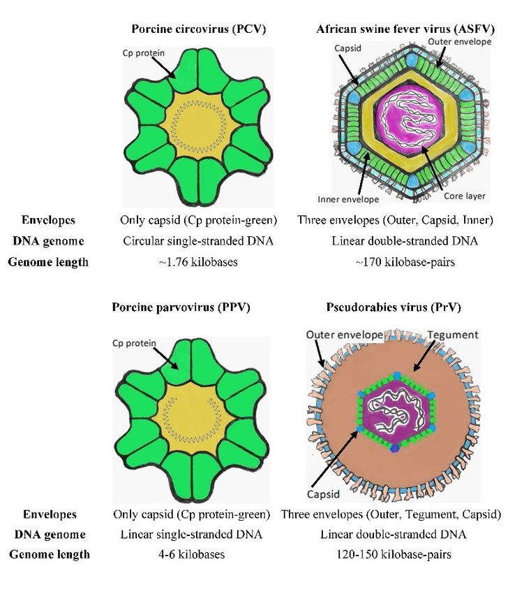
Virus structure. PCV virions are small isometric while ORF2 was considered a critical phylogenetic
particles with a diameter of 17 nm containing circular marker in Germany (85).
ssDNA which only contains three protein-coding genes PCV4. This type was only discovered in April 2019
(114). The virus particle of both PCV1 and PCV2 is (124). Type 4 contains 1.77 kbp long DNA and shares
composed of a single structural protein called the capsid 67% homology with mink circovirus, which is the
protein (Cp), with a molecular mass of 30 kDa and highest homology across circoviruses, and 43–52%
which is responsible for spontaneous capsid formation homology with other porcine circoviruses (124).
(62) (Fig. 1). The size of two crucial genes was predicted at 891
PCV1. This was the first identified porcine nucleotides for the rep gene and 687 nucleotides for the
circovirus. It was designated PCV PK-15 after its cp gene (124). For the understanding of porcine
discovery and characterisation as a contaminant in the circovirus’ pathogenicity and infection, further
PK-15 porcine kidney cell line (113). Interestingly, it investigations will be necessary.
was also found in lymph nodes from piglets affected by Clinical syndromes. Postweaning multisystemic
a wasting syndrome in France (4, 60). wasting syndrome was first described in 1996 and a year
PCV2. In 1998, Meehan et al. (74) observed that later was associated with PCV2 (46). The precise
monoclonal antibodies raised to circoviruses causing definition of PMWS was proposed by Sorden in 2000
post-weaning multisystemic wasting syndrome (109). For pigs to be diagnosed with PMWS, they must
(PMWS) were different from those raised to the PCV show all of the following conditions: firstly, clinical
PK-15 isolate. They also published the first nucleotide signs like wasting, weight loss or failure to thrive;
sequences of the circoviruses associated with PMWS, secondly, histological lesions, which are signs of
which showed less than 80% identity with the PCV PK-15 depletion of lymphoid tissues and organs, and
isolate, and they thus provided evidence for a new inflammation of the lungs and lymphoid tissues in usual
pathogenic type of porcine circovirus, referred to as cases and less often the liver, kidneys, pancreas or
PCV2 (74). Based on the results of the phylogenetic intestine; and thirdly, PCV2 infection inside the lesions.
study using the capsid protein gene region as a marker, The effect of PMWS on the host immune system is
PCV2 sequences were divided into two main groups: the pronounced, causing virus-induced lymphocyte
first group, which subdivides into three clusters 1A to depletion. In the work of Mandrioli et al. (69), the
1C, and the second group, which branches into five presence of activated macrophages was described as
clusters 2A to 2E (40, 85). There is also another grouping an essential factor for the development of the syndrome.
method considering the geographic localisation of Although mainly CD4+ T-lymphocyte counts were
the virus, dividing PCV2 into PCV2a for the North decreased during the infection, a dramatic decline in CD8+
American-like isolates (which also fall into the first and CD4+/CD8+ T-lymphocyte and B-lymphocyte
capsid protein gene region-differentiated group of numbers was also observed, associated with the loss
PCV2), and PCV2b for the European-like isolates (also of lymphoid follicles (69). The reduced proliferation
in the second capsid protein group of PCV2) (86). of lymphocytes thus results in a reduction of cytokines
PCV3. This is a recently discovered type, and yet it as positive growth factors, which can affect the further
has been detected and characterised in many countries expression of major histocompatibility complex I
throughout the world, including China (55), Italy (31), antigens type I and II (MHC I and MHC II) and thus
Brazil (115), and Sweden (121). PCV3 was first impair the immune response (72). Interestingly,
identified in 2015 in North Carolina (USA) in isolates apoptosis was not observed in lymphoid tissues that
from sows showing high mortality, low conception rates showed a decreased rate of virus proliferation (69).
and typical signs of PDNS (90). Therefore, PCV3 was However, the work of Shibahara et al. (106) showed that
associated with PDNS and reproductive failure (90) and apoptosis occurred only in B-lymphocytes and not in
it has also been linked to congenital tumours in piglets macrophages (106). This can be explained by the yet-
as well after Chinese PDNS cases were investigated unknown cause of the apoptosis in lymphoid tissues of
(21). This new type of PCV shares only a small PMWS in swine (69).
percentage of homology in genomic DNA sequence with Another disease associated with porcine
those of PCV1 and PCV2 (90). The homology between circoviruses is PDNS. Pigs affected by this syndrome are
PCV3 and PCV2 found by sequencing in the rep gene slightly febrile, depressed, and have ventrocaudal
sequence is 55% and in the cp gene only 37% (90). In subcutaneous oedema (100). The incubation time of thisC. Díaz et al./J Vet Res/65 (2021)
disease is very short, and most swine die within three piglets in the early stage of growth, the vaccination
days. There are some similarities between PMWS and should be administered pre-farrowing, when colostrum
PDNS, such as lymphoid depletion and the presence of contains more specific antibodies (66). The two vaccines
syncytial cells and others, suggesting that PCV2 may be described are currently used frequently for controlling
responsible for this disease. Typically, this disease leads PCV2 infection.
to skin lesions on the hind legs, however PCV2 has not A useful way of combating PCV can also be the
been confirmed as the causative agent of this application of vaccines or drugs which could block the
phenomenon (100). attachment of viral particles to host cells. Recently
Porcine respiratory disease complex (PRDC) is two studies have reported two different components
a disease that affects mainly 2–8-month-old pigs. PRDC which can accomplish that. Li et al. (63) found that
is characterised by poor appetite, weight loss, or weak epigallocatechin gallate from green tea can inhibit the
growth accompanied by clinical signs like anorexia, infection of PCV by interfering with the capsid protein
fever, cough and dyspnoea (19, 52, 86). and thus inhibiting its binding to the host cells. Another
Development of vaccines. PCVs are highly option could be therapeutically neutralising antibodies.
resistant to conventional detergents and disinfectants, In the study of Huang et al. (49), a new neutralising
which makes decontamination problematic (4). To cope monoclonal antibody was prepared capable of blocking
with the negative impacts on pig livestock, scientists the capsid protein attachment to PK15 cells. These
have developed vaccines for combating these viruses. findings can provide useful information for the
The first step in producing an efficient vaccine against development and synthesis of new vaccines and drugs
pathogenic PCV2 is creating and characterising against porcine circoviruses.
monoclonal antibodies against the pathogen. In 2001, Recent approaches to vaccines mostly target the
McNeilly et al. (72) prepared and characterised sole capsid protein (Cp), recognising it as the most
monoclonal antibodies against six PCV2 isolates. One important. This protein was either expressed in
year later, Fenaux et al. (32) reported the first bacterial strains (Lactobacillus lactis) (116) or viruses
construction of a DNA clone containing an inserted (adenoviruses) (127) or used to produce PCV2 virus-like
infectious PCV2 genome and its subsequent use for particles in insect cells in a baculoviral expression
in vivo transfection of pigs. The results from transfection system (18, 70).
testing showed that the cloned PCV2 genomic DNA
could be used for future pathogenesis testing, replacing
the virulent virus for greater safety (32). The same African swine fever virus (ASFV)
research group observed that not only PCV2 genomic
DNA could enhance the production of specific ASFV is a large DNA virus that is the sole member
monoclonal antibodies, but also that a DNA clone of the Asfivirus genus within the Asfarviridae family
containing a capsid gene from PCV2 inserted into the (64) affecting all species of swine and predominantly
backbone of PCV1 could achieve the same (34). This vectored by ticks from the Ornithodoros genus (37).
DNA clone was further tested as a live attenuated ASFV causes a highly infectious disease called African
vaccine, which enhanced cell-mediated immune swine fever (ASF). Even though ASF was first identified
response and thus protected pigs against a pathogenic in 1921, its first occurrence had already been observed
PCV2 challenge (33). in 1910 in British East Africa (the Kenya Colony) as
The first preparation which came onto the market an infectious disease affecting domestic pigs (78).
(Circovac®, now produced by Ceva, France) Virus structure. ASFV is a large virus, of which
successfully vaccinated sows and piglets older than three the viral particle has icosahedral symmetry (Fig. 1).
weeks (87). Interestingly, the two-dose vaccine was The size of ASFV derives from the trilayer viral
observed to enable the transfer of specific PCV2 envelope protecting the core that contains linear dsDNA.
antibodies from sow to offspring via colostrum (66). Each of the layers is composed of different structural
This type of vaccination was named dam vaccination. proteins playing not only a protective role but also
Another preparation used for immunisation of pregnant an infective one. A brief description of each envelope
sows was a baculovirus-expressed PCV2 vaccine layer and the most important structural proteins follows.
(Ingelvac CircoFLEX®, Boehringer Ingelheim, Outer envelope. The outer layer is composed of
Germany). Only a single dose of the vaccine could the structural proteins p12 (pO61R), p22 (KP177R) and
develop neutralising antibodies against PCV2, but 10% CD2v (EP402R) (3, 17, 98). The p12 protein (pO61R) is
of the piglets born to those vaccinated sows contracted a late structural protein which attaches the viral particle
in utero infection (67, 68). These studies also suggest to the host cell (3), and the p22 (KP177R) protein is
that the timing of vaccination is crucial, selection of the an early structural protein which is localised on the outer
life stage for administration depending on the desired envelope of the viral particle (17). CD2v is a more
result. For example, if a farm with sows wants to prevent complex protein which plays different roles during ASFV
in utero infection in the next generation, they will infection. It is a transmembrane protein containing 402
specify pre-breeding and post-farrowing vaccinations amino acids showing a high degree of similarity to CD2,
(66, 68). As another example, in the case of protecting an adhesion receptor of T lymphocytes, particularlyC. Díaz et al./J Vet Res/65 (2021)
sharing the immunoglobulin Ig domain with 28–30 a common ancestor with the genotypes IX and X (2).
highly glycosylated sites (76, 99). This protein functions In Europe, two types of genotypes caused outbreaks:
in the adsorption of red blood cells on the surface of genotype I on Sardinia and genotype II in Eastern
infected host cells (13, 99) and was found to interact Europe (11).
with an adaptor protein complex (AP-1) through the Clinical syndromes. The clinical signs caused by
diLeu motif in the C-terminal domain (91). Adaptor ASFV infection include lesions, high fever, skin
protein complex 1 is a group of cytosolic heterotetramers haemorrhages and neurological diseases (117).
which sort membrane proteins to endosomes by the Although these clinical signs may be similar to those of
formation of clathrin-coated vesicles using clathrin as other diseases like classical swine fever virus and
a scaffold protein (80). In this way, CD2v helps ASFV porcine reproductive and respiratory syndrome, African
to enter into the host cells. swine fever is manifested by additional symptoms
Capsid envelope. The major capsid p72 protein including depression, apathy, anorexia, vomiting, and
(encoded by the viral B646L gene) is knowable by its red skin on the ears, abdomen and chest (117).
assembly in the area of the inner core matrix and outer ASFV and host immune system. The primary
capsid layer of the viral particle (24). This assembly is target cells of ASFV include macrophages and
mediated by a chaperone encoded by B602L and takes monocytes (45). ASFV uses macropinocytosis and
place on the membrane of the endoplasmic reticulum, clathrin-mediated endocytosis as two different
where the process of envelopment is localised (24). mechanisms to enter the host cells (47). When the virus
Another crucial structural protein is p49 (B438L), which enters the cell, the lower pH inside late endosomes
forms the icosahedral shape of the viral particles by causes the disruption of the outer envelope and capsid
localising in the vertices of the capsid (29). (47). Thus, the inner envelope is exposed and
Inner envelope. The inner envelope contains five subsequently fused with the endosomal membrane to
structural proteins: the abundant transmembrane p17 release the viral genome into the cytosol (6). This fusion
(D117L); the late structural pE248R (E248R), j5R is mediated by the pE248R transmembrane protein of the
(H108R) and j18L (E199L); and p54 or j13L (E183L) inner envelope (6). Cholesterol from the endosome is
(15, 96, 97, 111, 112). Their functions have also been also essential for the ASFV genome release to the
characterised, and it was learned that j5R and j13L/p54 cytosol (26). The further transport of the genome is
are involved in the assembly of viral particles in which mediated by p54 protein, which interacts with the light
j13L is accumulated on the endoplasmic reticulum chain of dynein until it reaches the perinuclear spot near
membrane, and involved in recruiting viral membrane the microtubular organizing centre (MTOC), where DNA
precursors (15, 98). Protein p17 is also involved in replication and transcription take place (5). Interestingly,
recruiting viral precursors (111). Although the function the ASFV genome replicates independently on the host
of pE248R is not precisely known, it has been cell (29). The next step of ASFV infection is forming
ascertained that it is an actor in the early phase during viral factories. These are formed near the nucleus at the
virus entry into the host cell (97). MTOC, where virus proteins and DNA are assembled to
Core layer. The first step in forming the viral form new viral particles (41). The integrity of the
particle is protecting the genomic DNA with a core layer microtubules is necessary for the formation of viral
of proteins. This layer is composed of structural proteins, factories (41). The last step is the release of completed
which originate from polyproteins pp62 (CP530R) and viral particles outside the cells. The pE120R virus
pp220 (CP2475L) (107, 108). Both polyproteins are protein helps in the microtubule-mediated transfer of
processed by SUMO-like protease (S273R) yielding viral particles from the viral factory to the plasma
different structural proteins, which in the case of pp62 membrane (7). The protein is attached to the surface of
are p15 and p35 and in the case of pp220 are p14, p34, intracellular virions by binding to the p72 major capsid
p37 and p150 (107, 108). protein, which helps to incorporate pE120R into the viral
Genomic DNA. ASFV genome is 170 kbp long and particle (7).
contains 151 ORFs (20, 120). The genome contains multiple Evasion from the host immune system. ASFV
genes with different functions. There are genes involved contains multiple genes that inhibit the function of
in DNA replication, genes encoding enzymes and factors interferon type I (IFN I), which results in inhibition of
involved in transcription and processing, genes encoding the antiviral state in infected host cells (30). One study
structural proteins and proteins involved in the assembly suggests that the MGF 360 and 505 multigene families
of viral particles, genes encoding proteins involved in are involved in evasion from the antiviral state, due to
host defences, and last but not least, multigene families, the sensitivity of the virus to IFN I when MGFs were
which correspond to the 30% of the genome (29). deleted (42). The essential part of the escape from the
Genetic classification. Distinct ASFV genotypes host immune system includes inhibition of cell death by
were identified based on the p72 structural protein. apoptosis. Here, many proteins from ASFV can disable
Phylogenetic analysis of the C-terminal end of the p72 the apoptosis mechanism of the host cell. One of these is
gene showed the presence of 22 different genotypes a protein encoded by the A179L gene, which belongs to
(I–XXII) (14). Recently two new genotypes were added, the B-cell lymphoma Bcl2 family (10). This family is
XXIII and XXIV (2, 94), of which XXIII shares characterised by an anti- or pro-apoptotic functionC. Díaz et al./J Vet Res/65 (2021)
depending on the type of homology region (BH1–BH4) provided by the non-virulent OURT88/3 isolate and
and the protein interactions (56, 122). This protein is virulent OURT88/1 isolate used in combination induced
known for its interaction with proteins containing the protection against two isolates, Benin 97/1 and genotype
BH3 domain (such as Bak and Bax) and resultant X Uganda 1965 (53). Interestingly, the mutant virus
inactivation of them (10). Bak and Bax are primary BA71ΔCD2v conferred protection to both parental
gatekeepers, which upon activation by apoptosis BA71 and heterologous E75 virulent strains, which are
inducers cause disruption of mitochondrial membranes, two genotype I strains (77). Furthermore, pigs also
and the subsequent release of cytochrome c activates the survived a lethal challenge with the virulent Georgia
caspase cascade resulting in apoptosis (56, 122). However, 2007/1 genotype II strain (77). In the study of Sánchez-
their inactivation by the A179L gene–encoded protein Córdon et al. (103), the immunisation technique was
causes the inhibition of apoptosis in infected host cells. observed to be crucial for protection against ASFV:
Another protein which can inactivate apoptosis is that vaccination through the intranasal route was markedly
encoded by the A224L gene. This protein belongs to the more effective than the intramuscular route (103).
inhibitors of apoptosis protein family, which is recognised Subunit vaccines. Subunit vaccines use
by the BIR motif, and uses tumour necrosis factor alpha biomacromolecules for immunisation, such as DNA or
(TNF-α) as a stimulus for inhibition of apoptosis (30). protein antigens. DNA vaccines have one main
That inhibition by this protein is accomplished by disadvantage, which is their reduced immunogenicity in
inhibition of caspase 3 and activation of the NF-κB large animals. This fact was confirmed by failed
nuclear factor (30), which then activates the expression immunisation with a DNA vaccine containing ASFV
of cFLIP, an inactivated caspase 8 homologue that genes (8). The study of Argilaguet et al. (8) attempted
subsequently blocks caspase 8 activity (30). However, the construction of a new DNA clone encoding ASFV
this protein is not essential for growth or viral virulence genes fused with a fragment of an antibody specific to
(81), which suggests that inhibition of apoptosis by a swine leukocyte antigen II and yielded the observation
TNF-α is not necessary for the replication of ASFV. that targeting antigens to the antigen-presenting cells
Development of vaccines. The development of induced an immune response in pigs. Unfortunately,
vaccines for combating ASFV began in the 1960s (9). protection against lethal challenge was not achieved (8).
During those early years, multiple vaccines were There was also protection by a DNA vaccine containing
developed, but none of them proved effective enough for ASFV genes encoding p54, p30 and the HA extracellular
commercial purposes. There are three main types of domain fused to ubiquitin against challenge with the
vaccines which were designed against ASFV: virulent E75 strain (57). Protein antigens are, however,
inactivated vaccines with a killed virus, live attenuated more effective than DNA vaccines; even if they do not
vaccines and subunit vaccines. Inactivated vaccine confer protection in all cases. For example,
approaches were not successful at all; such vaccines immunisation with baculovirus-expressed p30, p54, p72
could not enhance the immune response in pigs, even and p22 ASFV antigens showed only a temporal delay
with the addition of different types of adjuvants (12). in the onset of disease and reduced viremia (82). It has
Live attenuated vaccines (LAVs). These been observed that neutralising antibodies were raised to
vaccines, containing viruses with deleted genes p54 and p72 antigens inhibiting virus attachment to the
responsible for host invasion, infectivity or immune surface of the host cells (44). Neutralising antibodies
system inhibitors, were found to enhance cellular and specific to the p30 antigen, which is the most
humoral immunity and further protected pigs against the immunogenic among ASFV antigens, were found to
virulent virus type (102). There are three successful inhibit virus internalisation (44, 92). Recently, new
LAVs, which derive from the OURT88/3, NH/P68 and p30-specific monoclonal antibodies were prepared, and
BA71ΔCD2v isolates (61, 77, 79). The OURT88/3 their binding epitopes were mapped (92). It was found
strain has been observed to enhance the production of that immunisation with either p30 or p54 recombinant
CDβ8+ lymphocytes, the part of CD8+ lymphocytes antigen was not successful because pigs were not
confirming the importance of cellular immunity in the protected and eventually died. However, when the
resistance to ASF (89). Interestingly, using the antigens were used together as a cocktail, immunisation
OURT88/3 isolate, it has been found that deletion of was successful and pigs raised neutralising antibodies,
genes involved in virulence such as DP71L, DP96R and which delayed the disease and even stopped the infection
the IFN I interferon modulators MGF 360 and (43). The study of Ruiz-Gonzalvo et al. (101) conducted
MGF530/505 weakened the infectivity of and conferred in 1996 showed that immunisation with recombinant
subsequent protection against the OURT88/1 virulent CD2v antigen inhibited the haemagglutination,
strain (1, 95). However, MGF360/505 and 9GL deletion restricted the infection temporally and in some cases also
in the ASFV Georgia 2007 isolate also reduced the conferred protection against lethal disease. A more
virulence of the isolate but without affording protection recent study from 2016 reports a similar result, which
against the parental virus (84). A similar result was was that serotype-specific CD2v or C-type lectin
observed using the Georgia isolate with the deletion of induced haemadsorption-inhibition serotype-specific
the thymidine kinase gene involved in the virulence of protective immunity. This shows that these antigens
ASFV (104). It has also been noted that cross-protection could be used for future vaccine development (16).C. Díaz et al./J Vet Res/65 (2021)
Fig. 1. Structural characteristics of viruses of interest
Porcine parvovirus (PPV) first identified in 2013 and 2014 in the USA and China,
respectively (83, 105, 118). The first occurrence of
PPV (58, 65) is a small ssDNA icosahedral PPV6 in Europe was observed in Poland in 2017 (28).
nonenveloped virus (Fig. 1) with 5 kbp-long genomic The last identified genotype was PPV7, which was
DNA, which belongs to the Parvoviridae family, found in the USA, China and Korea in 2016 and 2017
Parvovirinae subfamily and Protoparvovirus genus. (88, 119).
PPV was first isolated in 1965 as a cell-culture Clinical syndromes. The pathogenicity of PPV1 is
contaminant (71) and this first isolate is designated the best known among the genotypes. PPV1 causes
PPV1. From 1965 onwards, different genotypes were a reproductive failure disease in pregnant sows with
identified, and recorded as PPV2 to PPV7, which were clinical signs called SMEDI, an acronym of stillbirth,
further classified based on their different mummification, embryonic death and infertility (54).
characterisation as a separate genus within the family The route of infection in gravidity can influence the
Parvoviridae. pathogenesis of the virus. The study by Joo et al. (50)
Genetic classification. PPV1 genotype is the first shows that the intramuscular route facilitated the transfer
identified genotype that was classified as the Parvovirus of the virus from the dam through placenta and caused
genus (65). PPV2 and PPV3 were both sorted into the infection of foetuses earlier than oral routes of infection.
Tetraparvovirus genus (27). PPV2 was identified for the However, the natural PPV entry path is oral, and such
first time during a study of the hepatitis E virus in swine infections occur only when dams are exposed in the first
sera collected in Myanmar in 2001 (48). The PPV3 part of the middle trimester of gestation (50).
genotype is closely related to human parvovirus 4 PPV and host immune system. Induction of
(PARV4) and porcine hokovirus that was identified for a cellular immune response to infection with PPV was
the first time in Hong Kong in 2008 (59). PPV4, PPV5 observed (58). More specifically, CD4+ CD8+ T-cells
and PPV6 were classified in the Copiparvovirus genus were found to proliferate, while the activity of cytotoxic
(83, 110). Even though PPV4 belongs to the T-lymphocytes (CTL) was weak during the infection,
Copiparvovirus genus, it is closely related to the indicating the role of humoral activity (58). The invasion
Bocavirus genus, containing an additional ORF3 (23) as by PPV also causes cell death by apoptosis, probably as
Bocavirus does. The PPV5 and PPV6 genotypes were a result of reactive oxygen species formation, whichC. Díaz et al./J Vet Res/65 (2021)
activates the Bax apoptosis regulator and translocates it piglets (93). Recently, it was found that the coinfection
to near the mitochondrial membrane, triggering the with PrV and PCV2 causes severe neurological and
subsequent release of cytochrome c and a caspase respiratory symptoms in pigs while damaging brain and
cascade (128). A recent study discovered that the NS1 lung tissue in piglets, resulting in higher mortality (126).
PPV non-structural protein is responsible for the Development of vaccines. Two different vaccine
induction of apoptosis and thus involved in placental types were developed for combating Aujeszky’s disease.
tissue damage and reproductive failure (125). Inactivated and live attenuated vaccines were explored
Development of vaccines. Vaccines designed against and live vaccines transpired to show higher efficiency
PPV infection are, in most cases, inactivated virus and be more genetically stable than inactivated vaccines
preparations based on PPV genotype 1 strains. It has (38). Furthermore, live attenuated vaccines were
been observed that inactivated vaccines can only prevent observed to exhibit no or minimal residual virulence,
the disease but not the infection and virus shedding of suggesting their safety (38). The development of live
PPV (35). In 2016, the study by Foerster et al. (35) attenuated vaccines against PrV is reviewed in the article
showed that this applies both to homologous heterologous by Freuling et al. (38).
challenges with virulent PPV. Several approaches in The main DNA viruses significantly affecting
vaccine development have been assessed. Vaccines based swine are divided into four groups: PCVs, ASFV, PPVs,
on genotype 1, including PPV-NADL2, PPV-IDT (MSV) and PrV. Both porcine circoviruses and parvoviruses are
and PPV-143a, and a vaccine based on the Stendal strain small viruses having one capsid protein (Cp) and short
(51), are used for combating the disease caused by PPV1 genomic ssDNA. Vaccines against both viruses have
(51, 123). It has been found that these vaccines were been developed. However, a new vaccine should be
able to protect pigs against the disease but not against designed, as a response to new genetically different
PPV-27a genotype 2 strain infection (51). PPV-27a was genotypes having been identified which either have
also used to prepare an inactivated vaccine, which demonstrably different or yet unknown pathogenicity. In
likewise was only successful in providing protection contrast, the African swine fever virus and pseudorabies
from the disease and not from the infection and DNA virus are large viruses composed of a trilayer envelope
replication (35). A vaccine against other genotype and long linear genomic dsDNA. In the case of the
strains was not designed mainly due to inadequate African swine fever virus, there are many approaches to
information on the pathogenicity of these strains. vaccine development. However, the effectiveness of
every preparation was not sufficient for commercial
purposes. In other words, there is no commercial vaccine
Pseudorabies Virus (PrV) for combating the viral infection and its disease. Further
research is needed in this area to rectify this deficit. In
PrV is a large enveloped virus with a size of the case of the pseudorabies virus, the majority of
approximately 180 nm containing dsDNA (25). This developed vaccines are live attenuated vaccines, due to
virus was first described by Aujeszky in Hungary in their efficiency.
1902 as the agent of a disease, and although that disease
was not related to rabies, its viral agent was named Conflict of Interests Statement: The authors declare
pseudorabies virus (the disease being termed Aujeszky’s that there is no conflict of interests regarding the
disease). The virus symptoms had already been observed publication of this article.
previously, however, in the USA in the 1800s (25).
Virus structure. The viral particle appears in diagram Financial Disclosure Statement: The study was
form in Fig. 1. It is composed of morphologically supported by the IGA_PrF_2019_022 grant from the
different layers including a capsid protecting the dsDNA Palacký University in Olomouc, Czech Republic.
in the centre of the particle and thus forming
a nucleocapsid and a protein matrix known as Animal Rights Statement: None required.
a tegument coated by the outer envelope, which contains
a lipid membrane with distinct glycoproteins (93).
A description of all structural proteins and their genes is References
given in detail in the article by Pomeranz et al. (93).
Genetic classification. Originally called suid 1. Abrams C.C., Goatley L., Fishbourne E., Chapman D., Cooke L.,
herpesvirus 1 or Aujeszky’s disease virus, PrV is classified Oura C.A., Netherton C.L., Takamatsu H.-H., Dixon L.K.:
into the Herpesviridae family and Alphaherpesvirinae Deletion of virulence associated genes from attenuated African
swine fever virus isolate OUR T88/3 decreases its ability to
subfamily containing a single serotype (36). A phylogenetic protect against challenge with virulent virus. Virology 2013, 443,
study based on sequences from the UL44 gene encoding 99–105, doi: 10.1016/j.virol.2013.04.028.
glycoprotein C (gC) divides PrV into five genotypes 2. Achenbach J.E., Gallardo C., Nieto-Pelegrín E., Rivera-Arroyo B.,
(A–E), which are neither country- nor continent-specific, Degefa-Negi T., Arias M., Jenberie S., Mulisa D.D., Gizaw D.,
in large part as a consequence of swine imports (36). Gelaye E., Chibssa T.R., Belaye A., Loitsch A., Forsa M.,
Yami M., Diallo A., Soler A., Lamien C.E., Sánchez-Vizcaíno
Clinical syndromes. Aujeszky’s disease is typified J.M.: Identification of a new genotype of African swine fever virus
by neurological and respiratory disorders resulting in in domestic pigs from Ethiopia. Transbound Emerg Dis 2017, 64,
weight loss, decreased growth and high mortality of 1393–1404, doi: 10.1111/tbed.12511.C. Díaz et al./J Vet Res/65 (2021)
3. Alcamí A., Angulo A., López-Otín C., Muñoz M., Freije J.M.P., 19. Chae C.: Porcine respiratory disease complex: Interaction of
Carrascosa A.L., Viñuela E.: Amino-acid-sequence and vaccination and porcine circovirus type 2, porcine reproductive
structural-properties of protein p12, an African swine fever virus and respiratory syndrome virus, and Mycoplasma
attachment protein. J Virol 1992, 66, 3860–3868, doi: hyopneumoniae. Vet J 2016, 212, 1–6, doi: 10.1016/j.tvjl.
10.1128/JVI.66.6.3860-3868.1992. 2015.10.030.
4. Allan G.M., Ellis J.A.: Porcine circoviruses: a review. J Vet Diagn 20. Chapman D.A.G., Tcherepanov V., Upton C., Dixon L.K.:
Invest 2000, 12, 3–14, doi: 10.1177/104063870001200102. Comparison of the genome sequences of nonpathogenic and
5. Alonso C., Miskin J., Hernáez B., Fernandez-Zapatero P., Soto L., pathogenic African swine fever virus isolates. J Gen Virol 2008,
Cantó C., Rodríguez-Crespo I., Dixon L., Escribano J.M.: African 89, 397–408, doi: 10.1099/vir.0.83343-0.
swine fever virus protein p54 interacts with the microtubular 21. Chen G.H., Mai K.J., Zhou L., Wu R.T., Tang X.Y., Wu J.L.,
motor complex through direct binding to light-chain dynein. He L.L., Lan T., Xie Q.M., Sun Y., Ma J.Y.: Detection and
J Virol 2001, 75, 9819–9827, doi: 10.1128/JVI.75.20.9819- genome sequencing of porcine circovirus 3 in neonatal pigs with
9827.2001. congenital tremors in South China. Transbound Emerg Dis 2017,
6. Andrés G.: African swine fever virus gets undressed: new insights 64, 1650–1654, doi: 10.1111/tbed.12702.
on the entry pathway. J Virol 2017, 91, e01906-16, doi: 22. Chen Y., Xu Q., Chen H., Luo X., Wu Q., Tan C., Pan Q.,
10.1128/JVI.01906-16. Chen J.L.: Evolution and genetic diversity of porcine circovirus 3
7. Andrés G., García-Escudero R., Viñuela E., Salas M.L., in China. Viruses 2019, 11, 786, doi: 10.3390/v11090786.
Rodríguez J.M.: African swine fever virus structural protein 23. Cheung A. K., Wu G., Wang D., Bayles D.O., Lager K.M.,
pE120R is essential for virus transport from assembly sites to Vincent A.L.: Identification and molecular cloning of a novel
plasma membrane but not for infectivity. J Virol 2001, 75, porcine parvovirus. Arch Virol 2010, 155, 801–806, doi:
6758–6768, doi: 10.1128/JVI.75.15.6758-6768.2001. 10.1007/s00705-010-0646-8.
8. Argilaguet J.M., Pérez-Martín E., Gallardo C., Salguero F.J., 24. Cobbold C., Wileman T.: The major structural protein of African
Borrego B., Lacasta A., Accensi F., Díaz I., Nofrarías M., swine fever virus, p73, is packaged into large structures, indicative
Pujols J., Blanco E., Pérez-Filgueira M., Escribano J.M., of viral capsid or matrix precursors, on the endoplasmic reticulum.
Rodríguez F.: Enhancing DNA immunization by targeting ASFV J Virol 1998, 72, 5215–5223, doi: 10.1128/JVI.72.6.5215-
antigens to SLA-II bearing cells. Vaccine 2011, 29, 5379–5385, 5223.1998.
doi: 10.1016/j.vaccine.2011.05.084. 25. Crandell R.A.: Pseudorabies (Aujeszky’s disease). Vet Clin North
9. Arias M., de la Torre A., Dixon L., Gallardo C., Jori F., Am Large Anim Pract 1982, 4, 321–331, doi: 10.1016/s0196-
Laddomada A., Martins C., Parkhouse R.M., Revilla Y., 9846(17)30108-8.
Rodriguez F., Sanchez-Vizcaino J.M.: Approaches and 26. Cuesta-Geijo M.A., Chiappi M., Galindo I., Barrado-Gil L.,
perspectives for development of African swine fever virus Muñoz-Moreno R., Carrascosa J.L., Alonso C.: Cholesterol flux
vaccines. Vaccines 2017, 5, 35, doi: 10.3390/vaccines5040035. is required for endosomal progression of African swine fever
10. Banjara S., Caria S., Dixon L.K., Hinds M.G., Kvansakul M.: virions during the initial establishment of infection. J Virol 2016,
Structural insight into African swine fever virus A179L-mediated 90, 1534–1543, doi: 10.1128/JVI.02694-15.
inhibition of apoptosis. J Virol 2017, 91, e02228-16, doi: 10.1128/ 27. Cui J., Biernacka K., Fan J., Gerber P.F., Stadejek T., Opriessnig T.:
JVI.02228-16. Circulation of porcine parvovirus types 1 through 6 in serum
11. Bellini S., Rutili D., Guberti V.: Preventive measures aimed samples obtained from six commercial Polish pig farms.
at minimizing the risk of African swine fever virus spread in pig Transbound Emerg Dis 2017, 64, 1945–1952, doi: 10.1111/
farming systems. Acta Vet Scand 2016, 58, 82, doi: 10.1186/ tbed.12593.
s13028-016-0264-x. 28. Cui J., Fan J., Gerber P.F., Biernacka K., Stadejek T., Xiao C.-T.,
12. Blome S., Gabriel C., Beer M.: Modern adjuvants do not enhance Opriessnig T.: First identification of porcine parvovirus 6 in
the efficacy of an inactivated African swine fever virus vaccine Poland. Virus Genes 2017, 53, 100–104, doi: 10.1007/s11262-
preparation. Vaccine 2014, 32, 3879–3882, doi: 10.1016/ 016-1386-y.
j.vaccine.2014.05.051. 29. Dixon L.K., Chapman D.A.G., Netherton C.L., Upton C.: African
13. Borca M.V., Carrillo C., Zsak L., Laegreid W.W., Kutish G.F., swine fever virus replication and genomics. Virus Res 2013, 173,
Neilan J.G., Burrage T.G., Rock D.L.: Deletion of a CD2-like 3–14, doi: 10.1016/j.virusres.2012.10.020.
gene, 8-DR, from African swine fever virus affects viral infection 30. Dixon L.K., Islam M., Nash R., Reis A.L.: African swine fever
in domestic swine. J Virol 1998, 72, 2881–2889, doi: 10.1128/ virus evasion of host defences. Virus Res 2019, 266, 25–33, doi:
JVI.72.4.2881-2889.1998. 10.1016/j.virusres.2019.04.002.
14. Boshoff C.I., Bastos A.D.S., Gerber L.J., Vosloo W.: Genetic 31. Faccini S., Barbieri I., Gilioli A., Sala G., Gibelli L.R., Moreno A.,
characterization of African swine fever viruses from outbreaks in Sacchi C., Rosignoli C., Franzini G., Nigrelli A.: Detection and
southern Africa (1973–1999). Vet Microbiol 2007, 121, 45–55, genetic characterization of porcine circovirus type 3 in Italy.
doi: 10.1016/j.vetmic.2006.11.007. Transbound Emerg Dis 2017, 64, 1661–1664, doi: 10.1111/
15. Brookes S.M., Sun H., Dixon L.K., Parkhouse R.M.E.: tbed.12714.
Characterization of African swine fever virion proteins j5R and 32. Fenaux M., Halbur P.G., Haqshenas G., Royer R., Thomas P.,
j13L: immuno-localization in virus particles and assembly sites. Nawagitgul P., Gill M., Toth T.E., Meng X.J.: Cloned genomic
J Gen Virol 1998, 79, 1179–1188, doi: 10.1099/0022-1317- DNA of type 2 Porcine circovirus is infectious when injected
79-5-1179. directly into the liver and lymph nodes of pigs: characterization of
16. Burmakina G., Malogolovkin A., Tulman E.R., Zsak L., Delhon G., clinical disease, virus distribution, and pathologic lesions. J Virol
Diel D.G., Shobogorov N.M., Morgunov Y.P., Morgunov S.Y., 2002, 76, 541–551, doi: 10.1128/jvi.76.2.541-551.2002.
Kutish G.F., Kolbasov D., Rock D.L.: African swine fever virus 33. Fenaux M., Opriessnig T., Halbur P.G., Elvinger F., Meng X.J.:
serotype-specific proteins are significant protective antigens for A chimeric porcine circovirus (PCV) with the immunogenic
African swine fever. J Gen Virol 2016, 97, 1670–1675, doi: capsid gene of the pathogenic PCV type 2 (PCV2) cloned into the
10.1099/jgv.0.000490. genomic backbone of the non-pathogenic PCV1 induces
17. Camacho A., Viñuela E.: Protein P22 of African swine fever virus protective immunity against PCV2 infection in pigs. J Virol 2004,
– an early structural protein that is incorporated into the membrane 78, 6297–6303, doi: 10.1128/JVI.78.12.6297-6303.2004.
of infected cells. Virology 1991, 181, 251–257, doi: 10.1016/ 34. Fenaux M., Opriessnig T., Halbur P.G., Meng X.J.:
0042-6822(91)90490-3. Immunogenicity and pathogenicity of chimeric infectious DNA
18. Cao W., Cao H., Yi X., Zhuang Y.: Development of a simple and clones of pathogenic porcine circovirus type 2 (PCV2) and
high‑yielding fed‑batch process for the production of porcine nonpathogenic PCV1 in weanling pigs. J Virol 2003, 77, 11232–
circovirus type 2 virus‑like particle subunit vaccine. AMB 11243, doi: 10.1128/JVI.77.20.11232-11243.2003.
Express 2019, 9, 164, doi: 10.1186/s13568-019-0880-8.C. Díaz et al./J Vet Res/65 (2021)
35. Foerster T., Streck A.F., Speck S., Selbitz H.-J., Lindner T., 52. Kedkovid R., Woonwong Y., Arunorat J., Sirisereewan C.,
Truyen U.: An inactivated whole-virus porcine parvovirus vaccine Sangpratum N., Lumyai M., Kesdangsakonwut S., Teankum K.,
protects pigs against disease but does not prevent virus shedding Jittimanee S., Thanawongnuwech R.: Porcine circovirus type 3
even after homologous virus challenge. J Gen Virol 2016, 97, (PCV3) infection in grower pigs from a Thai farm suffering from
1408–1413, doi: 10.1099/jgv.0.000446. porcine respiratory disease complex (PRDC). Vet Microbiol 2018,
36. Fonseca Jr.A.A., Camargos M.F., Sales M.L., Heinemann M.B., 215, 71–76, doi: 10.1016/j.vetmic.2018.01.004.
Leite R.C., Reis J.K.P.: Pseudorabies virus can be classified 53. King K., Chapman D., Argilaguet J.M., Fishbourne E., Hutet E.,
into five genotypes using partial sequences of UL44. Braz Cariolet R., Hutchings G., Oura C.A., Netherton C.L., Moffat K.,
J Microbiol 2012, 43, 1632–1640, doi: 10.1590/S1517- Taylor G., Le Potier M.F., Dixon L.K., Takamatsu H.H.:
838220120004000048. Protection of European domestic pigs from virulent African
37. Frant M., Woźniakowski G., Pejsak Z.: African swine fever (ASF) isolates of African swine fever virus by experimental
and ticks. No risk of tick-mediated ASF spread in Poland and immunisation. Vaccine 2011, 29, 4593–4600, doi: 10.1016/
Baltic states. J Vet Res 2017, 61, 375–380, doi: 10.1515/jvetres- j.vaccine.2011.04.052.
2017-0055. 54. Kresse J.I., Taylor W.D., Stewart W.W., Eernisse K.A.:
38. Freuling C.M., Müller T.F., Mettenleiter T.C.: Vaccines against Parvovirus infection in pigs with necrotic and vesicle-like lesions.
pseudorabies virus (PrV). Vet Microbiol 2017, 206, 3–9, doi: Vet Microbiol 1985, 10, 525–531, doi: 10.1016/0378-
10.1016/j.vetmic.2016.11.019. 1135(85)90061-6.
39. Fux R., Söckler C., Link E.K., Renken C., Krejci R., Sutter G., 55. Ku X., Chen F., Li P., Wang Y., Yu X., Fan S., Qian P., Wu M.,
Ritzmann M., Eddicks M.: Full genome characterization of He Q.: Identification and genetic characterization of porcine
porcine circovirus type 3 isolates reveals the existence of two circovirus type 3 in China. Transbound Emerg Dis 2017, 64,
distinct groups of virus strains. Virol J 2018, 15, 25, doi: 703–708, doi: 10.1111/tbed.12638.
10.1186/s12985-018-0929-3. 56. Kvansakul M., Caria S., Hinds M.G.: The Bcl-2 family in host-
40. Gagnon C., Tremblay D., Tijssen P.: PCV2 strain variation: What virus interactions. Viruses 2017, 9, 290, doi: 10.3390/v9100290.
does it mean? Proc Am Assoc Swine Practitioners 2007, 38, 57. Lacasta A., Ballester M., Monteagudo P.L., Rodríguez J.M.,
535–540. Salas M.L., Accensi F., Pina-Pedrero S., Bensaid A., Argilaguet J.,
41. Galindo I., Alonso C.: African swine fever virus: a review. Viruses López-Soria S., Hutet E., Le Potier M.F., Rodríguez F.:
2017, 9, 103, doi: 10.3390/v9050103. Expression library immunization can confer protection against
42. Golding J.P., Goatley L., Goodbourn S., Dixon L.K., Taylor G., lethal challenge with African swine fever virus. J Virol 2014, 88,
Netherton C.L.: Sensitivity of African swine fever virus to type I 13322–13332, doi: 10.1128/JVI.01893-14.
interferon is linked to genes within multigene families 360 and 58. Ladekjær-Mikkelsen A.S., Nielsen J.: A longitudinal study of cell-
505. Virology 2016, 493, 154–161, doi: 10.1016/j.virol. mediated immunity in pigs infected with porcine parvovirus. Vir
2016.03.019. Immunol 2002, 15, 373–384, doi: 10.1089/08828240260066297.
43. Gómez-Puertas P., Rodríguez F., Oviedo J.M., Brun A., 59. Lau S.K., Woo P.C., Tse H., Fu C.T., Au W.K., Chen X.C.,
Alonso C., Escribano J.M.: The African swine fever virus proteins Tsoi H.W., Tsang T.H., Chan J.S., Tsang D.N., Li K.S., Tse C.W.,
p54 and p30 are involved in two distinct steps of virus attachment Ng T.K., Tsang O.T., Zheng B.J., Tam S., Chan K.H., Zhou B.,
and both contribute to the antibody-mediated protective immune Yuen K.Y.: Identification of novel porcine and bovine
response. Virology 1998, 243, 461–471, doi: 10.1006/viro. parvoviruses closely related to human parvovirus 4. J Gen Virol
1998.9068. 2008, 89, 1840–1848, doi: 10.1099/vir.0.2008/000380-0.
44. Gómez-Puertas P., Rodríguez F., Oviedo J.M., Ramiro-Ibáñez F., 60. LeCann P., Albina E., Madec F., Cariolet R., Jestin A.: Piglet
Ruiz-Gonzalvo F., Alonso C., Escribano J.M.: Neutralizing wasting disease. Vet Rec 1997, 141, 660.
antibodies to different proteins of African swine fever virus inhibit 61. Leitão A., Cartaxeiro C., Coelho R., Cruz B., Parkhouse R.M.E.,
both virus attachment and internalization. J Virol 1996, 70, Portugal F.C., Vigário J.D., Martins C.L.V.: The non-
5689–5694, doi: 10.1128/JVI.70.8.5689-5694.1996. haemadsorbing African swine fever virus isolate ASFV/NH/P68
45. Gómez-Villamandos J.C., Bautista M.J., Sánchez-Cordón P.J., provides a model for defining the protective anti-virus immune
Carrasco L.: Pathology of African swine fever: the role of response. J Gen Virol 2001, 82, 513–523, doi: 10.1099/0022-
monocyte-macrophage. Virus Res 2013, 173, 140–149, doi: 1317-82-3-513.
10.1016/j.virusres.2013.01.017. 62. Nawagitgul P., Morozov I., Bolin S.R., Harms P.A., Sorden S.D.,
46. Harding J.C., Clark E.G., Strokappe J.H., Willson P.I., Ellis J.A.: Paul P.S.: Open reading frame 2 of porcine circovirus type 2
Postweaning multisystemic wasting syndrome: Epidemiology and encodes a major capsid protein. J Gen Virol 2000, 81, 2281–2287,
clinical presentation. Swine Health Prod 1998, 6, 249–254, doi: 10.1099/0022-1317-81-9-2281.
https://www.aasv.org/jshap/issues/v6n6/v6n6p249.pdf. 63. Li J., Song D., Wang S., Dai Y., Zhou J., Gu J.: Antiviral effect of
47. Hernáez B., Guerra M., Salas M.L., Andrés G.: African swine epigallocatechin gallate via impairing porcine circovirus type 2
fever virus undergoes outer envelope disruption, capsid attachment to host cell receptor. Viruses 2020, 12, 176, doi:
disassembly and inner envelope fusion before core release from 10.3390/v12020176.
multivesicular endosomes. PLoS Pathog 2016, 12, e1005595, doi: 64. MacLachlan N.J., Dubovi E.J.: Chapter 8: Asfarviridae and
10.1371/journal.ppat.1005595. Iridoviridae, In: Fenner’s Veterinary Virology, 5th ed., edited by
48. Hijikata M., Abe K., Win K.M., Shimizu Y.K., Keicho N., N.J. MacLachlan, E.J. Dubovi, Academic Press, Cambridge, MA,
Yoshikura H.: Identification of new parvovirus DNA sequence in 2011, pp. 167–177.
swine sera from Myanmar. Jpn J Infect Dis 2001, 54, 244–245. 65. MacLachlan N.J., Dubovi E.J.: Chapter 12: Parvoviridae, In:
49. Huang L., Sun Z., Xia D., Wei Y., Sun E., Liu C., Zhu H., Fenner’s Veterinary Virology, 5th ed., edited by N.J. MacLachlan,
Bian H., Wu H., Feng L., Wang J., Liu C.: Neutralization E.J. Dubovi, Academic Press, Cambridge, MA, 2011, pp. 225–237.
mechanism of a monoclonal antibody targeting a porcine 66. Madson D.M., Opriessnig T.: Effect of porcine circovirus type 2
circovirus type 2 cap protein conformational epitope. J Virol 2020, (PCV2) infection on reproduction: disease, vertical transmission,
94, e01836-19, doi: 10.1128/JVI.01836-19. diagnostics and vaccination. Anim Health Res Rev 2011, 12,
50. Joo H.S., Donaldson-Wood C.R., Johnson R.H.: Observations on 47–65, doi: 10.1017/S1466252311000053.
the pathogenesis of porcine parvovirus infection. Arch Vir 1976, 67. Madson D.M., Patterson A.R., Ramamoorthy S., Pal N.,
51, 123–129, doi: /10.1007/BF01317841. Meng X.J., Opriessnig T.: Effect of porcine circovirus type 2
51. Jóźwik A., Manteufel J., Selbitz H.-J., Truyen U.: Vaccination (PCV2) vaccination of the dam on PCV2 replication in utero. Clin
against porcine parvovirus protects against disease, but does not Vac Immunol 2009, 16, 830–834, doi: 10.1128/CVI.00455-08.
prevent infection and virus shedding after challenge infection with 68. Madson D.M., Patterson A.R., Ramamoorthy S., Pal N.,
a heterologous virus strain. J Gen Virol 2009, 90, 2437–2441, doi: Meng X.J., Opriessnig T.: Reproductive failure experimentally
10.1099/vir.0.012054-0. induced in sows via artificial insemination with semen spiked withC. Díaz et al./J Vet Res/65 (2021)
porcine circovirus type 2 (PCV2). Vet Pathol 2009, 46, 707–716, 84. O’Donnell V., Holinka L.G., Sanford B., Krug P.W., Carlson J.,
doi: 10.1354/vp.08-VP-0234-O-FL. Pacheco J.M., Reese B., Risatti G.R., Gladue D.P., Borca M.V.:
69. Mandrioli L., Sarli G., Panarese S., Baldoni S., Marcato P.S.: African swine fever virus Georgia isolate harboring deletions of
Apoptosis and proliferative activity in lymph node reaction in 9GL and MGF360/505 genes is highly attenuated in swine but
postweaning multisystemic wasting syndrome (PMWS). Vet does not confer protection against parental virus challenge. Virus
Immunol Immunopathol 2004, 97, 25–37, doi: 10.1016/j.vetimm. Res 2016, 221, 8–14, doi: 10.1016/j.virusres.2016.05.014.
2003.08.017. 85. Olvera A., Cortey M., Segalés J.: Molecular evolution of porcine
70. Masuda A., Lee J.M., Miyata T., Sato T., Hayashi S., Hino M., circovirus type 2 genomes: phylogeny and clonality. Virology
Morokuma D., Karasaki N., Mon H., Kusakabe T.: Purification 2007, 357, 175–185, doi: 10.1016/j.virol.2006.07.047.
and characterization of immunogenic recombinant virus-like 86. Opriessnig T., Meng X.J., Halbur P.G.: Porcine circovirus type 2–
particles of porcine circovirus type 2 expressed in silkworm associated disease: Update on current terminology, clinical
pupae. J Gen Virol 2018, 99, 917–926, doi: 10.1099/jgv.0.001087. manifestations, pathogenesis, diagnosis, and intervention
71. Mayr A., Bachmann P.A., Siegl G., Mahnel H., Sheffy B.E.: strategies. J Vet Diagn Invest 2007, 19, 591–615, doi: 10.1177/
Characterization of a small porcine DNA virus. Arch Gesamte 104063870701900601.
Virusforsch 1968, 25, 38–51, doi: 10.1007/BF01243088. 87. Opriessnig T., Patterson A.R., Madson D.M., Pal N.,
72. McNeilly F., Allan G.M., Foster J.C., Adair B.M., McNulty M.S.: Ramamoorthy S., Meng X.J., Halbur P.G.: Comparison of the
Effect of porcine circovirus infection on porcine alveolar effectiveness of passive (dam) versus active (piglet) immunization
macrophage function. Vet Immunol Immunopathol 1996, 49, against porcine circovirus type 2 (PCV2) and impact of passively
295–306, doi: 10.1016/0165-2427(95)05476-6. derived PCV2 vaccine-induced immunity on vaccination. Vet Microbiol
73. McNeilly F., McNair I., Mackie D.P., Meehan B.M., Kennedy S., 2010, 142, 177–183, doi: 10.1016/j.vetmic.2009.09.056.
Moffett D., Ellis J., Krakowka S., Allan G.M.: Production, 88. Ouh I.-O., Park S., Lee J.-Y., Song J.-Y.: Cho I.-S., Kim H.-R.,
characterisation and applications of monoclonal antibodies to Park C.-K.: First detection and genetic characterization of porcine
porcine circovirus 2. Arch Virol 2001, 146, 909–922, doi: parvovirus 7 from Korean domestic pig farms. J Vet Sci 2018, 19,
10.1007/s007050170124. 855–857, doi: 10.4142/jvs.2018.19.6.855.
74. Meehan B.M., McNeilly F., Todd D., Kennedy S., Jewhurst V.A., 89. Oura C.A.L., Denyer M.S., Takamatsu H., Parkhouse R.M.E.:
Ellis J.A., Hassard L.E., Clark E.G., Haines D.M., Allan G.M.: In vivo depletion of CD8+ T lymphocytes abrogates protective
Characterization of novel circovirus DNAs associated with immunity to African swine fever virus. J Gen Virol 2005, 86,
wasting syndromes in pigs. J Gen Virol 1998, 79, 2171–2179, doi: 2445–2450, doi: 10.1099/vir.0.81038-0.
10.1099/0022-1317-79-9-2171. 90. Palinski R., Piñeyro P., Shang P., Yuan F., Guo R., Fang Y.,
75. Meehan B.M., Todd D., Creelan J.L., Earle J.A P., Hoey E.M., Byers E., Hause B.M.: A novel porcine circovirus distantly related
McNulty M.S.: Characterization of viral DNAs from cells infected to known circoviruses is associated with porcine dermatitis and
with chicken anaemia agent: sequence analysis of the cloned nephropathy syndrome and reproductive failure. J Virol 2017, 91,
replicative form and transfection capabilities of cloned genome e01879-16, doi: 10.1128/JVI.01879-16.
fragments. Arch Virol 1992, 124, 301–319, doi: 10.1007/ 91. Pérez-Núñez D., García-Urdiales E., Martínez-Bonet M., María L.,
BF01309811. Nogal M.L., Barroso S., Revilla Y., Madrid R.: CD2v interacts
76. Mima K.A., Burmakina G.S., Titov I.A., Malogolovkin A.S.: with adaptor protein AP-1 during African swine fever infection.
African swine fever virus glycoproteins p54 and CD2v in the PLoS One 2015, 10, e0123714, doi: 10.1371/journal.pone.0123714.
context of immune response modulation: bioinformatic analysis 92. Petrovan V., Yuan F., Li Y., Shang P., Murgia M.V., Misra S.,
of genetic variability and heterogeneity. Agrobiol 2015, 50, Rowland R.R.R., Fang Y.: Development and characterization of
785–793, doi: 10.15389/agrobiology.2015.6.785eng. monoclonal antibodies against p30 protein of African swine fever
77. Monteagudo P.L., Lacasta A., López E., Bosch L., Collado J., virus. Virus Res 2019, 269, 197632, doi: 10.1016/j.virusres.2019.05.010.
Pina-Pedrero S., Correa-Fiz F., Accensi F., Navas M.J., Vidal E., 93. Pomeranz L.E., Reynolds A.E., Hengartner C.J.: Molecular
Bustos M.J., Rodríguez J.M., Gallei A., Nikolin V., Salas M.L., biology of pseudorabies virus: impact on neurovirology and
Rodríguez, F.: BA71ΔCD2: A new recombinant live attenuated veterinary medicine. Microbiol Mol Biol Rev 2005, 69, 462–500,
African swine fever virus with cross-protective capabilities. doi: 10.1128/MMBR.69.3.462–500.2005.
J Virol 2017, 91, e01058-17, doi: 10.1128/JVI.01058-17. 94. Quembo C.J., Jori F., Vosloo W., Heath L.: Genetic characterization
78. Montgomery R.E.: On a form of swine fever occurring in British of African swine fever virus isolates from soft ticks at the
East Africa (Kenya colony). J Comp Pathol Therap 1921, 34, wildlife/domestic interface in Mozambique and identification of
159–191, doi: 10.1016/S0368-1742(21)80031-4. a novel genotype. Transbound Emerg Dis 2018, 65, 420–431, doi:
79. Mulumba-Mfumu L.K., Goatley L.C., Saegerman C., Takamatsu H.H., 10.1111/tbed.12700.
Dixon L.K.: Immunization of African indigenous pigs with 95. Reis A.L., Abrams C.C., Goatley L.C., Netherton C., Chapman D.G.,
attenuated genotype I African swine fever virus OURT88/3 Sanchez-Cordon P., Dixon L.K.: Deletion of African swine fever
induces protection against challenge with virulent strains of virus interferon inhibitors from the genome of a virulent isolate
genotype I. Transbound Emerg Dis 2015, 63, e323–7, doi: reduces virulence in domestic pigs and induces a protective
10.1111/tbed.12303. response. Vaccine 2016, 34, 4698–4705, doi: 10.1016/j.vaccine.
80. Nakatsu F., Ohno H.: Adaptor protein complexes as the key 2016.08.011.
regulators of protein sorting in the post-Golgi network. Cell Struct 96. Rodriguez F., Alcaraz C., Eiras A., Yáñez R.J., Rodriguez J.M.,
Funct 2003, 25, 419–429, doi: 10.1247/csf.28.419. Alonso C., Rodriguez J.F., Escribano J.M.: Characterization and
81. Neilan J.G., Lu Z., Kutish G.F., Zsak L., Burrage T.G., molecular-basis of heterogeneity of the African swine fever virus
Borca M.V., Carrillo C., Rock D.L.: A BIR motif containing gene envelope protein P54. J Virol 1994, 68, 7244–7252, doi:
of African swine fever virus, 4CL, is nonessential for growth in 10.1128/JVI.68.11.7244-7252.1994.
vitro and viral virulence. Virology 1997, 230, 252–264, doi: 97. Rodríguez I., Nogal M.L., Redrejo-Rodríguez M., Bustos M.J.,
10.1006/viro.1997.8481. Salas M.L.: The African swine fever virus virion membrane
82. Neilan J.G., Zsak L., Lu Z., Burrage T.G., Kutish G.F., Rock D.L.: protein pE248R is required for virus infectivity and an early
Neutralizing antibodies to African swine fever virus proteins p30, postentry event. J Virol 2009, 83, 12290–12300, doi:
p54, and p72 are not sufficient for antibody-mediated protection. 10.1128/JVI.01333-09.
Virology 2004, 319, 337–342, doi: 10.1016/j.virol.2003.11.011. 98. Rodríguez J.M., García-Escudero R., Salas M.L., Andrés G.:
83. Ni J., Qiao C., Han X., Han T., Kang W., Zi Z., Cao Z., Zhai X., African swine fever virus structural protein p54 is essential for the
Cai X.: Identification and genomic characterization of a novel recruitment of envelope precursors to assembly sites. J Virol 2004,
porcine parvovirus (PPV6) in China. Virol J 2014, 11, 203, doi: 78, 4299–4313, doi: 10.1128/jvi.78.8.4299-4313.2004.
10.1186/s12985-014-0203-2. 99. Rodríguez J.M., Yáñez R.J., Almazán F., Viñuela E., Rodriguez J.F.:
African swine fever virus encodes a CD2 homolog responsible forYou can also read


