Molecular Diversity in Venom from the Australian Brown Snake, Pseudonaja textilis* S
←
→
Page content transcription
If your browser does not render page correctly, please read the page content below
Research

Molecular Diversity in Venom from the
Australian Brown Snake, Pseudonaja textilis*□                                                                                           S

Geoff W. Birrell‡, Stephen Earl‡§, Paul P. Masci¶, John de Jersey储, Tristan P. Wallis**,
Jeffrey J. Gorman**, and Martin F. Lavin‡¶‡‡

Venom from the Australian elapid Pseudonaja textilis                          These include postsynaptic ␣-neurotoxins (2– 4), the presyn-
(Common or Eastern Brown snake), is the second most                           aptic neurotoxin textilotoxin (5–7), phospholipase A2s
toxic snake venom known and is the most common cause                          (PLA2s)1 (6, 8); serine protease inhibitors (9, 10), and a pro-
of death from snake bite in Australia. This venom is known                    thrombin activator (11, 12). Fry (13) has written a comprehen-
to contain a prothrombin activator complex, serine pro-                       sive review on the properties of venom components from
teinase inhibitors, various phospholipase A2s, and pre-                       Australian elapids.
and postsynaptic neurotoxins. In this study, we performed
                                                                                 Barnett et al. (2) isolated a postsynaptic ␣-neurotoxin from
a proteomic identification of the venom using two-dimen-
                                                                              the venom and named it pseudonajatoxin A. This protein is

                                                                                                                                                  Downloaded from https://www.mcponline.org at UQ Library on April 27, 2020
sional gel electrophoresis, mass spectrometry, and de
novo peptide sequencing. We identified most of the                            117 amino acids in length, which is considerably larger than
venom proteins including proteins previously not known                        other snake neurotoxins, and like other ␣-neurotoxins acts by
to be present in the venom. In addition, we used immu-                        binding to acetylcholine receptors. Pseudonajatoxin B by
noblotting and post-translational modification-specific                       contrast (3) contains only 71 amino acids and has consider-
enzyme stains and antibodies that reveal the complexity                       able homology with other postsynaptic long ␣-neurotoxins.
and regional diversity of the venom. Modifications ob-                        Six other ␣-neurotoxins have been cloned and expressed
served include phosphorylation, ␥-carboxylation, and gly-                     from venom gland RNA (4). These have fewer amino acids (57
cosylation. Glycoproteins were further characterized by                       or 58 residues) and possess lower neurotoxic activities than
enzymatic deglycosylation and by lectin binding specific-                     the short-chain ␣-neurotoxins found in other snakes. Textilo-
ity. The venom contains an abundance of glycoproteins
                                                                              toxin is the most potent neurotoxin yet isolated from the
with N-linked sugars that include glucose/mannose, N-
                                                                              venom of a land snake. It represents 3% by weight of the
acetylgalactosamine, N-acetylglucosamine, and sialic ac-
ids. Additionally there are multiple isoforms of mammalian                    crude venom and 70% of its total lethality (1). It is comprised
coagulation factors that comprise a significant proportion                    of five PLA2 subunits, two of which are identical, and acts by
of the venom. Indeed two of the identified proteins, a                        blocking the release of acetylcholine following the arrival of an
procoagulant and a plasmin inhibitor, are currently in de-                    action potential at a nerve terminal (14). Armugam et al. (8)
velopment as human therapeutic agents. Molecular &                            isolated four other PLA2s from the venom and found them to
Cellular Proteomics 5:379 –389, 2006.                                         be group 1B PLA2s. In that study, analysis of DNA from the
                                                                              venom gland showed that only two genes and two cDNAs
                                                                              were responsible for the four PLA2 proteins produced. This is
  Snakes from the Australian elapid genus Pseudonaja are                      similar to the short-chain ␣-neurotoxins (4) where alternative
fast moving and highly venomous and are responsible for                       splicing of the P. textilis short neurotoxin 1 gene (PT sntx1)
most deaths by snake bite in Australia. Of the eight classified               gives rise to two mRNAs and two neurotoxins. Masci et al. (9)
species of Pseudonaja, Pseudonaja textilis, the Common or                     identified and characterized serine protease inhibitors from
Eastern Brown snake, has the most lethal venom, surpassed                     the venom, named them textilinins, and showed that they
only by Oxyuranus microlepidotus, the Inland Taipan. The                      significantly reduce bleeding in an animal model. More re-
venom is strongly procoagulant and has little hemolytic or                    cently, six isoforms of textilinin have been identified in P.
myolytic activity (1). Several researchers have examined the                  textilis venom gland-derived cDNA (10). A prothrombin-acti-
venom from P. textilis and have isolated numerous toxins.                     vating complex was identified by Masci et al. (11) and was
                                                                              found to comprise a large proportion of the venom. This
  From ‡The Queensland Institute of Medical Research, P. O. Royal             complex was subsequently named pseutarin C and found to
Brisbane Hospital, Brisbane 4029, §Central Clinical Division and
¶Southern Clinical Division, Faculty of Health Sciences, University of
Queensland, Brisbane 4029, and 储School of Molecular and Microbial               1
                                                                                 The abbreviations used are: PLA2, phospholipase A2; 2D, two-
Sciences and **Institute for Molecular Biosciences, University of             dimensional; Gla, ␥-carboxyglutamate, mAb, monoclonal antibody;
Queensland, Brisbane 4072, Australia                                          PNGase F, peptidyl-N-glycosidase F; ConA, concanavalin A; WGA,
  Received, August 19, 2005, and in revised form, November 9, 2005            wheat germ agglutinin; RCA120, Ricinus communis agglutinin 120;
  Published, MCP Papers in Press, November 10, 2005, DOI                      GalNAc, N-acetylgalactosamine; GlcNAc, N-acetylglucosamine;
10.1074/mcp.M500270-MCP200                                                    GRP78, glucose-regulated protein 78; HSP, heat shock protein.

© 2006 by The American Society for Biochemistry and Molecular Biology, Inc.              Molecular & Cellular Proteomics 5.2                379
This paper is available on line at http://www.mcponline.org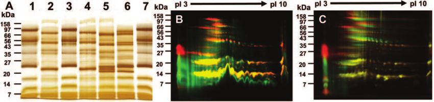
Proteomic Analysis of Venom from the Australian Brown Snake
be a group C prothrombin activator consisting of a Factor venom samples with IEF conditions scaled down to 40,000 V-h total
Xa-like enzymatic subunit and a non-enzymatic Factor Va-like and samples run on 12% SDS-PAGE gels (Bio-Rad Criterion format
gels).
subunit (12). Injection of purified pseutarin C results in mas-
Cy Dye Labeling—Pooled P. textilis venom samples from Queen-
sive disseminated intravascular coagulation within the body of sland or South Australia were precipitated in 8:1 acetone:methanol for
the snake’s prey, ultimately resulting in death (11). In addition 16 h at ⫺20 °C. Samples were centrifuged for 30 min at 16,000 ⫻ g
to the identification of a number of toxins, it has been reported to pellet protein, the supernatants were removed and discarded, and
that the venom composition from a single specimen can vary the protein pellets were allowed to air dry for 10 min. Proteins were
substantially from season to season (15). resuspended in 20 l of 20 mM Tris in UTC buffer to bring the pH to
⬃9. Cy3 and Cy5 monofunctional reactive Cy dyes (Amersham Bio-
In this study, we performed the first comprehensive pro- sciences) were used to label proteins according to the manufacturer’s
teomic analysis of the venom composition using two-dimen- instructions. Labeling reactions were quenched by the addition of 2 l
sional (2D) PAGE, mass spectrometry, and immunoblotting to of 10 mM lysine. Reaction mixtures were combined and loaded onto
identify the toxins. We also used post-translational modifica- 11-cm pH 3–10 IEF strips as described under “2D PAGE” above. After
tion-specific enzyme stains and antibodies that reveal the electrophoresis, gels were scanned on a Typhoon laser scanner
(Amersham Biosciences) for both Cy3 and Cy5, and the resultant
complexity and regional diversity of the venom. Post-transla-
images were overlaid and analyzed using PDQuest software
tional modifications examined include phosphorylation, (Bio-Rad).
acetylation, ␥-carboxylation, and glycosylation. Glycoproteins Identification of Venom Proteins Using MALDI-TOF and TOF-TOF
were further characterized by chemical and enzymatic degly- Mass Spectrometry and de Novo Peptide Sequencing—Protein spots
Downloaded from https://www.mcponline.org at UQ Library on April 27, 2020
cosylation and by lectin binding specificity. from silver-stained 2D PAGE were excised, washed in water, and
destained as described by Gharahdaghi et al. (18). Trypsin was
EXPERIMENTAL PROCEDURES added, and proteins were allowed to digest overnight at 37 °C prior to
extraction. Extracted peptides were dried, resuspended in 50% ACN,
Materials: Snake Venoms and Chemicals—Venom samples were
0.1% TFA, mixed 1:1 with matrix (␣-cyano-4-hydroxycinamic acid),
obtained from Venom Supplies Pty. Ltd. (Tanunda, South Australia).
and spotted on a MALDI plate. Samples were analyzed using a
Venom samples were collected over the course of a year and pooled
Voyager-DE STR mass spectrophotometer (Applied Biosystems) in
from over 40 individual snakes to reduce seasonal and individual
positive reflector mode with an accelerating voltage of 20,000 V. One
variation. Sample collection was performed for both Queensland and
hundred and fifty laser shots per spectrum were acquired in the mass
South Australian specimens to enable comparison of geographic
range 600 – 4000 Da. The 50 most intense peptide masses were
variation. Venoms were lyophilized and reconstituted at 10 mg/ml in
searched against the Chordata taxonomic subset of the National
PBS, 50% glycerol. Water was prepared using a Milli-Q system
Center for Biotechnology Information (NCBI) protein database using
(Millipore, Bedford, MA). FITC-labeled lectins were from Vector Lab-
oratories (Burlingame, CA). Cy dyes were from Amersham Bio- the Mascot search engine. MS peptide tolerance was 100 ppm, the
sciences. Pro-Q Diamond and Pro-Q Emerald were from Molecular search allowed for carbamidomethylated cysteine, and no other post-
Probes Inc. (Eugene, OR). Neuraminidase and peptidyl-N-glycosi- translational modifications were taken into account.
dase F (PNGase F) were from New England Biolabs (Beverly, MA). For TOF-TOF analysis, peptides were analyzed using a 4700 Pro-
O-Glycosidase was from Roche Applied Science. Antisera to various teomics Analyzer (Applied Biosystems) operated in positive ion re-
venom components were raised in rabbits or sheep in University of flectron mode. MS data were acquired using 2000 shots of a neo-
Queensland animal house facilities with appropriate ethical clear- dymium:yttrium-aluminum-garnet laser at 355 nm with a 200-Hz
ances. Acetylated lysine monoclonal antibody (mAb) was from Cell repetition rate and fixed intensity. The top 50 most intense peptides
Signaling Technology (Beverly, MA). mAb against ␥-carboxygluta- detected for each spot in the MS mode were automatically selected
mate (Gla) residues was from American Diagnostica Inc. (Greenwich, for MS/MS analysis using 3000 laser shots at a fixed intensity, which
CT). All other chemicals were from Sigma unless stated otherwise. was ⬃20% greater than that used for MS. MS/MS data were cali-
2D PAGE—Venom samples (500 g) were diluted to 400 l with brated against the MS/MS fragments of the m/z ⫽ 1296.685 Angio-
UTC buffer (7 M urea, 2 M thiourea, 4% CHAPS with a trace of tensin I peptide in the standards. MALDI-TOF/TOF MS/MS data were
bromphenol blue), 50 mM DTT, and 1% ampholytes (Biolytes 3–10, automatically analyzed using the GPS Explorer suite of software
Bio-Rad). Samples were mixed and centrifuged for 5 min at 13,000 ⫻ (Applied Biosystems). For each spot a combined MS and MS/MS
g to pellet insoluble material, and the supernatant was loaded onto analysis was performed in-house using a Mascot search engine and
IEF strips (Bio-Rad ReadyStrip, pH 3–10, 24 cm) for 16-h passive the Chordata taxonomic subset of Celera Discovery System data-
rehydration. Proteins were focused on a Bio-Rad IEF cell. The IEF base. MS peptide tolerance was 100 ppm, and MS/MS tolerance was
running conditions were: 250 V for 15 min, ramp to 8000 V for 3 h, 0.3 Da. The search allowed for carbamidomethylated cysteine and
hold at 8000 V for 90,000 V-h. After IEF, IPG strips were equilibrated oxidized methionine. For the purposes of protein identification, no
for 15 min in an equilibration buffer (50 mM Tris-HCl, pH 8.8, 6 M urea, other post-translational modifications were taken into account.
2% SDS, 30% glycerol, 2% DTT) followed by 15 min in an equilibra- For de novo peptide sequencing, MALDI-TOF/TOF MS/MS data
tion buffer that had DTT replaced with 2.5% iodoacetamide. IPG were opened in Data Explorer (Applied Biosystems, Version 4.2) and
strips were briefly rinsed in SDS-PAGE running buffer and embedded deisotoped, and raw text peak lists were exported. The peak lists
on top of 12% SDS-PAGE gels (Bio-Rad Protean Plus, 25 ⫻ 20 cm) were analyzed using the automatic de novo function of PEAKS Studio
and covered with 0.5% agarose. Gels were run at 200 V constant software Version 2.4 (19) (Bioinformatics Solutions Inc., Ontario, Can-
voltage until the bromphenol blue dye front reached the bottom of the ada). Contiguous stretches of seven or more amino acids with 100%
gel. Silver staining was performed as described by Shevchenko et al. confidence call using the default parameters of the softwares were
(16). Colloidal Coomassie staining was performed as described by collected and matched to the NCBI non-redundant protein database
Neuhoff et al. (17). For immunoblotting; amino-terminal sequencing; using the protein BLAST algorithm (www.ncbi.nlm.nih.gov/BLAST/).
phosphoprotein, glycoprotein, and lectin binding studies 11-cm IEF Amino-terminal Sequence Determination—Venom proteins were
strips (Bio-Rad ReadyStrip, pH 3–10, 11 cm) were used with 50-g electroblotted from 2D PAGE onto PVDF membrane (Pall Scientific,
380 Molecular & Cellular Proteomics 5.2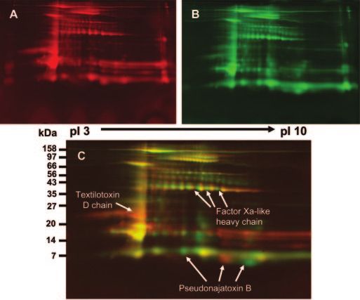
Proteomic Analysis of Venom from the Australian Brown Snake
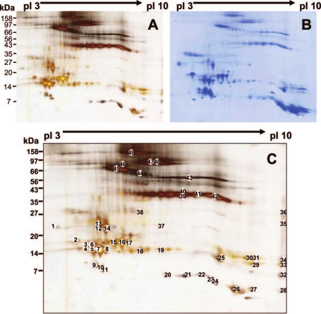
FIG. 1. 2D PAGE of P. textilis
(Queensland) venom. Duplicate sam-
ples of 500 g of venom proteins were
subjected to isoelectric focusing on
24-cm pH 3–10 IEF strips and subse-
quently resolved by 12% SDS-PAGE.
The silver-stained gel (A) shows stronger
staining intensity of high molecular size
proteins compared with the Coomassie-
stained equivalent gel (B). Forty-nine
protein spots were excised (C) for iden-
tification by mass spectrometry, de novo
Downloaded from https://www.mcponline.org at UQ Library on April 27, 2020
peptide sequencing, and NH2-terminal
sequencing (Supplemental Table 1).
Melbourne, Australia) in Towbin buffer at 15 V for 16 h. The membrane Enzymatic Deglycosylation—Venom samples were precipitated in
was briefly stained with Coomassie Blue, destained, washed exten- methanol:acetone (8:1) as above and air-dried prior to exposure to
sively with several changes of water for 2 h, and dried. Selected spots neuraminidase (New England Biolabs), O-glycosidase (New England
were excised from the membrane with a scalpel blade, and the Biolabs), or PNGase F (New England Biolabs) in buffers and condi-
NH2-terminal sequences were analyzed by Edman degradation using tions recommended by the manufacturer. Reactions were allowed to
an Applied Biosystems Model 492 sequencer (Applied Biosystems). proceed for 1 h at 37 °C.
The observed sequences were matched to the NCBI non-redundant
protein database using the protein BLAST algorithm (www.ncbi.nlm. RESULTS
nih.gov/BLAST/).
Immunoblotting—Proteins from 2D PAGE were electrotransferred Separation and Comparative Analysis of P. textilis Venom
to nitrocellulose membranes (Pall Scientific, Brisbane, Australia), and Proteins—Venom proteins from P. textilis (Queensland spec-
membranes were blocked by immersion in 5% skim milk in PBS, imen pool) were initially analyzed using 2D PAGE. Sample
0.05% Tween 20 (SM/PBS/T). Membranes were probed with rabbit or
sheep polyclonal antisera raised against specific venom proteins or
processing and isoelectric focusing conditions were chosen
venom protein complexes (26). The antisera were diluted 1:2000 in to give the greatest number of resolved protein spots per gel.
SM/PBS/T, and membranes were incubated overnight with gentle Over the pH range 3–10, 219 protein spots were detected
agitation. After washing in PBS/T for 3 ⫻ 10 min, membranes were using PDQuest software (Bio-Rad) from the silver-stained gel
incubated for 1 h in secondary antisera (goat anti-rabbit Ig/horserad- (Fig. 1A), whereas 192 spots were identified on the corre-
ish peroxidase or rabbit anti-sheep Ig/horseradish peroxidase,
Chemicon, Melbourne, Australia) diluted 1:5000 in PBS/T. After 5 ⫻
sponding Coomassie Blue-stained gel (Fig. 1B). The proteins
10-min washes in PBS/T, blots were developed using ECL reagent ranged in size from 5 kDa to greater than 200 kDa. Numerous
(Western Lightning, Perkin Elmer Life Sciences) and visualized with a horizontal trains of spots were evident especially in the mo-
chemiluminescence detector (FujiImager 3000, Fuji). lecular mass range greater than 30 kDa. These are suggestive
Lectin Binding Specificity—Glycosylation profiles of 2D PAGE-sep- of post-translational modifications of individual proteins. The
arated proteins transferred to nitrocellulose membrane were exam-
ined using a panel of eight lectins with FITC labels (Fluorescein Lectin
abundance of spots in the molecular mass range 10 –15 kDa
Kit I, Vector Laboratories) as described by Nawarak et al. (20). with low pI corresponds to that expected of PLA2s, which
Phosphoprotein and Glycoprotein Staining—Detection of phos- have already been shown to be present in P. textilis venom (6,
phoproteins and glycoproteins in 2D PAGE was performed using the 8). The overall pattern of staining was similar when colloidal
ProQ Diamond and ProQ Emerald fluorescent reagents, respectively Coomassie Blue staining was used; however, the relative
(Molecular Probes Inc.) according to the manufacturer’s instructions.
ProQ Emerald is a fluorescent-based form of the periodic acid-Schiff
intensity of some protein spots was different compared with
stain for carbohydrates that has a broad range of specificity for detection by silver staining. Both methods of staining are
glycol-containing molecules. compatible with further analysis by mass spectrometry and
Molecular & Cellular Proteomics 5.2 381Proteomic Analysis of Venom from the Australian Brown Snake
are likely between them to facilitate characterization of most
proteins in the venom.
Protein Identification by Mass Spectrometry—To identify
venom proteins, gel spots were excised from 2D gels and
digested with trypsin, and peptides were analyzed by MALDI-
TOF MS and MALDI-TOF/TOF MS/MS. Forty-nine spots were
selected from the 2D gels across a range of molecular
masses. The distribution of these is indicated in Fig. 1C, and
a complete list of the peptides and proteins identified appears
in Supplemental Table 1. A prominent protein represented as
a train of ⬃10 spots at 40 kDa (Fig. 1C, spots 39 – 42 and
adjacent spots) was identified as the Factor Xa-like heavy
chain. Another protein also present in the prothrombin acti-
vator complex of P. textilis, Factor Va-like protein, was iden-
tified at its expected molecular mass of ⬃160 kDa also as a
train of spots (Fig. 1C, spot 49 and adjacent spots). There
were several other Factor Va-like polypeptides corresponding
Downloaded from https://www.mcponline.org at UQ Library on April 27, 2020
to the A1 domain (heavy chain, 100 kDa; Fig. 1C, spots 47 and
FIG. 2. Regional diversity of P. textilis venom. Pooled venoms
48), A3 domain (light chain, 65 kDa; Fig. 1C, spot 45), and A2 from Queensland specimens (A) were labeled with Cy 3 dye, whereas
domain (52 kDa; Fig. 1C, spots 43 and 44). These were those from South Australia (B) were labeled with Cy 5 dye. Both Cy3
identified by the presence of tryptic peptides that mapped and Cy5 samples were combined and subjected to 2D gel electro-
specifically to the respective domains (Supplemental Table 1). phoresis. The gel was scanned at wavelengths specific for each Cy
The discrepancy between expected and observed molecular dye, and the images were false colored and superimposed (C) to
reveal protein spots unique to each region. Proteins common to both
size is thought to be due to potential N-glycosylation sites in regions are colored yellow. Red spots are unique to Queensland;
the A2 and A3 domains (12). Another protein identified in the green spots are unique to South Australia. Arrows indicate proteins
high molecular mass region of the gel was a single protein that appear at noticeably different levels.
spot identified as glucose-regulated protein 78 (GRP78; Fig.
1C, spot 46), a member of the heat shock protein 70 (HSP70) identified using specific antisera in a corresponding immuno-
family. The presence of GRP78 in snake venom has not been blot (Fig. 4E). Somewhat surprisingly, two isoforms of textilinin
reported previously. were identified migrating at an approximate molecular size of
A pseudechetoxin-like protein was identified by de novo 10 kDa (Fig. 1C, spots 9 and 10). Although multiple textilinin
peptide sequencing as a protein spot of 25 kDa and pI ⬃10 cDNAs have been reported for P. textilis, the size of predicted
(Fig. 1C, spot 35). Pseudechetoxin and its homolog pseudecin proteins based on these cDNAs is 6 –7 kDa, which is consid-
are members of the cysteine-rich secreted proteins that were erably lower than that observed here. As expected, several
previously identified in two related Australian snakes, neurotoxins were identified at their anticipated pI and molec-
Pseudechis australis and Pseudechis porphyriacus, respec- ular size (Fig. 1C, spots 20, 22, 24, 25, 26, 27, 28, 33, and 34).
tively (21). More recently, a cDNA clone corresponding to this Geographic Variation in Venom Composition—Previous re-
protein was identified from the venom gland of P. textilis in a ports have shown that the toxicity of P. textilis venom can vary
separate study (22). Additional members of this family have in a geographic manner (1, 23). To examine for geographic
also been identified in several other Australian snake species variation in venom composition we compared venom proteins
using cDNA cloning (22). Two de novo sequenced peptides in P. textilis from two geographically separated regions,
were used to identify protein spot 35 in the present study as Queensland and South Australia. Proteins from the two sam-
the pseudechetoxin-like protein from P. textilis. We also iden- ples were differentially labeled with Cy3 and Cy5 dyes to allow
tified what appeared to be several members of the PLA2 comparison within a single gel. Similar numbers of proteins
family at ⬃15 kDa and over a pI range of 4 – 6. Due to the were observed in both samples with greater intensity of label-
limited amount of sequence information in the NCBI data- ing of proteins in the lower molecular mass region compared
base, it was not possible to identify all PLA2s as individual with the silver or Coomassie Blue stain (Fig. 2, A and B).
isoforms. However, the differences in pI and size between the Overlay of the two images revealed that there was concord-
isoforms and previously reported data on the presence of ance between the labeled proteins in greater than 95% of
multiple PLA2 isoforms in P. textilis venom (8) supports the cases (Fig. 2C). There were notable differences in proteins in
presence of sequence variants. The four subunits of the tex- the molecular mass range of 5–10 kDa. It has been reported
tilotoxin complex were identified in multiple protein spots (Fig. that short and long neurotoxins and textilinins are known to
1C, spots 1, 3, 6, 12, 13, 14, 29, 30, 31, 33, and 36). These occur in this size range (3, 4, 9). By reference to Fig. 1C, we
molecules are members of the PLA2 family and were also propose the most notable differences to be an increase in
382 Molecular & Cellular Proteomics 5.2Proteomic Analysis of Venom from the Australian Brown Snake
Downloaded from https://www.mcponline.org at UQ Library on April 27, 2020
FIG. 3. Immunoblots of 2D separated venom proteins. Venom proteins were separated on 2D gels, and proteins were transferred to
nitrocellulose membrane for immunoblotting with antisera to prothrombinase complex (A), heavy chain of the Factor Xa-like protease (B), Gla
residues (C), textilinins (D), and textilotoxin (E).
abundance of the phospholipase A2, textilotoxin D chain (Fig. though the heavy chain was readily detectable in 2D gels
2C, arrow), and differences in the abundance of forms of the using silver and Coomassie Blue staining, the Factor Xa-like
long neurotoxin pseudonajatoxin B. Small differences were light chain was not evident in these gels. However, with the
also observed for the Factor Xa-like heavy chain suggesting prothrombin activator antisera, a complex staining pattern
subtle differences in post-translational modifications of this was also evident in a region corresponding to the light chain
train of spots. Our findings suggest that overall there is great of Factor Xa-like molecule. Because it has been reported that
similarity between the venoms from the two regions and that the light chains of Factor Xa from both mammalian species
the differences are unlikely to account for significant changes and snakes are post-translationally modified at their NH2 ter-
in overall toxicity. minus by ␥-carboxylation of glutamic acid residues (11), we
Protein Identification by 2D Immunoblotting—As an addi- used a monoclonal antibody that detects this modification.
tional approach to identifying proteins, antisera specific to The pattern observed with this antibody corresponded to a
various components of the venom were used to identify the region that was also observed with the prothrombin activator
composition and location of the individual proteins (Fig. 3). complex antiserum (Fig. 3C). This region also corresponds
The prothrombin activator complex in P. textilis is composed with the expected molecular size and pI of the Factor Xa-like
of a Factor Va-like molecule and a Factor Xa-like molecule. light chain.
Three different antibodies were used to examine the pro- A series of textilinin spots were detected using rabbit anti-
thrombin activator complex in the venom. These include a sera raised against purified textilinin 1. This pattern was con-
rabbit polyclonal antisera raised against the purified complex sistent with the presence of multiple isoforms of textilinin
(Fig. 3A), a sheep polyclonal antisera raised against a recom- identified in P. textilis venom gland using cDNA cloning (10).
binant form of the Factor Xa-like heavy chain (Fig. 3B), and a These spots corresponded very well to those identified by
mouse monoclonal antibody specific for Gla residues (Amer- mass spectrometry (see Fig. 1C). Multiple spots were de-
ican Diagnostica Inc.). tected for textilotoxin using rabbit antisera raised against the
Antisera against the prothrombin activator complex (Fig. purified complex at both acidic and basic pI values. This was
3A) detected three isoforms of the Factor Va-like molecule. consistent with mass spectrometric identification of textilo-
This corresponded well with the data in Fig. 1C where these toxin, which is composed of four related PLA2 molecules,
isoforms were identified by mass spectrometry, although the textilotoxins A–D (7). Textilotoxins A and B were identified by
antisera failed to detect isoforms corresponding to the A1 mass spectrometry at high pI (pH 9 –10). Antisera used here
domain (Fig. 1C, spots 47 and 48) and the A2 domain (Fig. 1C, detected spots in this region (Fig. 3E). The multiple forms
spot 43). The Factor Xa-like molecule was also identified as a detected at a lower pI (pH 3–5) corresponded well to textilo-
series of discrete spots as observed above using silver stain- toxins C and D (7), also identified by mass spectrometry. It is
ing (Fig. 1C, spots 39 – 42). Antisera raised against a recom- of interest that textilotoxins B and D were identified from
binant form of the heavy chain of Factor Xa-like molecule several distinct spots.
detected a train of protein spots identical to that seen with Post-translational Modifications—One of the intriguing ob-
antisera against prothrombin activator complex (Fig. 3B). Al- servations of the 2D gel separations of the P. textilis venom
Molecular & Cellular Proteomics 5.2 383Proteomic Analysis of Venom from the Australian Brown Snake
FIG. 4. Post-translational modification stains. Venom proteins were separated on 2D gels, and proteins were transferred to nitrocellulose
membrane for staining with fluorescent stains specific for phosphoproteins (A), total protein (B), and glycoproteins (C). Asterisks in A and B
indicate casein run as a phosphoprotein positive control lane on the edge of the second dimension gel.
proteins was that there were multiple horizontal trains of spots hydrolysis to remove sugars from the venom proteins resulted
in different regions of the gels. These were most apparent in in the appearance of protein smearing in both one-dimen-
the higher molecular mass region where both Factor Va-like sional and 2D gels (data not shown). This may have been due
and Factor Xa-like components were identified (Fig. 1C, spots to cleavage of peptide bonds as well as glycosidic bonds.
39 – 42). These suggested the presence of multiple isoforms To relate the presence of glycosylation to the appearance of
Downloaded from https://www.mcponline.org at UQ Library on April 27, 2020
differentiated either by amino acid composition, post-transla- trains of spots on 2D gels, we used Cy dye methodology. We
tional modifications, or a combination of both. One of the treated samples with and without neuraminidase and with and
most common post-translational modifications of proteins is without PNGase F and subjected them to 2D PAGE (Fig. 5, B
phosphorylation. We assayed for this modification using a and C, respectively). Both results showed a reduction in mo-
fluorescent stain, Pro-Q Diamond (Molecular Probes Inc.), lecular size for several trains of spots after deglycosylation
reported to be specific for phosphoproteins (24). The results with very little effect on the pI of the proteins. The reduction in
in Fig. 4A revealed the presence of several apparently phos- molecular size after deglycosylation was particularly evident
phorylated proteins. When the same gel was subsequently above 20 kDa where these trains were most apparent. Al-
stained with a total protein stain (Deep Purple, Amersham though not as clear, the lower molecular mass region showed
Biosciences), the phosphorylated spots correlated well with direct overlay (yellow) between sets of spots. We also exam-
some of the proteins detected (Fig. 4B). Several of the high ined blots for the presence of acetylated lysine residues using
molecular mass phosphorylated proteins correlated well with a mAb specific for this modification. Acetylation of lysine is an
Factor Va-like and Factor Xa-like isoforms on the basis of size important reversible modification controlling protein activity
and isoelectric point. A set of spots in the 15–20-kDa region (25). We saw no evidence of lysine acetylation on 2D blots
with pI ⬃4 –5 were consistent with phosphorylated PLA2s. (data not shown).
Glycoproteins were also detected in a separate gel (Fig. 4C) Lectin Binding Specificity—Fluorescently labeled (FITC)
using a specific stain (Pro-Q Emerald, Molecular Probes Inc.). lectins were used to detect glycoproteins on nitrocellulose
This revealed evidence of multiple proteins modified across membranes after 2D PAGE of venom proteins from P. textilis.
the range of different sizes and pI values. Because it was possible that peripheral sugars may be lost
Treatment of crude venom with neuraminidase and sepa- during the preparation and separation of samples for 2D
ration by single dimension SDS-PAGE revealed a shift in PAGE and subsequent electrotransfer to nitrocellulose, we
molecular size for most proteins above 20 kDa (Fig. 5, lanes 1 initially spotted different dilutions of crude venom onto nitro-
and 2) suggesting an abundance of sialic acids. We also used cellulose membrane and probed it with a selection of eight
the glycosidases PNGase F and O-glycosidase to distinguish lectins chosen to identify a wide range of specific sugar
between N-glycosidic and O-glycosidic linked sugars (Fig. 5, groups. Under these conditions, four of eight different lectins
lanes 6 and 3, respectively). PNGase F treatment resulted in a used showed no binding to venom proteins (results not
number of band shifts above 20 kDa in the treated (Fig. 5, lane shown). These four non-binding lectins were soybean agglu-
6) compared with untreated (lane 7) samples. On the other tinin (specific for N-acetylgalactosamine and galactose), Ulex
hand, no change was evident after treatment with O-glycosi- europeus I lectin (specific for fucose), elderberry bark lectin
dase alone (lane 3). Because it has been reported that O- (specific for ␣2–3-linked sialic acid), and peanut agglutinin
glycosidase cleavage is often more efficient after pretreat- (specific for galactose). It should also be noted that lectins can
ment with a sialidase, we used this combination of enzymes have additional structural requirements for binding (www.vec-
(lane 4). The pattern of proteins observed was identical to that torlabs.com/infopage.asp?dpID⫽24&locID⫽146). The four
observed with sialidase alone, providing additional support for lectins that showed binding by dot blot analysis were subse-
the lack of O-linked sugars in venom proteins. The combina- quently used to probe 2D separated venom proteins (Fig. 6,
tion of the three enzymes (lane 5) supports the presence of A–D). Incubation with ConA, which is specific for glucose and
sialic acid residues and N-linked sugars. The use of acid mannose, revealed the presence of multiple binding proteins
384 Molecular & Cellular Proteomics 5.2Proteomic Analysis of Venom from the Australian Brown Snake
FIG. 5. Enzymatic deglycosylation. Venom samples were treated with various glycosidic enzymes and resolved by SDS-PAGE to examine
for proteins with altered mobility. A, lane 1, untreated; lane 2, neuraminidase-treated; lane 3, O-glycosidase-treated; lane 4, neuraminidase- and
O-glycosidase-treated; lane 5, neuraminidase-, O-glycosidase-, and PNGase F-treated; lane 6, PNGase F-treated; lane 7, untreated. B, venom
samples were treated without and with neuraminidase and subsequently labeled with Cy3 and Cy5 dyes, respectively. The samples were
combined and subjected to 2D PAGE, and the gel was scanned at wavelengths specific for the two Cy dyes. The images were false colored
and superimposed to reveal proteins affected by deglycosylation. (Red, Cy3, without neuraminidase treatment; green, Cy5, with neuraminidase
treatment. Overlap of red and green spots appear as yellow.) C, venom samples were treated as in B except neuraminidase was replaced with
PNGase F.
Downloaded from https://www.mcponline.org at UQ Library on April 27, 2020
FIG. 6. Lectin binding. Venom pro-
teins were separated on 2D gels, and
proteins were transferred to nitrocellu-
lose membrane to examine binding of
various lectins. Lectin binding specifici-
ties are shown in parentheses: A, ConA
(mannose, glucose); B, WGA (sialic acid,
GlcNAc); C, D. biflorus agglutinin (Gal-
NAc); D, RCA120 (GalNAc, Gal).
above 30 kDa in size (Fig. 6A). Comparison of the pattern of Ricinus communis agglutinin (RCA120), which detects GalNAc
ConA binding with 2D silver-stained gels showed a strong and galactose. Again the Factor Xa-like protease heavy chain
resemblance indicating that Factor Va-like and Factor Xa-like was detected as well as discrete fragments corresponding to
proteins were being modified by glycosylation. Use of wheat Factor Va-like protein (Fig. 6D).
germ agglutinin (WGA), which detects GlcNAc and sialic ac-
ids, revealed a simpler and more defined pattern of staining. DISCUSSION
The lower molecular mass train of spots (pI 7–9) appeared to The venoms of Australian elapid snakes represent a rich
correspond to the Factor Xa-like heavy chain (Fig. 6B). It was source of highly active molecules that affect a variety of
notable that the train of spots immediately above the Factor homeostatic mechanisms, including the coagulation cascade,
Xa-like heavy chain, revealed by ConA staining, was unde- neuromuscular signaling, and the cardiovascular system (13).
tectable with the WGA lectin. A second lectin known to have A greater understanding of the mechanism of action of these
specificity for ␣2–3-linked sialic acid, Sambucus nigra agglu- molecules has potential for the development of human ther-
tinin, failed to stain any proteins (results not shown). Dolichos apeutic agents. To understand how these proteins function, it
biflorus agglutinin, which detects GalNAc, bound to the train is necessary to fractionate and identify the individual factors
of spots corresponding to the Factor Xa-like heavy chain (Fig. involved. In this study we carried out a proteomic profiling of
6C). The only other lectin that detected protein binding was the venom proteins from P. textilis and showed that although
Molecular & Cellular Proteomics 5.2 385Proteomic Analysis of Venom from the Australian Brown Snake
⬃200 of the protein spots could be identified by 2D PAGE and 2D gels could be due to the presence of post-translational
MS, as few as 20 individual proteins are represented due to modifications, proteolysis, protein splicing, or a combination
isoform variation. The number of proteins detected here was of these. Furthermore several cDNAs coding for the different
comparable to that observed in bovine milk, another secreted forms could be another explanation. The only evidence of
fluid where ⬎95% of spots on 2D PAGE are represented by as proteolysis observed was fragments of the Factor Va-like
few as six proteins again with extensive isoform variation (27). protein (Fig. 1C, spots 21, 23, and 32) corresponding to the
Series of horizontal trains of spots on 2D PAGE often rep- NH2-terminal region of the light chain. In the case of Factor
resent glycoforms of specific proteins. Glycoforms can vary in Va-like and Factor Xa-like proteins it is unlikely that multiple
site occupation and diversity of glycan structures, both of cDNA isoforms exist because we have isolated full-length
which contribute to the separation of protein isoforms in 2D cDNAs for these genes and found only two isoforms for
PAGE (28). These were especially apparent in the present Factor Xa-like and a single species for Factor Va-like (32, 33).
study in regions of the gels where the Factor Va-like and the In the present study, the use of mass spectrometry failed to
Factor Xa-like molecules were identified. The identity of these distinguish between these isoforms due to limited tryptic pep-
proteins was confirmed by selecting a series of individual tide sequence coverage. The Factor Xa-like protein is in de-
spots across the trains and analyzing them by MALDI-TOF velopment as a single agent procoagulant (33) (www.qrxphar-
MS, MALDI-TOF/TOF MS/MS, and NH2-terminal sequence ma.com/Q8010.htm) useful in situations for control of
analysis. Multiple trains of spots have also been observed in bleeding and tissue sealing following surgery. It has also been
Downloaded from https://www.mcponline.org at UQ Library on April 27, 2020
venoms from the Asian snakes Naja naja atra and Agkistrodon shown to induce blood clotting both in vitro and in vivo in a
halys (29) and for several snakes of the Elapidae and Viperidae calcium-independent manner.
families (20). Li et al. (29) also used 2D PAGE and observed The presence of different post-translational modifications,
trains of multiple spots that were subsequently shown to largely in the higher molecular mass region, was evident after
contain protein glycoforms using tandem MS. It is notable that treatment with both sialidase and PNGase F using SDS-PAGE
in the case of the A. halys venomous proteins, the spots were in a single dimension. Bands corresponding in size to both
distributed over a narrow range of isoelectric points of 4 – 6, Factor Va and Factor Xa-like proteins were clearly shifted in
whereas in the case of Naja, most proteins migrated to basic their migration after treatment by either enzyme (see Fig. 5A).
regions (pH ⬎ 7). In the present study of P. textilis, the venom This was confirmed using 2D gel electrophoresis and Cy dye
proteins were distributed over the entire pH range of 3–10. labeling (Fig. 5, B and C). When venom proteins were treated
Furthermore the molecular mass range for the A. halys pro- with PNGase F and subsequently labeled with Cy dyes, min-
teins extended from ⬍20 kDa to a maximum of ⬃60 kDa imal overlap between the trains of spots that correspond to
compared with a range observed in the present study from 5 Factor Va-like protein was seen (Fig. 5C). This treatment did
to 160 kDa. The difference in the higher molecular mass not reduce the number of individual species but simply led to
region can be largely accounted for by the presence of Factor a shift in their overall migration indicative of a loss of acidic
Va-like proteins in P. textilis that are not present in A. halys. sugar groups. The continuing presence of multiple spots un-
This is consistent with the different mechanisms used by the der these conditions supports the presence of other post-
two snakes to interfere with the mammalian coagulation proc- translational modifications on this molecule. We provided ev-
ess. The most abundant protein complex in P. textilis venom idence here that Factor Va-like protein is also modified by
is the prothrombin activator complex, which is composed of N-glycosylation and to a lesser extent by phosphorylation. It
Factor Va-like and Factor Xa-like proteins that activate pro- seems likely that the trains of spots observed for P. textilis
thrombin to initiate coagulation (11). On the other hand A. Factor Xa-like proteins represent similar post-translational
halys has largely anticoagulant activity. The major factors modifications. The multiple species observed for other pro-
responsible for this anticoagulant activity are a 65-kDa inhib- teins is unlikely to be explained by such modifications. For
itor of platelet aggregation (30) and an L-amino acid oxidase example in the case of PLA2s, it is more likely to be due to
that impairs intrinsic clotting by inhibiting Factor IX (31). multiple isoforms for which there is evidence in P. textilis (7, 8).
Although the prothrombinase complex is abundant in P. Furthermore in the Cy dye overlay experiments there seemed
textilis, it has been reported that variation in the composition to be a high degree of concordance between treated and
of the venom from a single snake undergoes seasonal varia- untreated samples for the PLA2s.
tion (15). In that study, coagulant activity dropped to a mini- One of the characteristics of the light chain of the Factor
mum during the summer months; however, they did not de- Xa-like protease is a series of Gla residues in the NH2-terminal
termine the levels of the individual proteins in the region of the molecule. This modification is conserved across
prothrombinase complex. By using venom pooled from over different species (34, 32). The Gla residues chelate calcium
40 individual snakes, collected over the course of a year, we ions and induce a conformational transition in the Gla domain
minimized the variation inherent in individual samples. The that results in phospholipid membrane binding and stimula-
multiple spots observed for Factor Va-like and Factor Xa-like tion of the procoagulant activity of the prothrombinase com-
proteins as well as for several other proteins separated on the plex (35). As well as mammalian coagulation factors, Gla
386 Molecular & Cellular Proteomics 5.2Proteomic Analysis of Venom from the Australian Brown Snake
domains have been found in the toxins of predatory cone 1), MALDI-TOF/TOF MS/MS analysis of tryptic peptides iden-
snails of the genus Conus (36, 37). Recently precursors of tified individual spots corresponding to known PLA2s, includ-
Gla-containing conotoxins have been found to contain a car- ing the four PLA2 subunits of the presynaptic neurotoxin
boxyl-terminal recognition site that directs ␥-carboxylation textilotoxin.
(38), thus providing evidence that ␥-carboxylation occurs as a We also identified textilinins and ␣-neurotoxins among the
post-translational rather than cotranslational event. The use of low molecular mass proteins and found ␣-neurotoxins among
antisera against the prothrombinase complex detected a dif- two PLA2 spots. Textilinins have been shown to inhibit plas-
fuse region of staining that corresponded in size to the light min, a key enzyme in the fibrinolytic pathway involved in
chain of Factor Xa-like protein. This was supported by the dissolving blood clots (9). Textilinins share ⬃45% protein
observation of coincident reactivity using a Gla-specific anti- sequence similarity with aprotinin (Trasylol姞, Bayer) the cur-
body (Fig. 3, A and C). The Factor Xa-like heavy chain was rent “protein-based” antifibrinolytic agent on the market.
readily detectable on 2D gels by protein staining and also Aprotinin is derived from bovine lung and is otherwise known
identified with antisera against this protein and the whole as bovine pancreatic trypsin inhibitor. As the name suggests
complex (Fig. 3, A and B). However, protein staining alone the inhibitor is able to inhibit trypsin and is also shown to
failed to detect the presence of the light chain possibly due to inhibit a broad range of trypsin-like proteases, many of which
the presence of the charged Gla residues. The Factor Va- and are involved in the control of fibrinolysis and hemostasis. We
Factor Xa-like proteins represented only a small proportion of have previously isolated textilinins from the venom by con-
Downloaded from https://www.mcponline.org at UQ Library on April 27, 2020
the total number of proteins separated on the 2D gels in this ventional chromatography and by cDNA cloning (10, 11). The
study yet comprised a significant proportion by mass. This 2D PAGE separation described here distributed the textilinins
was evident in the prothrombinase complex Western blot into two distinct spots of ⬃9 kDa. However, use of immuno-
(Fig. 3A). blotting detected what appeared to be four separate species
Another protein identified in the higher molecular mass in the corresponding region (Fig. 4D). This is not surprising
region was GRP78, a member of the HSP70 family of chap- because six separate cDNAs coding for textilinins have been
erones and protein folding catalysts (39). GRP78 may play a described in P. textilis (10) with four of them having pI values
role in facilitating the assembly of multimeric protein com- (pI 4 –5) corresponding with those observed here. The ob-
plexes known to exist in the venom. GRP78 has not been served molecular size of 9 kDa corresponds to the precursor
identified previously in snake venom; however, protein se- protein predicted from the full-length cDNA, which contains
quences for the related proteins HSP70 (NCBI accession an open reading frame of 61 amino acids and a propeptide of
number AAY33973.1) and HSP90 (NCBI accession number 22 amino acids (10). On the other hand the mature textilinin
AAY67995.1) have been found in the Australian taipan, proteins would be expected to migrate at ⬃6 –7 kDa in size
Oxyuranus scutellatus. However, these share only 50 and once the propeptide had been removed. This discrepancy
21% sequence identity, respectively, with the GRP78 se- might be explained by failure to completely reduce the three
quence identified by tandem mass spectrometry in the pres- disulphide linkages that occur in these proteins, post-trans-
ent study. lational modifications, inaccurate sizing in this molecular
The majority of venom proteins were observed in the ⬍5– mass range, or abnormal migration of the protein. Seven
25-kDa region. Mass spectrometry identified a number of spots were identified as neurotoxins based on NH2-terminal
PLA2s across the entire pI range at a molecular size of 12–25 sequencing and MALDI-TOF/TOF MS/MS analysis. These
kDa. These enzymes catalyze the hydrolysis of insoluble correspond to both long-chain and short-chain ␣-neurotoxins
phospholipid substrates at the sn-2 position of the glycerol previously identified in P. textilis (3, 4). Five spots were iden-
backbone giving rise to fatty acids and lysophospholipids. In tified as the long-chain neurotoxin pseudonajatoxin B, which
addition, PLA2s from Australian elapids inhibit platelet aggre- is consistent with reports of at least four isoforms in the
gation, they have procoagulant and myotoxic activities, and venom of P. textilis (42). These protein spots were observed at
they are presynaptic neurotoxins (for a review, see Ref. 40). a range of molecular size from 6 to 15 kDa, whereas the
Accelerated evolution within the snake PLA2 family has gen- mature proteins are expected to be 8 kDa in size. Long-chain
erated multiple isoforms that have resulted in diverse biolog- ␣-neurotoxins typically have five disulphide bridges that if not
ical activities (41). Armugam et al. (8) have reported four new fully denatured may account for altered electrophoretic mi-
PLA2s in the venom of P. textilis bringing the total number of gration. Short-chain neurotoxins contain four disulphide
isoforms reported for this snake to eight. RT-PCR analysis of bonds and share a similar three-finger-loop structure with
P. textilis venom gland RNA has also identified additional long-chain neurotoxins (43). The two short-chain neurotoxins
PLA2 isoforms (23). Of 49 protein spots from 2D PAGE ana- identified correspond to ␣-neurotoxin 6, one of six isoforms
lyzed by mass spectrometry and NH2-terminal sequencing, previously identified by cDNA cloning (4). Interestingly both
21 contained peptides from PLA2s. Although it was not pos- were identified in 2D gel spots that also contained a PLA2, the
sible to identify them as individual isoforms due to incomplete A-chain of textilotoxin. A complex containing an ␣-neurotoxin,
peptide sequence coverage (Fig. 1 and Supplemental Table a PLA2, and a serine protease inhibitor has been identified in
Molecular & Cellular Proteomics 5.2 387Proteomic Analysis of Venom from the Australian Brown Snake
the related Australian elapid O. scutellatus (44). However, venom of P. textilis and to extend these studies to a variety of
purification of the major serine protease inhibitor present in other elapid snake venoms.
P. textilis venom, textilinin, has found no evidence of a similar
Acknowledgment—We acknowledge Chris Wood for NH2-terminal
complex.2 The two ␣-neurotoxin 6 spots were identified at a pI
sequencing.
of ⬎10 and at molecular sizes of 12 and 14 kDa, which are
significantly greater than their predicted size of 8.5 kDa. * This work was supported by QRxPharma and the Australian Re-
Our lectin binding studies showed the venom to be rich in search Council Linkage Program. The costs of publication of this
ConA-binding (mannose- and glucose-specific) glycoproteins article were defrayed in part by the payment of page charges. This
article must therefore be hereby marked “advertisement” in accord-
in the higher molecular size region. This is consistent with a
ance with 18 U.S.C. Section 1734 solely to indicate this fact.
study by Nawarak et al. (20) who used ConA-agarose to □ S The on-line version of this article (available at http://www.
isolate such glycoproteins from N. naja venom. The WGA mcponline.org) contains supplemental material.
lectin (sialic acid- and GlcNAc-specific) and RCA120 lectin ‡‡ To whom correspondence should be addressed: The Queens-
(galactose- and GalNAc-specific) showed similar binding of land Cancer Fund Research Unit, The Queensland Inst. of Medical
Research, P. O. Box Royal Brisbane Hospital, Herston, Brisbane
both the Factor Va-like and Factor Xa-like molecules. It seems
4029, Australia. Tel.: 617-3362-0341; Fax: 617-3362-0106; E-mail:
unlikely that these glycosylations are required for prothrom- martin.lavin@qimr.edu.au.
binase activity as neither of the mammalian homologs are
glycosylated. The large amount of glycosylation present, as REFERENCES
Downloaded from https://www.mcponline.org at UQ Library on April 27, 2020
confirmed by the glycosidase experiments (Fig. 5, A, B, and 1. Sutherland, S., and Tibballs, J. (2001) in Australian Animal Toxins. The
C), may be required for enzyme stability rather than function. Creatures, Their Toxins and Care of the Poisoned Patient, 2nd Ed., pp.
17–26, Oxford University Press, Melbourne, Australia
It is interesting to note that the mammalian coagulation factor 2. Barnett, D., Howden, M. E. H., and Spence, I. (1979) Pre- and post-synaptic
homologs are found in the activated form in snake venoms neurotoxins in the venom of the common brown snake (Pseudonaja
(12), which is not the case in mammalian circulation. These textilis). Proc. Aust. Physiol. Pharmacol. Soc. 10, 240
3. Tyler, M. I., Spence, I., Barnet, D., and Howden, M. E. (1987) Pseudonaja-
procoagulant proteins are useful in treating various throm- toxin b: unusual amino acid sequence of a lethal neurotoxin from the
botic and hemostatic conditions (45). venom of the Australian common brown snake, Pseudonaja textilis. Eur.
Examples of clinically useful venom proteins from other J. Biochem. 166, 139 –143
4. Gong, N., Armugam, A., and Jeyaseelan, K. (1999) Postsynaptic short-
snakes include contortrostatin from the Southern Copper- chain neurotoxins from Pseudonaja textilis. cDNA cloning, expression
head viper that is being tested as an anticancer agent and and protein characterization. Eur. J. Biochem. 265, 982–989
Ancrod from American pit viper that is being tested as an 5. Coulter, A. R., Broad, A. J., and Sutherland, S. K. (1979) Isolation and
properties of a high molecular weight neurotoxin from the eastern brown
anticoagulant for stroke victims. Another drug, Integrilin, is a snake (Pseudonaja textlilis), in Neurotoxins, Fundamental and Clinical
small molecule based on the RGD peptide motif found in a Advances (Chubb, I. W., and Geffen, L. B., eds) p. 260, Adelaide Univer-
platelet aggregation inhibitor from rattlesnake venom and has sity Union Press, Adelaide, Australia
6. Su, M. J., Coulter, A. R., Sutherland, S. K., and Chang, C. C. (1983) The
been approved by the United States Food and Drug Admin- presynaptic neuromuscular blocking effect and phospholipase A2 activ-
istration to treat patients with severe chest pain, minor heart ity of textilotoxin, a potent toxin isolated from the venom of the Australian
attacks, and other cardiovascular conditions. brown snake, Pseudonaja textilis. Toxicon 21, 143–151
7. Pearson, J. A., Tyler, M. I., Retson, K. V., and Howden, M. E. (1993) Studies
The results described here represent the first approach to on the subunit structure of textilotoxin, a potent presynaptic neurotoxin
identifying all of the venom proteins in P. textilis. Represen- from the venom of the Australian common brown snake (Pseudonaja
tatives of all the major classes were identified after 2D gel textilis). 3. The complete amino-acid sequences of all the subunits.
Biochim. Biophys. Acta 1161, 223–229
electrophoresis using a combination of immunoblotting, 8. Armugam, A., Gong, N., Li, X., Siew, P. Y., Chai, S. C., Nair, R., and
MALDI-TOF MS and MALDI-TOF/TOF MS/MS analysis, and Jeyaseelan K. (2004) Group IB phospholipase A2 from Pseudonaja tex-
NH2-terminal sequencing. A common characteristic of these tilis. Arch. Biochem. Biophys. 421, 10 –20
9. Masci, P. P., Whitaker, A. N., Sparrow, L. G., de Jersey, J., Winzor, D. J.,
2D gel separations of venom proteins was the detection of Watters, D. J., Lavin, M. F., and Gaffney, P. J. (2000) Textilinins from
multiple trains of spots corresponding to a single protein. This Pseudonaja textilis textilis. Characterization of two plasmin inhibitors that
was shown to be the case by immunoblotting and by mass reduce bleeding in an animal model. Blood Coagul. Fibrinolysis 11,
385–393
spectrometry. We also demonstrated that these were at least 10. Filippovich, I., Sorokina, N., Masci, P. P., de Jersey, J., Whitaker, A. N.,
partially generated by post-translational modifications and by Winzor, D. J., Gaffney, P. J., and Lavin, M. F. (2002) A family of textilinin
amino acid variation. Two of the identified proteins, the pro- genes, two of which encode proteins with antihaemorrhagic properties.
Br. J. Haematol. 119, 376 –384
coagulant Factor Xa-like protein and the plasmin inhibitor 11. Masci, P. P., Whitaker, A. N., and de Jersey, J. (1988) Purification and
textilinin, are currently in development as human therapeutic characterization of a prothrombin activator from the venom of the Aus-
agents (www.qrxpharma.com/pipeline.htm) and are the sub- tralian brown snake, Pseudonaja textilis textilis. Biochem. Int. 17,
825– 835
ject of international patent filings. Both of these proteins were 12. Rao, V. S., and Kini, R. M. (2002) Pseutarin C, a prothrombin activator from
shown to have multiple isoforms expressed in the venom. The Pseudonaja textilis venom: its structural and functional similarity to mam-
challenge ahead is to identify all of the other proteins in the malian coagulation factor Xa-Va complex. Thromb. Haemostasis 88,
611– 619
13. Fry, B. G. (1999) Structure-function properties of venom components from
2
P. Masci, personal communication. Australian elapids. Toxicon 37, 11–32
388 Molecular & Cellular Proteomics 5.2Proteomic Analysis of Venom from the Australian Brown Snake
14. Southcott, R. N., and Coulter, A. R. (1979) The action of textilon on neuro- Purification and characterization of platelet aggregation inhibitor com-
muscular transmission in the murine diaphragm, in Neurotoxins, Funda- ponent from venom of Agkistrodon halys pallas. Zhongguo Shi Yan Xue
mental and Clinical Advances (Chubb, I. W., and Geffen, L. B., eds) p. Ye Xue Za Zhi 12, 194 –198
272, Adelaide University Union Press, Adelaide, Australia 31. Sakurai, Y., Shima, M., Matsumoto, T., Takatsuka, H., Nishiya, K., Kasuda,
15. Williams, V., and White, J. (1992) Variation in the composition of the venom S., Fujimura, Y., and Yoshioka, A. (2003) Anticoagulant activity of M-
from a single specimen of Pseudonaja textilis (common brown snake) LAO, L-amino acid oxidase purified from Agkistrodon halys blomhoffii,
over one year. Toxicon 30, 202–206 through selective inhibition of factor IX. Biochim. Biophys. Acta 1649,
16. Shevchenko, A., Wilm, M., Vorm, O., and Mann, M. (1996) Mass spectro- 51–57
metric sequencing of proteins from silver-stained polyacrylamide gels. 32. St Pierre, L., Masci, P. P., Filippovich, I., Sorokina, N., Marsh, N., Miller,
Anal. Chem. 68, 850 – 858 D. J., and Lavin, M. F. (2005) Comparative analysis of prothrombin
17. Neuhoff, V., Arold, N., Taube, D., and Ehrhardt, W. (1988) Improved staining activators from the venom of Australian elapids. Mol. Biol. Evol. 22,
of proteins in polyacrylamide gels including isoelectric focusing gels with 1853–1864
clear background at nanogram sensitivity using Coomassie Brilliant Blue 33. Filippovich, I., Sorokina, N., St Pierre, L., Flight, S., de Jersey, J., Perry, N.,
G-250 and R-250. Electrophoresis 9, 255–262 Masci, P. P., and Lavin, M. F. (2005) Cloning and functional expression
18. Gharahdaghi, F., Weinberg, C. R., Meagher, D. A., Imai, B. S., and Mische, of venom prothrombin activator protease from Pseudonaja textilis with
S. M. (1999) Mass spectrometric identification of proteins from silver- whole blood procoagulant activity. Br. J. Haematol. 131, 237–246
stained polyacrylamide gel: a method for the removal of silver ions to 34. Joseph, J. S., Valiyaveettil, M., Gowda, D. C., and Kini, R. M. (2003)
enhance sensitivity. Electrophoresis 20, 601– 605 Occurrence of O-linked Xyl-GlcNAc and Xyl-Glc disaccharides in troca-
19. Ma, B., Zhang, K., Hendrie, C., Liang, C., Li, M., Doherty-Kirby, A., and rin, a factor Xa homolog from snake venom. J. Thromb. Haemostasis 1,
Lajoie, G. (2003) PEAKS: powerful software for peptide de novo se- 545–550
quencing by tandem mass spectrometry. Rapid Commun. Mass Spec- 35. Freedman, S. J., Blostein, M. D., Baleja, J. D., Jacobs, M., Furie, B. C., and
trom. 17, 2337–2342 Furie, B. (1996) Identification of the phospholipid binding site in the
Downloaded from https://www.mcponline.org at UQ Library on April 27, 2020
20. Nawarak, J., Phutrakul, S., and Chen, S. T. (2004) Analysis of lectin-bound vitamin K-dependent blood coagulation protein factor IX. J. Biol. Chem.
glycoproteins in snake venom from the Elapidae and Viperidae families. 271, 16227–16236
J. Proteome Res. 3, 383–392 36. McIntosh, J. M., Olivera, B. M., Cruz, L. J., and Gray, W. R. (1984) ␥-Car-
21. Yamazaki, Y., Brown, R. L., and Morita, T. (2002) Purification and cloning of boxyglutamate in a neuroactive toxin. J. Biol. Chem. 259, 14343–14346
toxins from elapid venoms that target cyclic nucleotide-gated ion chan- 37. Hansson, K., Furie, B., Furie, B. C., and Stenflo, J. (2004) Isolation and
nels. Biochemistry 41, 11331–11337 characterization of three novel Gla-containing Conus marmoreus venom
22. St Pierre, L., Woods, R., Earl, S., Masci, P. P., and Lavin, M. F. (2006) peptides, one with a novel cysteine pattern. Biochem. Biophys. Res.
Identification and analysis of venom gland-specific genes from the Commun. 319, 1081–1087
coastal taipan (Oxyuranus scutellatus) and related species. Cell. Mol. Life 38. Brown, M. A., Begley, G. S., Czerwiec, E., Stenberg, L. M., Jacobs, M.,
Sci., in press Kalume, D. E., Roepstorff, P., Stenflo, J., Furie, B. C., and Furie, B. (2005)
23. Flight, S., Mirtschin, P., and Masci, P. (2006) Comparison of active venom Precursors of novel Gla-containing conotoxins contain a carboxy-termi-
components between Eastern brown snakes collected from South Aus- nal recognition site that directs ␥-carboxylation. Biochemistry 44,
tralia and Queensland. Ecotoxicology, in press 9150 –9159
24. Steinberg, T. H., Agnew, B. J., Gee, K. R., Leung, W. Y., Goodman, T., 39. Mayer, M. P., and Bukau, B. (2005) Hsp70 chaperones: cellular functions
Schulenberg, B., Hendrickson, J., Beechem, J. M., Haugland, R. P., and and molecular mechanism. Cell. Mol. Life Sci. 62, 670 – 684
Patton, W. F. (2003) Global quantitative phosphoprotein analysis using 40. Valentin, E., and Lambeau, G. (2000) What can venom phospholipases A2
Multiplexed Proteomics technology. Proteomics 3, 1128 –1144 tell us about the functional diversity of mammalian secreted phospho-
25. Polevoda, B., and Sherman F. (2002) The diversity of acetylated proteins. lipases A2? Biochimie (Paris) 82, 815– 831
Genome Biol. 3, reviews0006 41. Jeyaseelan, K., Armugam, A., Donghui, M., and Tan, N. H. (2000) Structure
26. Masci, P. (2000) Studies of Coagulation and Fibrinolysis Using Australian and phylogeny of the venom group I phospholipase A2 gene. Mol. Biol.
Snake Venoms: from Molecular Toxinology to Novel Therapeutic Agents. Evol. 17, 1010 –1021
Ph.D. thesis, University of Queensland, Brisbane, Australia 42. Gong, N., Armugam, A., Mirtschin, P., and Jeyaseelan, K. (2001) Cloning
27. Holland, J. W., Deeth, H. C., and Alewood, P. F. (2005) Analysis of O- and characterization of the pseudonajatoxin b precursor. Biochem. J.
glycosylation site occupancy in bovine -casein glycoforms separated 358, 647– 656
by two-dimensional gel electrophoresis. Proteomics 5, 990 –1002 43. Endo, T., and Tamiya, N. (1991) Structure-function relationships of post
28. Packer, N. H., Lawson, M. A., Jardine, D. R., Sanchez, J. C., and Gooley, synaptic neurotoxins from snake venoms, in Snake Toxins (Harvey, A. L.,
A. A. (1998) Analyzing glycoproteins separated by two-dimensional gel ed) pp. 165–222, Pergamon Press, New York
electrophoresis. Electrophoresis 19, 981–988 44. Possani, L. D., Martin, B. M., Yatani, A., Mochca-Morales, J., Zamudio,
29. Li, S., Wang, J., Zhang, X., Ren, Y., Wang, N., Zhao, K., Chen, X., Zhao, C., F. Z., Gurrola, G. B., and Brown, A. M. (1992) Isolation and physiological
Li, X., Shao, J., Yin, J., West, M. B., Xu, N., and Liu, S. (2004) Proteomic characterization of taicatoxin, a complex toxin with specific effects on
characterization of two snake venoms: Naja naja atra and Agkistrodon calcium channels. Toxicon 30, 1343–1364
halys. Biochem. J. 384, 119 –127 45. Kini, R. M., Rao, V. S., and Joseph, J. S. (2001) Procoagulant proteins from
30. Liu, P., Xu, J. M., Jin, Q., Wang, Y. Q., Zhu, H., and Zhou, Y. C. (2004) snake venoms. Haemostasis 31, 218 –224
Molecular & Cellular Proteomics 5.2 389You can also read