Characterization of Chenopodin Isoforms from Quinoa Seeds and Assessment of Their Potential Anti-Inflammatory Activity in Caco-2 Cells
←
→
Page content transcription
If your browser does not render page correctly, please read the page content below
biomolecules

Article
Characterization of Chenopodin Isoforms from
Quinoa Seeds and Assessment of Their Potential
Anti-Inflammatory Activity in Caco-2 Cells
Jessica Capraro 1,† , Stefano De Benedetti 1,† , Marina Di Dio 1 , Elisa Bona 2 , Ambra Abate 3 ,
Paola Antonia Corsetto 4 and Alessio Scarafoni 1, *
 1   Department of Food, Environmental and Nutritional Sciences, Università degli Studi di Milano, 20122
     Milano, Italy; jessica.capraro@unimi.it (J.C.); stefano.debenedetti@unimi.it (S.D.B.);
     marinadidio93@gmail.com (M.D.D.)
 2   Department of Sciences and Technology Innovation, Università del Piemonte Orientale, 13100 Vercelli, Italy;
     elisa.bona@uniupo.it
 3   Ambulatorio Polispecialistico Casazza, Casazza 24060, Italy; ambra.abate@postecert.it
 4   Department of Pharmacological and Biomolecular Sciences, Università degli Studi di Milano,
     20122 Milano, Italy; paola.corsetto@unimi.it
 *   Correspondence: alessio.scarafoni@unimi.it
 †   Both Authors contributed equally to this work.

 Received: 21 April 2020; Accepted: 18 May 2020; Published: 21 May 2020

 Abstract: Several food-derived molecules, including proteins and peptides, can show bioactivities
 toward the promotion of well-being and disease prevention in humans. There is still a lack of
 information about the potential effects on immune and inflammatory responses in mammalian
 cells following the ingestion of seed storage proteins. This study, for the first time, describes the
 potential immunomodulation capacity of chenopodin, the major protein component of quinoa seeds.
 After characterizing the molecular features of the purified protein, we were able to separate two
 different forms of chenopodin, indicated as LcC (Low charge Chenopodin, 30% of total chenopodin)
 and HcC (High charge Chenopodin, 70% of total chenopodin). The biological effects of LcC and HcC
 were investigated by measuring NF-κB activation and IL-8 expression studies in undifferentiated
 Caco-2 cells. Inflammation was elicited using IL-1β. The results indicate that LcC and HcC show
 potential anti-inflammatory activities in an intestinal cell model, and that the proteins can act
 differently, depending on their structural features. Furthermore, the molecular mechanisms of action
 and the structural/functional relationships of the protein at the basis of the observed bioactivity were
 investigated using in silico analyses and structural predictions.

 Keywords: seed storage proteins; protein structure and function; food bioactives; Chenopodium
 quinoa Willd

1. Introduction
     Quinoa (Chenopodium quinoa Willd.) seeds store about 12%–15% of proteins with high biological
value [1,2]. The 11S globulins (about 37%–38% of the total proteins) and the 2S albumins (25%–31%)
are the two major storage proteins in quinoa seed. The 11S globulin of quinoa seeds is also known
by the trivial name of chenopodin, with an estimated native Mr of about 320 kDa [1,3]. The 3D
structure of chenopodin has not been yet determined. However, it likely assumes the typical hexameric
quaternary structure of all 11S seed storage globulins, made of nearly identical subunits. Each subunit
is composed of two polypeptides (designated as α and β chains) with Mr around 30–40 and 20–23 kDa,
respectively, linked by one disulfide bond [4,5]. The two chains of one subunit are encoded by a

Biomolecules 2020, 10, 795; doi:10.3390/biom10050795                         www.mdpi.com/journal/biomolecules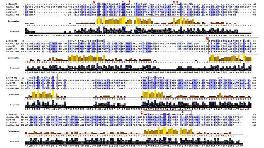
Biomolecules 2020, 10, 795                                                                        2 of 15

single gene, whose primary translation product (precursor) is later cleaved by a specific protease [6].
Like other seed storage proteins, chenopodin is codified by a family of genes, all expressed during
seed development, and the native assembly of the holoprotein originates from the association of the
resulting chains [5]. The hexameric structure arises from the assembly of two trimers formed by
precursors only. The proteolytic processing of the precursors is the signal for two trimers to interact
and form the hexamer [6].
     Quinoa is an important domesticated crop of the Andean region, which recently has had a growing
consumption in many countries [7,8]. The health benefits of quinoa seeds in human nutrition have
been extensively reported [9], such that this crop is now indicated as a promising source for the
development of functional foods and nutraceutical products [9–11]. Indeed, it is well-established that
several food-derived molecules, including proteins and peptides, can show bioactivities toward the
promotion of well-being and disease prevention in humans [12,13]. However, there is still a lack of
information about the potential effects on immune and inflammatory responses in mammalian cells of
proteins from quinoa seeds [12,14].
     Inflammation is a protective nonspecific response of the immune system that can be triggered by
a variety of factors, playing an important role in the body defense [15]. The nuclear factor κB (NF-κB)
is a key agent in the beginning and maintenance of the inflammatory response in various tissues,
including the intestinal mucosa [16,17]. NF-κB may target inflammation by inducing expression of
inflammatory cytokines, chemokines, and adhesion molecules, and by regulating cell proliferation
and differentiation [16,17]. Usually, inflammation resolves in a timely manner to avoid deleterious
consequences [18]. Complete resolution of an acute inflammatory response and return to homeostasis
are key processes in maintaining good health. Increased activation of NF-κB has often been detected
as a hallmark of chronic inflammation, a condition ultimately leading to different inflammatory
diseases [17,18]. Therefore, the downregulation of NF-κB activation may represent an attractive
approach for anti-inflammatory therapies of the gut [19].
     The present work pushes ahead with our line of research on the possible metabolic effects
of food-derived molecules. In particular, this study aimed to investigate the potential capacity of
chenopodin to modulate the NF-kb pathway in a human intestinal cell model following the trigger
of inflammation. Furthermore, we speculated on the molecular mechanisms of action and on the
structural/functional relationships of the protein at the basis of the observed bioactivity.

2. Materials and Methods

2.1. Chenopodin Extraction and Purification
     The procedure was a modification of an unpublished protocol [20], in turn adapted from the
original method for legume globulin extraction [21]. Quinoa seeds (var. Titicaca) were milled to a flour
and sieved through a 60-mesh filter. All following procedures were performed at 4 ◦ C. The flour was
extracted twice with distilled water (1:20, w/v) for 4 h under stirring. The insoluble pellet obtained
following centrifugation was then resuspended in the ratio 1:20 (w/v) in a 50 mM sodium phosphate
buffer with pH of 7.5 containing 500 mM NaCl. The salt soluble globulins were extracted overnight
under stirring. The suspension was centrifuged at 10,000× g for 30 min. The ppt was discarded.
     The supernatant was desalted using a Sephadex G-50 column, equilibrated in the 50 mM sodium
phosphate buffer with pH of 7.5, and the proteins were separated by ion exchange chromatography
(IEC) using a DEAE-cellulose column (20 × 180 mm) equilibrated with a 50 mM Tris-HCl buffer with
pH of 8.0 (2 mg protein/mL resin). The protein elution was obtained using the same buffer with
stepwise addition of 100 and 250 mM NaCl to avoid any chenopodin fractionation. Alternatively,
chenopodin was fractionated adopting the same conditions but with stepwise addition of 100, 150,
and 250 mM NaCl. The fractions containing the unretained proteins and that eluted with 100 mM
NaCI were discarded. Those eluted with 150 and 250 mM NaCl were dialyzed against ammonium
carbonate, freeze-dried, and kept in sealed tubes at 4 ◦ C until used.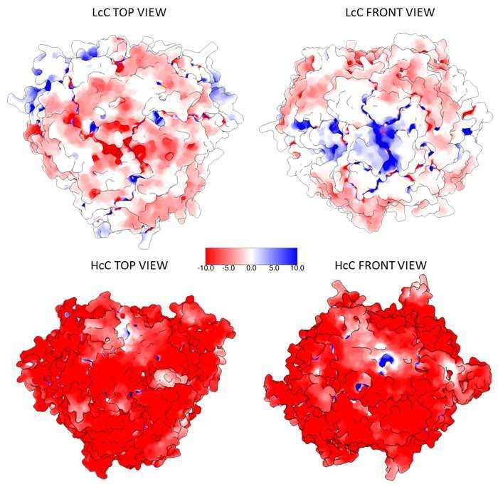
Biomolecules 2020, 10, 795                                                                       3 of 15

2.2. SDS-PAGE and IEF/SDS-PAGE
     SDS-PAGE was carried out according to [22] on 12% polyacrylamide gel. Gels were stained
with Coomassie Blue G-250 (Bio-Rad, Hercules, CA, USA) and the relative molecular masses of the
polypeptides were determined by comparison with standard protein (GE Healthcare, Chicago, IL,
USA): β-phosphorilase (94 kDa), BSA (66 kDa), egg albumin (45 kDa), carbonic anidrase (30 kDa),
and trypsin inhibitor (20 kDa).
     Bi-dimensional separations have been carried out according to [23]. Isoelectrofocalization (IEF)
was performed on 11 cm, pH 3–10 linear Readystrips IPG strips (Bio-Rad), whereas the second
dimension was performed on 12% polyacrylamide. Gels were stained with Coomassie Blue G-250.

2.3. Protein Identification
     Bands were cut from the CBB-stained SDS-PAGE gel and destained overnight with a solution of
25 mM ammonium bicarbonate and 50% acetonitrile. The proteins were in-gel digested with trypsin
(Roche, Segrate, Milano, Italy) as described in [24]. Mass spectrometry analyses were performed using
a MicroLC 200 Plus Eksigent Technologies system (Sciex, Dublin, CA, USA) with a Halo Fused C18
column (0.5 × 100 mm, 2.7 µm). The LC system was interfaced with a 5600+ TripleTOF system equipped
with a DuoSpray Ion Source (AB Sciex, Concord, Canada). Mass data were analyzed using Mascot server
(Matrix Science Inc., Boston, MA, USA), available on-line at http://www.matrixscience.com/index.html,
against an in-house sequence database prepared downloading all protein sequences present in NCBInr
related to Chenopodium quinoa. Setting of possible variable modifications of proteins was as previously
described [25].

2.4. Structural Predictions
      Sequence analyses were performed with the online ProtParam tool [26] to determine isoelectric
points and SignalP 5.0 to predict signal peptides [27].
      Structural predictions were performed using the Swiss Model homology modelling pipeline [28],
a tool available online via ExPASy server at https://swissmodel.expasy.org.
      Electrostatic calculations were performed with APBS (Adaptive Poisson–Boltzmann Solver) [29]
after structure preparation assigning atomic charges and radii with PDB2PQR [30]. Molecular surfaces
were created with the MSMS package [31]. All these applications are part of the UCSF Chimera
software (Version 1.13.1, The Regents of the University of California, Oakland, CA, USA), used for
molecular graphics as well [32]. Docking analyses were performed with Patchdock and Firedock
programs [33–35].
      Sequence alignments were performed with T-Coffee Web Service through Jalview 2.11.1.0 [36,37],
available at http://www.tcoffee.org and http://www.jalview.org/jalview-js/, respectively.

2.5. Caco-2 Cell Cultivation
     Human intestinal epithelial Caco-2 cells were from the European Collection of Authenticated Cell
Cultures (ECACC) (Merck, Milan, Italy). Cells were routinely grown in vented TC-treated 75 cm2
flasks (Star-lab, Milano, Italy) at 37 ◦ C in a humidified atmosphere of 95% air and 5% CO2 , using
DMEM supplemented with 10% inactivated fetal bovine serum, 2mM L-glutamine, 100 U/mL penicillin,
and 0.1 mg/mL streptomycin. All reagents were purchased from Sigma-Aldrich (Milan, Italy).

2.6. Transient Transfection and Immunomodulation Assay in Transfected Caco-2 Cells
     The day before transfection, Caco-2 cells were seeded in 24-multiwell plates at a density of
2 × 10⁵ cells/cm2 . Caco-2 cells were transiently transfected with the plasmid pNiFty2-Luc (InvivoGen,
Rho, Italy). This plasmid combines five NF-κB binding sites with the luciferase reporter gene luc,
thus the presence of NF-κB-activating molecules stimulates the expression of the luciferase gene.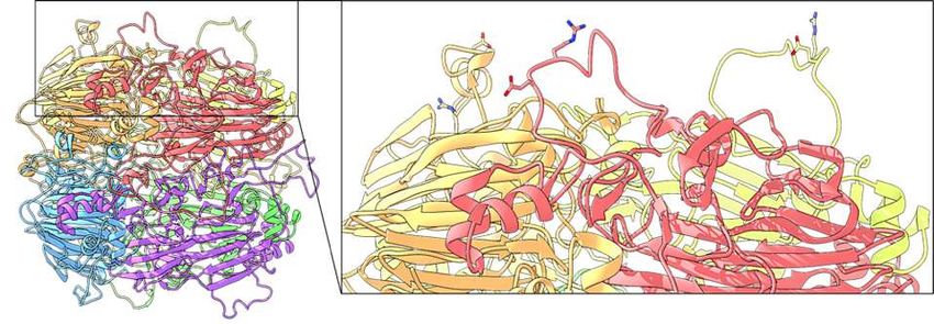
Biomolecules 2020, 10, 795                                                                           4 of 15

Transfection was performed using the StoS transfection kit (GeneSpin, Milan, Italy) following the
manufacturer’s protocol. After transfection, cells were grown in complete medium as described before.
     Immunomodulation assays were performed 24 h after transfection. Transfected Caco-2 cells were
incubated with complete DMEM containing protein samples at a final concentration of 0.5 mg/mL and
interleukin 1β (IL-1β) (10 ng/mL) for 4 h at 37 ◦ C. After incubation, the plate was chilled on ice for
15 min and cells were scrapped mechanically from the bottom of wells, transferred into 1.5 mL tubes,
and subjected to sonication for 10 s using a Soniprep 150 Ultrasonic Disintegrator (MSE) (Heathfield,
East Sussex, UK). Insoluble particles were removed using centrifugation. One hundred µL of each
supernatant were placed in a 96-well microtiter plate (PerkinElmer, Milano, Italy) and added with
12 µL of a 10 mM adenosine triphosphate (ATP) solution and 12 µL of 0.1 mM D-luciferin. The emitted
bioluminescence was monitored every 120 s using a VICTOR3 1420 Multilabel Counter (PerkinElmer,
Waltham, MA, USA). Each individual treatment was carried out in triplicate and each sample was
analyzed in triplicate.

2.7. Gene Expression Studies by qPCR
      Caco-2 cells were seeded in 12-multiwell plates at a density of 2 × 10⁵ cells/cm2 and allowed
to reach confluence. Caco-2 cells were treated by adding quinoa proteins at a final concentration of
0.5 and 1.0 mg/mL, with or without IL-1β (20 ng/mL). Each individual treatment was carried out in
triplicate and each sample was analyzed in triplicate.
      After treatments, total RNA from Caco-2 cells was isolated using the Aurum Total RNA Kit
(Bio-Rad). RNA was quantified at 260 nm using a fiber-optic ultra-micro cell (TrayCell, Hellma,
Germany). One µg of total RNA was reverse transcribed to cDNA in 20 µL total volume using the
iScript Reverse Transcription Supermix for RT-qPCR kit (Bio-Rad). Reaction conditions were: 25 ◦ C for
5 min, followed by incubation at 46 ◦ C for 20 min. Reactions were blocked at 95 ◦ C for 1 min. cDNA
samples were diluted 1:100 with sterile water before their use as templates for qPCR.
      qPCR was carried out using a CFX Connect Real-Time PCR detection system (Bio-Rad,
Hercules, CA, USA). Two µL of diluted cDNA samples were added to 10 µL of iQ SYBR Green
Supermix (Bio-Rad). Each primer was added to a final concentration of 250 nM. Primers for
amplification of IL-8 expressed gene were [38]: 50 -ATGACTTCCAAGCTGGCCGTGGCT-30 and
50 -TCTCAGCCCTCTTCAAAAACTTCTC-30 . The GAPDH reference gene was amplified with the
primer pair [39]: 50 -GGAAGGTGAAGGTCGGAGTC-30 and 50 -CACAAGCTTCCCGTTCTCAG-30 .
The reaction final volume was 20 µL. Cycling conditions were as follows: 3 min at 95 ◦ C, then 40 cycles
of denaturation (20 s at 95 ◦ C), annealing (20 s at 55 ◦ C), and extension (20 s at 72 ◦ C).
      Expression levels of the target genes in the test sample relative to the calibrator sample (untreated
cells) were calculated according to the Livak method [40]. Each sample was analyzed in triplicate.

2.8. Determination of Saponins
    Saponins were extracted according to [41] with minor modifications. Briefly, lyophilized protein
samples were extracted with three chloroform/methanol mixtures (1:2, 2:1, and 1:1, v/v), dried,
and partitioned with 1 vol of water and 2 vol of chloroform/methanol/water 3:48:47 (v/v/v). The organic
phase was resuspended in chloroform/methanol 2:1 and then separated and detected using UPLC
coupled to an ACQUITY QDa Mass Detector (Waters Corporation, Milford, MA, USA) on a BEH C18
column (Waters) according to [42].

2.9. Cell Vitality Assays
     The assessment of Caco-2 cells vitality was determined as previously reported [43], by using both
the (3-(4,5-dimethylthiazol-2-yl)-2,5-diphenyltetrazolium bromide (MTT) method and direct counts
following staining with Trypan Blue.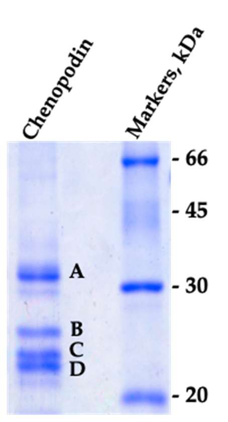
Biomolecules 2020, 10, 795 5 of 15
2.10. Statistical Analysis
Biomolecules 2020, 10, x 5 of 15
Data reported in the histograms are expressed as the means ± S.E. Data were analyzed by a t-test;
2.10. Statistical Analysis
p values < 0.05 were considered statistically significant. Data from RT-qPCR were analyzed using the
Data reported in the histograms are expressed as the means ± S.E. Data were analyzed by a t-
CFX Maestro 1.1 software (Bio-Rad, Hercules, CA, USA).
test; p values < 0.05 were considered statistically significant. Data from RT-qPCR were analyzed using
the CFX Maestro 1.1 software (Bio-Rad, Hercules, CA, USA).
3. Results
3. Results
3.1. Characterization of the Isolate Chenopodin
3.1. Characterization of the Isolate Chenopodin
The electrophoretic pattern of chenopodin isolated by a single-step elution with 0.25 M NaCl
The electrophoretic pattern of chenopodin isolated by a single-step elution with 0.25 M NaCl
(see Section 2.1)
(see is shown
paragraph 2.1) isinshown
Figure 1. The
in Figure result
1. The resultisis in perfect
in perfect accordance
accordance with previous
with previous data [5]. data [5].
Two main Two
groupsmainofgroups of polypeptides
polypeptides withwith
MrMr35–37
35–37 (band
(band A) A)and
and22–25 kDa kDa
22–25 (bands(bands
B, C andB,D)Care
and D) are
visible andvisible and correspond
correspond to the to α the
and and chains
β chains ofof11S
11S seed
seed globulin,
globulin,respectively [3,5]. [3,5].
respectively
Figure 1. The SDS-PAGE showing the polypeptide composition of the purified chenopodin. The gel
Figure 1. The SDS-PAGE showing the polypeptide composition of the purified chenopodin. The gel
was run under reducing conditions. Letters indicate the bands analyzed by mass spectrometry for
was run under reducing conditions. Letters indicate the bands analyzed by mass spectrometry
identification.
for identification.
The four main bands (A, B, C and D) were excised from the gel and analyzed by mass
spectrometry
The four main bands for protein
(A, B, identification.
C and D) were Theexcised
results indicate thatgel
from the theand
bands all belongby
analyzed to mass
the 11–13S
spectrometry
globulin family (Table 1). More than one protein was present (up to four different gene products) in
for protein identification. The results indicate that the bands all belong to the 11–13S globulin family
each of the four separated bands.
(Table 1). More than one protein was present (up to four different gene products) in each of the four
separated bands.
Table 1. List of proteins identified by mass spectrometry (MS). More than one protein was found in
each of the four (A–D) separated bands. MWs refers to the unprocessed precursors as reported by data
banks. Statistical information, experimental data, and sequences of the MS attributions are provided in
Supplementary Table S1.
Band Accession ID Description MW
AAS67036.1 11S seed storage globulin 54007
ABI94736.1 11S seed storage globulin B 53942
A
XP_021768838.1 11S globulin seed storage protein 2-like 52806
XP_021752668.1 13S globulin seed storage protein 2-like 61191
B XP_021752668.1 13S globulin seed storage protein 2-like 61191
AAS67036.1 11S seed storage globulin 54007
ABI94736.1 11S seed storage globulin B 53942
C
XP_021768838.1 11S globulin seed storage protein 2-like 52806
XP_021752668.1 13S globulin seed storage protein 2-like 61191
AAS67036.1 11S seed storage globulin 54007
D
ABI94736.1 11S seed storage globulin B 53942Biomolecules 2020, 10, 795 6 of 15
Chenopodin was further separated into two fractions, eluted from the IEC resin at distinct
ionic strengths (0.15 and 0.25 M NaCl), thus differing for the net protein surface charge. Mrs of
the bands of these two fractions appear identical following SDS-PAGE (Supplementary Figure S1A),
but differences concerning the isoelectric points (pIs) of the constituent polypeptides are clearly visible
in IEF/SDS-PAGE bi-dimensional gels (Supplementary Figure S1B). From now on, the two fractions
will be indicated as LcC (Low charge Chenopodin) and HcC (High charge Chenopodin). The relative
amounts of LcC and HcC were about 30% and 70%, respectively. Starting from the mass spectrometry
results, an attribution to LcC or HcC class was hypothesized based on the isoelectric point of the
proteins identified, calculated with ProtParam tool from the sequence of the mature protein without
the signal peptide, and predicted with SignalP-5.0 Server (Table 2).
Table 2. Isoelectric point and charge calculated at pH 8.0 of the identified chenopodin isoforms; e is the
charge of one electron.
Accession ID Description pI Charge
XP_021752668.1 13S globulin seed storage protein 2-like 5.96 −101 e
XP_021768838.1 11S globulin seed storage protein 2-like 6.35 −79 e
ABI94736.1 11S seed storage globulin B 6.47 −45 e
AAS67036.1 11S seed storage globulin 7.34 −38 e
Structures for the four chenopodin isoforms were generated by the SWISS-MODEL server,
submitting the relative sequences with default parameters. For all structures, the model representing
the homo-hexamer was chosen in order to fit the experimental data [3,5]. All the predicted models
were obtained from the crystal structure of almond Pru1 protein (PDB: 3FZ3), which shares sequence
identity ranging from 42.7% to 49.8%. Starting from the models, PDB2PQR software was used to
calculate the proteins’ total charge at pH 8.0 to provide a further clue about the attribution of LcC and
HcC classification. In the following modelling analyses, AAS67036.1 and XP_021752668.1 are assumed
to represent LcC and HcC, respectively, in order to maximize and enhance the differences between the
two classes.
Proteins’ charges at pH 8.0 and atom radii were calculated with PDB2PQR in order to visualize
the electrostatic surface potential with the APBS Electrostatic plugin implemented in the Chimera
Biomolecules 2020, 10, x 7 of 15
software. The results show that the negative charges of HcC are distributed all over the surface of the
protein,protein,
allowing the interaction
allowing with
the interaction withthe
thepositively charged
positively charged resin;
resin; conversely,
conversely, LcC atLcC at presents,
pH 8.0 pH 8.0 presents,
in comparison,
in comparison, less solvent-accessible
less solvent-accessible negatively charged
negatively chargedresidues
residues(Figure 2; Table
(Figure 2). 2).
2; Table
Figure 2. Electrostatic surface potential representation of the putative LcC (AAS67036.1) and HcC
Figure 2. Electrostatic surface potential representation of the putative LcC (AAS67036.1) and HcC
(XP_021752668.1) proteins, calculated at pH 8.0. Red: negative potential, blue: positive potential.
(XP_021752668.1) proteins, calculated at pH 8.0. Red: negative potential, blue: positive potential.
3.2. Immunomodulation Effects of Native Chenopodins
To study NF-κB activity, we transiently transfected Caco-2 cells with a NF-κB luciferase reporter
construct. The pNiFty2-Luc plasmid includes five NF-κB binding sites, driving the expression of the
luciferase reporter gene luc, thus the presence of NF-κB-activating molecules stimulates the
expression of the luciferase gene. Following incubation with LcC and HcC proteins in the absence ofBiomolecules 2020, 10, 795 7 of 15
Figure 2. Electrostatic surface potential representation of the putative LcC (AAS67036.1) and HcC
(XP_021752668.1) proteins, calculated at pH 8.0. Red: negative potential, blue: positive potential.
3.2. Immunomodulation Effects of Native Chenopodins
3.2. Immunomodulation Effects of Native Chenopodins
To study NF-κB activity, we transiently transfected Caco-2 cells with a NF-κB luciferase reporter
To study NF-κB activity, we transiently transfected Caco-2 cells with a NF-κB luciferase reporter
construct. The pNiFty2-Luc plasmid includes five NF-κB binding sites, driving the expression of the
construct. The pNiFty2-Luc plasmid includes five NF-κB binding sites, driving the expression of the
luciferase reporter gene luc, thus the presence of NF-κB-activating molecules stimulates the expression
luciferase reporter gene luc, thus the presence of NF-κB-activating molecules stimulates the
of the luciferase gene. Following incubation with LcC and HcC proteins in the absence of IL-1β, NF-κB
expression of the luciferase gene. Following incubation with LcC and HcC proteins in the absence of
activation was found to be similar to that of the control cells (Figure 3A). The proteins themselves
IL-1β, NF-κB activation was found to be similar to that of the control cells (Figure 3A). The proteins
arethemselves
not able toarestimulate
not ableinflammation responses, and
to stimulate inflammation exert noand
responses, cytotoxic
exert noeffects, since
cytotoxic insignificant
effects, since
losses of cell vitality were observed (Supplementary Figure S2). On the other hand, when
insignificant losses of cell vitality were observed (Supplementary Figure S2). On the other hand, when the cells
werethestimulated by IL-1β, LcC
cells were stimulated and HcC
by IL-1β, wereHcC
LcC and ablewere
to decrease NF-κB activation
able to decrease by about
NF-κB activation by 30%
aboutand
45%,30% and 45%, respectively.
respectively. These dataThese datathat
suggest suggest
LcC that
andLcC
HcCand areHcC
ableare
to able to reduce
reduce NF-κB-mediated
NF-κB-mediated cellular
cellular inflammation with different capacities. As expected, bovine serum albumin (BSA), used control
inflammation with different capacities. As expected, bovine serum albumin (BSA), used as a as a
control
protein, protein,
did did any
not exert not exert
NF-κB anymodulating
NF-κB modulating
activity.activity.
Figure 3. Inflammatory response of Caco-2 cells elicited by treatment with IL-1β alone and incubated
Figure 3. Inflammatory response of Caco-2 cells elicited by treatment with IL-1β alone and incubated
with 0.5 mg/mL
with 0.5 mg/mLofofLcC,
LcC, HcC, orBSA
HcC, or BSAwith
with
(+)(+) or without
or without (−) IL-1β.
(−) IL-1β. LcC or LcC
HcC orandHcC and
IL-1β wereIL-1β were
added
added to the cell growing medium at the same time. Response to IL-1β alone was set
to the cell growing medium at the same time. Response to IL-1β alone was set as 100%. See text for as 100%.
Seeexperimental
text for experimental
details. (A)details. (A) NF-kBpercentage
NF-kB activation activationofpercentage
transfectedofcells.
transfected
(B) IL-8cells. (B) IL-8
expression
percentage.
expression percentage.
As a complementary approach, we also assessed the modulation of interleukin 8 (IL-8). The results
are shown in Figure 3B and validate previous preliminary data [20]. IL-8 expression decreased to about
53% and 38% in the presence of LcC and HcC, respectively, confirming the capacity of chenopodin
isoforms to protect Caco-2 cells from the inflammatory stimulus. No significant increase of IL-8 mRNA
levels was detected in cells without stimulation by IL-1β, confirming that quinoa proteins do not exert
pro-inflammatory effects on cells under the adopted experimental conditions. On the contrary, as
expected, IL-8 expression markedly increased upon addition of IL-1β to the cell medium. A very similar
result was obtained when the cells were treated with proteins at a final concentration of 1.0 mg/mL
(Supplementary Figure S3).
The decreased activity of NF-κB caused by chenopodin induced the downregulation of IL-8
expression, being downstream from the NF-κB signaling pathway. IL-8 is a chemotactic cytokine
produced by different cell lines, including intestinal epithelial and Caco-2 cells, following the NF-κB
pathway activation [44,45]. Quinoa seeds contain high amount of saponins [46] that strongly
interact with proteins [47] and have been associated with the inhibition of inflammatory mediator
overproduction, including NO, TNF-α, and IL-6 [48]. We checked the presence of saponins in LcC and
HcC samples, and they were not detected at the protein concentration used in the assays.
We then assessed the expression of IL-8 in Caco-2 cells incubated with IL-1β and quinoa proteins
according to different experimental protocols. In the previous experiments (Figure 3B), either LcC or
HcC and IL-1β were added to the cell growing medium at the same time. Otherwise, when chenopodins
were added to the cell medium 30 min before the stimulation with IL-1β, expression of IL-8 was
markedly decreased in cells treated with HcC, whereas the presence of LcC slightly affected IL-8
expression (Figure 4A). Finally, either LcC or HcC was first pre-incubated with IL-1β for 30 min andand HcC samples, and they were not detected at the protein concentration used in the assays.
We then assessed the expression of IL-8 in Caco-2 cells incubated with IL-1β and quinoa proteins
according to different experimental protocols. In the previous experiments (Figure 3B), either LcC or
HcC and IL-1β were added to the cell growing medium at the same time. Otherwise, when
chenopodins
Biomolecules 2020, 10,were
795 added to the cell medium 30 min before the stimulation with IL-1β, expression8of of 15
IL-8 was markedly decreased in cells treated with HcC, whereas the presence of LcC slightly affected
IL-8 expression (Figure 4A). Finally, either LcC or HcC was first pre-incubated with IL-1β for 30 min
thenand
added
then to the cell
added medium.
to the In this In
cell medium. latter
thiscondition, LcC showed
latter condition, higherhigher
LcC showed anti-inflammatory effects
anti-inflammatory
witheffects
respectwith respect to HcC (Figure 4B).
to HcC (Figure 4B).
Figure
Figure 4. IL-8
4. IL-8 relative
relative expression
expression in Caco-2
in Caco-2 cells cells incubated
incubated with with LcC
LcC or HcCor(0.5
HcC (0.5 mg/mL)
mg/mL) under
under different
different experimental
experimental conditions.toResponse
conditions. Response to IL-1β
IL-1β alone was setalone was set(A)
as 100%. as LcC
100%. or (A)
HcCLcCwasoradded
HcC was
to the
celladded
medium to the cell medium
30 min 30 addition
before the min before ofthe addition
IL-1β and thenof IL-1β and then
incubated forincubated
1h. (B) LcC for or
1h.HcC
(B) LcC
wasorfirst
HcC was first pre-incubated with IL-1β for 30 min and then added to the cell
pre-incubated with IL-1β for 30 min and then added to the cell medium and incubated for 1h.medium and incubated
for 1h.
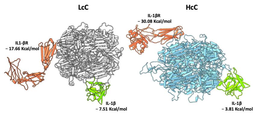
3.3. In Silico Protein–Protein Interaction Analyses
3.3. In Silico Protein–Protein Interaction Analyses
Molecular docking analyses were performed with the 3D models created for HcC and LcC to
Molecular
study their docking
interaction analyses
with were
possible performed
target withIL-1β
molecules. the 3Dand
models created for HcC
the extracellular and LcC
domain to
of IL-1β
study (IL-1R,
receptor their interaction
PDB: 1ITB) with possible target
structures molecules.
[49] were split inIL-1β and the extracellular
two different PDB files todomain
obtain oftheIL-1β
single
receptor
chain molecules(IL-1R,
forPDB: 1ITB) structures
the interaction [49] with
analyses werechenopodins.
split in two different
Both HcCPDBandfiles to obtain
LcC are ablethe
tosingle
interact
chain molecules for the interaction analyses with chenopodins. Both HcC and LcC are
with either IL-1β or soluble IL-1R with different portions of the proteins. In greater detail, IL-1β able to interact
binds
with either IL-1β or soluble IL-1R with different portions of the proteins. In greater detail,
to LcC with a more favorable binding energy (−7.51 Kcal/mol vs. −3.81 Kcal/mol), while conversely, IL-1β binds
to LcC with a more favorable binding energy (−7.51 Kcal/mol vs −3.81 Kcal/mol), while conversely,
binding of HcC to IL-1R is favored (−30.08 Kcal/mol vs. −17.66 Kcal/mol). Binding of the chenopodins
binding of HcC to IL-1R is favored (−30.08 Kcal/mol vs −17.66 Kcal/mol). Binding of the chenopodins
to IL-1R is predicted to occur far from the binding site of IL-1β in a region close to the transmembrane
to IL-1R is predicted to occur far from the binding site of IL-1β in a region close to the transmembrane
domain, thus the potential mechanism of action could be exerted through the distortion of the receptor,
domain, thus the potential mechanism of action could be exerted through the distortion of the
rather than a2020,
Biomolecules competition
10, x with IL-1β for the binding site (Figure 5). 9 of 15
receptor, rather than a competition with IL-1β for the binding site (Figure 5).
Figure
Figure 5. 5. Moleculardocking
Molecular dockingmodels.
models. Pipes
Pipes and
and plank
plankrepresentation
representationofofIL-1β (green)
IL-1β and
(green) thethe
and soluble
soluble
IL-1β receptor (orange) binding to LcC (grey) and HcC (light blue). The binding energy
IL-1β receptor (orange) binding to LcC (grey) and HcC (light blue). The binding energy for each for each
complex
complex is reported.
is reported.
TheThe human
human interleukin-1receptor
interleukin-1 receptor antagonist
antagonist (IL-1RA)
(IL-1RA)isisananantagonist
antagonist ofof
IL-1R
IL-1R[50], which
[50], actsacts
which
by by disrupting
disrupting oror preventingthe
preventing theformation
formation of
of the
the complex
complexbetween
betweenIL-1R
IL-1Rand
andIL-1R accessory
IL-1R protein
accessory protein
essential
essential forforthe
thesignal
signaltransduction
transduction [51].
[51].Anakinra
Anakinra is aisrecombinant formform
a recombinant of IL-1RA approved
of IL-1RA for the for
approved
thetreatment
treatmentofofautoinflammatory
autoinflammatory disorders
disorders[52]. ItsIts
[52]. mechanism
mechanism of of
action involves
action involvesthethe
competitive
competitive
inhibition of the local inflammatory effects of IL-1β. In light of this, we compared the amino acid
sequences of the four chenopodin isoforms and IL1-RA to investigate if chenopodins could mimic
the agonist effect exerted by IL-1RA (Figure 6). Interestingly, three regions showed a high level of
sequence similarity (Figure 6, boxes A, B, and C). These, although located quite far apart from one
another within the sequences, are predicted to lie close to one another in the native conformation of
chenopodin homo-hexamer (Supplementary Figure S4), suggesting a possible propensity of theBiomolecules 2020, 10, 795 9 of 15
inhibition of the local inflammatory effects of IL-1β. In light of this, we compared the amino acid
sequences of the four chenopodin isoforms and IL1-RA to investigate if chenopodins could mimic
the agonist effect exerted by IL-1RA (Figure 6). Interestingly, three regions showed a high level of
sequence similarity (Figure 6, boxes A, B, and C). These, although located quite far apart from one
another within the sequences, are predicted to lie close to one another in the native conformation of
chenopodin homo-hexamer (Supplementary Figure S4), suggesting a possible propensity of the region
to be functional. In addition, five amino acids are crucial for the interaction of IL-1RA with IL-1R [53].
In chenopodins, all these amino acids are located in the regions with the highest similarities and four
out of five are either conserved or substituted with similar ones (Figure 6).
Biomolecules 2020, 10, x 10 of 15
Figure
Figure 6. 6. Alignmentperformed
Alignment performed with
with the
the sequences
sequencesofofthe mature
the form
mature (without
form signal
(without peptides)
signal of
peptides)
IL-1RA (P18510) and the studied chenopodin isoforms. Sequence coloring represents percentage
of IL-1RA (P18510) and the studied chenopodin isoforms. Sequence coloring represents percentage
identity; conservation and consensus logos are shown. Regions with the highest homology are
identity; conservation and consensus logos are shown. Regions with the highest homology are
represented into boxes (A, B, and C), and amino acids crucial for IL1-RA interaction with IL-1R are
represented into boxes (A, B, and C), and amino acids crucial for IL1-RA interaction with IL-1R are
marked with an asterisk. The acronyms of chenopodin sequences are those reported in Table 1.
marked with an asterisk. The acronyms of chenopodin sequences are those reported in Table 1.
Intriguingly, lunasin, a small water-soluble peptide of about 5000 Da initially isolated from
Intriguingly, lunasin, a small water-soluble peptide of about 5000 Da initially isolated from
soybeans but also identified in quinoa and other seeds, exerts anti-inflammatory activity on
soybeans but also identified in quinoa and other seeds, exerts anti-inflammatory activity on macrophage
macrophage cells [54,55]. Lunasin can bind αVβ3 integrin through an Arg-Gly-Asp (RGD) motif [56].
cells [54,55]. Lunasin can bind αVβ3 integrin through an Arg-Gly-Asp (RGD) motif [56]. This interaction
This interaction has been associated with inhibition of inflammatory pathways involving NF-κB
has been associated with inhibition of inflammatory pathways involving NF-κB [56,57]. αVβ3 integrin
[56,57]. αVβ3 integrin is a receptor present in virtually all cells, including Caco-2 cells [58]. The amino
is a acid
receptor present
sequence in virtually
analysis all cells, isoforms
of chenopodin includinginvestigated
Caco-2 cellshere
[58].indicated
The amino theacid sequence
presence analysis
of RGD in
of chenopodin isoforms investigated here indicated the presence of RGD in sequence
sequence XP_021752668.1. A similar motif Arg-Gly-Glu (RGE) is present in sequences AAS67036.1 XP_021752668.1.
and ABI94736.1.
A similar It has been(RGE)
motif Arg-Gly-Glu demonstrated
is present that
in RGE of lipidAAS67036.1
sequences phosphate phosphohydrolase-3
and ABI94736.1. It hasis able
been
to interact with both α5β1 and αVβ3 integrins, acting as a possible cell ligand site in humans and
demonstrated that RGE of lipid phosphate phosphohydrolase-3 is able to interact with both α5β1 and
αVβ3 mice [59]. Theacting
integrins, 3D structural modelcell
as a possible of AAS67036.1
ligand site inshowed
humans that
andRGEmicemotif is predicted
[59]. to be located
The 3D structural model
in a very flexible
of AAS67036.1 andthat
showed exposed
RGEloop
motifatisthe protein surface
predicted (Figurein7).a very flexible and exposed loop at
to be located
the protein surface (Figure 7).[56,57]. αVβ3 integrin is a receptor present in virtually all cells, including Caco-2 cells [58]. The amino
acid sequence analysis of chenopodin isoforms investigated here indicated the presence of RGD in
sequence XP_021752668.1. A similar motif Arg-Gly-Glu (RGE) is present in sequences AAS67036.1
and ABI94736.1. It has been demonstrated that RGE of lipid phosphate phosphohydrolase-3 is able
to interact with both α5β1 and αVβ3 integrins, acting as a possible cell ligand site in humans and
mice [59].2020,
Biomolecules The10,
3D structural model of AAS67036.1 showed that RGE motif is predicted to be located
795 10 of 15
in a very flexible and exposed loop at the protein surface (Figure 7).
Figure7.7.Predictive
Figure Predictive 3D 3D structure
structure of
of the
thehomo-hexamer
homo-hexamerAAS67036
AAS67036chenopodin.
chenopodin.In the magnification
In the magnification
panelon
panel onthe
theright,
right, aa detail
detail shows
shows the
thesurface
surfacelocations
locationsofof
RGE sequences,
RGE oneone
sequences, for for
eacheach
monomer, as as
monomer,
stick representations.
stick representations.
XP_021752668.1 sequence analysis revealed the presence of an RGD motif in a non-homologous
region at protein C-terminal that, despite being not modeled, could be, however, solvent-accessible.
4. Discussion
To date, few studies have reported on the anti-inflammatory properties of proteins and peptides
from plant seeds. This study, for the first time, describes the potential immunomodulation capacity and
anti-inflammatory effects of chenopodin, the major protein component of quinoa seeds. The results
indicate that chenopodin may exert biological effects on intestinal cells and that the protein can act
differently, depending on its structural features. Moreover, our results add new information about the
structural characterization of chenopodin. The typical heterogenicity of seed storage proteins is clearly
evident in chenopodin due to the multigenic origin and to post-translation proteolytic processing of
precursor polypeptides [60]. Chenopodin is formed by a number of different polypeptides, as evidenced
by electrophoretic analysis. We separated two main fractions, LcC and HcC, that differ for their net
surface charge. All the constituent polypeptides are codified by at least four different genes. Structural
modeling and electrostatic surface potential analysis support the possible attribution of the gene
products to LcC and HcC.
Biological effects of LcC and HcC were investigated by measuring NF-κB activation and IL-8
expression studies in undifferentiated Caco-2 cells. This cell line, originally derived from colon
carcinomatous enterocytes, has been exploited for a range of studies aimed to elucidate the molecular
mechanisms of food-derived compounds, including the ability to elicit a reaction in response to
pro-inflammatory stimuli [61]. Caco-2 cells express the IL-1R receptor [62,63]. In our case, inflammation
was elicited using IL-1β, a pro-inflammatory cytokine able to strongly induce the release of IL-8 in
Caco-2 cells by triggering the NF-κB signaling pathway [64]. Cytokine IL-1β is able to stimulate IL-8
release at both the mRNA and protein levels in Caco-2 cells [65]. Several reports have demonstrated
the abilities of different natural compounds to regulate IL-8 expression by either transcriptional or
post-transcriptional mechanisms in intestinal epithelium [14,61,66]. It was observed in IL-1β stimulated
undifferentiated Caco-2 cells that the effects of olive oil phenols on IL-8 mRNA mirrored those observed
on IL-8 release [61].
Overall, the results outline possible mechanisms through which LcC and HcC may modulate
inflammation response. In our experimental conditions, LcC likely binds to IL-1β, and prevents
cytokine interaction with the target membrane receptor. HcC seems to instead interact directly with
the cells, possibly reducing the accessibility to IL-1β. The carried out predictive molecular modelling
supports this possible mechanism of action.Biomolecules 2020, 10, 795 11 of 15
Saponins in LcC and HcC samples were not detected. Thus, the protein purification process
effectively removed saponins and, therefore, the observed biological activities are attributable only to
the proteins.
Although our results suggest the activation of the canonical NF-κB signaling pathway,
the involvement of the non-canonical pathway [67] cannot be excluded. However, several studies
showed that some food proteins may stimulate members of the Toll-like receptor family (TLRs) such
as pea proteins and peptides obtained from milk [68]. It has been shown that the inhibitory effect on
IL-8 cell expression can be due to a direct interaction of whey protein hydrolysates with TLR4 [69].
Plant extracts were able to modulate TLR signaling as well through the direct activation of receptors
or further downstream of TLRs’ pathways [70]. On the other hand, though, other events or factors
cannot be ruled out. One possibility is that LcC and HcC may be internalized in the cytoplasm, as it
occurs for other seed proteins [71], exerting their activity in intracellular pathways such as in IkB
translocation [72], for example.
Since the evaluation of the long-term effects of chenopodins on the inflammatory response was
out of the scope of the work, protein accumulation of inflammatory effectors such as IL-8, IL-6 and
TNF-α has not been investigated.
Whether or not chenopodin can actually exert biological effects on the human body following
ingestion remains to be established. Conformational changes or denaturations induced by food
processing and gastrointestinal digestion may have important consequences on the observed activity
that need to be deeply investigated. Digestibility of isolated quinoa proteins is, indeed, higher if
compared to those of whole meal quinoa flour [73]. Further studies have been undertaken to shed
light on the molecular mechanisms of action and the long-term effects of chenopodin on inflammatory
response using different intestinal cell models and assessing a panel of inflammation protein markers.
This assessment is envisaged because the correspondence between transcript and protein levels is not
always maintained. A little variation in transcription levels, indeed, could have remarkable effects on
protein accumulation and, in the end, on the cellular events involved in prolonged inflammation.
Supplementary Materials: The following are available online at http://www.mdpi.com/2218-273X/10/5/795/
s1, Figure S1: Mono-dimensional and bi-dimensional electrophoresis of LcC and HcC chenopodin fractions,
Figure S2: Assessment of Caco-2 cell vitality following treatment with LcC and HcC, Figure S3: Relative expression
of Il-8 in Caco-2 cells incubated with 1.0 mg/mL LcC or HcC, Figure S4: Magnification of the predictive 3D
structure of the homo-hexamer AAS67036 chenopodin (HcC) showing the regions with the highest homology
with IL-1RA, Table S1: List of the proteins identified by MS.
Author Contributions: A.S. and J.C. conceived and designed the experiments; J.C., M.D.D., E.B., and P.A.C.
performed lab experiments; S.D.B. performed structural predictions and modelling; A.S., A.A., S.D.B. and J.C.
analyzed and discussed the data; and A.S., A.A. and J.C. wrote and revised the paper. All authors have read and
agreed to the published version of the manuscript.
Funding: J.C. was supported by Università degli Studi di Milano (Assegno di Ricerca tipo A, 2018-RPDF-0048).
The open access APC was partially covered by Università degli Studi di Milano.
Acknowledgments: Authors are grateful to Dr. Stefania Arioli for assistance with luminometer determinations,
Prof. M.A: Pagani for the generous gift of the quinoa seeds and Dr. Annalisa Bellucci for helpful assistance.
All experiments involving cultured Caco-2 cells have been carried out at the DeFENS Cell Culture Laboratory, a core
facility of the Department of Food, Environmental and Nutritional Sciences (University of Milan, Italy). Molecular
graphics and analyses performed with UCSF Chimera, developed by the Resource for Biocomputing, Visualization,
and Informatics at the University of California, San Francisco, with support from NIH P41-GM103311.
Conflicts of Interest: The authors declare no conflict of interest.
References
1. Toapanta, A.; Carpio, C.; Vilcacundo, R.; Carrillo, W. Analysis of protein isolate from quinoa
(Chenopodium Quinoa Willd). Asian J. Pharm. Clin. Res. 2016, 9, 332–334.
2. Repo-Carrasco, R.; Espinoza, C.; Jacobsen, S.E. Nutritional value and use of the Andean crops quinoa
(Chenopodium quinoa) and kaniwa (Chenopodium pallidicaule). Food Rev. Int. 2003, 19, 179–189. [CrossRef]Biomolecules 2020, 10, 795 12 of 15
3. Fairbanks, D.J.; Burgener, K.W.; Robinson, L.R.; Andersen, W.R.; Ballon, E. Electrophoretic characterization
of quinoa seed proteins. Plant Breed. 1990, 104, 190–195. [CrossRef]
4. Utsumi, S. Plant Food Protein Engineering. Adv. Food Nutr. Res. 1992, 36, 89–208.
5. Brinegar, C.; Goundan, S. Isolation and characterization of chenopodin, the 11S seed storage protein of
quinoa (Chenopodium quinoa). J. Agric. Food Chem. 1993, 41, 182–185. [CrossRef]
6. Srivastava, L.M. Seed food reserves and their accumulation. In Plant Growth and Development; Academic
Press: Oxford, UK, 2002; pp. 503–520.
7. Jacobsen, S.-E. The worldwide potential for quinoa (Chenopodium quinoa, Willd.). Food Rev. Int. 2003,
19, 167–177. [CrossRef]
8. Bazile, D.; Jacobsen, S.-E.; Verniau, A. The global expansion of quinoa: Trends and limits. Front. Plant Sci.
2016, 7, 622. [CrossRef]
9. Gordillo-Bastidas, E.; Díaz-Rizzolo, D.; Roura, R.; Rizzolo, D.A.D.; Massanés, T.; Gomis, R. Quinoa
(Chenopodium quinoa Willd), from nutritional value to potential health benefits: An integrative review. J. Nutr.
Food Sci. 2016, 6, 497.
10. Graf, B.L.; Rojas-Silva, P.; Rojo, L.E.; Delatorre-Herrera, J.; Baldeón, M.E.; Raskin, I. Innovations in health
value and functional food development of quinoa (Chenopodium quinoa Willd.). Compr. Rev. Food Sci. Food Saf.
2015, 14, 431–445. [CrossRef]
11. Maradini-Filho, A.M. Quinoa: Nutritional aspects. J. Nutraceuticals Food Sci. 2017, 2, 1–5.
12. Liu, R.H. Dietary bioactive compounds and their health implications. J. Food Sci. 2013, 78 (Suppl. S1),
A18–A25. [CrossRef] [PubMed]
13. Ribeiro, P.V.M.; Andrade, P.A.; Hermsdorff, H.H.M.; Dos Santos, C.A.; Cotta, R.M.M.; Estanislau, J.A.S.G.;
Campos, A.A.O.; Rosa, C.O.B. Dietary non-nutrients in the prevention of non-communicable diseases:
Potentially related mechanisms. Nutrition 2019, 66, 22–28. [CrossRef] [PubMed]
14. Reyes-Díaz, A.; Del-Toro-Sánchez, C.L.; Rodríguez-Figueroa, J.C.; Valdéz-Hurtado, S.; Wong-Corral, F.J.;
Borboa-Flores, J.; González-Osuna, M.F.; Perez-Perez, L.M.; González-Vega, R.I. Legume proteins as a
promising source of anti-inflammatory peptides. Curr. Protein Pept. Sci 2019, 20, 1204–1217.
15. Medzhitov, R. Origin and physiological roles of inflammation. Nature 2008, 454, 428–435. [CrossRef]
16. Ben-Neriah, Y.; Karin, M. Inflammation meets cancer, with NF-κB as the matchmaker. Nat. Immunol. 2011,
12, 715–723. [CrossRef]
17. Liu, T.; Zhang, L.; Joo, D.; Sun, S.C. NF-κB signaling in inflammation. Signal Transduct. Target Ther. 2017,
2, 17023. [CrossRef]
18. Ariel, A.; Timor, O. Hanging in the balance: Endogenous anti-inflammatory mechanisms in tissue repair and
fibrosis. J. Pathol. 2013, 229, 250–263. [CrossRef]
19. Zhang, L.; Wei, X.; Zhang, R.; Petitte, J.N.; Si, D.; Li, Z.; Cheng, J.; Du, M. Design and development of a novel
peptide for treating intestinal inflammation. Front. Immunol. 2019, 10, 1841. [CrossRef]
20. Artusa, V. Development of a RT-qPCR Assay to Determine the Anti-Inflammatory Potential of Quinoa
(Chenopodium quinoa Willd.) Seed Proteins in Human Intestinal Caco-2 Cells. Master’s Thesis, Università
degli Studi di Milano, Milano, Italy, 2018.
21. Scarafoni, A.; Giani, D.; Cerletti, P. An endopeptidase in dormant lupin seed. Phytochemistry 1992,
37, 3715–3723. [CrossRef]
22. Laemmli, U.K. Cleavage of structural proteins during the assembly of the head of bacteriophage T4. Nature
1970, 227, 680–685. [CrossRef]
23. Scarafoni, A.; Ronchi, A.; Prinsi, B.; Espen, L.; Assante, G.; Venturini, G.; Duranti, M. The proteome of
exudates from germinating Lupinus albus seeds is secreted through a selective dual-step process and contains
proteins involved in plant defense. FEBS J. 2013, 280, 1443–1459. [CrossRef]
24. Hellman, U.; Wernstedt, C.; Gonez, J.; Heldin, C. Improvement of an ‘In-Gel’ digestion procedure for the
micropreparation of internal protein fragments for amino acid sequencing. Anal. Biochem. 1995, 224, 451–455.
[CrossRef]
25. Bona, E.; Massa, N.; Novello, G.; Boatti, L.; Cesaro, P.; Todeschini, V.; Magnelli, V.; Manfredi, M.; Marengo, E.;
Mignone, F.; et al. Metaproteomic characterization of the Vitis vinifera rhizosphere. FEMS Microbiol. Ecol.
2019, 95, fiy204. [CrossRef]Biomolecules 2020, 10, 795 13 of 15
26. Gasteiger, E.; Hoogland, C.; Gattiker, A.; Duvaud, S.; Wilkins, M.R.; Appel, R.D.; Bairoch, A. Protein
identification and analysis tools on the ExPASy server. In The Proteomics Protocols Handbook; Walker, J.M., Ed.;
Humana Press: Totowa, NY, USA, 2005; pp. 571–607.
27. Almagro Armenteros, J.J.; Tsirigos, K.D.; Sønderby, C.K.; Petersen, T.N.; Winther, O.; Brunak, S.; von Heijne, G.;
Nielsen, H. SignalP 5.0 improves signal peptide predictions using deep neural networks. Nat. Biotechnol.
2019, 37, 420–423. [CrossRef]
28. Waterhouse, A.; Bertoni, M.; Bienert, S.; Studer, G.; Tauriello, G.; Gumienny, R.; Heer, F.T.; de Beer, T.A.P.;
Rempfer, C.; Bordoli, L.; et al. SWISS-MODEL: Homology modelling of protein structures and complexes.
Nucl. Acids Res. 2018, 46, 296–303. [CrossRef]
29. Baker, N.; Sept, D.; Joseph, S.; Holst, M.; McCammon, J. Electrostatics of nanosystems: Application to
microtubules and the ribosome. Proc. Natl. Acad. Sci. USA 2001, 98, 10037–10041. [CrossRef]
30. Dolinsky, T.J.; Czodrowski, P.; Li, H.; Nielsen, J.E.; Jensen, J.H.; Klebe, G.; Baker, N.A. PDB2PQR: Expanding
and upgrading automated preparation of biomolecular structures for molecular simulations. Nucl. Acids Res.
2007, 35, 522–525. [CrossRef]
31. Sanner, M.F.; Olson, A.J.; Spehner, J.C. Reduced surface: An efficient way to compute molecular surfaces.
Biopolymers 1996, 38, 305–320. [CrossRef]
32. Pettersen, E.F.; Goddard, T.D.; Huang, C.C.; Couch, G.S.; Greenblatt, D.M.; Meng, E.C.; Ferrin, T.E. UCSF
Chimera-a visualization system for exploratory research and analysis. J. Comput. Chem. 2004, 25, 1605–1612.
[CrossRef]
33. Duhovny, D.; Nussinov, R.; Wolfson, H.J. Efficient unbound docking of rigid molecules. In Algorithms in
bioinformatics; Guigó, R., Gusfield, D., Eds.; Springer: Berlin, Germany, 2002; Volume 2452, pp. 185–200.
34. Schneidman-Duhovny, D.; Inbar, Y.; Nussinov, R.; Wolfson, H.J. PatchDock and SymmDock: Servers for
rigid and symmetric docking. Nucl. Acids Res. 2005, 33, 363–367. [CrossRef]
35. Andrusier, N.; Nussinov, R.; Wolfson, H.J. FireDock: Fast interaction refinement in molecular docking.
Proteins 2007, 69, 139–159. [CrossRef]
36. Di Tommaso, P.; Moretti, S.; Xenarios, I.; Orobitg, M.; Montanyola, A.; Chang, J.M.; Taly, J.F.; Notredame, C.
T-Coffee: A web server for the multiple sequence alignment of protein and RNA sequences using structural
information and homology extension. Nucl. Acids Res. 2011, 39, 13–17. [CrossRef]
37. Waterhouse, A.M.; Procter, J.B.; Martin, D.M.A.; Clamp, M.; Barton, G.J. Jalview Version 2-A multiple
sequence alignment editor and analysis workbench. Bioinformatics 2009, 25, 1189–1191. [CrossRef]
38. Li, J.; Moran, T.; Swanson, E.; Julian, C.; Harris, J.; Bonen, D.K.; Hedl, M.; Nicolae, D.L.; Abraham, C.;
Cho, J.H. Regulation of IL-8 and IL-1 expression in Crohn’s disease associated NOD2/CARD15 mutations.
Hum. Mol. Genet. 2004, 13, 1715–1725. [CrossRef]
39. Bottero, V.; Imbert, V.; Frelin, C.; Formento, J.L.; Peyron, J.F. Monitoring NF-κB transactivation potential via
Real-Time PCR quantification of IκB-α Gene Expression. Mol. Diagn. 2003, 7, 187–194. [CrossRef]
40. Livak, K.J.; Schmittgen, T.D. Analysis of relative gene expression data using real-time quantitative PCR and
the 2(-Delta Delta C(T)) method. Methods 2001, 25, 402–408. [CrossRef]
41. Aoun, M.; Corsetto, P.A.; Nugue, G.; Montorfano, G.; Ciusani, D.C.; Hogarth, P.; Gregory, A.; Hayflick, S.;
Zorzi, G.; Rizzo, A.M.; et al. Changes in red blood cell membrane lipid composition: A new perspective into
the pathogenesis of PKAN. Mol. Genet. Metab. 2017, 121, 180–189. [CrossRef]
42. McDonald, J.G.; Thompson, B.M.; Mc Crum, E.C.; Russell, D.W. Extraction and analysis of sterols in
biological matrices by high performance liquid chromatography electrospray ionization mass spectrometry.
Methods Enzymol. 2007, 432, 145–170.
43. Barbiroli, A.; Capraro, J.; Marulo, S.; Gamba, M.; Scarafoni, A. Effects on the Caco-2 cells of a hypoglycemic
protein from lupin seeds in a solution and adsorbed on polystyrene nanoparticles to mimic a complex food
matrix. Biomolecules 2019, 9, 606. [CrossRef]
44. Daig, R.; Rogler, G.; Aschenbrenner, E.; Vogl, D.; Falk, W.; Gross, V.; Schölmerich, J.; Andus, T. Human
intestinal epithelial cells secrete interleukin-1 receptor antagonist and interleukin-8 but not interleukin-1 or
interleukin-6. Gut 2000, 46, 350–358. [CrossRef]
45. Philpott, D.J.; Yamaoka, S.; Israël, A.; Sansonetti, P.J. Invasive Shigella flexneri activates NF-kappa B through a
lipopolysaccharide-dependent innate intracellular response and leads to IL-8 expression in epithelial cells.
J. Immunol. 2000, 165, 903–914. [CrossRef]Biomolecules 2020, 10, 795 14 of 15
46. Jarvis, D.E.; Ho, Y.S.; Lightfoot, D.J.; Schmöckel, S.M.; Li, B.; Borm, T.J.A.; Ohyanagi, H.; Mineta, K.;
Michell, C.T.; Saber, N.; et al. The genome of Chenopodium quinoa. Nature 2017, 542, 307–312. [CrossRef]
47. Tang, Y.; Tsao, R. Phytochemicals in quinoa and amaranth grains and their antioxidant, anti-inflammatory,
and potential health beneficial effects: A review. Mol. Nutr. Food Res. 2017, 61, 1600767. [CrossRef]
48. Yao, Y.; Yang, X.; Shi, Z.; Ren, G. Anti-inflammatory activity of saponins from quinoa (Chenopodium quinoa
Willd.) seeds in lipopolysaccharide-stimulated RAW 264.7 macrophages cells. J. Food Sci. 2014, 79, 1018–1023.
[CrossRef]
49. Vigers, G.P.A.; Anderson, L.J.; Caffes, P.; Brandhuber, B.J. Crystal structure of the type-I interleukin-1 receptor
complexed with interleukin-1β. Nature 1997, 386, 190–194. [CrossRef]
50. Dinarello, C.A. The interleukin-1 family: 10 years of discovery. FASEB J. 1994, 8, 1314–1325. [CrossRef]
51. Greenfeder, S.A.; Nunes, P.; Kwee, L.; Labow, M.; Chizzonite, R.A.; Ju, G. Molecular cloning and
characterization of a second subunit of the interleukin 1 receptor complex. J. Biol. Chem. 1995, 270, 13757–13765.
[CrossRef]
52. Castañeda, S.; Atienza-Mateo, B.; Martín-Varillas, J.L.; Serra López-Matencio, J.M.; González-Gay, M.A.
Anakinra for the treatment of adult-onset Still’s disease. Expert Rev. Clin. Immunol. 2018, 14, 979–992.
[CrossRef]
53. Schreuder, H.; Tardif, C.; Trump-Kallmeyer, S.; Soffientini, A.; Sarubbi, E.; Akeson, A.; Bowlin, T.; Yanofsky, S.;
Barrett, R.W. A new cytokine-receptor binding mode revealed by the crystal structure of the IL-1 receptor
with an antagonist. Nature 1997, 386, 194–200. [CrossRef]
54. Ren, G.; Zhu, Y.; Shi, Z.; Li, J. Detection of lunasin in quinoa (Chenopodium quinoa Willd.) and the in vitro
evaluation of its antioxidant and anti-inflammatory activities. J. Sci. Food Agric. 2017, 97, 4110–4116.
[CrossRef]
55. Hernández-Ledesma, B.; Hsieh, C.C.; de Lumen, B.O. Antioxidant and anti-inflammatory properties of
cancer preventive peptide lunasin in RAW 264.7 macrophages. Biochem. Biophys. Res. Commun. 2009,
390, 803–808. [CrossRef]
56. Cam, A.; de Mejia, E.G. RGD-peptide lunasin inhibits Akt-mediated NF-κB activation in human macrophages
through interaction with the αVβ3 integrin. Mol. Nutr. Food Res. 2012, 56, 1569–1581. [CrossRef]
57. Aguzzi, M.S.; Fortugno, P.; Giampietri, C.; Ragone, G.; Capogrossi, M.C.; Facchiano, A. Intracellular targets
of RGDS peptide in melanoma cells. Mol. Cancer 2010, 9, 84. [CrossRef]
58. Kim, C.; Ye, F.; Ginsberg, M.H. Regulation of integrin activation. Annu. Rev. Cell Dev. Biol. 2011, 27, 321–345.
[CrossRef]
59. Humtsoe, J.O.; Bowling, R.A., Jr.; Feng, S.; Wary, K.K. Murine lipid phosphate phosphohydrolase-3 acts as a
cell-associated integrin ligand. Biochem. Biophys. Res. Commun. 2005, 335, 906–919. [CrossRef]
60. Shewry, P.R.; Napier, J.A.; Tatham, A.S. Seed storage proteins: Structures and biosynthesis. Plant Cell 1995,
7, 945–956.
61. Muto, E.; Dell’Agli, M.; Sangiovanni, E.; Mitro, N.; Fumagalli, M.; Crestani, M.; De Fabiani, E.; Caruso, D.
Olive oil phenolic extract regulates interleukin-8 expression by transcriptional and posttranscriptional
mechanisms in Caco-2 cells. Mol. Nutr. Food Res. 2015, 59, 1217–1221. [CrossRef]
62. Varilek, G.W.; Neil, G.A.; Bishop, W.P. Caco-2 cells express type I interleukin-1 receptors: Ligand binding
enhances proliferation. Am. J. Physiol. 1994, 267, G1101–G1107. [CrossRef]
63. Böcker, U. Influence of therapeutic intervention with interleukins on epithelial cell function. In Cytokines and
cell Homeostasis in the Gastroinstestinal Tract, Proceedings of Falk Symposium 113 Held in Regensburg, Germany,
16–18 September 1999; Andus, T., Rogler, G., Schlottmann, K., Frick, E., Adler, G., Schmiegel, W., Zeitz, M.,
Schölmerich, J., Eds.; Kluwer Academic Publishers: Berlin, Germany, 1999; pp. 61–71.
64. Klampfer, L. Cytokines, inflammation and colon cancer. Curr. Cancer Drug Targets. 2011, 11, 451–464.
[CrossRef]
65. Leonard, F.; Collnot, E.M.; Lehr, C.M. A three-dimensional coculture of enterocytes, monocytes and dendritic
cells to model inflamed intestinal mucosa in vitro. Mol. Pharm. 2010, 7, 2103–2119. [CrossRef]
66. Romier, B.; Van De Walle, J.; During, A.; Larondelle, Y.; Schneider, Y.J. Modulation of signalling nuclear
factor-kappaB activation pathway by polyphenols in human intestinal Caco-2 cells. Br. J. Nutr. 2008,
100, 542–551. [CrossRef]
67. Sun, S.C. Non-canonical NF-κB signaling pathway. Cell Res. 2011, 21, 71–85. [CrossRef]You can also read


