A decellularized human corneal scaffold for anterior corneal surface reconstruction
←
→
Page content transcription
If your browser does not render page correctly, please read the page content below
www.nature.com/scientificreports
OPEN A decellularized human corneal
scaffold for anterior corneal surface
reconstruction
Naresh Polisetti1,4*, Anke Schmid1,4, Ursula Schlötzer‑Schrehardt2, Philip Maier1,
Stefan J. Lang1, Thorsten Steinberg3, Günther Schlunck1* & Thomas Reinhard1
Allogenic transplants of the cornea are prone to rejection, especially in repetitive transplantation and
in scarred or highly vascularized recipient sites. Patients with these ailments would particularly benefit
from the possibility to use non-immunogenic decellularized tissue scaffolds for transplantation,
which may be repopulated by host cells in situ or in vitro. So, the aim of this study was to develop
a fast and efficient decellularization method for creating a human corneal extracellular matrix
scaffold suitable for repopulation with human cells from the corneal limbus. To decellularize human
donor corneas, sodium deoxycholate, deoxyribonuclease I, and dextran were assessed to remove
cells and nuclei and to control tissue swelling, respectively. We evaluated the decellularization
effects on the ultrastructure, optical, mechanical, and biological properties of the human cornea.
Scaffold recellularization was studied using primary human limbal epithelial cells, stromal cells, and
melanocytes in vitro and a lamellar transplantation approach ex vivo. Our data strongly suggest that
this approach allowed the effective removal of cellular and nuclear material in a very short period
of time while preserving extracellular matrix proteins, glycosaminoglycans, tissue structure, and
optical transmission properties. In vitro recellularization demonstrated good biocompatibility of the
decellularized human cornea and ex vivo transplantation revealed complete epithelialization and
stromal repopulation from the host tissue. Thus, the generated decellularized human corneal scaffold
could be a promising biological material for anterior corneal reconstruction in the treatment of corneal
defects.
Allogenic corneal transplantation is a highly successful technique to restore vision in patients with corneal
diseases or after ocular trauma1. In eyes with moderate damage, the ocular immune privilege and the lack of
corneal blood vessels decrease the risk for immunological transplant rejection. This is different in eyes with severe
corneal damage, e.g. in patients suffering from alkaline burns or corneal stromal ulceration due to inflammation
or infection, where allogenic transplants placed in an inflamed recipient bed are prone to transplant rejection2,3.
These patients in particular would benefit from the possibility to use non-immunogenic decellularized tissue
scaffolds which may be repopulated by host cells in situ. A large number of studies used porcine tissue for
decellularization4,5. However, its clinical implementation may be hampered by issues of xenotransplantation
such as immunocompatibility and the possible presence of porcine viruses. Human donor corneas unsuitable for
transplantation due to a low endothelial cell count have therefore been advocated as an excellent source of the
tissue6,7. With the broad use of Descemet membrane endothelial keratoplasty (DMEK), where only a 20–30 µm
thick tissue membrane and the endothelial cell layer are being transplanted, a large number of corneal tissue
remnants remain after DMEK transplant preparation and could also be used for scaffold preparation to treat
corneal ulcers and superficial defects.
Several methods have been described to decellularize corneal tissue using physical or chemical means8,9. For
decellularization, hypertonic sodium chloride (NaCl), sodium dodecyl sulfate (SDS) or non-ionic detergents
such as Triton X-100 have been used alone or in combination with nucleases to eradicate cells while preserving
protein content and structure of the extracellular matrix (ECM) scaffold to varying degrees6,7. Drawbacks may
1
Eye Center, Medical Center ‑ Faculty of Medicine, University of Freiburg, Killianstrasse 5, 79106 Freiburg,
Germany. 2Department of Ophthalmology, University Hospital Erlangen, Friedrich-Alexander-University of
Erlangen-Nürnberg, Schwabachanlage 6, 91054 Erlangen, Germany. 3Department of Operative Dentistry and
Periodontology, Division of Oral Biotechnology, University of Freiburg, Hugstetter Strasse 55, 79106 Freiburg,
Germany. 4These authors contributed equally: Naresh Polisetti and Anke Schmid. *email: naresh.polisetti@
uniklinik‑freiburg.de; guenther.schlunck@uniklinik‑freiburg.de
Scientific Reports | (2021) 11:2992 | https://doi.org/10.1038/s41598-021-82678-3 1
Vol.:(0123456789)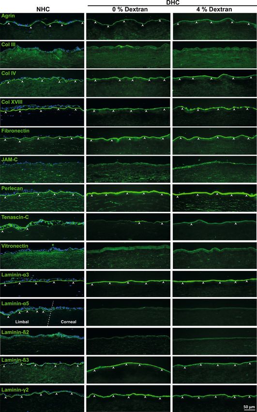
www.nature.com/scientificreports/
Figure 1. Decellularization efficiency and role of dextran: (A) Histological evaluation of human cornea ▸
decellularized by various concentrations of sodium deoxycholate (SD) with deoxyribonuclease I in comparison
to the normal human cornea (NHC) using hematoxylin and eosin (H&E) staining. The higher magnification
images of H&E staining showing the stromal cells in NHC (arrowheads) and cellular debris in 0.5% SD (
arrowheads). Scale bar = 100 µm. HLA-ABC staining (green) of NHC and DHC showing cellular expression
of HLA in NHC and 0.5% SD treated DHC (white arrows); no HLA expression in 1% and 4% SD treated
DHC cornea. DAPI staining reveals nuclei in NHC without remaining nuclei/nuclear debris in DHC. (B) The
role of dextran during decellularization in both DM− and DM+ cornea in comparison to respective controls.
Graphs represent the thickness of DHC in various dextran conditions compared to NHC. Data are expressed
as means ± S.E.M. (n = 5). * p < 0.05; ** p < 0.01; *** p < 0.001; **** p < 0.0001; (Wilcoxon signed-rank test).
(C) AS-OCT images of D M− and DM+ cornea in varying concentrations of dextran with respective controls.
(D) Histological evaluation by staining with H&E on NHC and DHC showing no cell remnants in DHC but
varying corneal thickness (in presence or absence of dextran) of both D M− and DM+ cornea. Scale bar = 100 µm
E) Confirmation of decellularization by quantification of residual DNA. The graph represents the quantity of
DNA in both DM− and DM+ cornea before and after decellularization (0 or 4% dextran). Data are expressed as
means ± S.E.M. (n = 4). * p < 0.05; Mann–Whitney U test. DAPI 4′,6‐diamidino‐2‐phenylindole, DM− cornea
with the absence of Descemet membrane, DM+ cornea with the presence of Descemet membrane, AS-OCT
anterior segment optical coherence tomography, HLA human leukocyte antigen.
be that SDS denatures proteins and a decrease in scaffold transparency was reported after treatment with SDS
or hypertonic s alt7. Moreover, protocols using SDS or hypertonic salt with nucleases are very time-consuming
(~ 5–8 days)6,7,10 and failure to fully elute SDS can have cytotoxic effects6,7. Sodium deoxycholate (SD), a naturally
occurring bile salt metabolite, has been used to decellularize various organs or tissues such as liver11, periph-
eral nerves12, ovary13, uterus14, conchal c artilage15, trachea16, lungs and heart valves17. The combination of SD/
deoxyribonuclease I (DNAse) was also used to generate decellularized human corneal limbus, however, a long
elution time for removal of cellular components was a drawback of the protocol suggested18. Moreover, there
are no reports on a combination of SD and DNAse as a decellularizing agent for human corneal tissue. Synthetic
SD is being applied clinically as an injectable dissolvent of subcutaneous body f at19 and was FDA-approved for
removal of submental fat, which may illustrate its biocompatibility. The cornea is endowed with high glycosa-
minoglycan content and it has been shown that tissue swelling during processing can cause structural damage,
which was prevented by adding dextran to the solutions used on porcine corneal t issue20. The role of dextran and
tissue swelling during human corneal decellularization has not specifically been addressed in earlier protocols6,7.
Moreover, published decellularization protocols often lack an extensive characterization of ECM integrity and
preservation of BM components6,7, which are essential to maintain tissue homeostasis and ensure cell adhesion,
growth, long term survival and function of corneal epithelium and epithelial progenitor cell p henotype8,21,22.
7,23
SDS and triton-x-100 were shown to be cytotoxic and limit b iocompatibility .
In light of these data, it was our goal to devise a further improved protocol for decellularization of human
corneal tissue remnants after removal of transplant material for DMEK (without Descemet membrane, labeled
DM−) using agents in clinical use and controlling for tissue swelling. We also wanted to clarify whether corneal
tissue unsuitable for transplantation due to a low endothelial cell count (with Descemet membrane, labeled DM+),
would be amenable to the same protocol with similar preservation of tissue integrity. We evaluated decellulariza-
tion effects on the basement membrane (BM) components and physical characteristics such as transparency and
mechanical properties of the decellularized human cornea (DHC). Furthermore, we evaluated the biocompat-
ibility of generated DHC scaffolds by repopulation with human corneal cells from the limbus.
Results
Decellularization of human corneas. Human DM− corneas decellularized with different concentrations
of SD were initially screened by hematoxylin and eosin (H&E) staining, immunofluorescent staining for human
leukocyte antigen (HLA)-ABC was used to assess the presence of residual cellular/membranous material, and
nuclear staining with 4′,6-diamidino-2-phenylindole (DAPI) (Fig. 1A). H&E staining showed distinctly visible
corneal epithelial and stromal cells (arrowheads indicating stromal cells at higher magnification) in normal
human cornea (NHC) (Fig. 1A). In DHC treated with 4% SD or 1% SD, no epithelial and stromal cells were
observed and HLA-ABC, as well as deoxyribonucleic acid (DNA) (DAPI), was also not detected (Fig. 1A). The
use of 0.5% SD did not result in complete removal of cellular debris as apparent in H&E (Fig. 1A, higher magni-
fication, arrowheads) and HLA-ABC staining’s (Fig. 1A, arrows). These results were reproduced in DM+ corneas
(data not shown). Thus, a concentration of 1% SD was used for decellularization in all subsequent experiments.
We did observe an increased thickness of DHC as compared to the respective NHC controls. Final swelling
states of DHC in the DM− and DM+ groups were also different depending on the swelling state at the outset of
the decellularization procedure (Table 1).
Effects of dextran during decellularization. In DM− corneas, the average thickness of control NHC
was 1243.4 ± 108.3 µm, whereas DHC processed in the absence of dextran (1487.6 ± 103.2 µm, p = 0.03) were
significantly thicker (Fig. 1B). Decellularization in 2% dextran yielded thinner scaffolds (1169.9 ± 71.7 µm), in
4% dextran (1043.7 ± 112.7 µm, p = 0.007) and 6% dextran (1005.8 ± 96.2 µm, p = 007) the reduction in corneal
thickness was significant when compared to (NHC) (Fig. 1B). As dextran control, the NHC were incubated in
a medium containing 6% dextran with an incubation time equivalent to the decellularization process (~ 24 h)
resulted in a significant decrease in thickness (702.6 ± 57.4 µm, p = 0.0001) (Table 1).
Scientific Reports | (2021) 11:2992 | https://doi.org/10.1038/s41598-021-82678-3 2
Vol:.(1234567890)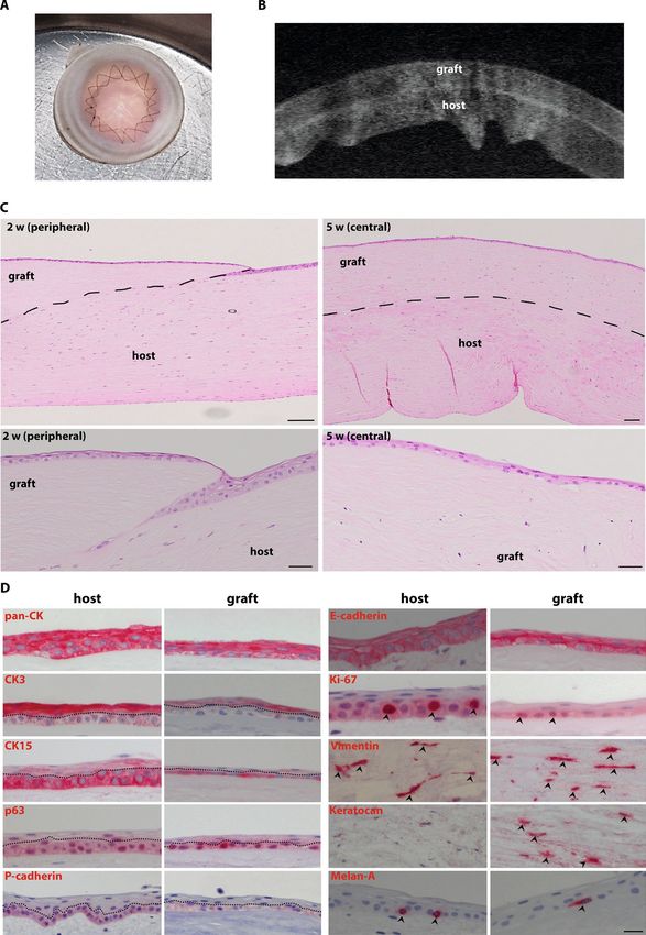
www.nature.com/scientificreports/

Scientific Reports |   (2021) 11:2992 |   https://doi.org/10.1038/s41598-021-82678-3        3

                                                                                       Vol.:(0123456789)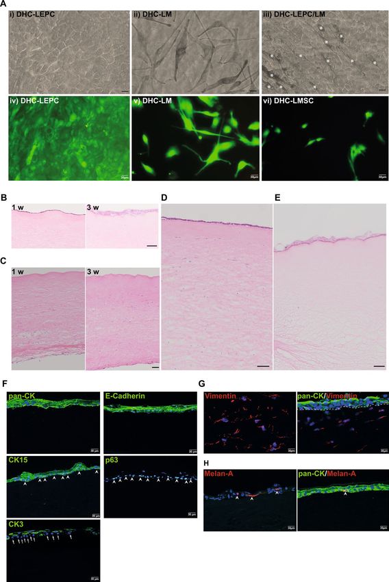
www.nature.com/scientificreports/
Thickness (µm) after additional 24 h in 6%
Corneal tissue Treatment Dextran during treatment (%) Thickness (µm) after treatment (mean ± S.D.) dextran (mean ± S.D.)
NHC 0 1243.4 ± 108.3
Dextran Control 6 702.6 ± 57.4 (24 h) 548.0 ± 19.2 (48 h)
0 1487.6 ± 103.2 686.3 ± 35.0
DM−
2 1169.9 ± 71.7 609.0 ± 11.9
DHC
4 1043.7 ± 112.7 536.6 ± 24.7
6 1005.8 ± 96.2 540.1 ± 30.5
NHC 6 554.1 ± 55.4
Dextran-free Control 0 711.2 ± 86.6 (24 h) 528.0 ± 31.9
0 1192.1 ± 125.2 477.0 ± 37.5
DM+
2 902.2 ± 95.4 581.3 ± 36.2
DHC
4 679.2 ± 59.7 536.3 ± 9.2
6 649.4 ± 76.9 593.6 ± 10.5
Table 1. Corneal thickness measurements by anterior segment optical coherence tomography.
Corneal samples Number Donor age (years)* Cultivation duration (days)* Cultivation in dextran (days)*
Total 183 70.2 ± 13.0 (24–93) 32.0 ± 9.5 (6–75) 4.4 ± 6.5 (0–43)
DM− 102 69.4 ± 10.3(42–87) 30.0 ± 6.4 (16–58) 1.4 ± 1.1 (0–3)
DM+ 51 77.3 ± 11.4 (50–93) 35.3 ± 11.7 (6–69) 12.5 ± 8.4 (3–43)
After PK 30 61.3 ± 16.7 (24–86) 35.0 ± 14.6 (20–75) 2.3 ± 1.1 (1–4)
Table 2. Organ cultured corneal sample details. *All values are expressed as mean ± standard deviation
(range).
In DM+ cornea, the average thickness of NHC was 554.1 ± 55.4 µm, whereas DHC were swollen in the absence
of dextran (1192.1 ± 125.2 µm, p = 0.0001) and less so in 2% dextran (902.2 ± 95.4 µm, p = 0.003, Fig. 1B). The mean
thickness of the scaffolds decellularized using 4% dextran (679.2 ± 59.7 µm) and 6% dextran (649.4 ± 76.9 µm) was
best comparable to NHC (Fig. 1B). As dextran-free control, the NHC were incubated in a medium containing
0% dextran with an incubation time equivalent to the decellularization process (~ 24 h) resulted in a significant
increase in thickness (711.2 ± 86.6 µm, p = 0.0001) (Table 1).
The corresponding anterior segment optical coherence tomography (AS-OCT) images also reflected the same
(Fig. 1C). When corneas (both D M− and DM+) were kept at 6% dextran for 24 h after decellularization, corneal
thickness was comparable to NHC irrespective of the dextran concentration used in decellularization (Table 1).
Based on these results, we performed all subsequent experiments using 1% SD with 4% dextran during the whole
decellularization process, compared to 1% SD without dextran. Even though the 2% dextran was effective to
reduce the thickness of D M− corneas (1169.9 ± 71.7 µm) to match NHC (1243.4 ± 108.3 µm), we used 4% dex-
tran for all further experiments as it was our goal to devise a protocol amenable to both D M− and DM+ corneas.
To test whether the decellularization in the presence of 4% dextran is as efficient as in its absence, histological
evaluations were performed in both D M− and D M+ corneas. H&E staining showed a decrease of corneal thick-
ness when dextran was present during decellularization, but no differences were noticed with respect to remnant
M− as well as D
cellular material. This was true in D M+ samples (Fig. 1D). Spectroscopic analyses of DNA content
showed significantly less residual DNA in DHC compared to NHC, irrespective of dextran used during decel-
lularization. SD and DNAse treatment removed about 98.6 ± 1.2% of the DNA content in the presence of dextran
and 98.4 ± 0.9% in its absence (Fig. 1E). We also noticed that the quality of D M+ corneas was poor compared
to DM− (loss of epithelial layers in (Fig. 1D), and showed lower DNA content (Fig. 1E) presumably due to long
time incubation in dextran containing medium as mentioned earlier (Table 2)24.
Scanning electron microscopy. The effect of decellularization treatment on the integrity of the ECM
structure was evaluated by scanning electron microscopy (SEM). As dextran control, the NHC were incubated in
a medium containing 4% dextran with an incubation time equivalent to the decellularization process. Epithelial
cells detectable in control corneas (Fig. 2A, i & iii) were absent in DHC (Fig. 2A, ii & iv), irrespective of dextran
use. On cross-sections, no significant gross changes of the collagen fibers were noted in DHC except that the
collagen bundles were slightly larger due to swelling and a slight increase in distance between bundles in absence
of dextran (Fig. 2A, vi). When decellularized in the presence of 4% dextran, DHC showed a decrease in collagen
bundles distance (Fig. 2A, viii) compared to NHC in dextran-free medium (Fig. 2A, v).
Optical properties. Macroscopic tissue evaluation suggested that decellularization in the presence of dex-
tran did not affect transparency or even slightly improved it, whereas decellularization in the absence of dextran
Scientific Reports | (2021) 11:2992 | https://doi.org/10.1038/s41598-021-82678-3 4
Vol:.(1234567890)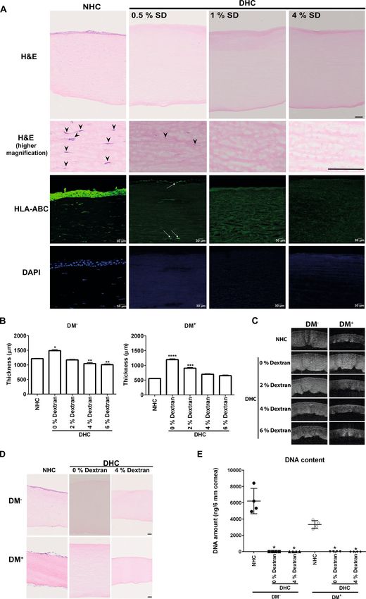
www.nature.com/scientificreports/
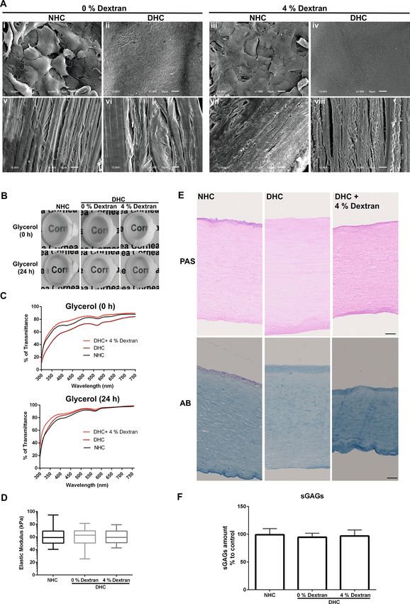
Figure 2. Physical and biochemical properties of the decellularized human cornea (DHC). (A) Scanning
electron microscopy micrographs of the normal human cornea (NHC) and DHC in presence or absence of
dextran; the anterior surface of the cornea (i–iv) showing cells on the NHC (I, iii) but no cells on the DHC (ii,
iv) and cross-section of the cornea (v–viii) showing collagen bundles and distance between the bundles being
larger without dextran (i, ii). Scale bar = 10 µm. (B) Macroscopic pictures of NHC & DHC (0, 4% dextran)
placed on the word “cornea” in the background for a visual representation of the tissue transparency: DHC
seems to be a bit cloudier without dextran than NHC and DHC with 4% dextran. (C) Quantitative analysis of
light transmittance through the NHC and DHC in glycerol before and after incubation at visible wavelengths
(n = 5). The graph represents the percentage of transmittance in NHC and DHC at different wavelengths. (D)
Mechanical properties of DHC by indentation in comparison to NHC. Data are expressed as means ± S.E.M.
(n = 4). (E) Histological evaluation of extracellular matrix content by periodic acid Schiff (PAS) and alcian blue
(AB) on DHC (with 0 or 4% dextran) compared with NHC. Scale bar = 100 µm. (F) Sulfated glycosaminoglycans
(sGAGs) content of DHC in the presence or absence of dextran in comparison to NHC. The graph represents a
percentage (%) of sGAGs content in corneal samples and data are expressed as means ± S.E.M. (n = 5).
Scientific Reports | (2021) 11:2992 | https://doi.org/10.1038/s41598-021-82678-3 5
Vol.:(0123456789)www.nature.com/scientificreports/
led to some tissue clouding (Fig. 2B, 0 h). Twenty-four hours of incubation in glycerol resulted in the recovery of
DHC transparency in the absence of dextran (Fig. 2B, 24 h). The spectroscopic analysis at 300–750 nm revealed
similar transparency of NHC and DHC processed in 4% dextran (Fig. 2C). In the absence of dextran, there was
a reduction in transparency by 10–15% (Fig. 2C, 0 h), which subsided after 24 h in glycerol (Fig. 2C, 24 h).
Mechanical properties. Instrumented indentation measurements indicated no major differences in
the elastic moduli of NHC (62.6 ± 15.9 kPa) and DHC irrespective of dextran (59.3 ± 13.6 kPa in 0% dextran;
59.2 ± 10.56 kPa in 4% dextran) used during decellularization (Fig. 2D).
ECM components. In periodic acid schiff (PAS) staining, the intensity of the glycoproteins of DHC seemed
markedly reduced in the absence of dextran as the thickness of the cornea also increased (Fig. 2E) whereas the
intensity was increased in the DHC in the presence of dextran (Fig. 2E), compared to NHC as thickness also
decreased (Fig. 2E).
Alcian Blue (AB) staining of NHC showed the presence of glycosaminoglycan (GAG) throughout the cornea.
In DHC, GAG staining intensity was much weaker when treated in the absence of dextran (Fig. 2E), whereas
in DHC treated in the presence of dextran GAG staining was more intense than in NHC (Fig. 2E). The levels of
sulfated GAG (sGAG) measured with biochemical assays were unaffected by decellularization and no significant
differences were observed among the samples (Fig. 2F).
The ECM of the corneal surface was evaluated in detail by immunostaining for various components including
agrin, collagen types (Col III, IV, XVIII), fibronectin (FN), junctional adhesion molecule C (JAM-C), perlecan,
tenascin C (TN-C), vitronectin (VN), and laminin (LN) chains (α3, α5, β2, β3, and γ2).
The major BM heparan sulfate proteoglycan, agrin, was expressed in BM with uniform and strong labeling
intensity in all samples (Fig. 3, arrowheads). Expression of Coll IV and Coll XVIII molecules were clearly noted
in BM of the cornea in both NHC and DHC samples (Fig. 3, arrowheads). No significant difference was noted
for Col IV, whereas Coll XVIII expression was reduced in DHC (no dextran) and discontinuous in DHC (4%
dextran) compared to NHC. The fibrillar Coll III was not observed in either NHC or DHC (Fig. 3).
FN was clearly detected in BM in all samples without any significant differences (arrowheads), whereas a
weak expression of vitronectin was observed (Fig. 3). Strong expression of the other major BM heparan sulfate
proteoglycan, perlecan, was noted similar to agrin (Fig. 3, arrowheads). TN-C was hardly expressed on the BM
of the corneal surface of all samples, whereas JAM-C expression was not observed (Fig. 3). Using Laminin chain
specific antibodies, the LN-α3, -ß3, -γ2 chains were found to be strongly expressed in the corneal BM of NHC
and DHC with or without dextran (Fig. 3, arrowheads). No significant differences were observed in staining pat-
terns among samples. The LN α5 chain and β2 chains were not detectable in corneal samples as their expression
is known to be limited to the limbal region (Fig. 3)25.
Recellularization. The characterization details of cultured limbal epithelial progenitor cells (LEPC), limbal
mesenchymal stromal cells (LMSC), and limbal melanocytes (LM) were provided in the Supplementary Data S1.
In vitro recellularization. The biocompatibility of the DHC to support LEPC, LMSC, and LM was evalu-
ated. The seeded LEPC and LMs attached to the surface of decellularized corneas (Fig. 4A, i-iii) and remained
viable after 48 h of cultivation as confirmed by live/ dead staining (Fig. 4A, iv&v). In DHC-LEPC/LM scaffolds,
the melanocytes intermingled with epithelial cells (Fig. 4A, iii, white asterisk). The LMSCs injected into DHC
spread and appeared healthy (Fig. 4A, vi). H&E staining showed a monolayer of LEPC on DHC after 1 week of
cultivation (Fig. 4B, 1 w). After 3 weeks in culture stratification of the epithelium was successfully induced by
lifting the tissues to the air–liquid interface and confirmed by H&E staining (Fig. 4B, 3 w). The injected LMSCs
were spread over the posterior side of the DHC after 1 week (Fig. 4C, 1 w). After 3 weeks, the LMSCs appeared
to migrate into the anterior part of the corneal stroma (Fig. 4C, 3 w). After 3 weeks of cultivation, the DHC-
LEPC/LM scaffolds showed a stratified surface epithelium and stromal cells in the anterior part of the stroma as
confirmed by H&E staining (Fig. 4D). The DHC-LEPC/LM scaffolds also showed a stratified epithelium after
3 weeks of cultivation (Fig. 4E).
Phenotypic evaluation of recellularized scaffolds by immunofluorescent staining confirmed pronounced pan-
cytokeratin (pan-CK) and E(epithelial)-cadherin staining in all epithelial layers; CK15 and p63 staining confined
to basal layers (Fig. 4F, arrowheads); corneal differentiation marker CK3 was expressed in superficial epithelial
layers but not in basal cells (Fig. 4F, arrows indicating basal cells) in DHC-LEPC scaffolds . Vimentin-positive
cells (red) were observed in the stromal region close to pan-CK-positive epithelial cells (green) of the DHC-
LEPC/LMSC scaffolds (Fig. 4G). Melan-A positive cells (red) were interspersed in the epithelial layers (pan-CK+,
green) of the DHC-LEPC/LM scaffolds (Fig. 4H, arrowhead). The results of recellularization by neighboring
tissue are provided in Supplementary Data S2.
Ex vivo transplantation. The anterior part of a DHC was successfully sutured onto the posterior bed of
a NHC (Fig. 5A) and both tissues were well adapted by sutures as confirmed by AS-OCT imaging (Fig. 5B).
The surgical handling of the DHC tissue was comparable to NHC and no significant tissue damage occurred
during suturing. After 2 weeks in culture, histological analyses showed complete epithelialization of the grafted
tissue scaffold with stratification and the absence of cells in the stromal region of the graft (Fig. 5C, 2 w). Stro-
mal cells were detectable in the graft 5 weeks after surgery (Fig. 5C, 5 w). Immunohistochemical analyses
revealed the expression of the epithelial markers cytokeratin (pan-CK) and E-cadherin in the epithelial cells on
the host (peripheral cornea/limbal region) and graft tissue (Fig. 5D). Expression of the LEPC markers, CK15,
P(placental)-cadherin, and p63, was apparent in basal epithelial cells on both, the grafted scaffold and host tis-
Scientific Reports | (2021) 11:2992 | https://doi.org/10.1038/s41598-021-82678-3 6
Vol:.(1234567890)www.nature.com/scientificreports/
Figure 3. Extracellular matrix (ECM) composition of decellularized human cornea (DHC). Immunostaining
analysis of various (ECM) and basement membrane (BM) molecules on the DHC (0 and 4% dextran) compared
to the normal human cornea (NHC). The white dashed line marked the boundary between the limbal and
corneal tissue in laminin (LN) α5 staining of NHC. Arrowheads indicating the expression of ECM and BM
molecules. Nuclei counterstained with 4′,6‐diamidino‐2‐phenylindole (blue). Col Collagen, JAM-C junctional
adhesion molecule C.
Scientific Reports | (2021) 11:2992 | https://doi.org/10.1038/s41598-021-82678-3 7
Vol.:(0123456789)www.nature.com/scientificreports/
Figure 4. In vitro recellularization of the decellularized human cornea (DHC) (A) Phase contrast micrographs of LEPC (i); LM (ii);
and LEPC/LM scaffolds showing intermingled melanocytes (iii, white asterisk) in the epithelial cell layer on DHC. Scale bar = 20 µm.
Live/dead viability assay after 24 h of cultivation of LEPC, LMSC, and LM on DHC (iv–vi). (B) Light microscopic analysis of DHC-
LEPC scaffolds showing cell monolayers after 1 week (1 w) and stratified epithelium (3 w) by hematoxylin and eosin staining (H&E)
on the anterior surface. Scale bar—100 µm. (C) The injected stromal cells in DHC showing the stromal cells on the posterior side at
1-week culture and migrated to the anterior side after 3 weeks by H&E staining. Scale bar—100 µm (D) DHC-LEPC/LMSC scaffold
after 3 weeks cultivation showing stratified epithelium and presence of stromal cells throughout the stroma by H&E staining (E)
DHC-LEPC/LM scaffold showing stratified epithelium after 3 weeks of cultivation. Scale bar—100 µm (F) Immunofluorescence
staining of DHL-LEPC scaffolds showing the expression of CK15 and p63 (green) at basal layers of epithelial cells (arrowheads); CK3
expression (green) in suprabasal cells but not in basal cells (arrows); E(epithelial)-cadherin and cytokeratin (pan-CK) expression
(green) in all human limbal epithelial cells on DHC. (G) DHL-LEPC/LMSC scaffolds showing the vimentin expression on stromal
cells and cytokeratin (pan-CK) expression (green) on epithelial layers. The dashed line separated the epithelium and stroma. (H) DHL-
LEPC/LM scaffolds showing the melan-A (red) positive melanocytes (arrowheads) in the epithelial layers (pan-CK, green). LEPC
limbal epithelial progenitor cells; LMSC limbal mesenchymal stromal cells, LM limbal melanocytes, pan-CK pan-cytokeratin, CK15
cytokeratin 15, CK3 cytokeratin 3, DAPI 4′,6‐diamidino‐2‐phenylindole.
Scientific Reports | (2021) 11:2992 | https://doi.org/10.1038/s41598-021-82678-3 8
Vol:.(1234567890)www.nature.com/scientificreports/
Figure 5. Ex Vivo transplantation of decellularized human cornea (DHC) (A) micrograph of DHC transplanted
ex vivo in the normal human cornea (NHC) as anterior lamellar grafts with double running cross-stitch
sutures. (B) AS-OCT micrograph showing the well-adapted graft (DHC) and host tissue (NHC) and no gap
was observed in between. (C) After 2 weeks (2 w) of transplantation, histological analyses by hematoxylin and
eosin (H&E) staining showed complete epithelialization of graft tissue with stratification and absence of stromal
cells in graft tissue (2 w) (dashed line marking the boundary between the graft and host tissue). The stromal
cells appeared in the graft region at 5 weeks samples confirmed by H&E staining (5 w). Scale bar—100 µm D)
Immunohistochemical staining on paraffin sections showing the cytokeratin (pan-CK) expression on epithelial
layers; CK3 expression on suprabasal cells; CK15, P(placental)-cadherin, p63 (dotted line separated the basal
and superficial cells), and Ki-67 expression at basal layers of epithelial cells (arrowheads); E(epithelial)-cadherin
expression in all epithelial layers; vimentin expression in the stromal region (arrowheads); and Melan-A
expression (magenta) (arrowheads) on melanocytes at basal layers of epithelium, in both host and graft tissue.
A keratocan expression was observed only in the graft tissue (arrowheads). AS-OCT anterior segment optical
coherence tomography, pan-CK pan-cytokeratin, CK3 cytokeratin 3, CK15 cytokeratin 15.
Scientific Reports | (2021) 11:2992 | https://doi.org/10.1038/s41598-021-82678-3 9
Vol.:(0123456789)www.nature.com/scientificreports/
sue, whereas expression of the corneal differentiation marker CK3 was present only in the superficial epithelial
cells of the graft and host tissue (Fig. 5D). Moreover, we also observed Ki-67 positive cells in the basal layer of the
epithelium covering the DHC tissue (Fig. 5D, arrowheads). The migrated stromal cells observed in the graft tis-
sue after 5 weeks showed expression of vimentin and keratocan, a corneal stromal marker (Fig. 5D, arrowheads).
Interestingly, we observed Melan-A positive melanocytes in association with basal epithelial cells in the graft
tissue similar to host tissue (Fig. 5D, arrowheads).
Discussion
Decellularized corneal scaffolds recently gained interest as an alternative material for corneal r eplacement26–28.
If corneal damage extends from the ocular surface to the upper stroma and an allotransplant is at high risk of
rejection due to vascularization or inflammation, decellularized corneal scaffolds may serve as an alternative
tissue source to reconstruct the corneal epithelium and its underlying stromal support. Therefore, it was our goal
to devise an efficient, fast, and gentle method of decellularization using well-tolerated substances to generate
human corneal scaffolds from readily available donor material.
Various protocols have been used to decellularize human corneal tissue, of them, SDS or hypertonic salt with
nucleases were most effective in removing cellular c omponents6,7. The combination of SD, a natural bile salt, and
DNAse was used to decellularize human corneal limbus. However, significant drawbacks were the long elution
times required for removal of cellular components by SDS/nucleases or NaCl/nucleases treatment reported for
human corneal decellularization (~ 5–8 days)6,7 and SD/DNAse for treatment of human limbal tissue (~ 5 days)18.
In this study, we established and validated a rapid alternative method (~ 1 day) for efficient decellularization of
the human cornea using SD and DNAse. Our data indicate that 1% or 4% SD with DNAse and 4% dextran allow
for efficient decellularization by removal of cellular components (HLA-ABC and nuclear debris) within 24hrs,
in line with earlier reports on other tissues11,17,18.
Due to its high glycosaminoglycan content, corneal tissue has a strong tendency to swell in cell culture
media29–31. A concentration of 5–6% dextran in a medium is therefore routinely used to maintain graft thickness
prior to keratoplasty31,32. The significant difference of the mean NHC thickness between DM− and DM+ groups
is due to the presence or absence of dextran in the respective culture medium used for tissue transfer from the
Cornea Bank to the laboratory. Most previous reports on human corneal decellularization have not specifically
addressed tissue s welling6,7, but dextran use has been reported on porcine corneas in conjunction with ultrahigh
hydrostatic pressure and chemical detergents20,29,30. We compared different concentrations of dextran to control
tissue swelling during decellularization of the human cornea and found that the presence (DM+) or absence
(DM−) of Descemet membrane had an additional influence on tissue thickness (Fig. 1B–D). In both, DM+ and
DM− cornea, a dextran concentration of 4% was sufficient to preserve corneal thickness during decellulariza-
tion. In line with earlier reports on porcine t issue7,20,29,30, SD/DNAse-mediated removal of cellular components
and DNA from human corneal tissue were unhampered by the presence of dextran (Fig. 1D,E). Moreover, our
decellularization protocol is also applicable to D M+ corneas, which might be usable for full-thickness corneal
transplantation in the future.
As optical properties are highly relevant for the selection of materials for corneal reconstruction, we next
assessed transparency and optical transmittance of DHC. It has been reported that ionic detergents solubi-
lize both cytoplasmic and nuclear cell membranes, damage collagen, and denature proteins by disrupting pro-
tein–protein interactions33,34. In a previous study, SDS/DNAse-treated corneas had the greatest loss of transpar-
ency with most ECM disruption as compared to corneas treated with non-ionic d etergents7. Even though SD is
an ionic detergent, ECM disruption, or reduction of the sGAGs content were not observed similar to previous
studies on other t issues35. In our own experiments, the transparency of DHC decreased with decellularization
in the absence of dextran and recovered after incubation in glycerol for 24 h (Fig. 2B). This suggests that the
decrease in corneal transparency was a result of increased corneal hydration rather than permanent structural
alteration6,29. However, it has also been reported that glycerol causes additional collagen disorganization36 and
may nevertheless mask structural damage inflicted by the decellularization p rocedure7. Herein, we suggest to
include dextran during decellularization of the human cornea to reduce tissue swelling, loss of transparency,
and to maintain tissue integrity.
Ex vivo cultivated limbal epithelial cells have been used to reconstruct the corneal epithelium in limbal stem
cell-deficient patients37–40. The long term clinical outcome of limbal epithelial cell grafts mainly depends on the
maintenance of stem/progenitor cells, which is regulated by the niche microenvironment including its ECM
components and mechanical properties in vivo39,41–44. Moreover, ECM molecules play a key role in cell–cell
interactions, cell adhesion, proliferation, migration, and r esponse25,45. Thus, the integrity and preservation of
ECM components are essential for further clinical use of the decellularized tissues. The published decellulariza-
tion protocols have not reported on ECM component preservation in much d etail6,7. To address this issue, we
extensively studied ECM components and their possible alteration. The corneal BM collagens type IV, important
in the structure and function of BM, and type XVIII, having an anti-angiogenic a ctivity46,47, were well preserved
by our decellularization protocol. Heparin sulfate proteoglycans (e.g. perlecan and agrin) and glycoproteins
(e.g. fibronectin), which were well preserved in this study, play important roles in epithelial cell migration and
proliferation48. LNs are among the best described BM components of the cornea, of which the cornea-specific
isoform LN-332 promotes adhesion, migration, and differentiation of LEPC25,49. Our own data clearly indicate
the preservation of a vast panel of ECM proteins including various laminin isoforms as detected by immuno-
fluorescence microscopy (Fig. 3).
Successful repopulation with cells is a prerequisite for the use of decellularized scaffolds in tissue engineering.
It has been reported that structurally damaged decellularized cornea and residual decellularizing agents (SDS)
detrimentally affect cell growth and proliferation7. It has been shown that SD-produced scaffolds were highly
Scientific Reports | (2021) 11:2992 | https://doi.org/10.1038/s41598-021-82678-3 10
Vol:.(1234567890)www.nature.com/scientificreports/
biocompatible compared to those decellularized using S DS23. We explored different strategies of DHC repopula-
tion by plating cells or by allowing cellular invasion from host tissue. With both approaches, the repopulating
epithelial cells maintained a proper phenotype as characterized by expression of the progenitor markers CK15,
P-cadherin, and p63 in basal layers and the marker for differentiated epithelial cells, CK3, in the superficial lay-
ers. Stromal repopulation is a slower process as has been clinically observed in keratoconus patients following
collagen cross-linking treatment. Activated keratocytes started to populate the corneal stroma at 2 months after
treatment and repopulation was almost complete after 6 m onths50. We observed stromal repopulation of a scaffold
by the 3rd week in vitro, whereas it took 5 weeks in an ex vivo model. Thus, the repopulation of DHC by stromal
cells could find an application treating anterior corneal lesions where corneal stroma is altered by disease, injury,
or scarring51. Currently, no data are available regarding the repopulation of decellularized tissue by LMs, which
are found in close proximity to LEPC at the limbus in vivo, where melanocytes appear to play a supporting role
in preserving the stemness state of corneal epithelial cells and modulating their migration towards the central
cornea during regeneration52,53. LMs were able to adhere and intermingle with epithelial cells on DHC tissue,
strongly suggesting the potential of the scaffold to facilitate LM survival. Moreover, in ex vivo lamellar grafting
experiments we have also observed the migration of LMs from donor limbal tissue onto decellularized corneal
grafts similar to reports on ocular surface regeneration in vivo52,54. All these observations indicate that the DHC
scaffold provides a niche microenvironment to keep basal epithelial cells in a less differentiated state. Thus, it may
urposes6,45. However, in vivo studies are necessary
serve as an ideal scaffold for future clinical transplantation p
to determine whether the DHC supports stromal dehydration, long-term transparency, and in vivo biocompat-
ibility. Moreover, it has to be tested whether a significant reduction of immunogenic epitopes was achieved to
allow for long-term immunological tolerance.
In conclusion, we have developed a fast and efficient corneal decellularization method using clinically appli-
cable SD with DNAse in the presence of dextran. Dextran prevented corneal swelling during decellularization.
The biomechanical and optical properties, ECM architecture and BM composition of the DHC were well pre-
served and resembled the properties of cultured NHC. The DHC scaffolds revealed excellent biocompatibility
and ex vivo transplanted corneas were completely epithelialized and effectively supported stromal cell ingrowth.
Hence, this could be a promising scaffold for anterior corneal reconstruction in the treatment of corneal defects.
Methods
Tissue. Organ cultured human corneoscleral tissue remnants after DMEK (DM−, n = 102) and corneal
buttons unsuitable for transplantation (DM+, n = 51) were used (Table 2). To isolate primary limbal cells for
repopulation experiments, organ cultured corneoscleral rims remaining after penetrating keratoplasty were used
(labeled after PK, n = 30, Table 2). The tissue was kindly provided by the LIONS Cornea Bank Baden-Württem-
berg, located at the Eye Center, University of Freiburg and informed consent for research use of remnant tissue
had been given by the donors or their next of kin. The study was approved by the institutional review board of
the University of Freiburg (25/20) and followed the tenets of the Declaration of Helsinki. No organs or tissues
from prisoners were used.
M+ corneas from the
Following the standard procedures for corneal grafts in the Eye Center, we obtained D
Cornea Bank cultured (12.5 ± 8.4 days) in Carry-C medium (Alchimia) containing 6% dextran, whereas D M− cor-
neas were cultured also in Carry-C medium (1.4 ± 1.1 days) before DMEK surgery and were supplied to the
laboratory in Tissue-C medium (Alchimia) without dextran. Due to higher availability, most experiments were
performed on DM− corneas except as otherwise indicated. DM+ corneas were especially used to comparatively
analyze the decellularization protocols in the course of initial experiments.
Decellularization. Corneas without any further processing were used as controls (NHC). For decellulariza-
tion, all washing steps and SD (Sigma-Aldrich, Hamburg, Germany) incubation were carried out under continu-
ous agitation (800 rpm) at room temperature. Corneoscleral tissue was washed in Dulbecco’s phosphate-buff-
ered saline (DPBS; 3 × 5 min), placed in 12-well plates with 0.5%, 1%, or 4% SD in ultrapure water for 30 min,
and then rinsed in DPBS (3 × 30 min). Subsequently, the tissues were incubated in DNAse I , 1 mg/ml in DPBS
(Roche, Mannheim, Germany) overnight under a sterile hood and terminally washed in DPBS (4 × 30 min). The
whole decellularization procedure was carried out under aseptic conditions to ensure tissue sterility.
Quantification of tissue swelling. To assess the effects of dextran on corneal swelling during decellulari-
zation, all decellularization steps were performed in the presence of 0, 2, 4, or 6% dextran 500 (Sigma-Aldrich,
Hamburg, Germany) in both DM+ and DM− corneas. To verify the effect of dextran alone on NHC, DM− corneas
in incubated in 6% dextran (dextran control) and DM+ corneas in dextran free medium (dextran-free control)
with an incubation time equivalent to the decellularization process. Corneal swelling was analyzed by AS-OCT
using a clinical AS-OCT device (SS-1000 CASIA, Tomey, Nuernberg, Germany). Measurements of the central
corneal thickness were performed with 3-dimensional cross-sectional imaging before and after decellularization
as described earlier55.
Histology. For bright field light microscopy, tissue was cryosectioned and stained as follows: After embed-
ding and freezing in optimal cutting temperature medium, 10 µm thick sections were cut with a cryostat (Leica,
Germany), mounted on adhesive slides and air-dried. Sections were stained with hematoxylin (Haematoxylin
Gill III, Surgipath, Leica, Germany) for 2 min and 1% eosin Y (Surgipath, Leica, Germany) for 1 min to observe
the gross tissue architecture and degree of decellularization. To visualize the proteoglycan content, cryosections
were stained with 1% alcian blue (Morphisto, Germany) for 30 min together with a counterstain of nuclear fast
red (Morhphisto, Germany). For glycoproteins, PAS staining was performed using 1% periodic acid (Fluka,
Scientific Reports | (2021) 11:2992 | https://doi.org/10.1038/s41598-021-82678-3 11
Vol.:(0123456789)www.nature.com/scientificreports/
Germany) for 10 min, and Schiff reagent (Roth, Germany) for 90 s. Samples were examined using either a bright
field fluorescence microscope (Axio Imager.A1, Zeiss) and images were processed using ProgRes CapturePro
Software (JENOPTIK, https://www.jenoptik.com/products/cameras-and-imaging-modules/microscope-camer
as/progres-usb-20-firewire/software-download-progres) or a Hamamatsu NanoZoomer S60 (Hamamatsu Pho-
tonics, Herrsching, Germany).
Immunostaining of frozen sections. Immunostaining on frozen sections performed as previously
described60. Briefly, human corneas in optimal cutting temperature medium were cut into 10 µm sections, fixed
in 4% paraformaldehyde (PFA) for 20 min or acetone for 10 min followed by permeabilization in 0.3% Triton
X-100 in PBS for 10 min. The sections were blocked with 10% normal goat serum (NGS) and incubated in
primary antibodies (Supplementary Table S1) diluted in 1% NGS in PBS overnight at 4 °C. Fluorescein iso-
thiocyanate-conjugated or rhodamine-conjugated anti-mouse or -rabbit immunoglobulins (Life Technologies,
Carlsbad, CA) were used for detection and nuclear staining was performed with DAPI (Vectashield antifade
mounting medium with DAPI; Vector, Burlingame CA). Immunolabeled cryosections were examined with a
laser scanning confocal microscope (TCS SP-8, Leica, Wetzlar, Germany). For negative controls, the primary
antibodies were replaced by equimolar concentrations of an irrelevant isotypic primary antibody of the same
species.
Immunostaining of paraffin sections. Immunohistochemistry was performed as previously described56.
The list of antibodies is provided in Supplementary Table S1.
Scanning electron microscopy. SEM was performed as described earlier57. Briefly, scaffolds were fixed
with 3.8% formaldehyde in PBS for at least 1 h and rinsed twice with PBS. Then, the scaffolds were dehydrated
by rinsing through graded ethanol/water mixtures (50%, 70%, 80%, 90%, and 100%; each step for 10 min at
room temperature). Next, ethanol was slowly exchanged by liquid C O2 (critical point dryer; Balzers CPD, Bal-
ter’s Union) and the samples were dried using the critical point method. For cross-sections, the specimens were
sectioned transversely. Finally, whole tissue and sections were sputtered with a thin layer of gold of ∼10 nm in
thickness (sputter coater, Balzers SLD; Balzers Union), and documented by a Leo 435VP (Zeiss) scanning elec-
tron microscope.
Elastic modulus determination by instrumented indentation. Biomechanical characterization was
performed as published e arlier58. In brief, the tissue was immersed in a medium containing 15% dextran for 24 h
to achieve dehydration similar to the situation in vivo. Central tissue samples were cut with a 7 mm diameter
trephine and the tissue was glued to the bottom of a cell culture dish. The indentation was performed using a
spherical ruby tip of a 500 µm radius on a Bioindenter (Anton Paar, Peseux, Switzerland). Load-controlled meas-
urements with loading and unloading rates of 300 µN/min and a maximum indentation load of 200 µN were
taken. The elastic modulus was calculated using the Hertz model and Bioindenter indentation software (Anton
Paar, https://wiki.anton-paar.com/en/indentation-testing-biological-soft-materials-using-bioindenter/).
DNA content. DNA was extracted using the DNeasy Blood & Tissue Kit (69,504, Qiagen, Hilden, Ger-
many). The central 6 mm diameter of NHC and DHC were trephined and incubated with proteinase K in ATL
buffer at 56 °C under continuous agitation and further processed for DNA elution according to the manufac-
turer’s protocol. The eluates were analyzed photometrically at 260 nm wavelength using the NanoDrop OneC
Microvolume UV–Vis spectrophotometer (Thermo Scientific).
Optical properties. The optical properties of the NHC and DHC were assessed by a UV–Vis spectro-
photometer as described previously59. Breifly, the 6 mm diameter central corneal pieces were trephined from
both NHC and DHC and the corneal pieces were placed in a 96-well UV-star microplate (Greiner, Germany)
filled with glycerol. The spectral transmittance of each sample was measured using a spark microplate reader
(TECAN) and the absorbance data were recorded at 1 nm wavelength increments (300–750 nm). The transmit-
tance of the samples was corrected with glycerol as a blank medium and data were analyzed as the mean percent-
age of transmittance. In addition, photographs (Digital camera, Canon EOS 700D) were taken with the word
“cornea” behind the corneal tissue to give a visual representation of the differences in transparency.
Sulfated glycosaminoglycans. The sGAG content of DHC was determined using a 1,9-dimethyl meth-
ylene blue (DMMB assay) (280,560-N, Proteoglycan detection kit, Amsbio) according to the manufacturer’s
protocol. The central 6 mm of the corneas were trephined and digested with papain as per manufacturer’s pro-
tocol (Tissue digestion kit, Amsbio). Briefly, the tissue was homogenized, and digested with papain at 60 °C for
1 h, and acetic acid and Tris-hydrochloric acid were added. The digested samples were added to DMMB and
absorbance was measured at 515 nm using a spark microplate reader (TECAN). The absorbance was expressed
in percentage (%) with reference to NHC (100%).
Cell culture. LEPC, LMSC, and LM were isolated, cultivated, and characterized as described e arlier60, details
are provided in the Supplementary Data S1.
Repopulation of decellularized corneas with cultured cells. For recellularization and ex vivo trans-
plantation experiments 4% dextran and 1% SD were used during the decellularization process. For DHC-LMSC
Scientific Reports | (2021) 11:2992 | https://doi.org/10.1038/s41598-021-82678-3 12
Vol:.(1234567890)www.nature.com/scientificreports/
scaffolds, the isolated LMSC (P2) were injected (20 µl) at 10 locations in the decellularized corneal stroma
(about 20% depth from the posterior side) with a 20-gauge needle on a 1 ml syringe (1 × 10⁶ cells/ml). The cor-
neas with injected cell suspensions were transferred to 12-well plates containing Mesencult media (Stem Cell
Technologies). For DHC-LEPC scaffolds, the isolated human LEPC (P1) were seeded (1 × 10⁶ cells/ml) on the
anterior surface of the cornea and cultured in corneal culture medium (CCM) containing DMEM/ Ham’s F12
(3:1) (Hyclone; GE Healthcare Life Sciences, Freiburg, Germany) supplemented with human corneal growth
supplement (Life Technologies), 5% FCS (GE Healthcare Life Sciences) and low calcium concentration (0.4 mM
Ca2+, labeled CCM-low). For DHC-LEPC/LMSC scaffolds, LMSCs were injected in DHC as mentioned earlier,
followed by LEPCs seeding 24 h later and cultivation of the resulting constructs in CCM-low. For DHC-LEPC/
LM scaffolds, both LEPC and LM were seeded together in a ratio of 3:1 on the decellularized corneal surface and
cultured in CCM-low medium. For stratification of DHC-LEPC, -LEPC/LMSC, and -LEPC/LM scaffolds, after
1 week, the tissues were raised to the air–liquid interface and the CCM was shifted to high-calcium concentra-
tions (2 mM Ca2+, labeled CCM-high) and cultured further 2 weeks. All cultures were maintained at 37 °C, 5%
CO2, and 95% humidity and medium was changed every alternative day. During culturing, recellularized scaf-
folds were examined using phase contrast microscope (Axio Vert.A1, Zeiss) and images were processed using
ProgRes CapturePro software (JENOPTIK, https://www.jenoptik.com/products/cameras-and-imaging-modul
es/microscope-cameras/progres-usb-20-firewire/software-download-progres). After terminating the cultiva-
tion, corneal samples were fixed for immunohistochemistry and light microscopy as described above.
Cell viability. Live/dead viability/cytotoxicity kit (MP 03,224, Molecular Probes) was used to visualize live
and dead cells in repopulated DHC as per manufacturer’s protocol. Briefly, the repopulated DHC was incu-
bated for 30 min at room temperature in calcein-AM (2 µM) and ethidium homodimer (4 µM) solution. After
incubation, the cornea was rinsed thrice in PBS and images were photographed using a inverted fluorescence
microscope with ZEN software (Axio Observer Z1, Zeiss; https://www.zeiss.com/microscopy/int/products/
microscope-software/zen.html).
Sutureless repopulation of decellularized corneas. In order to mimic the in-vivo-system in a suture-
less setup to avoid damage to the ECM, examined the gradual repopulation of the DHC by mounting a native
corneoscleral tissue quarter in close contact to a decellularized scaffold quarter. Details of the experiment are
provided in the Supplementary Data S2.
Ex vivo transplantation. The feasibility of the DHC to serve as donor tissue for corneal transplantation
was examined by performing anterior lamellar keratoplasty (ALK) ex vivo on non-decellularized human cadav-
eric corneoscleral tissue. For ALK, a microkeratome (mk-DSAEK, Gebauer, Neuhausen, Germany) was used
to dissect both the NHC (DM+) and the DHC (DM−) tissue using a 450 µm keratome head. Using a surgical
microscope (OPMI VISU 140, Zeiss, Oberkochen, Germany) and an artificial anterior chamber (Barron Artifi-
cial Anterior Chamber, Katena Products, INC., Parsippany, USA) to fix the host tissue, the anterior corneal part
of the DHC tissue (graft) was placed epithelial side up on the posterior bed of the NHC (host). These constructs
were fixed temporarily with four opposing single stitches 90 degrees apart from each other using polyamide
suture material (10–0 Ethilon, ETHICON, Johnson & Johnson Medical Devices, Ohio, USA). Subsequently, the
tissues were definitely attached with double running cross-stitch s utures61. Finally, the four temporary sutures
were removed. Thereafter, the corneal samples were transferred into 12-well plates and cultured in CCM-high
medium for 5 weeks.
Statistics. Statistical analyses were performed using GraphPad Prism software (Version 6.0; Graphpad Soft-
ware Inc., La Jolla, CA; https://www.graphpad.com/). Data is represented as a mean ± standard deviation (S.D.)
from individual experiments (Table 1 & 2) or as mean ± standard error of the mean (S.E.M.) (graphs). The statis-
tical significance (p ≤ 0.05) was evaluated by the Wilcoxon signed-rank test or Mann–Whitney U test.
Data availability
The datasets generated during and /or analyzed during the current study are available from the corresponding
author on reasonable request.
Received: 14 November 2020; Accepted: 14 January 2021
References
1. Singh, R., Gupta, N., Vanathi, M. & Tandon, R. Corneal transplantation in the modern era. Indian J. Med. Res. 150(1), 7–22 (2019).
2. Reinhard, T. et al. Long-term results of allogeneic penetrating limbo-keratoplasty in total limbal stem cell deficiency. Ophthalmol-
ogy 111(4), 775–782 (2004).
3. Sundmacher, R. & Reinhard, T. Central corneolimbal transplantation under systemic ciclosporin A cover for severe limbal stem
cell insufficiency. Graefes Arch. Clin. Exp. Ophthalmol. 234(S1), S122–S125 (1996).
4. Dong, M. et al. Rapid porcine corneal decellularization through the use of sodium N-lauroyl glutamate and supernuclease. J. Tissue
Eng. 10, 2041731419875876. https://doi.org/10.1177/2041731419875876 (2019).
5. Isidan, A. et al. Decellularization methods for developing porcine corneal xenografts and future perspectives. Xenotransplantation
26(6), e12564. https://doi.org/10.1111/xen.12564 (2019).
6. Shafiq, M. A., Gemeinhart, R. A., Yue, B. Y. & Djalilian, A. R. Decellularized human cornea for reconstructing the corneal epithe-
lium and anterior stroma. Tissue Eng. Part C Methods. 18(5), 340–348 (2012).
Scientific Reports | (2021) 11:2992 | https://doi.org/10.1038/s41598-021-82678-3 13
Vol.:(0123456789)www.nature.com/scientificreports/
7. Wilson, S. L., Sidney, L. E., Dunphy, S. E., Dua, H. S. & Hopkinson, A. Corneal decellularization: a method of recycling unsuitable
donor tissue for clinical translation?. Curr. Eye Res. 41(6), 769–782 (2016).
8. Fernández-Pérez, J. & Ahearne, M. Decellularization and recellularization of cornea: progress towards a donor alternative. Methods
171, 86–96 (2020).
9. Ahearne, M., Fernandez-Perez, J., Masterton, S., Madden, P. W. & Bhattacharjee, P. Designing scaffolds for corneal regeneration.
Adv. Funct. Mater. https://doi.org/10.1002/adfm.201908996 (2020).
10. Alió Del Barrio, J. L. et al. Corneal stroma enhancement with decellularized stromal laminas with or without stem cell recellulari-
zation for advanced keratoconus. Am. J. Ophthalmol. 186, 47–58 (2018).
11. Ansari, T. et al. Development and characterization of a porcine liver scaffold. Stem Cells Dev. 29(5), 314–326 (2020).
12. McCrary, M. W. et al. Novel sodium deoxycholate-based chemical decellularization method for peripheral nerve. Tissue Eng. Part
C Methods. 26(1), 23–36 (2020).
13. Alshaikh, A. B. et al. Decellularization of the mouse ovary: comparison of different scaffold generation protocols for future ovarian
bioengineering. J. Ovarian Res. 12(1), 58. https://doi.org/10.1186/s13048-019-0531-3 (2019).
14. Tiemann, T. T. et al. Towards uterus tissue engineering: a comparative study of sheep uterus decellularisation. Mol. Hum. Reprod.
26(3), 167–178 (2020).
15. Das, P., Singh, Y. P., Mandal, B. B. & Nandi, S. K. Tissue-derived decellularized extracellular matrices toward cartilage repair and
regeneration. Methods Cell Biol. 157, 185–221 (2020).
16. Guimaraes, A. B. et al. Evaluation of a physical-chemical protocol for porcine tracheal decellularization. Transpl. Proc. 51(5),
1611–1613 (2019).
17. Luo, Y., Lou, D., Ma, L. & Gao, C. Optimizing detergent concentration and processing time to balance the decellularization effi-
ciency and properties of bioprosthetic heart valves. J. Biomed. Mater. Res. A. 107(10), 2235–2243 (2019).
18. Spaniol, K. et al. Generation and characterisation of decellularised human corneal limbus. Graefes Arch. Clin. Exp. Ophthalmol.
256(3), 547–557 (2018).
19. Dayan, S. H. et al. Overview of ATX-101 (deoxycholic acid injection): a nonsurgical approach for reduction of submental fat.
Dermatol. Surg. 42(S1), S263–S270. https://doi.org/10.1097/DSS.0000000000000870 (2016).
20. Lynch, A. P., Wilson, S. L. & Ahearne, M. Dextran preserves native corneal structure during decellularization. Tissue Eng. Part C
Methods. 22(6), 561–572 (2016).
21. Gattazzo, F., Urciuolo, A. & Bonaldo, P. Extracellular matrix: a dynamic microenvironment for stem cell niche. Biochim. Biophys.
Acta. 1840(8), 2506–2519 (2014).
22. Swinehart, I. T. & Badylak, S. F. Extracellular matrix bioscaffolds in tissue remodeling and morphogenesis. Dev. Dyn. 245(3),
351–360 (2016).
23. Syed, O., Walters, N. J., Day, R. M., Kim, H. W. & Knowles, J. C. Evaluation of decellularization protocols for production of tubular
small intestine submucosa scaffolds for use in oesophageal tissue engineering. Acta Biomater. 10(12), 5043–5054 (2014).
24. Redbrake, C., Salla, S., Nilius, R., Becker, J. & Reim, M. A histochemical study of the distribution of dextran 500 in human corneas
during organ culture. Curr. Eye Res. 16(5), 405–411. https://doi.org/10.1076/ceyr.16.5.405.7044 (1997).
25. Polisetti, N. et al. Laminin-511 and -521-based matrices for efficient ex vivo-expansion of human limbal epithelial progenitor cells.
Sci. Rep. 7(1), 5152. https://doi.org/10.1038/s41598-017-04916-x (2017).
26. Moffatt, S. L., Cartwright, V. A. & Stumpf, T. H. Centennial review of corneal transplantation. Clin. Exp. Ophthalmol. 33(6), 642–657
(2005).
27. Mamalis, N., Anderson, C. W., Kreisler, K. R., Lundergan, M. K. & Olson, R. J. Changing trends in the indications for penetrating
keratoplasty. Arch. Ophthalmol. 110(10), 1409–1411 (1992).
28. Alió Del Barrio, J. L. & Alió, J. L. Cellular therapy of the corneal stroma: a new type of corneal surgery for keratoconus and corneal
dystrophies. Eye Vis. (Lond.) 5, 28. https://doi.org/10.1186/s40662-018-0122-1 (2018).
29. Hashimoto, Y. et al. Ultrastructural analysis of the decellularized cornea after interlamellar keratoplasty and microkeratome-assisted
anterior lamellar keratoplasty in a rabbit model. Sci. Rep. 6, 27734. https://doi.org/10.1038/srep27734 (2016).
30. Sasaki, S. et al. In vivo evaluation of a novel scaffold for artificial corneas prepared by using ultrahigh hydrostatic pressure to
decellularize porcine corneas. Mol. Vis. 15, 2022–2028 (2009).
31. Pogorelov, P., Cursiefen, C., Bachmann, B. O. & Kruse, F. E. Changes in donor corneal lenticule thickness after Descemet’s stripping
automated endothelial keratoplasty (DSAEK) with organ-cultured corneas. Br. J. Ophthalmol. 93(6), 825–829 (2009).
32. Heinzelmann, S., Böhringer, D., Eberwein, P., Reinhard, T. & Maier, P. Graft dislocation and graft failure following Descemet
membrane endothelial keratoplasty (DMEK) using precut tissue: a retrospective cohort study. Graefes Arch. Clin. Exp. Ophthalmol.
255(1), 127–133 (2017).
33. Gilbert, T. W., Sellaro, T. L. & Badylak, S. F. Decellularization of tissues and organs. Biomaterials 27(19), 3675–3683 (2006).
34. Seddon, A. M., Curnow, P. & Booth, P. J. Membrane proteins, lipids and detergents: not just a soap opera. Biochim. Biophys. Acta.
1666(1–2), 105–117 (2004).
35. Gilpin, A. & Yang, Y. Decellularization strategies for regenerative medicine: from processing techniques to applications. Biomed.
Res. Int. 2017, 9831534. https://doi.org/10.1155/2017/9831534 (2017).
36. Zhao, S. et al. Glycerol-mediated nanostructure modification leading to improved transparency of porous polymeric scaffolds for
high performance 3D cell imaging. Biomacromol 15(7), 2521–2531 (2014).
37. Tsai, R. J., Li, L. M. & Chen, J. K. Reconstruction of damaged corneas by transplantation of autologous limbal epithelial cells. N.
Engl. J. Med. 343(2), 86–93 (2000).
38. Shortt, A. J. et al. Transplantation of ex vivo cultured limbal epithelial stem cells: a review of techniques and clinical results. Surv.
Ophthalmol. 52(5), 483–502 (2007).
39. Rama, P. et al. Limbal stem-cell therapy and long-term corneal regeneration. N. Engl. J. Med. 363(2), 147–155 (2010).
40. Utheim, T. P. Limbal epithelial cell therapy: past, present, and future. Methods Mol Biol. 1014, 3–43 (2013).
41. Spelsberg, H., Reinhard, T., Henke, L., Berschick, P. & Sundmacher, R. Penetrating limbo-keratoplasty for granular and lattice
corneal dystrophy: survival of donor limbal stem cells and intermediate-term clinical results. Ophthalmology 111(8), 1528–1533
(2004).
42. Eberwein, P., Böhringer, D., Schwartzkopff, J., Birnbaum, F. & Reinhard, T. Allogenic limbo-keratoplasty with conjunctivoplasty,
mitomycin C, and amniotic membrane for bilateral limbal stem cell deficiency. Ophthalmology 119(5), 930–937 (2012).
43. Pellegrini, G., Rama, P., Di Rocco, A., Panaras, A. & De Luca, M. Concise review: hurdles in a successful example of limbal stem
cell-based regenerative medicine. Stem Cells. 32(1), 26–34 (2014).
44. Pellegrini, G. et al. Biological parameters determining the clinical outcome of autologous cultures of limbal stem cells. Regen. Med.
8(5), 553–567 (2013).
45. Polisetti, N., Zenkel, M., Menzel-Severing, J., Kruse, F. E. & Schlötzer-Schrehardt, U. Cell adhesion molecules and stem cell-niche-
interactions in the limbal stem cell niche. Stem Cells. 34(1), 203–219 (2016).
46. Pöschl, E. et al. Collagen IV is essential for basement membrane stability but dispensable for initiation of its assembly during early
development. Development 131(7), 1619–1628 (2004).
47. Massoudi, D., Malecaze, F. & Galiacy, S. D. Collagens and proteoglycans of the cornea: importance in transparency and visual
disorders. Cell Tissue Res. 363(2), 337–349 (2016).
Scientific Reports | (2021) 11:2992 | https://doi.org/10.1038/s41598-021-82678-3 14
Vol:.(1234567890)You can also read